Dr.Sepideh Falah-kooshki
|
|
|
- Rudolph Gardner
- 6 years ago
- Views:
Transcription
1
2 Dr.Sepideh Falah-kooshki
3 MAXILLA
4 Premaxillary/median palatal suture (radiolucent). Incisive fossa and foramen (radiolucent). Nasal passages (radiolucent). Nasal septum (radiopaque). Anterior nasal spine (radiopaque). Soft tissues of nose and lips (radiopaque).

5 Nasal septum Nasal passage Anterior nasal spine *Premaxillary suture a thin radiolucent line in the midline between the two portions of the premaxilla limited by two parallel radiopaque borders of thin cortical bone uniform width the inferior **border of the fossa aperture appears as a radiopaque line extending bilaterally away from the base of the ANS Above this line is the radiolucent space of the inferior portion of the cavity.

6 Incisive foramen Premaxillary suture

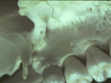
7 *Anterior nasal spine Incisive fossa Lamina dura Pulp Premaxillary suture Periodontal ligament space Located in the midline, it lies approximately 1.5 to 2 cm above the alveolar crest. below the junction of the inferior end of the nasal septum and the inferior outline of the nasal aperture V shape-opaque.

8 Nasal septum Nasal passage Anterior nasal spine Soft tissue shadow of nose Premaxillary suture Dental caries

9 LATERAL FOSSA incisive fossa a gentle depression in the maxilla near the apex of the lateral incisor appear diffusely radiolucent pathologic condition: 1. Intact LD 2. absence of clinical symptoms
the differing angles at which the x-ray beam is directed for the maxillary central incisors and (2) some")
10 NPC oral terminus of the nasopalatine canal in the innervation of the maxillary central incisors CBCT the borders of the nasopalatine canal placing an implant Incisive foramen between the roots and in the region of the middle and apical thirds of the central incisors 1/3 apical Cyst (1)the differing angles at which the x-ray beam is directed for the maxillary central incisors and (2) some variability in its anatomic size.
11 The incisive fossa can be superimposed on central incisor apices. The periodontal ligament spaces are intact. Same Lingual Opposite Buccal: The incisive fossa being lingually situated moves on the resulting image in the same direction as the movement of the tubehead.


12 Note how shadow of the incisive fossa moves in the direction of the movement of the tubehead. The fossa becomes superimposed over the root apex of the central incisor.the periodontal ligament space is intact.
13 Structures found on central incisor view are displaced - lingual structures appearing more posteriorly and facial structures more anteriorly. Inverted Y : Lateral wall of nasal passage and anterior wall of maxillary sinus (if you look carefully it is really an X!).

14 Nasal passage Anterior nasal spine

15 Anterior wall of maxillary sinus Maxillary sinus Lateral wall of nasal fossa

16 Lateral wall of nasal fossa Edentulous maxillary canine region indicating the two parts of the Inverted Y radiographic landmark Anterior wall of maxillary sinus (antrum)

17 Inverted Y Locule in maxillary sinus: note tooth periodontal ligament space intact Septum in maxillary sinus Bridge unit in porcelain Fused to metal Radiolucent anterior filling material
18 Maxillary sinus (radiolucent). Maxillary sinus floor and septums (radiopaque). Nutrient canals (radiolucent). Occasionally: lateral wall of nasal passage (radiopaque). Soft tissue shadow of lips/cheeks.

19 Premolar region: maxilla
20 Maxillary sinus The largest of the paranasal sinuses Borders: a thin, delicate, tenuous radiopaque line continuous BM They enlarge during childhood, achieving mature size by age 15 to 18 years. IN ADULTS: distal aspect of the canine to the posterior wall of the maxilla above the tuberosity. The right and left sinuses usually appear similar in shape and size. The flors of the maxillary sinus and nasal cavity are seen at the same level. In older individuals, the sinus may extend farther into the alveolar process, in the posterior region of the maxilla, its flor may appear considerably below the level of the flor of the nasal

21 Floor of maxillary sinus

22 When the rounded sinus flor dips between the buccal and palatal molar roots and is medial to the premolar roots, the projection of the apices is superior to the flor. This appearance conveys the impression that the roots project into the sinus cavity, which is an illusion. As the positive vertical angle of the projection is increased, the roots medial to the sinus appear to project farther into the sinus cavity. In contrast, the roots that are lateral to the sinus appear to move either out of the sinus or farther away from it as the angle is increased.

23 neurovascular canals in the lateral sinus walls any direction (including vertically), they are usually seen running a curved posteroanterior course that is convex toward the alveolar process. CYSTS

24 one or several radiopaque lines traverse the image of the Maxillary sinus.these opaque lines are called septa. vertically Septa warrant attention because they sometimes mimic periapical disease, and the chambers they create in the alveolar recess may complicate the search for a root fragment displaced into the sinus.

25 The flor of the maxillary sinus occasionally shows small radiopaque projections, which are nodules of bone root tips, which they resemble in shape. 1.In contrast to a root fragment, which is quite homogeneous in appearance, the bony nodules often show trabeculation; 2.although they may be quite well defied, at certain points on their surface they blend with the trabecular pattern of adjacent bone. 3.A root fragment may also be recognized by the presence of a root canal.
26 Maxillary sinus floor and septums (radiopaque). Maxillary sinus (radiolucent). Nutrient canals (radiolucent). Zygomatic process of the maxilla ( U - shaped radiopacity). Zygomatic arch/zygoma (radiopaque). Less commonly: Lateral wall of nasal passage (radiopaque).

27 Articular eminence Structures anterior to green line seen on intraoral radiographs Zygoma Zygomatic process of temporal bone Zygomatic process of maxilla

28 ZYGOMATIC PROCESS an extension of the lateral maxillary surface Apex of first & second molars a U-shaped radiopaque line with its open end directed superiorly. When the sinus is recessed deep within the process the image of the air space within the process is dark. Typically the walls of the process are thin and well defined. When the sinus exhibits relatively little penetration of the maxillary process (usually in younger individuals or individuals who have maintained their posterior teeth and vigorous masticatory function), the image of the walls of the zygomatic process of the maxilla tends to be thicker, and the appearance of the sinus in this region is smaller and more opaque.

29 Endodontically treated tooth Zygomatic process of the maxilla Zygomatic arch shadow Floor of maxillary sinus Maxillary tuberosity
30 Coronoid process of mandible (radiopaque). Maxillary tuberosity (radiopaque). Posterior wall of maxillary sinus (radiopaque). Maxillary sinus (radiolucent). Pterygoid hamulus (radiopaque). Pterygoid notch (radiolucent). Lateral pterygoid plate (radiopaque).
 Maxillary")
31 Lateral pterygoid plate Pterygoid hamulus (medial Pterygoid) Maxillary tuberosity

32 Pterygoid hamulus
33 PTERYGOID PLATES -Posterior to the tuberosity of the maxilla - radiopaque homogeneous shadow without any evidence of trabeculation. - Extending inferiorly from the medial pterygoid plate is the hamular process which on close inspection can show trabeculae

34 Posterior wall of maxillary sinus Pterygoid hamulus Air space
35 MANDIBLE
36 Symphysis Lingual foramen (radiolucent). Genial tubercles (radiopaque). Soft tissue shadow of lower lip (radiopaque). Mental ridges (radiopaque). Nutrient canals (radiolucent).


37 a radiolucent line through the midline of the jaw between the images of the forming deciduous central incisor fuses by the end of the fist year fracture cleft


38 Facial Lingual Mental depression Mental ridge Lingual foramen Genial tubercles

39 lingual surface slightly above the inferior border and in the midline. Right and left- superior and inferior A single round radiolucent canal with a welldefied opaque border lying in the midline below the level of the apices of the incisors.

40 lingual surface midline two or even more A single round radiolucent canal with a welldefied opaque border

41 Embossed dot Lingual foramen

42 two radiopaque lines sweeping bilaterally forward and upward toward the midline low in the premolar incisor tooth roots The image of the mental ridge is most prominent: 1. parallel with the surface of the mental tubercle 2.as when using the bisectingangle technique

43 a depression on the labial above the mental ridge thinness of jawbone in this area Mistaken for periapical disease

44 The occlusal wear on the incisive edges of the teeth is attrition. The patient also evidences moderate to severe periodontal bone loss. Soft tissue shadow of lower lip Mental ridge Lingual foramen Cortical plate of lower border of mandible Genial tubercles

45 Nutrient canals
46 Mental foramen (radiolucent) - usually situated between and just beneath roots of the premolars. Soft tissue shadow of reflected cheek (radiopaque). Mandibular canal (radiolucent).

47 Mandibular premolar region Mental foramen


48 Mental Foramen anterior limit of IANC opening of the mental canal is directed superiorly and posteriorly about halfway between the lower border of the mandible and the crest of the alveolar process, usually in the region of the apex of the second premolar mesial of the permanent fist molar roots to as far anterior as mesial of the fist premolar root periapical disease : 1. detectable LD 2.IANC 3. second radiograph from another angle

49 Mental foramen Periapical granuloma, abscess or cyst (periodontal ligament space not intact)

50 MANDIBULAR CANAL a dark linear shadow with thin radiopaque superior and inferior borders The width of the canal shows some interpatient variability but is usually constant anterior to the third molar region. the canal is in contact with the apex of the third molar, and the distance between it and the other roots increases as it progresses anteriorly. When the apices of the molars are projected over the canal: 1. missing lamina 2. thickened PDL *Vitality test *Because the canal is usually located just inferior to the apices of the posterior teeth, altering the vertical angle for a second fim of the area is not likely to separate the images of the apices and canal Bifid canal: 1. risk of inadequate anesthesia 2. diffiulties with jaw surgery, including implants, or trauma


51 NUTRIENT CANALS radiolucent lines of fairly uniform width. vertically from the IANC to the apex of a tooth or into the interdental space between the mandibular incisors black patients; male patients; older patients; and patients with high blood pressure, diabetes mellitus, or advanced periodontal disease.

52 Internal oblique ridge (mylohyoid ridge)
53 MYLOHYOID RIDGE internal oblique ridge Lingual surface Third molar. Midline mylohyoid muscle quite diffuse and of variable width SUBMANDIBULAR GLAND FOSSA lingual surface below the mylohyoid ridge radiolucent (mylohyoid ridge and inferior border of the mandible) Ill defined+ sparse trabecular pattern Superiorlyby the mylohyoid ridge inferiorly by the lower border of the mandible anteriorly (in the premolar region) posteriorly (at about the ascending ramus).

54 Mental foramen Mandibular canal Mylohyoid ridge Submandibular fossa

55 Mylohyoid ridge Mental foramen Submandibular fossa
56 Mandibular canal (radiolucent). External oblique ridge (radiopaque). Mylohyoid ridge - also known as internal oblique ridge (radiopaque). Submandibular fossa (radiolucent). Cortex of lower border (radiopaque).

57 External oblique ridge of mandible
58 External oblique ridge continuation of the anterior border of the mandibular ramus Disappear below the fist molar buccinator muscle superior to the mylohyoid ridge- paralel radiopaque line of varying width, density, and length, blending at its anterior end with the shadow of the alveolar bone INFERIOR BORDER OF THE MANDIBLE dense, broad radiopaque band of bone
")
59 External oblique ridge Internal oblique (mylohyoid) ridge

60 External oblique ridge Internal oblique ridge Lower cortex of mandible Submandibular fossa

61 Coronoid process of mandible Zygomatic process of maxilla Zygomatic arch
62 CORONOID PROCESS periapical radiographs of the maxillary molar region a triangular radiopacity, with its apex directed superiorly and anteriorly superimposed on the region of the third molar Homogeneous,although internal trabeculation results from the downward and forward movement of the mandible when the mouth is open. mouth minimally open root fragment

63 Zygomatic process of maxilla Zygomatic arch Pterygoid hamulus Coronoid process of mandible Floor of maxillary sinus
Upper arch. 1Prosthodontics. Dr.Bassam Ali Al-Turaihi. Basic anatomy & & landmark of denture & mouth
 1Prosthodontics Lecture 2 Dr.Bassam Ali Al-Turaihi Basic anatomy & & landmark of denture & mouth Upper arch Palatine process of maxilla: it form the anterior three quarter of the hard palate. Horizontal
Bones Ethmoid bone Inferior nasal concha Lacrimal bone Maxilla Nasal bone Palatine bone Vomer Zygomatic bone Mandible
 splanchnocranium - Consists of part of skull that is derived from branchial arches - The facial bones are the bones of the anterior and lower human skull Bones Ethmoid bone Inferior nasal concha Lacrimal
Normal Radiographic Anatomy Maxillary Lateral Area. Carmen Elena Georgescu1, Gabriela Tãnase 2, Augustin Mihai 3. Objectives.
 Normal Radiographic Anatomy Maxillary Lateral Area Carmen Elena Georgescu1, Gabriela Tãnase 2, Augustin Mihai 3 Bucharest, Romania Summary Intraoral examinations are the backbone of dental radiography.
高雄醫學大學 口腔醫學院 口腔病理影像科 牙科 X 光影像判讀 教學範例
 高雄醫學大學 口腔醫學院 口腔病理影像科 牙科 X 光影像判讀 教學範例 Content Image No. 001 Dentigerous cyst over left upper embedded canine--------------------- 頁 1 Image No. 002---------------------------------------------------------------
Techniques of local anesthesia in the mandible
 Techniques of local anesthesia in the mandible The technique of choice for anesthesia of the mandible is the block injection and this is attributed to the absence of the advantages which are present in
Oral cavity landmarks
 By: Dr. Ahmed Rabah Oral cavity landmarks The knowledge of oral anatomy and physiology will help the operator and provides enough landmarks to act as positive guide during denture construction. This subject
Infratemporal fossa: Tikrit University college of Dentistry Dr.Ban I.S. head & neck Anatomy 2 nd y.
 Infratemporal fossa: This is a space lying beneath the base of the skull between the lateral wall of the pharynx and the ramus of the mandible. It is also referred to as the parapharyngeal or lateral pharyngeal
Figure (2-6): Labial frenum and labial notch.
: Labial frenum and labial notch.") The anatomy of the edentulous ridge in the maxilla and mandible is very important for the design of a complete denture. The consistency of the mucosa and architecture of the underlying bone is different
The anatomy of the edentulous ridge in the maxilla and mandible is very important for the design of a complete denture. The consistency of the mucosa and architecture of the underlying bone is different
Dentalelle Tutoring - Faulty Radiographs
 Dentalelle Tutoring - Faulty Radiographs Errors in improperly exposing or processing dental films can produce undesirable dental radiographs of nondiagnostic quality. These are known as faulty radiographs.
Oral Surgery. Basic Techniques of Dental Local Anesthesia. A variety of techniques used in administration and deposition of local anesthesia:
 Oral Surgery Lecture: 9 Dr. Saif Saadedeen Basic Techniques of Dental Local Anesthesia A variety of techniques used in administration and deposition of local anesthesia: 1. Topical anesthesia 2. Infiltration
Normal Radiographic Anatomy Maxillary Central Area
 VARIA Normal Radiographic Anatomy Maxillary Central Area Carmen Elena Georgescu 1, Gabriela Tãnase 2, Augustin Mihai 3 Bucharest, Romania Summary The radiopgraphic recognition of disease requires knowledge
Lips and labial mucosa
 Lips and labial mucosa External portion of the lips: the vermilion border and the skin Vermilion border : the exposed red portion of the lip, covered by mucous membrane, no mucous glands Boundary: the
3. The Jaw and Related Structures
 Overview and objectives of this dissection 3. The Jaw and Related Structures The goal of this dissection is to observe the muscles of jaw raising. You will also have the opportunity to observe several
TRAUMA TO THE FACE AND MOUTH
 Dr.Yahya A. Ali 3/10/2012 F.I.C.M.S TRAUMA TO THE FACE AND MOUTH Bailey & Love s 25 th edition Injuries to the orofacial region are common, but the majority are relatively minor in nature. A few are major
 www.oralradiologists.com CONE BEAM CT REPORT CASE XXXX Patient information Patient Name: - Referring Doctor: - Patient DOB: - Scan Date: [Start date] Reason for Exam: Maxillary facial pain Doctor Notes:
6610 NE 181st Street, Suite #1, Kenmore, WA
 660 NE 8st Street, Suite #, Kenmore, WA 9808 www.northshoredentalacademy.com.08.900 READ CHAPTER The Professional Dental Assistant (p.-9) No Key Terms Recall Questions:,,,, and 6 CLASS SYLLABUS DAY READ
Dental Morphology and Vocabulary
 Dental Morphology and Vocabulary Palate Palate Palate 1 2 Hard Palate Rugae Hard Palate Palate Palate Soft Palate Palate Palate Soft Palate 4 Palate Hard Palate Soft Palate Maxillary Arch (Maxilla) (Uppers)
Radiology. & supporting structures. Lec. 14 Common diseases of teeth Dr. Areej
 Radiology Lec. 14 Common diseases of teeth Dr. Areej & supporting structures A radiograph is only one part of the diagnostic process. Usually one does NOT make a diagnosis solely from a radiograph. A diagnosis
Fundamentals of technique Types of local anaesthesia Topical or surface anaesthesia
 Fundamentals of technique The importance of a quiet, confident, and friendly manner towards all patients so physical comfort is also essential for the co-operation of the patient and the ease of operation
INTERPRETATION RADIOGRAPHIC INTERPRETATION. Law of Symmetry. We will be reviewing: 7/30/16
 RADIOGRAPHIC INTERPRETATION Pam Wood, CDA, RDH, M.Ed, CAGS Community College of Rhode Island pwood@ccri.edu INTERPRETATION the ability to read what is revealed on a dental radiograph any dental professional
Trigeminal Nerve Anatomy. Dr. Mohamed Rahil Ali
 Trigeminal Nerve Anatomy Dr. Mohamed Rahil Ali Trigeminal nerve Largest cranial nerve Mixed nerve Small motor root and large sensory root Motor root Nucleus of motor root present in the pons and medulla
Fundamental & Preventive Curvatures of Teeth and Tooth Development. Lecture Three Chapter 15 Continued; Chapter 6 (parts) Dr. Margaret L.
 Dr. Margaret L.") Fundamental & Preventive Curvatures of Teeth and Tooth Development Lecture Three Chapter 15 Continued; Chapter 6 (parts) Dr. Margaret L. Dennis Proximal contact areas Contact areas are on the mesial and
Fundamental & Preventive Curvatures of Teeth and Tooth Development Lecture Three Chapter 15 Continued; Chapter 6 (parts) Dr. Margaret L. Dennis Proximal contact areas Contact areas are on the mesial and
Morphology of an Anatomic Crown. By: Assistant Professor Dr. Baydaa Ali Al - Rawi
 Morphology of an Anatomic Crown By: Assistant Professor Dr. Baydaa Ali Al - Rawi October 4, 2009 Elevated landmarks Depressed landmarks A) Elevated landmarks : 1. Dental lobe : is one of the primary centers
Arrangement of the artificial teeth:
 Lecture Prosthodontic Dr. Osama Arrangement of the artificial teeth: It s the placement of the teeth on a denture with definite objective in mind or it s the setting of teeth on temporary bases. Rules
The Skull and Temporomandibular joint II Prof. Abdulameer Al-Nuaimi. E. mail:
 The Skull and Temporomandibular joint II Prof. Abdulameer Al-Nuaimi E-mail: a.al-nuaimi@sheffield.ac.uk E. mail: abdulameerh@yahoo.com Temporal fossa The temporal fossa is a depression on the temporal
Lec [8]: Mandibular nerve:
![Lec [8]: Mandibular nerve:](/thumbs/94/121295776.jpg "Lec [8]: Mandibular nerve:") Lec [8]: Mandibular nerve: The mandibular branch from the trigeminal ganglion lies in the middle cranial fossa lateral to the cavernous sinus. With the motor root of the trigeminal nerve [motor roots lies
Lec [8]: Mandibular nerve: The mandibular branch from the trigeminal ganglion lies in the middle cranial fossa lateral to the cavernous sinus. With the motor root of the trigeminal nerve [motor roots lies
PTERYGOPALATINE FOSSA
 PTERYGOPALATINE FOSSA Outline Anatomical Structure and Boundaries Foramina and Communications with other spaces and cavities Contents Pterygopalatine Ganglion Especial emphasis on certain arteries and
Lecture 2 Maxillary central incisor
 Lecture 2 Maxillary central incisor Generally The deciduous tooth appears in the mouth at 3 18 months of age, with 6 months being the average and is replaced by the permanent tooth around 7 8 years of
Temporal region. temporal & infratemporal fossae. Zhou Hong Ying Dept. of Anatomy
 Temporal region temporal & infratemporal fossae Zhou Hong Ying Dept. of Anatomy Temporal region is divided by zygomatic arch into temporal & infratemporal fossae. Temporal Fossa Infratemporal fossa Temporal
DEVELOPING ANALOGUE/SUBTITUTE FOR THE MANDIBULAR DENTURE BEARING AREA. Dr Muhammad Rizwan Memon FCPS Assistant Professor
 DEVELOPING ANALOGUE/SUBTITUTE FOR THE MANDIBULAR DENTURE BEARING AREA Dr Muhammad Rizwan Memon FCPS Assistant Professor Crest of Residual Ridge Buccal Shelf Shape of supporting structure Mylohyoid Ridge
Muscles of mastication [part 1]
![Muscles of mastication [part 1]](/thumbs/76/73586850.jpg "Muscles of mastication [part 1]") Muscles of mastication [part 1] In this lecture well have the muscles of mastication, neuromuscular function, and its relationship to the occlusion morphology. The fourth determinant of occlusion is the
Muscles of mastication [part 1] In this lecture well have the muscles of mastication, neuromuscular function, and its relationship to the occlusion morphology. The fourth determinant of occlusion is the
Prosthetic Options in Implant Dentistry. Hakimeh Siadat, DDS, MSc Associate Professor
 Prosthetic Options in Dentistry Hakimeh Siadat, DDS, MSc Associate Professor Dental Research Center, Department of Prosthodontics & Dental s Faculty of Dentistry, Tehran University of Medical Sciences
Basic Anatomy and Physiology of the Lips and Oral Cavity. Dr. Faghih
 Basic Anatomy and Physiology of the Lips and Oral Cavity Dr. Faghih It is divided into seven specific subsites : 1. Lips 2. dentoalveolar ridges 3. oral tongue 4. retromolar trigone 5. floor of mouth 6.
The future of health is digital
 Dated: XX/XX/XXXX Name: XXXXXXXX XXXXXXXXXXX Birth Date: XX/XX/XXXX Date of scan: XX/XX/XXXX Examination of the anatomical volume: The following structures are reviewed and evaluated for bilateral symmetry,
Temporal fossa Infratemporal fossa Pterygopalatine fossa Terminal branches of external carotid artery Pterygoid venous plexus
 Outline of content Temporal fossa Infratemporal fossa Pterygopalatine fossa Terminal branches of external carotid artery Pterygoid venous plexus Boundary Content Communication Mandibular division of trigeminal
Elevators. elevators:- There are three major components of the elevator are:-
 Elevators Elevators:- Are exo-levers, instrument designed to elevate or luxate the teeth or roots from their bony socket in close or surgical method of extraction to force a tooth or root along the line
Mandibular and Maxillary Anesthesia
 Mandibular and Maxillary Anesthesia Uses of the Conduction Technique JACK H. SELTSAM, D.D.S., M.D., Los Angeles THE ARMAMENTARIUM of a surgeon who operates on the head and neck should include the ability
1. What is the highest and sharpest cusp on the lower first deciduous molar? 2. Which of the following is NOT the correct location of an embrasure?
 1 1. What is the highest and sharpest cusp on the lower first deciduous molar? a. mesiobuccal b. distobuccal c. distolingual d.mesiolingual 2. Which of the following is NOT the correct location of an embrasure?
Unusual transmigration of canines report of two cases in a family
 ISSN: Electronic version: 1984-5685 RSBO. 2014 Jan-Mar;11(1):88-92 Case Report Article Unusual transmigration of canines report of two cases in a family Sulabha A. Narsapur 1 Sameer Choudhari 2 Shrishal
Bones of the skull & face
 Bones of the skull & face Cranium= brain case or helmet Copyright The McGraw-Hill Companies, Inc. Permission required for reproduction or display. The cranium is composed of eight bones : frontal Occipital
Periapical Radiography
 Periapical Radiography BARBARA E. DIXON B.D.S., M.Sc., D.P.D.S. Main Indications Detection of Apical infection/inflammation Assessment of the periodontal status After trauma Assessment of Unerupted teeth
Anatomic Relations Summary. Done by: Sohayyla Yasin Dababseh
 Anatomic Relations Summary Done by: Sohayyla Yasin Dababseh Anatomic Relations Lecture 1 Part-1 - The medial wall of the nose is the septum. - The vestibule lies directly inside the nostrils (Nares). -
MAXILLARY INJECTION TECHNIQUE. Chinthamani Laser Dental Clinic
 MAXILLARY INJECTION TECHNIQUE Chinthamani Laser Dental Clinic Introduction A number of injection techniques are available to aid in providing clinically adequate anesthesia of the teeth and soft and hard
DENTAL RADIOGRAPH INTERPRETATION
 DENTAL RADIOGRAPH INTERPRETATION Brook A. Niemiec, DVM Diplomate, American Veterinary Dental College Fellow, Academy of Veterinary Dentistry www.vetdentaltraning.com www.vetdentalrad.com Interpreting dental
THE BIOMECHANICAL BASIS OF RETENTION IN COMPLETE DENTURES
 THE BIOMECHANICAL BASIS OF RETENTION IN COMPLETE DENTURES Factors affecting the retention of dentures Retention is the resistance of the denture to removal along its path of insertion. Strictly speaking,
Medical NBDE-II. Dental Board Exams Part I.
 Medical NBDE-II Dental Board Exams Part I http://killexams.com/exam-detail/nbde-ii Question: 149 Anatomically, the term "clinical root" can be defined as which of the following: A. The space in the tooth
Anatomy of the Trigeminal Nerve
 19 Anatomy of the Trigeminal Nerve.1 Introduction 0. The Central Part of the Trigeminal Nerve 1..1 Origin 1.. Trigeminal Nuclei.3 The Peripheral Part of the Trigeminal Nerve 4.3.1 Ophthalmic Nerve 4.3.
Trigeminal Nerve (V)
") Trigeminal Nerve (V) Lecture Objectives Discuss briefly how the face is developed. Follow up the course of trigeminal nerve from its point of central connections, exit and down to its target areas. Describe
Trigeminal Nerve (V) Lecture Objectives Discuss briefly how the face is developed. Follow up the course of trigeminal nerve from its point of central connections, exit and down to its target areas. Describe
Nose & Mouth OUTLINE. Nose. - Nasal Cavity & Its Walls. - Paranasal Sinuses. - Neurovascular Structures. Mouth. - Oral Cavity & Its Contents
 Dept. of Human Anatomy, Si Chuan University Zhou hongying eaglezhyxzy@163.com Nose & Mouth OUTLINE Nose - Nasal Cavity & Its Walls - Paranasal Sinuses - Neurovascular Structures Mouth - Oral Cavity & Its
Large Dentigerous Cyst
 Volume 16.2.1 Feb 2016 This Lecture Series qualifies for 0.5 Informal CPD Learning Hours Large Dentigerous Cyst By Dr Hassem Geha A 55 year-old male presented with a painless swelling in the right mandible.
You know you would like to stop swearing at the computer after each shot. Troubleshooting oral radiography
 You know you would like to stop swearing at the computer after each shot Troubleshooting oral radiography Goals of oral radiology Achieve diagnostic images of the teeth and surrounding bone. Images should
Essentials in Head and Neck Embryology. Part 3 Development of the head, face, and oral cavity
 Essentials in Head and Neck Embryology Part 3 Development of the head, face, and oral cavity Outline General overview of prenatal development Embryonic period phase 1 Formation of bilaminar disk Formation
IMPACTED CANINES. Unfortunately, this important tooth is the second most common tooth to be impacted after third molars
 IMPACTED CANINES After we talked about impacted third molars, today we ll discuss about maxillary impacted canines in upper dental arch, how to manage these cases as a dental surgeon. You will study about
Anatomy and Physiology. Bones, Sutures, Teeth, Processes and Foramina of the Human Skull
 Anatomy and Physiology Chapter 6 DRO Bones, Sutures, Teeth, Processes and Foramina of the Human Skull Name: Period: Bones of the Human Skull Bones of the Cranium: Frontal bone: forms the forehead and the
Intraoral radiographic techniques Introduction There are three main types of intraoral radiographs: Periapical radiograph Bitewing radiograph
 Intraoral radiographic techniques Introduction There are three main types of intraoral radiographs: Periapical radiograph Bitewing radiograph Occlusal radiograph The anatomic area of interest and type
Chapter 7. Skeletal System
 Chapter 7 Skeletal System 1 Skull A. The skull is made up of 22 bones: 8 cranial bones, 13 facial bones, and the mandible. B. The Cranium encloses and protects the brain, provides attachments for muscles,
Maxilla, ORBIT and infratemporal fossa. Neophytos C Demetriades MD, DDS, MSc Associate professor European University of Cyprus School of Medicine
 Maxilla, ORBIT and infratemporal fossa Neophytos C Demetriades MD, DDS, MSc Associate professor European University of Cyprus School of Medicine MAXILLA Superior, middle, and inferior meatus Frontal sinus
Extraoral Radiology October 10th, 2008
 Extraoral Radiology October 10th, 2008 Steven R. Singer, DDS srs2@columbia.edu 212.305.5674 November 8 th, 1895 Extraoral Projections Images can be produced in the dental office X-ray source can be Intraoral
Periodontal Disease. Radiology of Periodontal Disease. Periodontal Disease. The Role of Radiology in Assessment of Periodontal Disease
 Radiology of Periodontal Disease Steven R. Singer, DDS srs2@columbia.edu 212.305.5674 Periodontal Disease! Includes several disorders of the periodontium! Gingivitis! Marginal Periodontitis! Localized
European Veterinary Dental College
 European Veterinary Dental College EVDC Training Support Document Preparation of Radiograph Sets (Cat and Dog) Document version : evdc-tsd-radiograph_positioning_(dog_and_cat)-20120121.docx page 1 of 13
Dental panoramic tomography: An approach for the general radiologist
 Pictorial Essay Australasian Radiology (2006) 50, 526 533 Dental panoramic tomography: An approach for the general radiologist R Boeddinghaus and A Whyte Perth Radiological Clinic, Perth, Western Australia,
04 Development of the Face and Neck. Development of the Face Development of the neck
 04 Development of the Face and Neck Development of the Face Development of the neck Development of the face Overview of facial development The fourth week ~ the twelfth week of prenatal development Between
Introduction to Occlusion and Mechanics of Mandibular Movement
 Introduction to Occlusion and Mechanics of Mandibular Movement Dr. Pauline Hayes Garrett Department of Endodontics, Prosthodontics, and Operative Dentistry University of Maryland, Baltimore Assigned reading
Disclosure. Educational Objectives. Terminology. Odontogenic Cysts. Terminology
 Disclosure Lisa J. Koenig BChD, DDS, MS Professor & Program Director, Oral Medicine and Oral Radiology Marquette University School of Dentistry Consultant to Soredex for the Scanora 3D and 3Dx Author/Editor
Subdivided into Vestibule & Oral cavity proper
 Extends from the lips to the oropharyngeal isthmus The oropharyngeal isthmus: Is the junction of mouth and pharynx. Is bounded: Above by the soft palate and the palatoglossal folds Below by the dorsum
IMAGING OF CYSTS OF THE JAWS July 2002 N. Serman
 IMAGING OF CYSTS OF THE JAWS July 2002 N. Serman This is an area where radiology plays an important role in assisting with the diagnosis, determining the size of the lesion and the relationship to adjacent
Remember from the first year embryology Trilaminar disc has 3 layers: ectoderm, mesoderm, and endoderm
 Development of face Remember from the first year embryology Trilaminar disc has 3 layers: ectoderm, mesoderm, and endoderm The ectoderm forms the neural groove, then tube The neural tube lies in the mesoderm
Nasal region. cartilages: septal cartilage (l); lateral nasal cartilage (2); greater alar cartilages (2); lesser alar cartilages (?
; lateral nasal cartilage (2); greater alar cartilages (2); lesser alar cartilages (?") Nasal region skull bones: nasal and frontal processes of maxilla cartilages: septal cartilage (l); lateral nasal cartilage (2); greater alar cartilages (2); lesser alar cartilages (?) 1 Nasal cavity Roof
Nasal region skull bones: nasal and frontal processes of maxilla cartilages: septal cartilage (l); lateral nasal cartilage (2); greater alar cartilages (2); lesser alar cartilages (?) 1 Nasal cavity Roof
Chapter 7 Part A The Skeleton
 Chapter 7 Part A The Skeleton Why This Matters Understanding the anatomy of the skeleton enables you to anticipate problems such as pelvic dimensions that may affect labor and delivery The Skeleton The
Dr.Noor Hashem Mohammad Lecture (5)
") Dr.Noor Hashem Mohammad Lecture (5) 2016-2017 If the mandible is discarded, the anterior part of this aspect of the skull is seen to be formed by the hard palate. The palatal processes of the maxillae
Dr.Noor Hashem Mohammad Lecture (5) 2016-2017 If the mandible is discarded, the anterior part of this aspect of the skull is seen to be formed by the hard palate. The palatal processes of the maxillae
DENTAL RADIOLOGY Identify basic facts and terms of radiology, to include fundamentals. with 70% accuracy.
 DENTAL RADIOLOGY Identify basic facts and terms of radiology, to include fundamentals of chemistry relating to radiology, with 70% accuracy. Radiation Physics Radiation Health and Safety Components of
Extraoral Imaging. Chapter 42. Copyright 2018, Elsevier Inc. All Rights Reserved. 1
 Extraoral Imaging Chapter 42 Copyright 2018, Elsevier Inc. All Rights Reserved. 1 Learning Objectives Lesson 42.1: Panoramic Imaging 1. Pronounce, define, and spell the key terms. 2. Discuss panoramic
Samantha W. Chou, D.M.D N. Southport Ave. Chicago, Illinois Phone: Fax:
 Samantha W. Chou, D.M.D. 2325 N. Southport Ave. Chicago, Illinois 60614 Phone: 312-608-6881 Fax: 773-296-0601 Samanthawchou@gmail.com What is our role as the dentist? "We live in a culture in which people
Primary Teeth Chapter 18. Dental Anatomy 2016
 Primary Teeth Chapter 18 Dental Anatomy 2016 Primary Teeth - Introduction Synonyms deciduous teeth, baby teeth, temporary teeth, milk teeth. There are 20 primary teeth, designated as A thru T in the Universal
Diagnosis. overt Examination. Definitive Examination. History. atient interview. Personal History. Clinical Examination.
 Diagnosis overt Examination History Definitive Examination atient interview Personal History Mental Attitude Medical History Dental History Clinical Examination Extra Oral Oral Radiographic Evaluation
Parotid Gland. Parotid Gland. Largest of 3 paired salivary glands (submandibular; sublingual) Ramus of Mandible. Medial pterygoid.
 Ramus of Mandible. Medial pterygoid.") Parotid region Parotid Gland Largest of 3 paired salivary glands (submandibular; sublingual) Ramus of Mandible Medial pterygoid Cross section of mandible Masseter D S SCM Parotid Gland Mastoid Process
Parotid region Parotid Gland Largest of 3 paired salivary glands (submandibular; sublingual) Ramus of Mandible Medial pterygoid Cross section of mandible Masseter D S SCM Parotid Gland Mastoid Process
Central Incisor DR.Ahmed Al-Jobory B.D.S.,M.Sc. Conservative Department
 Dental Anatomy Lecture 3 Central Incisor DR.Ahmed Al-Jobory B.D.S.,M.Sc. Conservative Department The permanent maxillary Incisors Maxillary incisor are four in number. The maxillary central incisor is
Component parts of Chrome Cobalt Removable Partial Denture
 Lec. 5 د.بسام الطريحي Component parts of Chrome Cobalt Removable Partial Denture Major connectors: Are either bars or plates, the difference between them is in the amount of tissue covers. Plates are broad
J. 0. AKINOSI, B.D.s., F.D.S.R.C.S.
 British Journal of Oral Surgery 15 (1977-78) 83-87 A NEW APPROACH TO THE MANDIBULAR NERVE BLOCK J. 0. AKINOSI, B.D.s., F.D.S.R.C.S. Department of Oral Surgery and Pathology, College of Medicine, Lagos
Maxillary LA: Techniques. Ra ed Salma BDS, MSc, JBOMFS, MFDRCSI
 Maxillary LA: Techniques Ra ed Salma BDS, MSc, JBOMFS, MFDRCSI dr.raedsalma@riyadh.edu.sa https://sites.google.com/a/riyadh.edu.sa/raed/ LA Options for the Maxilla Infiltration Submucosal Supraperiosteal
Comprehensive AOCMF Classification System. Cornelius CP, Kunz C, Prein J, Audigé L. Mandibular fractures Level-2 system (cases 1 to 18)
") Comprehensive AOCMF Classification System Cornelius CP, Kunz C, Prein J, Audigé L Mandibular fractures Level-2 system (cases 1 to 18) Case 1: Body fracture traversing anterior transition zone Imaging:
Comprehensive AOCMF Classification System Cornelius CP, Kunz C, Prein J, Audigé L Mandibular fractures Level-2 system (cases 1 to 18) Case 1: Body fracture traversing anterior transition zone Imaging:
Omran Saeed. Luma Taweel. Mohammad Almohtaseb. 1 P a g e
 2 Omran Saeed Luma Taweel Mohammad Almohtaseb 1 P a g e I didn t include all the photos in this sheet in order to keep it as small as possible so if you need more clarification please refer to slides In
-Ibrahim Al-Naser. -Dr Al- Muhtaseb. 1 P a g e
 -1 -Ibrahim Al-Naser - -Dr Al- Muhtaseb 1 P a g e The Digestive System The doctor started the lecture by talking about the class rules. The GI system is an organ system, it is divided into: The Alimentary
The Pharynx. Dr. Nabil Khouri MD. MSc, Ph.D
 The Pharynx Dr. Nabil Khouri MD. MSc, Ph.D Introduction The pharynx is the Musculo-fascial halfcylinder that links the oral and nasal cavities in the head to the larynx and esophagus in the neck Common
Panoramic Radiology. Seminars on Maxillofacial Imaging and Interpretation. Bearbeitet von Allan G Farman
 Panoramic Radiology Seminars on Maxillofacial Imaging and Interpretation Bearbeitet von Allan G Farman 1. Auflage 2007. Buch. xiv, 232 S. Hardcover ISBN 978 3 540 46229 3 Format (B x L): 19,3 x 27 cm Gewicht:
University of Palestine. Midterm Exam 2013/2014 Total Grade:
 [ Course No: DNTS2208 Course Title: Head and Neck Anatomy Date: 17/11/1024 No. of Questions: (52) Time: 2hours Using Calculator (No) University of Palestine Midterm Exam 2013/2014 Total Grade: Instructor
LOCAL ANESTHESIA IN PEDIATRIC DENTISTRY
 Disclaimer This movie is an educational resource only and should not be used to manage your health. All decisions about the management of local anesthesia in pediatric dentistry must be made in conjunction
Mohammad Hisham Al-Mohtaseb. Lina Mansour. Reyad Jabiri. 0 P a g e
 2 Mohammad Hisham Al-Mohtaseb Lina Mansour Reyad Jabiri 0 P a g e This is only correction for the last year sheet according to our record. If you already studied this sheet just read the yellow notes which
Concepts of occlusion Balanced occlusion. Monoplane occlusion. Lingualized occlusion. Figure (10-1)
") Any contact between teeth of opposing dental arches; usually, referring to contact between the occlusal surface. The static relationship between the incising or masticatory surfaces of the maxillary or
Any contact between teeth of opposing dental arches; usually, referring to contact between the occlusal surface. The static relationship between the incising or masticatory surfaces of the maxillary or
Dental Anatomy and Occlusion
 CHAPTER 53 Dental Anatomy and Occlusion Ma Lou C. Sabino DDS, and Emily G. Smythe, DDS What numerical system is used most commonly in the United States for designating the adult dentition? Pediatric dentition?
University of Palestine. Midterm Exam 2013/2014 Total Grade:
 Course No: DNTS2208 Course Title: Head and Neck Anatomy Date: 09/11/2013 No. of Questions: (50) Time: 1hour Using Calculator (No) University of Palestine Midterm Exam 2013/2014 Total Grade: Instructor
Chapter 7: Head & Neck
 Chapter 7: Head & Neck Osteology I. Overview A. Skull The cranium is composed of irregularly shaped bones that are fused together at unique joints called sutures The skull provides durable protection from
Skull basic structures. Neurocranium
 Assoc. Prof. Květuše Lovásová, M.V.D., PhD. Skull basic structures Skull consists of two groups of bones: neurocranium (bones forming the brain box) splanchnocranium (bones forming the facial skeleton)
Dr. Sami Zaqout, IUG Medical School
 The skull The skull is composed of several separate bones united at immobile joints called sutures. Exceptions? Frontal bone Occipital bone Vault Cranium Sphenoid bone Zygomatic bones Base Ethmoid bone
Case Study. Case # 1 Author: Dr. Suheil Boutros (USA) 2013 Zimmer Dental, Inc. All rights reserved. 6557, Rev. 03/13.
 2013 Zimmer Dental, Inc. All rights reserved. 6557, Rev. 03/13.") Placement of a Zimmer Trabecular Metal Dental Implant with Simultaneous Ridge Augmentation and Immediate Non-Functional Loading Following Tooth Extraction and Orthodontic Treatment for Implant Site Development
Placement of a Zimmer Trabecular Metal Dental Implant with Simultaneous Ridge Augmentation and Immediate Non-Functional Loading Following Tooth Extraction and Orthodontic Treatment for Implant Site Development
Visibility of Maxillary and Mandibular Anatomical Landmarks in Digital Panoramic Radiographs: A Retrospective Study
 Visibility of Maxillary and Mandibular Anatomical Landmarks in Digital Panoramic Radiographs: A Retrospective Study Srisha Basappa, Smitha JD, Nishath Khanum*, Santosh Kanwar, Mahesh MS and Archana Patil
Cephalometric Analysis
 Cephalometric Analysis of Maxillary and Mandibular Growth and Dento-Alveolar Change Part III In two previous articles in the PCSO Bulletin s Faculty Files, we discussed the benefits and limitations of
PH-04A: Clinical Photography Production Checklist With A Small Camera
 PH-04A: Clinical Photography Production Checklist With A Small Camera Operator Name Total 0-49, Passing 39 Your Score Patient Name Date of Series Instructions: Evaluate your Series of photographs first.
*in general the blood supply of the nose comes from branches of the internal and external carotid arteries.
 In the previous lecture we talked about the anatomy of the nasal cavity, today we will talk about its blood supply, venous drainage, innervations, and finally about the paranasal sinuses. When we describe