Differential Diagnosis of Oral Masses. Gingival Lesions
|
|
|
- Alan Bertram Stevenson
- 5 years ago
- Views:
Transcription
1 Differential Diagnosis of Oral Masses Gingival Lesions
2 Gingival/Alveolar Ridge Masses Parulis Periodontal Abscess Tori and Exostoses Reactive Proliferations Peripheral Odontogenic Cysts Peripheral Odontogenic Tumors Squamous Cell Carcinoma Expansile Central Bone Tumors
3 Parulis Buccal or Lingual Odontogenic Source of Infection Nonvitall Tooth Periapical Radiolucency Purulent exudate Gutta purcha/radiographic tracer
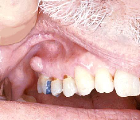
4 Parulis
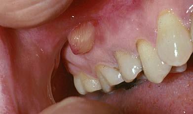
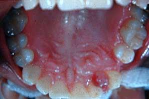
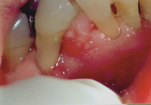
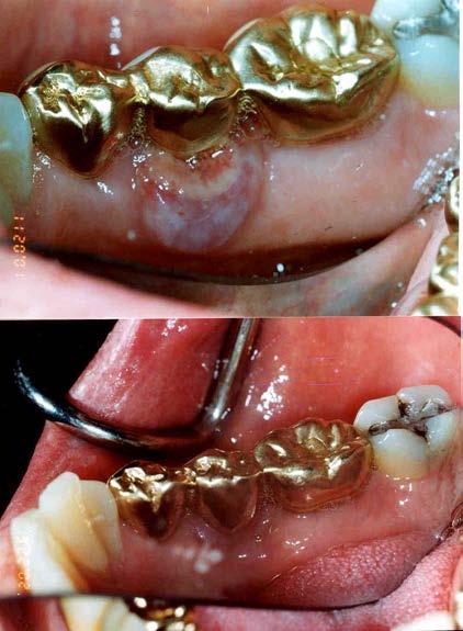
5 Periodontal Abscess Erythematous Deep Periodontal Pocket Alveolar Bone Loss Vital Teeth Pululence upon Probing Diabetes Mellitus
6 Periodontal Abscess
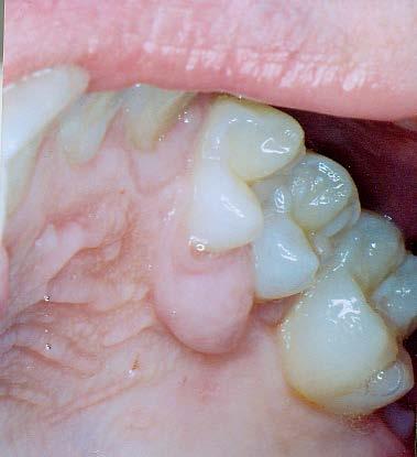
7 Torus Mandibularis Bilateral Lingual Premolar Region Adult Onset Lobulated, Bone Hard May become ulcerated May interfere with prostheses
8 Mandibular Tori
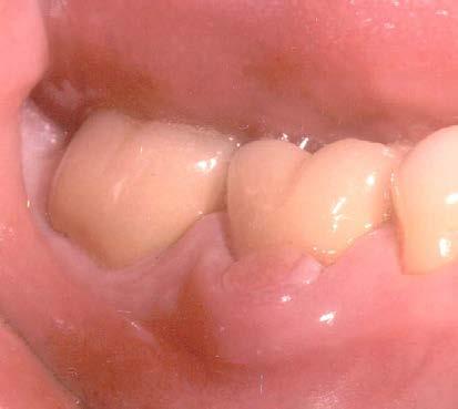
9 Exostoses Typically buccal posterior May occur anywhere on alveolus Adult onset Bone hard May interfere with prostheses
10 Exostoses
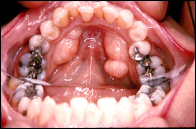
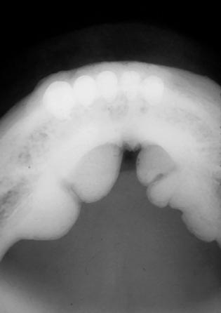
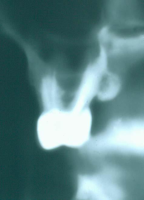
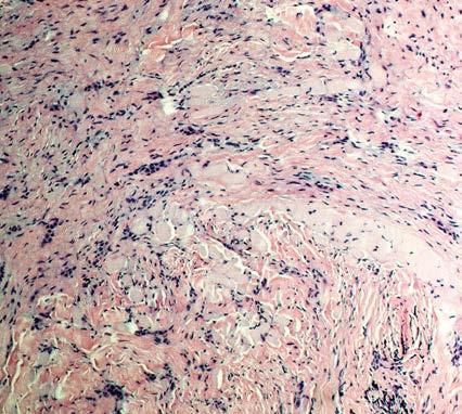
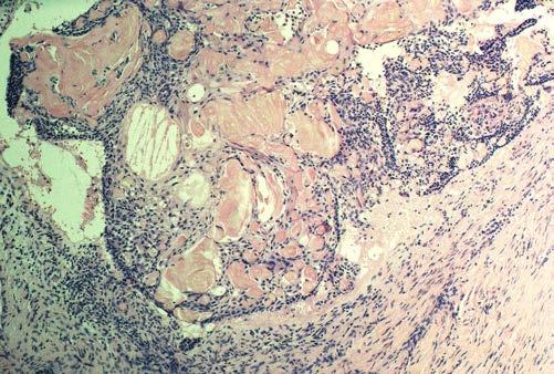
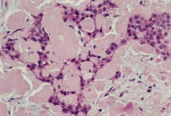
11 Congenital Epulis Bosselated tumor of the anterior alveolar ridge in newborns Maxilla > Mandibular ridge Microscopic: tumor is comprised of large granular cells with small nuclei. IHC staining is suggestive of smooth muscle origin Tx:Simple excision, excellent prognosis
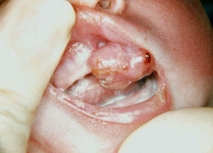
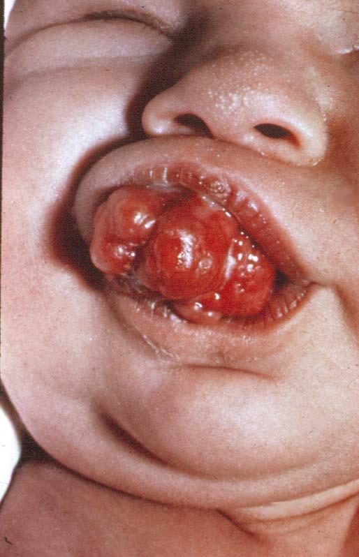
12 Congenital Epulis of the Newborn
13 Reactive Lesions of the Gingiva Pyogenic Granuloma Pregnancy Tumor Peripheral Fibroma Peripheral Ossifying Fibroma Peripheral Giant Cell Granuloma
14 Reactive Lesions of the Gingiva All tend to occur during 2 nd /3 rd decades Females>Males Interdental Papilla most commonly Irritant in Sulcus Foreign substance, physical irritant Calculus Vary in aggressiveness
15 Color Characteristics of Reactive Gingival Masses RED Pyogenic Granuloma PINK Peripheral Ossifying Fibroma Peripheral Fibroma BLUE/PURPLE Peripheral Giant Cell Granuloma
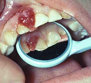
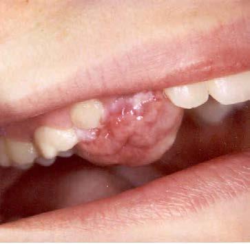
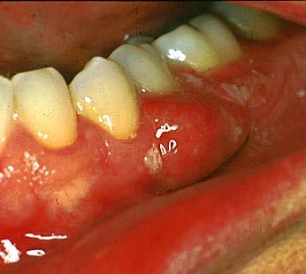
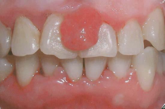
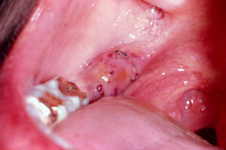
16 Pyogenic Granuloma Bright red, often ulcerated pseudomembrane Granulation tissue Pyogenic Bacteria are not etiologic Growth is superfical, rarely causing underlying bone loss Treatment: excision, thorough curettage

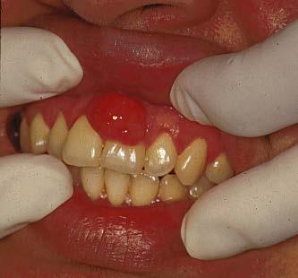
17 Pyogenic Granuloma

18

19 Pyogenic Granuloma
20 Peripheral Fibroma De Novo, or sclerosis of a Pyogenic Granuoma Interdental Papilla Coral Pink or White Noninvasive Histology Reactive Fibrous Hyperplasia Giant Cell Variant Fasciculated Spindle Cell variant

21 Peripheral Fibroma Giant Cell Fibroma

22 Retrocuspid Papilla A fibrous papule, mandibular cuspid lingual gingiva


23 Peripheral Fibroma Fibrous Hyperplasia Fasiculated Variant Giant Cell Variant
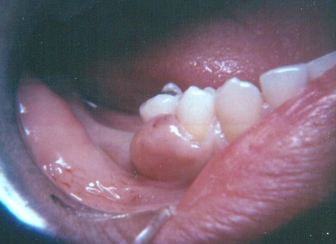
24 Peripheral Ossifying Fibroma Arises from PDL Not seen in edentulous regions Coral Pink or White Opacities may be seen on Xray Hypercellular Fibroblastic Osseous, cemental, dystrophic calcifications Excise down to PDL

25 Peripheral Ossifying Fibroma

26 Peripheral Ossifying Fibroma
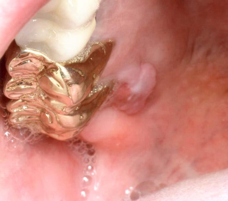
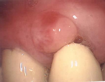
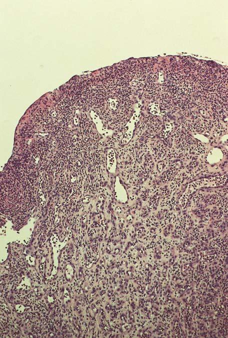
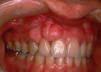
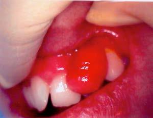
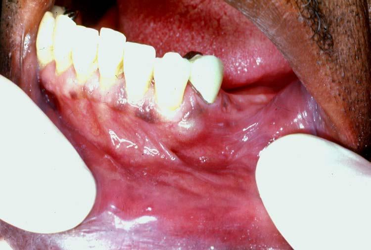
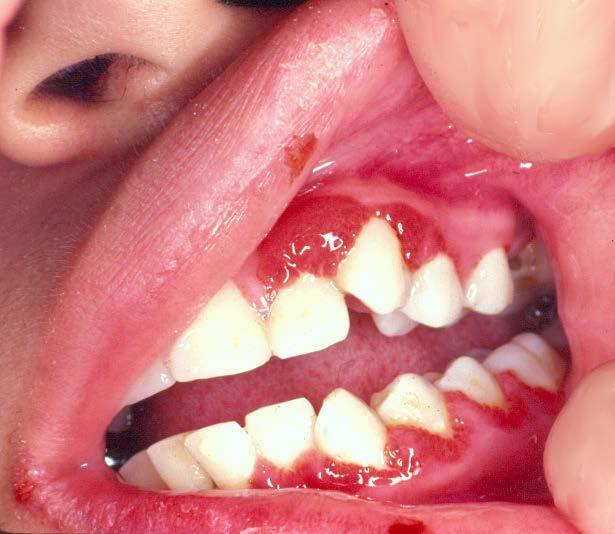
27 Peripheral Giant Cell Granuloma Arises from Periostium Dentulous or Edentulous Regions Bluish Purple Invasive, erodes underlying bone Hypercellular Fibrovascular Multinucleated Giant Cells Excise deeply, subperiosteal
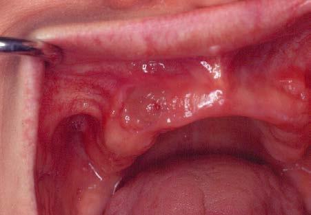
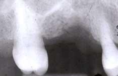
28 Peripheral Giant Cell Granuloma erosion Early recurrence
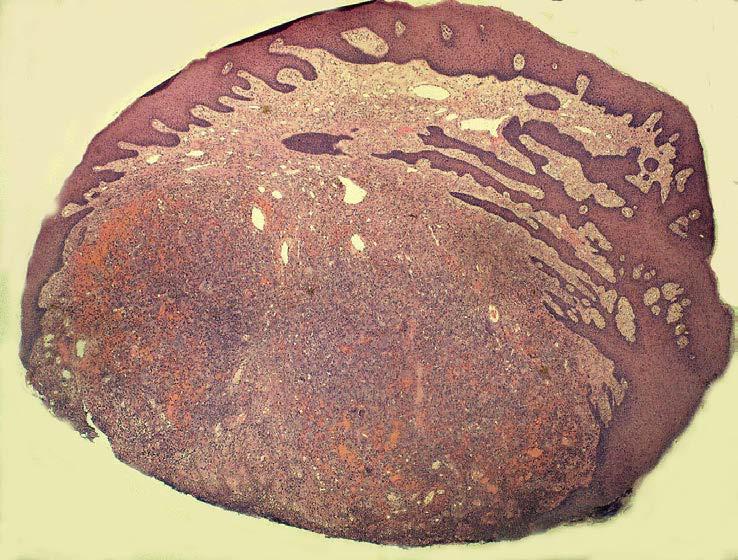
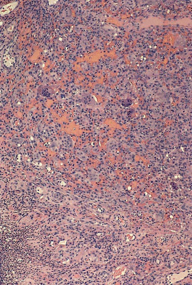
29 Peripheral Giant Cell Granuloma
30 Peripheral Odontogenic Cysts Cysts and Tumors Dental Lamina Cysts of the Newborn Adult Gingival Cyst Peripheral CEOC (Gorlin) Tumors Ameloblastoma Calcifying Epithelial Odontogenic Tumor Dentinogenic Ghost Cell Tumor Odontogenic fibroma
31 Gingival Cysts Dental Lamina Cyst Edentulous ridges of newborn Keratinizing diminutive cysts Spontaneous resolution Adult Gingival Cyst Buccal Attached Gingiva Peripheral Counterpart to Lateral Periodontal Cyst Nonkeratinizing (squamous/cuboidal)
32 Dental Lamina Cyst of the Newborn

33 Adult Gingival Cyst
34 Mesenchymal tumors neurofibroma hemangioma
35 Peripheral Odontogenic Tumors are Not Aggressive Gingiva and Tooth Bearing Alveolar Ridge May erode underlying bone Simple excision Histologic Types: Ameloblastoma Calcifying Epithelial Odontogenic Tumor Dentinogenic Ghost Cell Tumor Odontogenic fibroma


36 Peripheral Odontogenic Tumors Odontogenic Ghost Cell tumor Odontogenic Fibroma Peripheral Ameloblastoma

37 Peripheral Odontogenic Tumors Ameloblastoma Gorlin Cyst (OGCT) Pindborg (CEOT) Odontogenic Fibroma
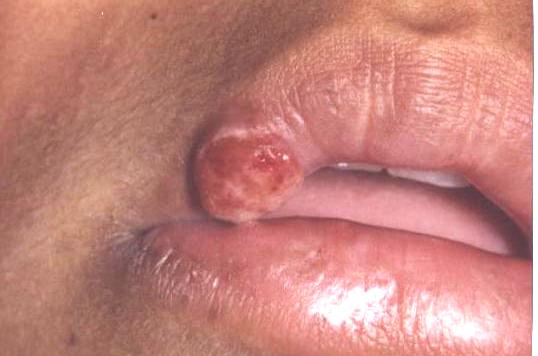
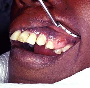
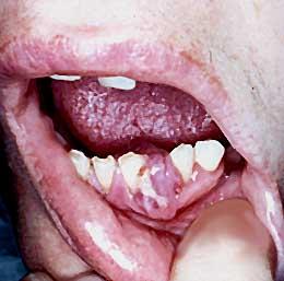
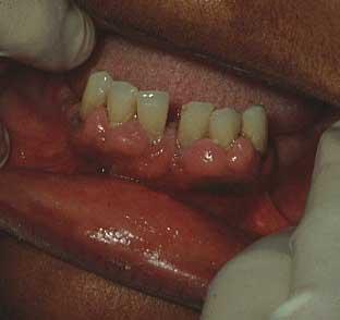
38 Squamous Cell Carcinoma Indurated and Ulcerated Mass Mandibular gingiva>maxillary Associated Risk Factors Periosteal and Osseous Invasion Regional and Distant Mets Resection and XRT


39 Squamous Cell Carcinoma

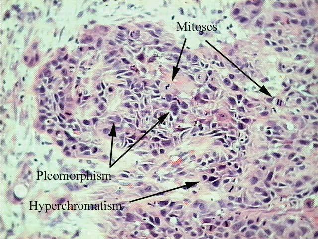
40 Squamous Cell Carcinoma
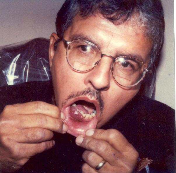
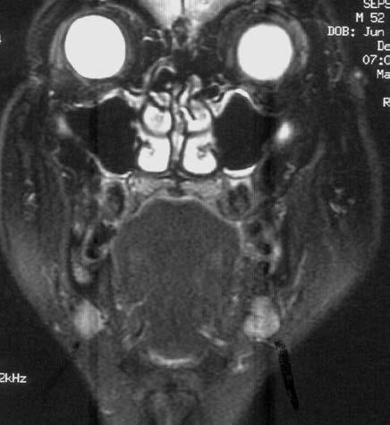
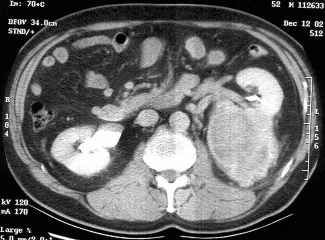
41 Metastatic Carcinoma Primary in kidney
42 Intraosseous Lesions Infections Cysts Odontogenic Tumors Nonodontogenic Tumors Most are diffuse, fusiforn enlargements of the alveolus Radiographs will disclose a central lesion

43 Diffuse Gingival Enlargments
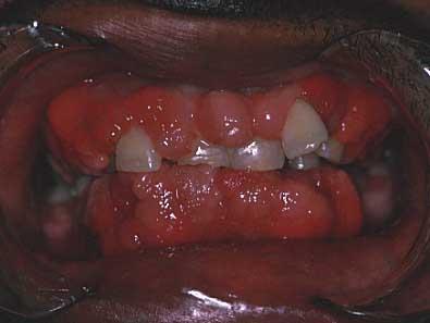
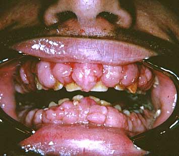
44 Diffuse Gingival Enlargement Hormonal Gingivitis Pregnancy, Puberty Drug Induced Hyperplasia Dilantin, Cyclosporin, Calcium Channel Blockers Fibromatosis Gingivae Plasma Cell Gingivitis Wegener s Granulomatosis Leukemia

45 Plasma Cell Gingivitis
46 Cyclosporin
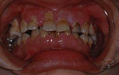
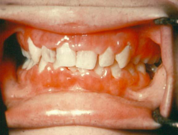
47 Dilantin
48 Pregnancy Gingivitis
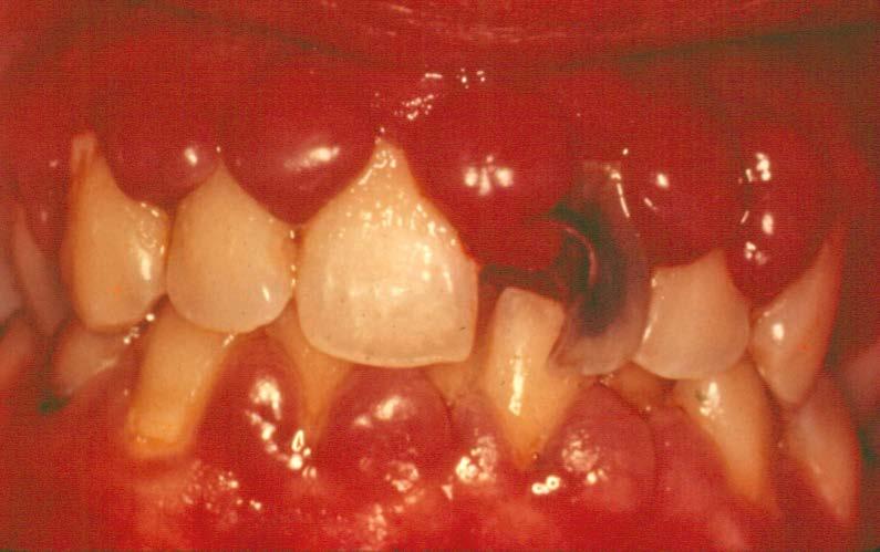
49 Leukemia

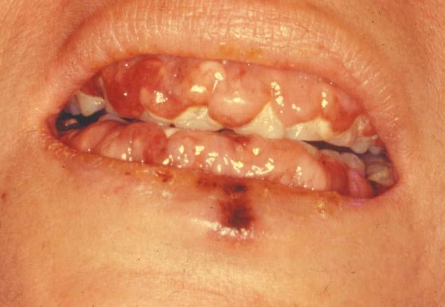
50 Wegener s Granulomatosis strawberry gums
Differential Diagnosis of Oral Lesions. An Interactive Lecture Using Audience Response Polling. John L. Alonge, MS, DDS
 Differential Diagnosis of Oral Lesions An Interactive Lecture Using Audience Response Polling John L. Alonge, MS, DDS Goals 1. Review the diagnostic process needed to formulate a differential diagnosis
Differential Diagnosis of Oral Masses. Palatal Lesions
 Differential Diagnosis of Oral Masses Palatal Lesions Palatal Masses Periapical Abscess Torus Palatinus Mucocele Lymphoid Hyperplasia Adenomatous Hyperplasia Benign Salivary Neoplasms Malignant Salivary
Differential Diagnosis of Radiolucent Lesions of the Jaws
 Differential Diagnosis of Radiolucent Lesions of the Jaws Multilocular Multilocular Radiolucencies Odontogenic Keratocyst Botryoid Odontogenic Cyst Glandular odontogenic Cyst Invasive Ameloblastoma Central
Case Report A Review and Report of Peripheral Giant Cell Granuloma in a 4-Year-Old Child
 Volume 2016, Article ID 7536304, 4 pages http://dx.doi.org/10.1155/2016/7536304 Case Report A Review and Report of Peripheral Giant Cell Granuloma in a 4-Year-Old Child Afsaneh Nekouei, Alireza Eshghi,
RADIOGRAPHIC INTERPRETATION Differential Diagnosis
 RADIOGRAPHIC INTERPRETATION Differential Diagnosis MODULE 1: The Introduction. Chief complaint Demographics Age Sex Race Historical findings Physical findings Clinical Radiographic Location Maxilla/mandible
Disclosure. Educational Objectives. Terminology. Odontogenic Cysts. Terminology
 Disclosure Lisa J. Koenig BChD, DDS, MS Professor & Program Director, Oral Medicine and Oral Radiology Marquette University School of Dentistry Consultant to Soredex for the Scanora 3D and 3Dx Author/Editor
Origin of Odontogenic Cysts & Tumors
 Origin of Odontogenic Cysts & Tumors Odontogenic Apparatus Origin of Odontogenic Cysts & Tumors Odontogenic Apparatus Remnants of dental lamina Reduced enamel epithelium Odontogenic rests Basal cell layer
Inter-radicular Radiolucencies
 Inter-radicular Radiolucencies Differential Diagnosis Laterally Displaced Radicular Cyst Accessory canals Root fracture Lateral Periodontal Cyst Botryoid variant Odontogenic Keratocyst Incisive Canal Cyst
Index. oralmaxsurgery.theclinics.com. Note: Page numbers of article titles are in boldface type.
 Index Note: Page numbers of article titles are in boldface type. A Adenomatoid odontogenic tumor, pediatric, 50 51 Ameloblastic carcinoma, pediatric, 17, 49 Ameloblastic fibro-odontoma, pediatric, 54 Ameloblastic
Case Report A Giant Cell Fibroma and Focal Fibrous Hyperplasia in a Young Child: A Case Report
 Hindawi Publishing Corporation Case Reports in Dentistry Volume 2012, Article ID 370242, 5 pages doi:10.1155/2012/370242 Case Report A Giant Cell Fibroma and Focal Fibrous Hyperplasia in a Young Child:
DISEASES OF THE JAWS I
 DISEASES OF THE JAWS I ODONTOGENIC AND PERIODONTAL INFECTIONS ODONTOGENIC INFECTIONS PERIAPICAL GRANULOMA PERIAPICAL ABSCESS APICAL PERIODONTAL CYST PHOENIX ABSCESS FISTULA, DRAINING SINUS SPACE INFECTIONS
Peripheral Ossifying Fibroma Mimicking Pyogenic Granuloma -A Case Report
 American Journal of Pharmacology and Pharmacotherapeutics Case Report Peripheral Ossifying Fibroma Mimicking Pyogenic Granuloma -A Case Report Sivakumarashankari, Gaurav*, Jayanthi K and Kamala R Dept.
IN THE NAME OF GOD. Dr.kheirandish DDS,MSC Oral and maxillofacial pathology
 IN THE NAME OF GOD Dr.kheirandish DDS,MSC Oral and maxillofacial pathology ODONTOGENIC CYSTS AND TUMORS Chapter 15 I. DENTIGEROUS CYST II. III. IV. ERUPTION CYST ODONTOGENIC KERATOCYST Orthokeratinized
Vascular. Extravasated blood. Melanocytic. Tattoo. Epidermolysis bullosa. Lichen planus. Pemphigoid Pemphigus Lupus. Candidosis. Surface Epithelial
 Oral Soft Tissue Pathology Epithelial Thickening (white) Combination Erythema migrans Epithelial atrophy (red) Surface Lesions Clinical Impression Enlargements Surface Debris Pigmented Vesicular Ulcerated
Problem diagnoses. Current issues in Anatomic pathology. Problem Diagnoses in Tumors of the Oral Cavity 5/29/2009
 Current issues in Anatomic pathology Problem Diagnoses in Tumors of the Oral Cavity Richard Jordan DDS PhD FRCPath Professor of Oral Pathology & Pathology Director, UCSF Oral Pathology Diagnostic Laboratory
Proceedings of the 36th World Small Animal Veterinary Congress WSAVA
 www.ivis.org Proceedings of the 36th World Small Animal Veterinary Congress WSAVA Oct. 14-17, 2011 Jeju, Korea Next Congress: http://www.ivis.org October 14(Fri) ~ 17(Mon) 2011 ICC Jeju, Korea 2011 WSAVA
Peripheral Ossifying Fibroma of the Maxilla: Case Report
 Case Report Article Peripheral Ossifying Fibroma of the Maxilla: Case Report José Carlos Martins Junior*, Frederico Santos Keim**, Mariana Schmidt Kreibich**. * Oral and maxillofacial surgeon at Hospital
Oral Tumors in Dogs Gingival Enlargement
 Oral Tumors in Dogs Is that lump you re seeing in your dog s mouth normal? Or is it something to be concerned about? The easiest way to know for sure is to have it evaluated by a veterinarian. When you
Focal Fibrous Hyperplasia: Two Case Reports
 American Journal of Oral and Maxillofacial Surgery Case Report http://ivyunion.org/index.php/maxillofacial Page 1 of 7 Focal Fibrous Hyperplasia: Two Case Reports Narjiss. Akerzoul 1*, Saliha. Chbicheb
Epithelial Sources. Rests of Serres Rests of Malassez Reduced Enamel Epithelium Surface Mucosa
 ODONTOGENIC CYSTS Epithelial Sources Rests of Serres Rests of Malassez Reduced Enamel Epithelium Surface Mucosa Epithelial Sources Surface Epithelium Rests of Serres Reduced Enamel Epithelium Rests of
EAR, NOSE, THROAT DISORDERS
 EAR, NOSE, THROAT DISORDERS A Case of Recurrent Pyogenic Granuloma of Gingiva ABSTRACT A case of pyogenic granuloma of gingiva is presented. Aetiology factors, clinical presentations and different treatment
Peripheral Odontogenic Fibroma : A Case Report
 Peripheral Odontogenic Fibroma : A Case Report Vinayak V Meharwade 1, Vidya V Meharwade 2 1- Assistant Professor, Department of Periodontology, Sinhgad Dental College and Hospital, Pune, Maharashtra, India.
Periodontal diagnoses (Armitage, 1999)
") Periodontal diagnoses (Armitage, 1999) Gingival diseases -Signs/symptoms confined to gingiva -Plaque may exacerbate/initiate severity of lesion -Colour: coral pink red/blue -Contour: scalloped + knife
Periodontal diagnoses (Armitage, 1999) Gingival diseases -Signs/symptoms confined to gingiva -Plaque may exacerbate/initiate severity of lesion -Colour: coral pink red/blue -Contour: scalloped + knife
Peripheral Odontogenic Fibroma: A rare case report
 Annals of Dental Research (2013) Vol 3 (1): 10-14 HATAM Publishers: All Rights Reserved Annals of Dental Research www.hgpub.com www.adres.yolasite.com Case Report Peripheral Odontogenic Fibroma: A rare
TANYA A. WRIGHT, DDS OBJECTIVES
 TANYA A. WRIGHT, DDS OBJECTIVES One will be able to recognize pathological entities One will be able to establish a reasonable differential diagnosis One will be able to identify various types of lesions
PERIPHERAL CEMENTO-OSSIFYING FIBROMA- A CASE REPORT
 PERIPHERAL CEMENTO-OSSIFYING FIBROMA- A CASE REPORT Dr.V.Gopinath, 1 Dr.V.Krishna, 2 Dr.Pramod kumar 3 1. Professor and Head, Department Of Periodontology, Department Of Periodontology, Rungta College
Dental Morphology and Vocabulary
 Dental Morphology and Vocabulary Palate Palate Palate 1 2 Hard Palate Rugae Hard Palate Palate Palate Soft Palate Palate Palate Soft Palate 4 Palate Hard Palate Soft Palate Maxillary Arch (Maxilla) (Uppers)
EAR, NOSE, THROAT DISORDERS
 EAR, NOSE, THROAT DISORDERS Clinico-Pathological Quiz ABSTRACT A case of a fleshy, granular, pedunculated growth from the oral cavity will be presented. A differential diagnosis of similar growths from
Jaws: Cysts and Odontogenic Neoplasms
 Topic 10: Jaw Cysts General Features of Jaw Cysts Sources of Epithelium in Cysts Radiographic Features of Jaw Cysts Microscopic Features of Jaw Cysts Treatment and Prognosis of Jaw Cysts Classification
COMBINED PERIODONTAL-ENDODONTIC LESION. By Dr. P.K. Agrawal Sr. Prof and Head Dept. Of Periodontia Govt. Dental College, Jaipur
 COMBINED PERIODONTAL-ENDODONTIC LESION By Dr. P.K. Agrawal Sr. Prof and Head Dept. Of Periodontia Govt. Dental College, Jaipur Differential diagnosis For differential diagnostic purposed the endo-perio
Case Report Peripheral ossifying fibroma in Pregnancy: A multifactorial consequence Suramya S 1, Gujjari SK 2, Sreeshyla HS 3
 Case Report Peripheral ossifying fibroma in Pregnancy: A multifactorial consequence Suramya S 1, Gujjari SK 2, Sreeshyla HS 3 1 Dr Suramya S MDS, Post graduate student Periodontology suramya.1986@gmail.com
Radiographic features of cysts and benign tumors of the jaws. Cyst. Effects on adjacent structures. Types. Odontogenic Cysts. Non-Odontogenic cysts
 Radiographic features of cysts and benign tumors of the jaws Cyst A Cyst is a benign pathologic cavity filled with fluid, lined by epithelium, and surrounded by a connective tissue wall Steven R. Singer,
IMAGING OF CYSTS OF THE JAWS July 2002 N. Serman
 IMAGING OF CYSTS OF THE JAWS July 2002 N. Serman This is an area where radiology plays an important role in assisting with the diagnosis, determining the size of the lesion and the relationship to adjacent
Peripheral Giant Cell Granuloma A Review and Case Report
 Case Report Peripheral Giant Cell Granuloma A Review and Case Report Dr. Somya Maheshwari *, Dr. Girish Bhutada, Dr Vaishakhi Baisane, Dr Devendra Palve Name & Address of Institution: Swargiya Dadasaheb
Case Report Soft Tissue Reconstruction with Free Gingival Graft Technique following Excision of a Fibroma
 Case Reports in Dentistry Volume 2015, Article ID 248363, 4 pages http://dx.doi.org/10.1155/2015/248363 Case Report Soft Tissue Reconstruction with Free Gingival Graft Technique following Excision of a
Clinical Management of an Unusual Case of Gingival Enlargement
 Clinical Management of an Unusual Case of Gingival Enlargement Abstract Aim: The purpose of this article is to report a case of conditioned gingival enlargement managed by nonsurgical periodontal therapy.
Pharmacologyonline 2: (2008) Young Researchers Gavasova and Budev EPULIS FISSURATUM CLINICAL APPEARANCE AND TREATMENT. Dr G.Gavasova, Dr I.
 Young Researchers Gavasova and Budev EPULIS FISSURATUM CLINICAL APPEARANCE AND TREATMENT. Dr G.Gavasova, Dr I.") EPULIS FISSURATUM CLINICAL APPEARANCE AND TREATMENT Dr G.Gavasova, Dr I.Budev Medical University, Faculty of Dental Medicine Oral Surgery Department Head: Prof. Dr D. Atanasov, Ph.D, MD. Summary Continuous
EPULIS FISSURATUM CLINICAL APPEARANCE AND TREATMENT Dr G.Gavasova, Dr I.Budev Medical University, Faculty of Dental Medicine Oral Surgery Department Head: Prof. Dr D. Atanasov, Ph.D, MD. Summary Continuous
Diagnosis. overt Examination. Definitive Examination. History. atient interview. Personal History. Clinical Examination.
 Diagnosis overt Examination History Definitive Examination atient interview Personal History Mental Attitude Medical History Dental History Clinical Examination Extra Oral Oral Radiographic Evaluation
Educational Cases EQA November T.J. Palmer Raigmore Hospital Inverness
 Educational Cases EQA November 2013 T.J. Palmer Raigmore Hospital Inverness Case 2 Clinical Details Dob 11 February 1951 PMH: 1964 Extraction of 45 aet 13 yr 1966 Cyst between 44 and 46 enucleated 1973
Name:XXX Sex: 男 Age:17 y/o Marital status: 未婚 Occupation: 學生
 General data Name:XXX Sex: 男 Age:17 y/o Marital status: 未婚 Occupation: 學生 Chief complaint A swelling mass over upper left arch for 3~4 months Present illness This 17 y/o male found an swelling over upper
Clinical Presentation and Management of Peripheral Giant Cell Granulomas in Children: 2 Cases Report
 10.1515/bjdm-2016-0007 BALKAN JOURNAL OF DENTAL MEDICINE ISSN 2335-0245 STOMATOLOGICAL SOCIETY Clinical Presentation and Management of Peripheral Giant Cell Granulomas in Children: 2 Cases Report SUMMARY
4Ps LUMPS AND BUMPS B.L.&T. BUMPS, LUMPS, AND TATTOOS. Most Common BUMP in the oral cavity Fibroma INTERDENTAL PAPILLAE LESIONS
 B.L.&T. BUMPS, LUMPS, AND TATTOOS LUMPS AND BUMPS DIFFERENTIAL DIAGNOSIS FOR LUMPS AND BUMPS Traumatic Fibroma Papilloma Epulis Fissuratum Inflammatory Papillary Hyperplasia Lesions of Attached Gingiva
PACIFIC JOURNAL OF MEDICAL SCIENCES ISSN:
 PACIFIC JOURNAL OF MEDICAL SCIENCES {Formerly: Medical Sciences Bulletin} ISSN: 2072 1625 Pac. J. Med. Sci. (PJMS) www.pacjmedsci.com. Email: pacjmedsci@gmail.com. A CASE REPORT DENTURE INDUCED INFLAMMATORY
PERIODONTAL CASE PRESENTATION - 1
 PERIODONTAL CASE PRESENTATION - 1 Overview A 32 year-old patient presented with generalized aggressive periodontitis. Treatment included non-surgical therapy with adjunctive antibiotics and surgical treatment.
Gingival and alveolar hyperplastic reactive lesions: clinicopathological study of 90 cases
 Braz J Oral Sci. JulySeptember 26 Vol. 5 Number 8 Pryscilla Giglio Peralles Ana Paula Borges Viana André Luiz da Rocha Azevedo 2 Fábio Ramôa Pires 3 DDS; formerly undergraduating students; School of Dentistry,
Pregnancy Tumor JOHSR CASE REPORT ABSTRACT CASE REPORT INTRODUCTION /jp-journals
 JOHSR CASE REPORT 10.5005/jp-journals-10042-1029 1 Shwetha Chikkaboraiah, 2 Rajiv Nidasale Puttaswamaiah, 3 Sushama R Galgali ABSTRACT Aim: The aim of this case report is to present a case of rapidly growing
Diseases of the breast (1 of 2)
") Diseases of the breast (1 of 2) Introduction A histology introduction Normal ducts and lobules of the breast are lined by two layers of cells a layer of luminal cells overlying a second layer of myoepithelial
Diseases of the breast (1 of 2) Introduction A histology introduction Normal ducts and lobules of the breast are lined by two layers of cells a layer of luminal cells overlying a second layer of myoepithelial
WHO Histological typing of odontogenic tumors, A. Epithelial Odontogenic Tumors
 Cheng-Chung Lin, Prof. in Oral Pathology College of Dental Medicine, KMU 2007 Classification: The following classification is based upon the inductive effect of one dental tissue upon another. In normal
A survey of biopsied oral lesions in pediatric dental patients
 PEDIATRIC DENTISTRY/Copyright 1986 by The American Academy of Pediatric Dentistry Volume 8 Number 2 A survey of biopsied oral lesions in pediatric dental patients Robert L. Skinner, MS W.D. Davenport,
Spectrum of clinical presentations
 Spectrum of clinical presentations Case History A 7-day-old male patient born full-term via uncomplicated vaginal delivery was seen for multiple erythematous red-brown purpuric lesions that were present
CENTRAL GIANT CELL GRANULOMA PRESENTING AS UNILOCULAR RADIOLUCENCY IN POSTERIOR MANDIBLE A CASE REPORT
 IJCRR Section: Healthcare Sci. Journal Impact Factor 4.016 Case Report CENTRAL GIANT CELL GRANULOMA PRESENTING AS UNILOCULAR RADIOLUCENCY IN POSTERIOR MANDIBLE A CASE REPORT S. Aruleena Shaminey 1, G.
Course Description 343 DDS- Clinical Oral and Maxillofacial Radiology II ( )
") King Saud University College of Dentistry Dept. of Oral Medicine & Diagnostic Sciences Division of Oral & Maxillofacial Radiology Course Description 343 DDS- Clinical Oral and Maxillofacial Radiology II
King Saud University College of Dentistry Dept. of Oral Medicine & Diagnostic Sciences Division of Oral & Maxillofacial Radiology Course Description 343 DDS- Clinical Oral and Maxillofacial Radiology II
Index. Note: Page numbers of article titles are in boldface type.
 Note: Page numbers of article titles are in boldface type. A Actinomycosis, 200 201 Adenoid cystic carcinoma, 148 150 Adenomatoid odontogenic tumors, 134, 135 Ameloblastic fibro-odontoma, 134 Ameloblastoma,
IN THE NAME OF GOD Dr. Kheirandish Oral and maxillofacial pathology
 IN THE NAME OF GOD Dr. Kheirandish Oral and maxillofacial pathology ORAL FOCAL MUCINOSIS Uncommon Tumorlike Cutaneous myxoid cyst Overproduction of hyaluronic acid by firoblasts Young adults Female Gingiva
That. Name QUIZ. 60 SEPTEMBER 2017 // dentaltown.com
 QUIZ Name That General dentists are first in the line of practitioners that patients see for an oral lesion evaluation; therefore, a sound understanding of oral mucosal diseases and their clinical presentation
Oral pathology. General structure of teeth
 Oral pathology Contents: 1. General structure of teeth 2. Common structural inflammatory lesions 3. Tumor like lesions 4. Infections 5. Oral manifestations of systemic disease 6. Tumors of oral cavity
PACIFIC JOURNAL OF MEDICAL SCIENCES ISSN:
 PACIFIC JOURNAL OF MEDICAL SCIENCES {Formerly: Medical Sciences Bulletin} ISSN: 2072 1625 Pac. J. Med. Sci. (PJMS) www.pacjmedsci.com. Email: pacjmedsci@gmail.com. ADENOMATOID ODONTOGENIC TUMOR WITH RARE
Fundamental & Preventive Curvatures of Teeth and Tooth Development. Lecture Three Chapter 15 Continued; Chapter 6 (parts) Dr. Margaret L.
 Dr. Margaret L.") Fundamental & Preventive Curvatures of Teeth and Tooth Development Lecture Three Chapter 15 Continued; Chapter 6 (parts) Dr. Margaret L. Dennis Proximal contact areas Contact areas are on the mesial and
Fundamental & Preventive Curvatures of Teeth and Tooth Development Lecture Three Chapter 15 Continued; Chapter 6 (parts) Dr. Margaret L. Dennis Proximal contact areas Contact areas are on the mesial and
Oral Health & HIV. Professor Sudeshni Naidoo Department of Community Dentistry University of the Western Cape
 Oral Health & HIV Professor Sudeshni Naidoo Department of Community Dentistry University of the Western Cape Importance & relevance of Oral HIV Lesions >70% of HIV+ve patients present with oral manifestations
Malignant Lesions Steven R. Singer, DDS
 Definitions Malignant Lesions Steven R. Singer, DDS srs2@columbia.edu 212.305.5674 Malignancies are uncontrolled growths of tissue Primary tumors represent de novo tumors in their initial site Metastatic
A Case Report of Odontogenic Keratocyst in Anterior Mandibule Position
 A Case Report of Odontogenic Keratocyst in Anterior Mandibule Position Malihe Moeini 1, Seyed Ehsan Anvar 2, Rasool Barzegari Bafghi 3* 1.Resident of Oral and Maxillofacial Radiology, Faculty of Dentistry,
Gingival Aggressiveness during Pregnancy
 www.jmscr.igmpublication.org ISSN (e)-2347-176x Gingival Aggressiveness during Pregnancy Authors Dr Arvind Garg 1, Dr Asmita Arora 2, Dr Shweta Aggarwal 3, Dr Arun Garg 4 Dr Viniti Goel 5 1 Professor &
PERIPHERAL'GIANT'CELL' GRANULOMA:'A'CASE'REPORT)
") KOTA,Kasim* KODANDA,Ram* JAISEKHARAN,VP* PERIPHERALGIANTCELL GRANULOMA:ACASEREPORT ABSTRACT Peripheral giant cell granuloma(pgcg is a non neoplastic reactive lesionofthegingiva,originatingfromtheperiosteumorperiodontal
KOTA,Kasim* KODANDA,Ram* JAISEKHARAN,VP* PERIPHERALGIANTCELL GRANULOMA:ACASEREPORT ABSTRACT Peripheral giant cell granuloma(pgcg is a non neoplastic reactive lesionofthegingiva,originatingfromtheperiosteumorperiodontal
NC Medicaid Dental Reimbursement Rates General Dentist, Oral Surgeon, Pediatric Dentist, Periodontist, & Orthodontist Effective Date: January 1, 2017
 NC Dental Reimbursement s Refer to the NC and Health Choice Clinical Coverage Policies on the DMA website. D0120 Periodic oral evaluation 24.51 D0140 Limited oral evaluation - problem focused 34.94 D0145
Overview of Periodontics for the General Practicioner
 Overview of Periodontics for the General Practicioner Nashville Area Dental Continuing Education August 27, 2008 Phillip D. Woods, DDS, MPH Commander, USPHS BOP National Periodontal Consultant Diplomate,
AMELOBLASTIC FIBROMA: A RARE CASE REPORT
 Case Report International Journal of Dental and Health Sciences Volume 04, Issue 03 AMELOBLASTIC FIBROMA: A RARE CASE REPORT Namratha Patil 1 1.Sr lecturer, dept of oral medicine and radiology, KAHES VK
CHRONIC PERIOSTITIS IN THE MANDIBLE UNDERNEATH ARTIFICIAL DENTURES
 CHRONIC PERIOSTITIS IN THE MANDIBLE UNDERNEATH ARTIFICIAL DENTURES P. H. D. LEWARS, M.B.B.S., B.D.S., F.D.S. Exeter INTRODUCTION FROM time to time patients wearing artificial lower dentures have presented
An Expansile Large Odontogenic Keratocyst Maxilla: A Case Report.
 RESEARCH AND REVIEWS: JOURNAL OF DENTAL SCIENCES An Expansile Large Odontogenic Keratocyst Maxilla: A Case Report. Nasib Chand Khabra 1, Ish Pandhi 1 *, Kiran DN 2, Sunil Alipuria 1, Bhawna Gulati 1, and
MANSOURA UNIVERSITY FACULTY OF DENTISTRY ORAL PATHOLOGY DEPT
 MANSOURA UNIVERSITY FACULTY OF DENTISTRY ORAL PATHOLOGY DEPT THIRD YEAR Course Director: Dr. Nadia M. Lotfy Professor of Oral Pathology Dr. Manal Mohamed Zyada Associate Professor of Oral Pathology Oral
Surgical Diode Laser Excision for Peripheral Cemento-Ossifying Fibroma: A Case Report and Literature Review
 Surgical Diode Laser Excision for Peripheral Cemento-Ossifying Fibroma: A Case Report and Literature Review Tanveer Alam a, Ali Azhar Dawasaz b, Naresh Thukral c, Daya Jangam d a Postgraduate Student,
Maxilla and mandible benign lesions: Radiologic Findings and Differential Diagnosis in CT
 Maxilla and mandible benign lesions: Radiologic Findings and Differential Diagnosis in CT Poster No.: C-0964 Congress: ECR 2012 Type: Scientific Exhibit Authors: N. Lopez 1, E. Marcos Naranjo 2, M. D.
Pre-reading - radiolucencies
 Pre-reading - radiolucencies Multiple radiolucencies o Suggests a systemic cause o Most likely: cherubism or KCOT s of nevoid basal cell carcinoma syndrome o Sometimes: florid osseous dysplasia (if limited
Peripheral ossifying fibroma: a clinical and immunohistochemical study of four cases
 95 Journal of Oral Science, Vol. 52, No. 1, 95-99, 2010 Original Peripheral ossifying fibroma: a clinical and immunohistochemical study of four cases José A. García de Marcos 1), María J. García de Marcos
Course Description 343 DDS- Clinical Oral and Maxillofacial Radiology II ( )
") King Saud University College of Dentistry Dept. of Oral Medicine & Diagnostic Sciences Division of Oral & Maxillofacial Radiology Course Description 343 DDS- Clinical Oral and Maxillofacial Radiology II
King Saud University College of Dentistry Dept. of Oral Medicine & Diagnostic Sciences Division of Oral & Maxillofacial Radiology Course Description 343 DDS- Clinical Oral and Maxillofacial Radiology II
Annals of RSCB Vol. XV, Issue 1
 CLINICAL MORPHOLOGICAL STUDY ON BENIGN TUMORAL LESIONS OF EPULIS TYPE Ana Maria Filioreanu 1, Eugenia Popescu 2, Monica Neamţu 1, Constantin Cotrutz, Carmen Elena Cotruz 1 1DEPARTMENT OF CELL AND MOLECULAR
RECORD or PRINT THE CONFIRMATION ID This unique ID is displayed upon successful submission of your answer form.
 2014 course two self-study course The Ohio State University College of Dentistry is a recognized provider for ADA, CERP, and AGD Fellowship, Mastership and Maintenance credit. ADA CERP is a service of
GINGIVAL SURGICAL TECHNIQUES
 Gingival Surgical Procedures GINGIVAL SURGICAL TECHNIQUES! limited to the gingival and not involving underlying osseous structures! Gingival Curettage! Gingivectomy! Gingivoplasty! Gingival Flap Gingival
Contents. Chapter 3: Principles of Surgery
 Contents Chapter 1: Medical History 1.1 Congestive Heart Failure.............. 1 1.2 Angina Pectoris..................... 1 1.3 Myocardial Infarction............................... 2 1.4 Rheumatic Heart
Periapical central giant cell granuloma misdiagnosed as odontogenic cyst
 doi: 10.1111/j.1365-2591.2006.01107.x CLINICAL ARTICLE Periapical central giant cell granuloma misdiagnosed as odontogenic cyst T. Lombardi 1, M. Bischof 1,2, R. Nedir 1,2, D. Vergain 1, C. Galgano 3,
Periodontal Disease. Radiology of Periodontal Disease. Periodontal Disease. The Role of Radiology in Assessment of Periodontal Disease
 Radiology of Periodontal Disease Steven R. Singer, DDS srs2@columbia.edu 212.305.5674 Periodontal Disease! Includes several disorders of the periodontium! Gingivitis! Marginal Periodontitis! Localized
Gingivectomy, excision gingival, each quadrant Gingivoplasty, each quadrant
 Dental in Nature Oral Surgery Effective CDT D3410 surgery - anterior D3421 surgery bicuspid (first root) D3425 surgery molar (first root) D3426 D3427 surgery (each additional root) Periradicular surgery
Objectives. 1. Recognizing benign skin lesions. 2.Know which patients will likely need surgical intervention.
 The Joy of Pediatric Skin Dr. Claire Sanger University of Kentucky Plastic & Reconstructive Surgery Objectives 1. Recognizing benign skin lesions 2.Know which patients will likely need surgical intervention.
A case report of cemento-ossifying fibroma presenting as a mass of. the ethmoid sinus
 Received: 27.3.2010 Accepted: 1.8.2010 Case Report A case report of cemento-ossifying fibroma presenting as a mass of the ethmoid sinus Ali Hekmatnia a, Amirhossein Ghazavi* b, Masih Saboori c, Parvin
Course Description 343 DDS- Clinical Oral and Maxillofacial Radiology II ( )
") King Saud University College of Dentistry Dept. of Oral Medicine & Diagnostic Sciences Division of Oral & Maxillofacial Radiology Course Description 343 DDS- Clinical Oral and Maxillofacial Radiology II
King Saud University College of Dentistry Dept. of Oral Medicine & Diagnostic Sciences Division of Oral & Maxillofacial Radiology Course Description 343 DDS- Clinical Oral and Maxillofacial Radiology II
MUCOCUTANEOUS LESIONS Normal structures in epithelium cell adhesion to each other and to underlying connective tissue:
 ORAL DERMATOSES AND MUCOSAL/GINGIVAL LESIONS MUCOCUTANEOUS LESIONS Normal structures in epithelium cell adhesion to each other and to underlying connective tissue: Diagram taken from: Oral and Maxillofacial
CHAPTER 8 SECTION 1.4 ORAL SURGERY TRICARE/CHAMPUS POLICY MANUAL M DEC 1998 SPECIAL BENEFIT INFORMATION
 TRICARE/CHAMPUS POLICY MANUAL 6010.47-M DEC 1998 SPECIAL BENEFIT INFORMATION CHAPTER 8 SECTION 1.4 Issue Date: October 8, 1986 Authority: 32 CFR 199.4(e)(10) I. DESCRIPTION There are certain oral surgical
Large Dentigerous Cyst
 Volume 16.2.1 Feb 2016 This Lecture Series qualifies for 0.5 Informal CPD Learning Hours Large Dentigerous Cyst By Dr Hassem Geha A 55 year-old male presented with a painless swelling in the right mandible.
The Oral Cavity. Image source:
 The Oral Cavity Anatomy Image source: http://anatomyforlayla.blogspot.co.za/2007/04/blog-post.html The major structures of the oral cavity are the lips, the teeth, the alveolar ridges (bony areas that
MALIGNANT TUMOURS OF THE JAWS
 MALIGNANT TUMOURS OF THE JAWS MALIGNANT TUMOURS OF THE JAWS Squamous cell carcinoma Osteogenic sarcoma Chondrosarcoma Fibrosarcoma Malignant lymphomas (incl. Burkitt s) Multiple myeloma Ameloblastoma Secondary
Tumors or Masses in the Mouth (Oral Masses) Basics
 Basics") Tumors or Masses in the Mouth (Oral Masses) Basics OVERVIEW Oral refers to the mouth; oral masses are tumors or growths located in the mouth Oral masses may be benign or malignant (that is, cancer); 4
Tumors or Masses in the Mouth (Oral Masses) Basics OVERVIEW Oral refers to the mouth; oral masses are tumors or growths located in the mouth Oral masses may be benign or malignant (that is, cancer); 4
Fibroma - A misnomer : Case Series
 Case Report Abstract Fibroma - A misnomer : Case Series NUJHS Vol. 5, No.4, December 2015, ISSN 2249-7110 Riya Verghese, Amitha Ramesh, Rahul Bhandary, Biju Thomas & Nishita L. Philip 1 2,3 4 5 Post Graduate,
Odontomes and Odontogenic tumours
 Odontomes and Odontogenic tumours Odontomes Developmental hamartoma Hamartoma: normal tissue in abnormal location Any cells to be neoplastic it must be able to replicate, which is not seen in hamartoma
Pictorial Essay. CT of Calcifying Jaw Bone Diseases
 Pictorial Essay CT of Calcifying Jaw one Diseases Koichi Yonetsu 1 and Takashi Nakamura Downloaded from www.ajronline.org by 46.3.204.207 on 01/08/18 from IP address 46.3.204.207. Copyright RRS. For personal
Common/Important Radiolucencies. B. Most Common Location Apex of permanent first molar, rare in primary teeth.
 Cincinnati Dental Association Breakfast at Tiffany s: The Jewels and Gems of Oral Pathology November 17, 2010 John A. Svirsky, DDS, MEd Virginia Commonwealth University 804-828-0547 FAX: 804-828-6234 EMAIL:
The clinical appearance and diagnosis of odontogenic cysts. SE Arc-Állcsont-Szájsebészeti és Fogászati Klinika BUDAPEST
 The clinical appearance and diagnosis of odontogenic cysts SE Arc-Állcsont-Szájsebészeti és Fogászati Klinika BUDAPEST DEFINITION A cyst is a sac with walls of connective tissue, lined by epithelium, containing
Let s start from the basics for a little review. The Mouth Is Like a Black Hole. But he s friendly at home. Always Wear Gloves!
 The Mouth Is Like a Black Hole Seek and ye shall find: Recognition of Oral Lesions in the Exam Room Lisa Fink, DVM, DAVDC Dentistry & Oral Surgery Service October 4, 2015 But he s friendly at home Part
Calcifying Odontogenic Cyst with Complex Odontoma: Histological and Immunohistochemical Features
 Case Report Calcifying Odontogenic Cyst with Complex Odontoma: Histological and Immunohistochemical Features Nooshin Mohtasham 1, Amin Rahpeyma 2, Saeedeh Khajeh Ahmadi 3, Mohsen Merati 4 1 Oral and Maxillofacial
Chapter 2 Variants of Normal and Common Benign Conditions
 Chapter 2 Variants of Normal and Common Benign Conditions Summary Fundamental to diagnosing oral pathologic conditions is the ability to recognize the spectrum of clinical findings that represents variation
Six cases report of differential diagnosis of periapical diseases
 Int J Oral Sci (2011) 3: 153-159. www.ijos.org.cn CLINICAL ARTICLE Six cases report of differential diagnosis of periapical diseases Wen-wei Xia, Ya-qin Zhu, Xiao-yi Wang* Department of Endodontics and