EXTENSIVE PERIAPICAL CYST IN THE MAXILLARY SINUS A CASE REPORT
|
|
|
- Jordan Green
- 5 years ago
- Views:
Transcription
1 May, 2012 INTERNATIONAL DENTAL JOURNAL OF STUDENT S RESEARCH: CASE REPORT Article Code: IDJSR 0002 EXTENSIVE PERIAPICAL CYST IN THE MAXILLARY SINUS A CASE REPORT Authors Bouguezzi Adel, DDS, Resident 1 Souid Kawthar, DDS, Assistant Professor 2 Boudegga Souha, DDS, Professor 3 Boughzala Abdellatif, DDS, Professor 4 E mail ID dr adel@live.fr Address of the Institution Department of Oral pathology, Oral surgery, University Hospital Farhat Hached, Sousse, TUNISIA
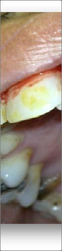
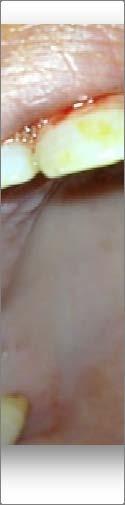
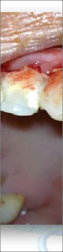
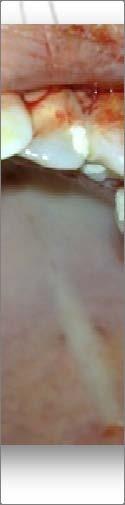
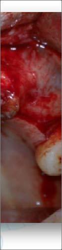
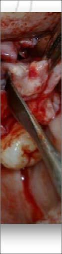
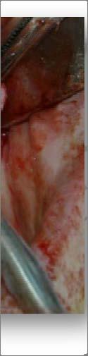
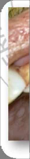
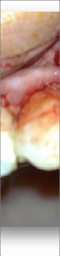
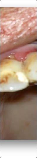
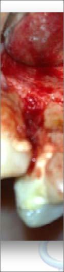
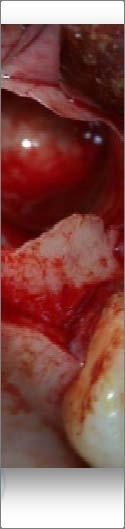
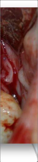
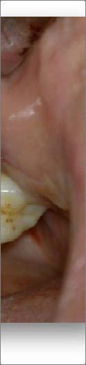
2 May, 2012 INTERNATIONAL DENTAL JOURNAL OF STUDENT S RESEARCH: CASE REPORT Introduction air flow was decreased on the left side. There were no other abnormalities identified on clinical examination. The radicular cyst is the most frequent cyst found in the jaw (between 38 % and 68 % of all the jaw cysts). The prevalence of periapical cysts varies between 8.7% and 37.7% of chronic inflammatory periapical lesions. (1) It is not uncommon, to find periapical lesions to extend to the surrounding tissues and not limit themselves to the apex of the involved tooth. In the literature most cases of unusually large periapical lesions of odontogenic origin are found in the maxilla where the bone is spongy. (1) Because of the bone consistency, it is easier for the lesion to occupy bony space and expand. Lesions have been found to occupy the entire sinus and even the floor of the nasal cavity. (2) Case report A 38 year old male with no past medical history was referred to the department of Oral pathology, Oral surgery of University Hospital Farhat Hached Sousse by a general dental practitioner for management of a painless swelling for six days in the left maxillary region with nasal obstruction. The patient had been treated with antibiotics as a case of chronic tooth abscess, but his complaints did not improve. Clinically, there was a well defined swelling with a smooth surface; the swelling was compressible on palpation which indicated a loss of integrity of buccal bone (Figure 1). On intraoral examination, the second premolar was decayed and nonvital. Also, external examination revealed that eye movements and visual acuities were normal. And according to the patient nasal Radiographs (panoramic & CT Scan) revealed an osteolytic radiolucency well delineated around the roots of the left second premolar, the large lesion measured 5,5cm vertically and 3,5cm horizontally, and expanded the buccal cortical and displaced the Schneiderian membrane (Figures 2,3,4). From history, clinical and radiographic examinations, a provisional diagnosis of radicular cyst was made. It was decided to surgically enucleate the lesion under local anesthesia. Reflection of a mucoperiosteal flap, followed by removal of bone and exposure of the lesion membrane was carried out. Aspiration of the contents of the cystic lesion was a valuable diagnostic aid; it revealed a yellow semi viscous fluid. The lesional wall was hypertrophic and adhered partly to the mucosa of the base of the maxillary sinus; therefore, the lesional mass was totally curetted to a maximum extent. Enucleation of the cystic lesion and extraction of the 25 with care of the wound and suturing were done (Figures 5 to 9). Enucleating biopsy of the periapical lesion was diagnosed histologically as radicular cyst with a layer of nonkeratinised stratified squamous epithelium (Figure 10).The healing was uneventful without swelling or other complications. Sutures were removed 1 week postoperatively, at the 8 months follow up, no complications or recurrence were noticed with complete bone healing and repneumatization of the antrum (Figure 11).


3 May, 2012 INTERNATIONAL DENTAL JOURNAL OF STUDENT SS RESEARCH: CASE REPORT Figure 1: Photograph of the patient s oral cavity showing a decayed second premolarr and a swelling at the premolar vestibular area Figure 2: Panoramic radiograph of the patient revealed an unusually large lesion invading the left maxillary sinus.


4 May, 2012 INTERNATIONAL DENTAL JOURNAL OF STUDENT SS RESEARCH: CASE REPORT Figure 3: Axial CT image shows process destroying lateral wall of the left maxillary sinus Figure 4: Computed tomography scan showing a cystic lesion occupying most of the left maxillary sinus
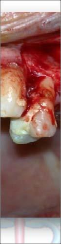
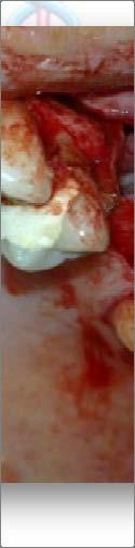
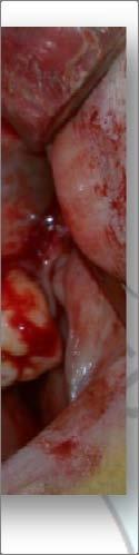
5 May, 2012 INTERNATIONAL DENTAL JOURNAL OF STUDENT SS RESEARCH: CASE REPORT Figure 5: Trans surgical view, note the marked lateral expansion of the lesion leading to loss of the lateral wall of the maxilla Figure 6: Enucleation of the cyst
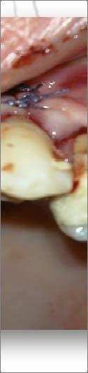
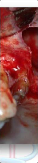
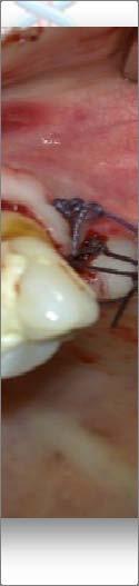
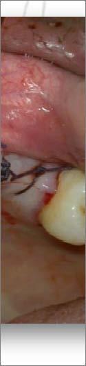
6 May, 2012 INTERNATIONAL DENTAL JOURNAL OF STUDENT SS RESEARCH: CASE REPORT Figure 7: A view of the maxilla removal of the cyst Figure 8 : Hermetic suture


7 May, 2012 INTERNATIONAL DENTAL JOURNAL OF STUDENT SS RESEARCH: CASE REPORT Figure 9: Enucleated cyst with the removed 25 Figure 10: A histological section of the lesion showing at the upper left the cholesterol crystal clefts (red arrows), the proliferative epithelium (black arrows), and the inflammation area (blue arrows) (Hematoxylin and Eosin)

8 May, 2012 INTERNATIONAL DENTAL JOURNAL OF STUDENT S RESEARCH: CASE REPORT Figure 11: A panoramic radiograph 8 months post surgery suggesting satisfactory bone radiopacity and neoformation Discussion The etiopathogenesis of cysts is particularly controversial; the formation has been explained by diverse theories, such as epithelial colonization, epithelial cavitations or the formation of microabscesses. The first is based on the formation of an epithelized fistulous tract up to the granuloma from a periapical abscess fistulized to the oral cavity; when the communication is closed, the epithelial cells have already fully colonized the abscess, epithelizing it and giving rise to a radicular cyst. In the theory of epithelial cavitation, accumulations of epithelial cells are created; those furthest from the connective tissue which feeds them are left without vascularization and undergo degeneration and necrosis, thus forming the central area of the cyst. The theory of microabscess formation is based on the degeneration of the connective tissue leading to the development of the cyst; the formation of a microabscess in the nucleus of the granuloma, with the presence of stimulated epithelial cells, would lead to their growth in an attempt to line the created cavity. (3) The pathogenesis of cysts has been described in three phases. During the first phase, the epithelial cell rests of Malassez begin to proliferate as a direct result of the inflammation and influenced by bacterial antigens, the epidermal growth factors, metabolic and cellular mediators. In the second, a cavity is formed, lined by epithelium (according to the above described theories), and in the third phase the cyst grows, probably by osmosis. (1) Radiographically, the radicular cyst is a unilocular radiolucent lesion with wellcircumscribed sclerotic borders that are often radiopaque. The lesion is associated with the apex of the tooth and a diameter of at least 1 cm is postulated to be
9 May, 2012 INTERNATIONAL DENTAL JOURNAL OF STUDENT S RESEARCH: CASE REPORT necessary to differentiate it from that of a normal follicular space. (4) Natkin and al.related radiographic lesion size to histological findings and concluded that with a radiographic lesion size of 200 mm2 or larger, the incidence of cysts was almost 100%. (5) Other odontogenic cysts like dentigerous cysts, odontogenic keratocysts, and odontogenic tumors such as ameloblastoma, Pindborg tumor, odontogenic fibroma, and cementomas may share the same radiologic features as radicular cysts. Microscopic evaluation is necessary most of the time to define the type of lesion.(6) Our specimen was diagnosed histologically as radicular cyst with a layer of nonkeratinised stratified squamous epithelium, in fact all radicular cysts are lined partially or completely by nonkeratinized stratified squamous epithelium. Keratinization is seen in approximately 2% of cases, and when present orthokeratinization is more common than parakeratinization. (7) When cysts are especially large, with maxillary sinus involvement as in our patient, the panoramic radiograph is often not of great aid. CT scans provide superior bony detail, allowing for the visualization of the size and extent of the lesion with determination of orbital or nasal invasion or involvement. Again, with larger lesions, it also aids in planning of a surgical approach. Mucoceles, retention cysts, and pseudocysts are also included in the differential diagnosis when a maxillary sinus cyst is visualized involving maxillary expansion; this is in addition to the array of radiolucent lesions mentioned above that can also be visualized on CT. (8) The treatment of pariapical cysts are still under discussion and many professionals opt for a conservative treatment by means of endodontic. However, in large lesions the endodontic treatment alone is not efficient and it should be associated to a decompression or a marsupialization (indicated when cyst is in close proximity to vital structures and where there is significant risk of injury with enucleation) or even to enucleation and extraction of the associated tooth. A large maxillary cyst may involve the whole sinus and can transmit pressure to the walls of the sinus; consequently, ophthalmologic and nasal symptoms may develop. With extensive lesions, it is important to carefully plan the surgical approach. The choice of treatment may be determined by some factors such as the extension of the lesion, relation with noble structures, evolution, origin, clinical characteristic of the lesion, cooperation and systemic condition of the patient. (9) Some authors suggested a nasal approach; however, in keeping with the law of gravity, it is reasonable to surmise that the content from maxillary cysts can be drained much more easily into the oral cavity. An oral vestibular approach is therefore more preferable than a nasal approach. (10 11) Conclusion It must be kept in mind that chronic periapical lesions (granuloma, cyst, and scar tissue) are usually asymptomatic and do not create soft tissue alterations. However, they can deteriorate, producing pain and fistulization. Dentists should be very careful on clinical examination and should not omit
10 May, 2012 INTERNATIONAL DENTAL JOURNAL OF STUDENT S RESEARCH: CASE REPORT any details. Before beginning any treatment a careful and complete clinical and radiographic examination is needed to supply all the required information. In extensive cases, radiography alone may not be sufficient to show the full extent of the lesions, and advanced imaging may be needed. References 1 Açikgöz A, Uzun Bulut E, Ozden B, Gündüz K. Prevalence and distribution of odontogenic and nonodontogenic cysts in a Turkish Population. Med Oral Patol Oral Cir Bucal 2012; 17(1):e Gibson GM, Pandolfi PJ, Luzader JO. Case report: a large radicular cyst involving the entire maxillary sinus. Gen Dent 2002; 50(1): García CC, Sempere FV, Diago MP, Bowen EM. The post endodontic periapical lesion: histologic and etiopathogenic aspects Med Oral Patol Oral Cir Bucal 2007; 1; 12(8):E Ricucci D, Mannocci F, Ford TR. A study of periapical lesions correlating the presence of a radiopaque lamina with histological findings. Oral Surg Oral Med Oral Pathol Oral Radiol Endod 2006; 101(3): Natkin E, Oswald RJ, Carnes LI. The relationship of lesion size to diagnosis, incidence, and treatment of periapical cysts and granulomas. Oral Surg Oral Med Oral Pathol 1984;57: FN Pekiner, O Borahan, F Uğurlu, S Horasan, B.M Şener, V Olgaç. Clinical and radiological reatures of a rarge radicular cyst involving the entire maxillary sinus. Journal of Marmara University Institute of Health Sciences Volume: 2, Number: 1, Joshi.N, Sujan.S, Rachappa.M. An unusual case report of bilateral mandibular radicular cysts. Contemporary Clinical Dentistry.2011;2(1): AS Tournas, MA Tewfik, PJ Chauvin, JJ Manoukian Multiple unilateral maxillary dentigerous cysts in a non syndromic patient: A case report and review of the literature. Int J Pediatric Otorhinol Extra 1,(2) 2006; Ribeiro, Paulo Domingos Jr. et al. Salusvita, Bauru. Surgical approaches of extensive Periapical cyst. Considerations about Surgical technique., v. 23, n. 2, p , Chaine A, Pitak Arnnop P, Dhanuthai K, et al. An asymptomatic radiolucent lesion of the maxilla. Clear cell odontogenic carcinoma. Oral Surg Oral Med Oral Pathol Oral Radiol Endod 2009;107: Pitak Arnnop P, Chaine A, Oprean N, et al. Management of odontogenic keratocysts of the jaws: a ten year experience with 120 consecutive lesions. J Craniomaxillofac Surg 2010;38:
Annals and Essences of Dentistry
 doi: 10.5958/0976-156X.2014.00005.7 LARGE RADICULAR CYST OF THE POSTERIOR MAXILLA A CASE REPORT 1 Manoj Meena 2 Nigel R Figueiredo 3 Ajit D Dinkar 4 Ena Mathur 5 Arum Khatkar 6 Sonam Malik 1 Senior lecturer
INFLAMMATORY DENTIGEROUS CYST OR INFLAMMATORY CYSTIC LESIONS OF MIXED DENTITION?: A REPORT OF THREE CASES
 Case Report International Journal of Dental and Health Sciences Volume 03, Issue 03 INFLAMMATORY DENTIGEROUS CYST OR INFLAMMATORY CYSTIC LESIONS OF MIXED DENTITION?: A REPORT OF THREE CASES Pritam K Mankapure
Dentigerous cyst associated with an impacted mesiodens: report of 2 cases
 Imaging Science in Dentistry 2012; 42 : 255-60 http://dx.doi.org/10.5624/isd.2012.42.4.255 Dentigerous cyst associated with an impacted mesiodens: report of 2 cases Neha Khambete, Rahul Kumar*, Mukund
Origin of Odontogenic Cysts & Tumors
 Origin of Odontogenic Cysts & Tumors Odontogenic Apparatus Origin of Odontogenic Cysts & Tumors Odontogenic Apparatus Remnants of dental lamina Reduced enamel epithelium Odontogenic rests Basal cell layer
INFECTED DENTIGEROUS CYST IN IMPACTED CANINE- A case report
 Case Report INFECTED DENTIGEROUS CYST IN IMPACTED CANINE- A case report Gazala Fatima Parveen, M.D. Akheel 1 Department of oral & maxillofacial surgery, MCDRC Lucknow, U.P.,India 1-Department of Oral &
Glandular Odontogenic Cyst Coexisting with a Dentigerous Cyst: Case Report
 SmyrnaMed Case 2018;2(1): 1-5 ISSN (Online): 2564-6869 www.smyrnamed.com Glandular Odontogenic Cyst Coexisting with a Dentigerous Cyst: Case Report Assist.Prof.Dr. Serap Keskin Tunç 1, Dt. Erkan Feslihan
The clinical appearance and diagnosis of odontogenic cysts. SE Arc-Állcsont-Szájsebészeti és Fogászati Klinika BUDAPEST
 The clinical appearance and diagnosis of odontogenic cysts SE Arc-Állcsont-Szájsebészeti és Fogászati Klinika BUDAPEST DEFINITION A cyst is a sac with walls of connective tissue, lined by epithelium, containing
Autologous Bone Augmentation in Combination with an Ameloblastoma in the Maxillary Region- A Case Report?
 Archives of Clinical and Medical Case Reports doi: 10.26502/acmcr.96550023 Volume 2, Issue 2 Case Report Autologous Bone Augmentation in Combination with an Ameloblastoma in the Maxillary Region- A Case
Large Dentigerous Cyst
 Volume 16.2.1 Feb 2016 This Lecture Series qualifies for 0.5 Informal CPD Learning Hours Large Dentigerous Cyst By Dr Hassem Geha A 55 year-old male presented with a painless swelling in the right mandible.
TRAUMATIC BONE CYST OF IDIOPATHIC ORIGIN? A REPORT OF TWO CASES
 Traumatic Bone Cyst Kumar S. et al 183 CASE REPORT TRAUMATIC BONE CYST OF IDIOPATHIC ORIGIN? A REPORT OF TWO CASES Kumar Satish 1, S. Padmashree 1, Jayalekshmy Rema 1 ABSTRACT BACKGROUND: Traumatic bone
Conservative management of a dentigerous cyst associated with eruption of teeth in a 7-year-old girl: a case report
 SE REPORT https://doi.org/10.5125/jkaoms.2017.43.s1.s1 pissn 2234-7550 eissn 2234-5930 onservative management of a dentigerous cyst associated with eruption of teeth in a 7-year-old girl: a case report
Case Report Aberrant Anatomical Variation of Maxillary Sinus Mimicking Periapical Cyst: A Report of Two Cases and Role of CBCT in Diagnosis
 Case Reports in Dentistry Volume 2013, Article ID 757645, 4 pages http://dx.doi.org/10.1155/2013/757645 Case Report Aberrant Anatomical Variation of Maxillary Sinus Mimicking Periapical Cyst: A Report
高雄醫學大學 口腔醫學院 口腔病理影像科 牙科 X 光影像判讀 教學範例
 高雄醫學大學 口腔醫學院 口腔病理影像科 牙科 X 光影像判讀 教學範例 Content Image No. 001 Dentigerous cyst over left upper embedded canine--------------------- 頁 1 Image No. 002---------------------------------------------------------------
Disclosure. Educational Objectives. Terminology. Odontogenic Cysts. Terminology
 Disclosure Lisa J. Koenig BChD, DDS, MS Professor & Program Director, Oral Medicine and Oral Radiology Marquette University School of Dentistry Consultant to Soredex for the Scanora 3D and 3Dx Author/Editor
Orthokeratinized Odontogenic Cyst: A Rarity
 aijoc AIJOC Case Report 1 Heena Sonawane, 2 Freny R Karjodkar, 3 Kaustubh Sansare, 4 Nimish Prakash ABSTRACT Orthokeratinized odontogenic cyst (OOC) was first identified as the rare variant of keratocystic
[ 06-10] Dr. B. Siva Reddy, Dr. B. Ajay Reginald, Dr. D. Sireesha, Dr. Meda Samatha India Abstract: Keywords ARTICLE 20/07/ /09/2018
![[ 06-10] Dr. B. Siva Reddy, Dr. B. Ajay Reginald, Dr. D. Sireesha, Dr. Meda Samatha India Abstract: Keywords ARTICLE 20/07/ /09/2018](/thumbs/87/95905171.jpg "[ 06-10] Dr. B. Siva Reddy, Dr. B. Ajay Reginald, Dr. D. Sireesha, Dr. Meda Samatha India Abstract: Keywords ARTICLE 20/07/ /09/2018") Dentigerous Cyst Associated with an Impacted Mesiodens: A Rare Case Report with Review of Literature [PP: 06-10] Dr. B. Siva Reddy, Dr. B. Ajay Reginald, Dr. D. Sireesha, Dr. Meda Samatha, Department of
Dentigerous Cyst Associated with an Impacted Mesiodens: A Rare Case Report with Review of Literature [PP: 06-10] Dr. B. Siva Reddy, Dr. B. Ajay Reginald, Dr. D. Sireesha, Dr. Meda Samatha, Department of
Keratocystic Odontogenic Tumor : What radiologist needs to know?
 Keratocystic Odontogenic Tumor : What radiologist needs to know? Poster No.: C-0444 Congress: ECR 2014 Type: Authors: Keywords: DOI: Educational Exhibit K. El Karzazi, J. M. Villanueva Rincón, R. Corrales,
Common/Important Radiolucencies. B. Most Common Location Apex of permanent first molar, rare in primary teeth.
 Cincinnati Dental Association Breakfast at Tiffany s: The Jewels and Gems of Oral Pathology November 17, 2010 John A. Svirsky, DDS, MEd Virginia Commonwealth University 804-828-0547 FAX: 804-828-6234 EMAIL:
Cholesterol granuloma of the jaws: report of two cases
 Cholesterol granuloma of the jaws: report of two cases Pages with reference to book, From 15 To 17 Alper Alkan, Osman Etoz ( Department of Oral and Maxillofacial Surgery, Erciyes University, Faculty of
Case Report An Unusual Case of Tooth in the Floor of the Orbit: The Libyan Experience
 Case Reports in Dentistry Volume 2012, Article ID 954789, 5 pages doi:10.1155/2012/954789 Case Report An Unusual Case of Tooth in the Floor of the Orbit: The Libyan Experience Y. Naresh Shetty, Irfan Adil
Clinical details: Details of scan: CONE BEAM CT REPORT: Name: H. B. Gender: Reason for referral: Referred by:
 Name: H. B. Gender: Male DOB: 11/12/1950 Age: 64 Date taken: 16/11/2015 Date reported: 19/11/2015 Clinical details: Reason for referral: Referred by: Investigate symptoms related to left TMJ. Reconstructed
Management of a Dentigerous Cyst Associated with Inverted and Fused Mesiodens: A Rare Case Report
 Management of a Dentigerous Cyst Associated with Inverted and Fused Mesiodens: A Rare Case Report Kiran Patel 1, Nishtha Patel 2, Karthik Venkataraghavan 3 1 Sr. Lecturer, Department of Oral & Maxillofacial
An unusual site of Adenomatoid Odontogenic Tumor: A rare case report
 J. Int Oral Health 2010 Case Report All right reserved An unusual site of Adenomatoid Odontogenic Tumor: A rare case report Sapna Panjwani*, Anjana Bagewadi**, Vaishali Keluskar*** *Post Graduate Student
Inter-radicular Radiolucencies
 Inter-radicular Radiolucencies Differential Diagnosis Laterally Displaced Radicular Cyst Accessory canals Root fracture Lateral Periodontal Cyst Botryoid variant Odontogenic Keratocyst Incisive Canal Cyst
PACIFIC JOURNAL OF MEDICAL SCIENCES ISSN:
 PACIFIC JOURNAL OF MEDICAL SCIENCES {Formerly: Medical Sciences Bulletin} ISSN: 2072 1625 Pac. J. Med. Sci. (PJMS) www.pacjmedsci.com. Email: pacjmedsci@gmail.com. ADENOMATOID ODONTOGENIC TUMOR WITH RARE
Rehabilitation of a large radicular cyst of the mandible without using any bone substitutes: case report
 Cumhuriyet Dental Journal Volume 17 Supplement 1 doi:10.7126/cdj.58140.1008002245 Cumhuriyet Dental Journal available at http://dergipark.ulakbim.gov.tr/cumudj/ Volume 16 Number 1 e-issn : 2146-2852 Official
Treatment of teeth with large cystic lesions can be problematic. For many years, it was
 Conventional Endodontic Therapy of Upper Central Incisor Combined with Cyst Decompression: A Case Report Scott A. Martin, DDS Abstract Treatment of a maxillary central incisor with an associated cystic
Dentigerous Cyst in Children: A Case Report and Outline of Clinical Management for Pediatric and General Dentists
 10.5005/jp-journals-10026-1051 M Zakirulla et al CASE REPORT Dentigerous Cyst in Children: A Case Report and Outline of Clinical Management for Pediatric and General Dentists M Zakirulla, CM Yavagal, DN
Key words: Third molar, Impacted tooth, Tooth Eruption, Molar, Mandible, Unerupted Tooth.
 JOURNAL OF CASE REPORTS 2014;4(2):286-290 OPG and CBCT Finding s of an Ectopic Third Molar in the Sub-condylar Region Tatu Joy E 1, Farakath Khan 1, Shameel Mohammed 2 From the Department of Oral Medicine
Epithelial Sources. Rests of Serres Rests of Malassez Reduced Enamel Epithelium Surface Mucosa
 ODONTOGENIC CYSTS Epithelial Sources Rests of Serres Rests of Malassez Reduced Enamel Epithelium Surface Mucosa Epithelial Sources Surface Epithelium Rests of Serres Reduced Enamel Epithelium Rests of
Cone Beam Computed Tomography Findings in Calcifying Cystic Odontogenic Tumor Associated with Odontome: A Case Report
 Phulambrikar T., et al. Dent Shiraz Univ Med Sci., December 2015; 16(4): 374-379. Case Report Cone Beam Computed Tomography Findings in Calcifying Cystic Odontogenic Tumor Associated with Odontome: A Case
Intraosseous Transmigration of Impacted Canines: Report of Five Cases Sulabha AN, Sachin Deshpande, Sameer C
 International Journal of Oral & Maxillofacial Pathology. 2012;3(3):56-60 ISSN 2231 2250 Available online at http://www.journalgateway.com or www.ijomp.org Case Report Intraosseous Transmigration of Impacted
Successful Conservative Surgical Treatment of Ameloblastic Fibroma in the Posterior Maxilla : A Case Report
 http://dx.doi.org/10.5933/jkapd.2013.40.4.321 ISSN (print) 1226-8496 Successful Conservative Surgical Treatment of Ameloblastic Fibroma in the Posterior Maxilla : A Case Report Youngeun Lee 1, Hyojung
IMAGING OF CYSTS OF THE JAWS July 2002 N. Serman
 IMAGING OF CYSTS OF THE JAWS July 2002 N. Serman This is an area where radiology plays an important role in assisting with the diagnosis, determining the size of the lesion and the relationship to adjacent
Odontogenic Cysts - An Overview
 Namita V Nayyer Michaelina Macluskey and William Keys Odontogenic Cysts - An Overview Abstract: This article aims to discuss the clinical features, radiological assessment, histopathology and management
Ortho keratinized Odontogenic Cyst of Mandible: A Rare Case Report
 Journal of Dental School 2015; 33(3): 233-237 Case Report Ortho keratinized Odontogenic Cyst of Mandible: A Rare Case Report 1 Soudabeh Sargolzaei 1 Fatemeh Mashhadi Abbas *2 Leila Hesami Moghadam 3 Sanaa
Radicular cyst associated with a primary first molar: A case report
 Case Report Radicular cyst associated with a primary first molar: A case report L. Toomarian 1, M. Moshref 2, M. Mirkarimi 3, A. Lotfi 4, M. Beheshti 5 1 Associate Professor, Department of Pediatric Dentistry,
Maxilla and mandible benign lesions: Radiologic Findings and Differential Diagnosis in CT
 Maxilla and mandible benign lesions: Radiologic Findings and Differential Diagnosis in CT Poster No.: C-0964 Congress: ECR 2012 Type: Scientific Exhibit Authors: N. Lopez 1, E. Marcos Naranjo 2, M. D.
Squamous cell carcinoma of the maxillary sinus mimicking periodontitis
 Squamous cell carcinoma of the maxillary sinus mimicking periodontitis 저자저널명발행기관 NDSL URL Na, Ji Yeon ; Kang, Joo Hyun ; Choi, Seong-Ho ; Jeong, Ho-Gul ; Han, Sang-Sun 大韓齒科醫師協會誌 = The journal of the Korean
Incidental finding of dentigerous cyst - a case report
 Case Report Incidental finding of dentigerous cyst - a case report Pulivarthi Sushma 1, Sowbhagya M.B 2, Balaji P 3, Mahesh Kumar T.S 4 1 Postgraduate, 2 Reader, 3 Professor and Head of department, 4 Senior
Treatment Modalities of Odontogenic Keratocyst of Maxilla and Mandible: Our Experience
 wjd Parveen Akhter Lone et al Original Research 10.5005/jp-journals-10015-1344 Treatment Modalities of Odontogenic Keratocyst of Maxilla and Mandible: Our Experience 1 Parveen Akhter Lone, 2 Mohan Singh,
A case of orthokeratinizing odontogenic cyst in the mandible
 114 J Meikai Dent Med 44 1, 114 118, 2015 1 1 1 2 1 1 1 1 1 1 1 1 1 1 1 2 1 1 2 1 46 A case of orthokeratinizing odontogenic cyst in the mandible Nobuaki TAMURA 1, Hiroshi TAKESHIMA 1, Kentaro KIKUCHI
Unusual transmigration of canines report of two cases in a family
 ISSN: Electronic version: 1984-5685 RSBO. 2014 Jan-Mar;11(1):88-92 Case Report Article Unusual transmigration of canines report of two cases in a family Sulabha A. Narsapur 1 Sameer Choudhari 2 Shrishal
Central odontogenic fibroma of the mandible: a case report
 457 Journal of Oral Science, Vol. 51, No. 3, 457-461, 2009 Case Report Central odontogenic fibroma of the mandible: a case report Ioanna Daskala 1), Demos Kalyvas 2), Markos Kolokoudias 2), Dimitris Vlachodimitropoulos
Pericoronal radiolucency associated with incomplete crown
 Imaging Science in entistry 2013; 43: 295-301 http://dx.doi.org/10.5624/isd.2013.43.4.295 Pericoronal radiolucency associated with incomplete crown Kyung-Soo Nah 1, * 1 epartment of Oral and Maxillofacial
Calcifying Odontogenic Cyst with Complex Odontoma: Histological and Immunohistochemical Features
 Case Report Calcifying Odontogenic Cyst with Complex Odontoma: Histological and Immunohistochemical Features Nooshin Mohtasham 1, Amin Rahpeyma 2, Saeedeh Khajeh Ahmadi 3, Mohsen Merati 4 1 Oral and Maxillofacial
Case Report An Extrafollicular Adenomatoid Odontogenic Tumor Mimicking a Periapical Cyst
 Hindawi Case Reports in Radiology Volume 2018, Article ID 6987050, 5 pages https://doi.org/10.1155/2018/6987050 Case Report An Extrafollicular Adenomatoid Odontogenic Tumor Mimicking a Periapical Cyst
A diagnostic dilemma: endodontic lesion or odontogenic keratocyst? A case presentation
 doi:10.1111/j.1365-2591.2008.01390.x CASE REPORT A diagnostic dilemma: endodontic lesion or odontogenic keratocyst? A case presentation R. Pace 1, F. Cairo 2, V. Giuliani 1,L.P.Prato 1 & G. Pagavino 1
Odontogenic cysts: a clinical study of 90 cases
 253 Journal of Oral Science, Vol. 46, No. 4, 253-257, 2004 Case Report Odontogenic cysts: a clinical study of 90 cases Banu Gurkan Koseoglu, Belir Atalay and Mehmet Ali Erdem Department of Oral Surgery,
A Case Report of Odontogenic Keratocyst in Anterior Mandibule Position
 A Case Report of Odontogenic Keratocyst in Anterior Mandibule Position Malihe Moeini 1, Seyed Ehsan Anvar 2, Rasool Barzegari Bafghi 3* 1.Resident of Oral and Maxillofacial Radiology, Faculty of Dentistry,
Arun V Subramaniam et al. / International Journal of Biopharmaceutics. 2014; 5(3): International Journal of Biopharmaceutics
: International Journal of Biopharmaceutics") 225 e- ISSN 0976-1047 Print ISSN 2229-7499 International Journal of Biopharmaceutics Journal homepage: www.ijbonline.com IJB ODONTOGENIC KERATOCYSTS: VARIOUS RADIOGRAPHIC APPEARANCES Arun Subramaniam*
225 e- ISSN 0976-1047 Print ISSN 2229-7499 International Journal of Biopharmaceutics Journal homepage: www.ijbonline.com IJB ODONTOGENIC KERATOCYSTS: VARIOUS RADIOGRAPHIC APPEARANCES Arun Subramaniam*
Jaws: Cysts and Odontogenic Neoplasms
 Topic 10: Jaw Cysts General Features of Jaw Cysts Sources of Epithelium in Cysts Radiographic Features of Jaw Cysts Microscopic Features of Jaw Cysts Treatment and Prognosis of Jaw Cysts Classification
Impacted third molars can be. Multiple Multilocular Dentigerous Cysts with Intra-osseous and Extra-osseous Third Molar Displacement: A Case Report
 Multiple Multilocular Dentigerous Cysts with Intra-osseous and Extra-osseous Third Molar Displacement: A Case Report Kevin E Lung, BSc, DDS, MSc, FRCD(C); Seema Ganatra, BSc, DDS, MSD, FRCD(C); Christopher
Radicular Cyst An Update with emphasis on Pathogenesis
 Review Article Radicular Cyst An Update with emphasis on Pathogenesis Purshotam Nainani, Gagandeep Kaur Sidhu Department of Oral Pathology and Microbiology, 1 Jodhpur Dental College Jodhpur 2 Maharaja
CENTRAL GIANT CELL GRANULOMA PRESENTING AS UNILOCULAR RADIOLUCENCY IN POSTERIOR MANDIBLE A CASE REPORT
 IJCRR Section: Healthcare Sci. Journal Impact Factor 4.016 Case Report CENTRAL GIANT CELL GRANULOMA PRESENTING AS UNILOCULAR RADIOLUCENCY IN POSTERIOR MANDIBLE A CASE REPORT S. Aruleena Shaminey 1, G.
Case Report Unicystic Ameloblastoma with Mural Proliferation Managed by Conservative Treatment
 Case Reports in Pathology Volume 2016, Article ID 3089540, 4 pages http://dx.doi.org/10.1155/2016/3089540 Case Report Unicystic Ameloblastoma with Mural Proliferation Managed by Conservative Treatment
International Journal of Health Sciences and Research ISSN:
 International Journal of Health Sciences and Research www.ijhsr.org ISSN: 2249-9571 Case Report Orthokeratinized Odontogenic Cyst: A Case Report- A Milder Variant of OKC or an Independent Entity Mariyam
Complex Odontoma in Both the Jaws: A Rare Case Report
 10.5005/jp-journals-10026-1013 Adit Srivastava et al CASE REPORT Complex Odontoma in Both the Jaws: A Rare Case Report Adit Srivastava, AG Annaji, Sanjay B Nyamati, Govind Singh, GC Shivakumar, S Sahana
MULTILOCULAR AMELOBLASTOMA OF MANDIBLE-A CASE REPORT
 International Journal of Advancements in Research & Technology, Volume 2, Issue2, February-2013 1 MULTILOCULAR AMELOBLASTOMA OF MANDIBLE-A CASE REPORT DR SANTOSH KUMAR SUBUDHI*, DR SUMIT DASH**, DR.K..PREMANANDA***,
Differential Diagnosis of Oral Lesions. An Interactive Lecture Using Audience Response Polling. John L. Alonge, MS, DDS
 Differential Diagnosis of Oral Lesions An Interactive Lecture Using Audience Response Polling John L. Alonge, MS, DDS Goals 1. Review the diagnostic process needed to formulate a differential diagnosis
 This article was published in an Elsevier journal. The attached copy is furnished to the author for non-commercial research and education use, including for instruction at the author s institution, sharing
An Expansile Large Odontogenic Keratocyst Maxilla: A Case Report.
 RESEARCH AND REVIEWS: JOURNAL OF DENTAL SCIENCES An Expansile Large Odontogenic Keratocyst Maxilla: A Case Report. Nasib Chand Khabra 1, Ish Pandhi 1 *, Kiran DN 2, Sunil Alipuria 1, Bhawna Gulati 1, and
Erupted odontomas: a report of two unusual cases
 ISSN: Printed version: 1806-7727 Electronic version: 1984-5685 RSBO. 2012 Apr-Jun;9(2):199-203 Case Report Article Erupted odontomas: a report of two unusual cases Santosh Patil¹ Farzan Rahman² Shoaib
Reconstruction of large oroantral defects using a pedicled buccal fat pad
 Yang et al. Maxillofacial Plastic and Reconstructive Surgery (2018) 40:7 https://doi.org/10.1186/s40902-018-0144-6 Maxillofacial Plastic and Reconstructive Surgery CASE REPORT Open Access Reconstruction
Management of Plexiform Ameloblastoma in a 12 year old female: A Case Report
 Article ID: ISSN 2046-1690 Management of Plexiform Ameloblastoma in a 12 year old female: A Case Report Corresponding Author: Dr. Sheeraz Badal, Senior Lecturer, Dept of Oral & Maxillofacial Surgery, MIDSR,
Pre-reading - radiolucencies
 Pre-reading - radiolucencies Multiple radiolucencies o Suggests a systemic cause o Most likely: cherubism or KCOT s of nevoid basal cell carcinoma syndrome o Sometimes: florid osseous dysplasia (if limited
Problem diagnoses. Current issues in Anatomic pathology. Problem Diagnoses in Tumors of the Oral Cavity 5/29/2009
 Current issues in Anatomic pathology Problem Diagnoses in Tumors of the Oral Cavity Richard Jordan DDS PhD FRCPath Professor of Oral Pathology & Pathology Director, UCSF Oral Pathology Diagnostic Laboratory
Case Report Cholesterol Granuloma in Odontogenic Cyst: An Enigmatic Lesion
 Volume 2016, Article ID 6105142, 5 pages http://dx.doi.org/10.1155/2016/6105142 Case Report Cholesterol Granuloma in Odontogenic Cyst: An Enigmatic Lesion Mala Kamboj, Anju Devi, and Shruti Gupta Department
Yoshihide OTA, Kazunari KARAKIDA, Daisuke WATANABE, Muneo MIYASAKA and Keiichi TSUKINOKI
 Tokai J Exp Clin Med., Vol. 23, No. 4, pp.157-165, 1998 A Case of Central Carcinoma of the Mandible Arising from a Recurrent Odontogenic Keratocyst : Delineation of Surgical Margins and Reconstruction
Management of Keratocystic Odontogenic Tumour With Marsupialisation, Enucleation and Carnoy s Solution Application: A Case Report
 Management of Keratocystic Odontogenic Tumour With Marsupialisation, Enucleation and Carnoy s Solution Application: A Case Report Aydin Ozkan 1, Gurkan Rasit Bayar 2, Hasan Ayberk Altug 2, Metin Sencimen
Treatment of giant maxillary dentigerous cyst and ectopic third molar with piezoelectric surgery
 Treatment of giant maxillary dentigerous cyst and ectopic third molar with piezoelectric surgery Ettore Marini*, Luca Marini**, Antonello Maria Messina** Private practitioners, Rome, Italy *MD, DDS **
Case Report Intraosseous Follicular Adenomatoid Odontogenic Tumour A Case Report
 International Dentistry Volume 2009, Article ID 597483, 4 pages doi:10.1155/2009/597483 Case eport Intraosseous Follicular Adenomatoid Odontogenic Tumour A Case eport Farhan Durrani 1 and oyana Singh 2
Central Poorly Differentiated Adenocarcinoma of the Maxilla: Report of a Case
 Kobe J. Med. Sci., Vol. 49, No. 2, pp. 45-49, 2003 Central Poorly Differentiated Adenocarcinoma of the Maxilla: Report of a Case MASAHIRO UMEDA 1), SATOSHI YOKOO 1), YASUYUKI SHIBUYA 1), TAKAHIDE KOMORI
MALIGNANT TUMOURS OF THE JAWS
 MALIGNANT TUMOURS OF THE JAWS MALIGNANT TUMOURS OF THE JAWS Squamous cell carcinoma Osteogenic sarcoma Chondrosarcoma Fibrosarcoma Malignant lymphomas (incl. Burkitt s) Multiple myeloma Ameloblastoma Secondary
Squamous Cell Carcinoma Arising in a Residual Cyst: A Case Report
 Squamous Cell Carcinoma Arising in a Residual Cyst: A Case Report Abstract Aim: The purpose of this report is to present a case of squamous cell carcinoma (SCC) arising from a mandibular residual cyst.
Role of MDCT and VR reconstructions in the diagnosis and characterization of maxillary cystic lesions.
 Role of MDCT and VR reconstructions in the diagnosis and characterization of maxillary cystic lesions. Poster No.: C-0704 Congress: ECR 2011 Type: Scientific Exhibit Authors: M. Coronella 1, P. V. Foti
ISSN: Volume 4 Issue CHOLESTEROL GRANULOMA: AN UNCOMMON CLINICAL ENTITY OF THE MAXILLARY SINUS
 ISSN: 2250-0359 Volume 4 Issue 4 2014 CHOLESTEROL GRANULOMA: AN UNCOMMON CLINICAL ENTITY OF THE MAXILLARY SINUS Sunita Singh Sonia Chhabra Pansi Gupta Priya Malik Pt B.D. Sharma PGIMS, Rohtak, Haryana
COMBINED PERIODONTAL-ENDODONTIC LESION. By Dr. P.K. Agrawal Sr. Prof and Head Dept. Of Periodontia Govt. Dental College, Jaipur
 COMBINED PERIODONTAL-ENDODONTIC LESION By Dr. P.K. Agrawal Sr. Prof and Head Dept. Of Periodontia Govt. Dental College, Jaipur Differential diagnosis For differential diagnostic purposed the endo-perio
PACIFIC JOURNAL OF MEDICAL SCIENCES ISSN:
 PACIFIC JOURNAL OF MEDICAL SCIENCES {Formerly: Medical Sciences Bulletin} ISSN: 2072 1625 Pac. J. Med. Sci. (PJMS) www.pacjmedsci.com. Email: pacjmedsci@gmail.com. CASE REPORT RADICULAR CYST: A CASE REPORT
Erupting Compound Odontome - A case report
 IOSR Journal of Dental and Medical Sciences (IOSR-JDMS) e-issn: 2279-0853, p-issn: 2279-0861.Volume 13, Issue 3 Ver. V. (Mar. 2014), PP 26-30 Erupting Compound Odontome - A case report Dr. Sahana Srinath
A survey of biopsied oral lesions in pediatric dental patients
 PEDIATRIC DENTISTRY/Copyright 1986 by The American Academy of Pediatric Dentistry Volume 8 Number 2 A survey of biopsied oral lesions in pediatric dental patients Robert L. Skinner, MS W.D. Davenport,
Dr.Sepideh Falah-kooshki
 Dr.Sepideh Falah-kooshki MAXILLA Premaxillary/median palatal suture (radiolucent). Incisive fossa and foramen (radiolucent). Nasal passages (radiolucent). Nasal septum (radiopaque). Anterior nasal spine
Supernumerary teeth in non-syndromic patients
 Imaging Science in Dentistry 2012; 42 : 41-5 http://dx.doi.org/10.5624/isd.2012.42.1.41 Supernumerary teeth in non-syndromic patients Santosh Mali, Freny Rashmiraj Karjodkar, Subodh Sontakke, Kaustubh
JOURNAL TAOMFR Mandiblular Ameloblastic fibro-odontoma
 Ameloblastic Fibro-Odontoma of the Mandible with Emphasis on Evaluation Using Cone Beam Computed Tomography Jing-Yi Chen 1, Frank Lei 2, Yu-Fong Chen 3,4 * Divisions of 1 Oral Pathology, 2 Periodotology,
Radiographic features of cysts and benign tumors of the jaws. Cyst. Effects on adjacent structures. Types. Odontogenic Cysts. Non-Odontogenic cysts
 Radiographic features of cysts and benign tumors of the jaws Cyst A Cyst is a benign pathologic cavity filled with fluid, lined by epithelium, and surrounded by a connective tissue wall Steven R. Singer,
AMELOBLASTIC FIBROMA: A RARE CASE REPORT
 Case Report International Journal of Dental and Health Sciences Volume 04, Issue 03 AMELOBLASTIC FIBROMA: A RARE CASE REPORT Namratha Patil 1 1.Sr lecturer, dept of oral medicine and radiology, KAHES VK
RADIOGRAPHIC INTERPRETATION Differential Diagnosis
 RADIOGRAPHIC INTERPRETATION Differential Diagnosis MODULE 1: The Introduction. Chief complaint Demographics Age Sex Race Historical findings Physical findings Clinical Radiographic Location Maxilla/mandible
Periapical central giant cell granuloma misdiagnosed as odontogenic cyst
 doi: 10.1111/j.1365-2591.2006.01107.x CLINICAL ARTICLE Periapical central giant cell granuloma misdiagnosed as odontogenic cyst T. Lombardi 1, M. Bischof 1,2, R. Nedir 1,2, D. Vergain 1, C. Galgano 3,
Impeded Eruption of Mandibular Canine
 10.5005/jp-journals-10024-1434 Case Report Gauri S Lele, Darshan Modi Abstract Odontome, tumor of odontogenic origin, is associated with disturbances in the eruption of teeth such as impaction, delayed
IN THE NAME OF GOD. Dr.kheirandish DDS,MSC Oral and maxillofacial pathology
 IN THE NAME OF GOD Dr.kheirandish DDS,MSC Oral and maxillofacial pathology ODONTOGENIC CYSTS AND TUMORS Chapter 15 I. DENTIGEROUS CYST II. III. IV. ERUPTION CYST ODONTOGENIC KERATOCYST Orthokeratinized
Clear cell odontogenic carcinoma mimicking a cystic lesion: a case of misdiagnosis
 CASE REPORT http://dx.doi.org/10.5125/jkaoms.2014.40.4.199 pissn 2234-7550 eissn 2234-5930 Clear cell odontogenic carcinoma mimicking a cystic lesion: a case of misdiagnosis Minkyu Kim 1, Eunae Cho 2,
A CLINICOPATHOLOGIC STUDY OF ODONTOGENIC KERATOCYST (OKC) AND THE ROLE OF AgNORs IN CELL PROLIFERATION
 AND THE ROLE OF AgNORs IN CELL PROLIFERATION") A CLINICOPATHOLOGIC STUDY OF ODONTOGENIC KERATOCYST (OKC) AND THE ROLE OF AgNORs IN CELL PROLIFERATION * Vindhya Savithri **Sudha S ***Shameena P.M ****Ipe Varghese Abstract : The histologic pattern of
A CLINICOPATHOLOGIC STUDY OF ODONTOGENIC KERATOCYST (OKC) AND THE ROLE OF AgNORs IN CELL PROLIFERATION * Vindhya Savithri **Sudha S ***Shameena P.M ****Ipe Varghese Abstract : The histologic pattern of
PACIFIC JOURNAL OF MEDICAL SCIENCES ISSN:
 PACIFIC JOURNAL OF MEDICAL SCIENCES {Formerly: Medical Sciences Bulletin} ISSN: 2072 1625 Pac. J. Med. Sci. (PJMS) www.pacjmedsci.com. Email: pacjmedsci@gmail.com. CASE REPORT ERUPTION CYST: A CASE REPORT
Garré s Osteomyelitis of the Mandible Caused by an Infected Wisdom Tooth
 Oral Science International, November 2008, p.150 154 Copyright 2008, Japanese Stomatology Society. All Rights Reserved. Garré s Osteomyelitis of the Mandible Caused by an Infected Wisdom Tooth Hiroyuki
Case Report Soft Tissue Reconstruction with Free Gingival Graft Technique following Excision of a Fibroma
 Case Reports in Dentistry Volume 2015, Article ID 248363, 4 pages http://dx.doi.org/10.1155/2015/248363 Case Report Soft Tissue Reconstruction with Free Gingival Graft Technique following Excision of a
Management of Radicular Cyst of Primary Molar: Case Report & Literature Review
 British Journal of Medicine & Medical Research 22(2): 1-6, 2017; Article no.bjmmr.33671 ISSN: 2231-0614, NLM ID: 101570965 Management of Radicular Cyst of Primary Molar: Case Report & Literature Review
Primary intraosseous squamous cell carcinoma of the maxilla possibly arising from an infected residual cyst: A case report
 ONCOLOGY LETTERS 9: 131-135, 2015 Primary intraosseous squamous cell carcinoma of the maxilla possibly arising from an infected residual cyst: A case report SHINTARO SUKEGAWA 1, HIDENOBU MATSUZAKI 2, NAOKI
Infected Dentigerous Cyst Associated with Mesiodens:
 CASE REPORT Infected Dentigerous Cyst Associated with Mesiodens: 10.5005/jp-journals-10037-1100 A Surprise for Maxillary Swellings Infected Dentigerous Cyst Associated with Mesiodens: A Surprise for Maxillary
Ameloblastic Carcinoma of the Mandible: a Rare Case Report
 IBIMA Publishing Journal of Research and Practice in Dentistry http://www.ibimapublishing.com/journals/dent/dent.html Vol. 2015 (2015), Article ID 672596, 6 pages DOI: 10.5171/2015.672596 Case Report Ameloblastic
SURGICAL'MANAGEMENT'OF' PLEXIFORM'AMELOBLASTOMA:'A' CASE'REPORT'
 HARSH,Ashutosh* PUROHIT,Pragya* PUROHIT,NC** ADLAKHA,Twisha*** SURGICALMANAGEMENTOF PLEXIFORMAMELOBLASTOMA:A CASEREPORT ABSTRACT Ameloblastoma is a benign but locally invasive neoplasm of odontogenicepithelium.patientsusuallypresentlateafterthetumor