THE EFFECT OF FLUORIDE GLASS SLOW-RELEASE DEVICES ON THE PROTECTION OF PRIMARY AND PERMANENT DENTAL ENAMEL TO EROSIVE CHALLENGE
|
|
|
- Louise Turner
- 5 years ago
- Views:
Transcription
1 THE EFFECT OF FLUORIDE GLASS SLOW-RELEASE DEVICES ON THE PROTECTION OF PRIMARY AND PERMANENT DENTAL ENAMEL TO EROSIVE CHALLENGE GIOULA KOTANTOULA Submitted in accordance with the requirements for the degree of Professional Doctorate of Paediatric Dentistry The University of Leeds Leeds School of Dentistry December 2016 The candidate confirms that the work submitted is her own and that appropriate credit has been given where reference has been made to the work of others. This copy has been supplied on the understanding that it is copyright material and that no quotation from the thesis may be published without proper acknowledgement.
2 Dedicated to my Family (MY PARENTS AND MY SIBLINGS) I
3 Acknowledgements I would like to extend my sincere gratitude to my supervisors Professor Kyriakos Jack Toumba, Professor Monty Duggal and Dr. Marina Malinowski for their continuous guidance, support, help and advice during these years. Also many thanks to Dr. Simon Strafford and my colleagues for their help and advice during this study. Also, I give my sincere appreciation to my family for their unconditional love, care, support and encouragement during this period without them I would not be able to complete this work. II
4 Abstract Aim: To investigate the use of fluoride glass slow-release devices (FGSRD) for the prevention of dental erosion of human dental enamel in vitro. Methods: Human teeth (permanent and primary) were used for this study. Enamel slabs were randomly allocated to four study groups: Group 1: 24 permanent enamel slabs with FGSRD, Group 2: 24 permanent enamel slabs with placebo non-fgsrd, Group 3: 20 primary enamel slabs with FGSRD, Group 4: 20 primary enamel slabs with placebo non-fgsrd. The glass slow-release devices were randomised into two groups. Test and placebo groups were coded until the end of the study. The enamel slabs were dipped in a citric acid solution for two minutes five times daily for 28 days and brushed twice a day. This was to create the erosive environment for this in vitro study. The slabs were kept in artificial saliva and stored in an incubator at 37 o C. The glass slow-release devices (fluoride and non-fluoride) were present in all containers. The surface profile was measured at baseline using surface profilometry and after 14 and 28 days of the cycling regime. Analysis: Simple t-tests were used to compare the permanent and primary teeth groups with 0.05 as the significance level and an ANOVA t-test with a Bonferroni correction to compare: primary and permanent teeth. Daily fluoride release of the FGSRD s was measured. Results: For enamel of primary teeth, after 14 days 40% less erosion was observed in the F group which decreased to 31% at the end of the study period, i.e., 28 days. This was highly statistically significant (p<0.001) at both time points. For permanent enamel, no significant differences were observed (p=0.091). Conclusion: FGSRD s have great potential for protection of primary human enamel against erosive challenge in addition to a number of other uses. III
5 Table of Contents Acknowledgements... II Abstract... III Table of Contents... IV List of Tables... VII List of Figures... IX List of Abbreviations... XI 1 Introduction Literature Review Dental Surface Loss Mechanism of dental erosion Aetiology of erosion Prevalence of dental erosion Erosion prevention methods Fluoride for erosion protection Mechanism of action of fluoride slow-release devices Copolymer membrane Hydroxyapatite-Eudragit RS Bioadhesive fluoride tablets Fluoride glass slow-release devices Model systems used in the study of dental erosion In vitro models In situ models Dental erosion evaluation techniques IV
6 2.5.1 Microhardness Surface profilometry Aim Null hypotheses Materials and Methods Ethical approval Materials Equipment Materials Methods Sample size calculation Enamel slab preparation Selection of slabs Blindness and Randomisation Experimental protocol/regime Data collection Daily Fluoride release analysis Statistics Results Baseline Measurements Profilometry measurements Microhardness measurements Tooth surface loss in permanent enamel Descriptive statistics after 14 days Independent t-test at 14 days Descriptive statistics after 28 days V
7 4.2.4 Independent t-test at 28 days Tooth surface loss in primary enamel Descriptive statistics after 14 days Independent t-test at 14 days Descriptive statistics after 28 days Independent t-test 28 days Intra-examiner correlation Daily fluoride release of the FGSRD devices Discussion Introduction Justification of study aims Appraisal of the methodology Evaluation of the results Tooth surface loss in permanent enamel Tooth surface loss in primary enamel Daily fluoride release of the FGSRD devices Study limitations and challenges encountered Recommendations for future research Conclusions References List of Appendices VI
8 List of Tables Table 1 Prevalence of erosion in 2-6 year old children Table 2 Prevalence of erosion in children and adolescents Table 3 Prevalence of erosion in adults Table 4 Day time artificial saliva Table 5 Night time artificial saliva Table 6 Means of baseline microhardness measurements Table 7 Means of tooth surface loss (µm) for permanent enamel slabs after 14 and 28 days for Placebo and FGSRD groups Table 8 Results of normality test for permanent enamel slabs Table 9 Descriptive statistics of placebo devices for the permanent enamel slabs at 14 days Table 10 Descriptive statistics of FGSRD devices for the permanent enamel slabs at 14 days Table 11 Comparing means of surface loss in permanent enamel slabs after 14 days Table 12 Descriptive statistics of placebo devices for the permanent enamel slabs at 28 days Table 13 Descriptive statistics of FGSRD devices for the permanent enamel slabs at 28 days Table 14 Comparing means of surface loss in permanent enamel slabs after 28 days VII
9 Table 15 Means of tooth surface loss (µm) for primary enamel slabs after 14 and 28 days for placebo and FGSRD groups Table 16 Results of normality tests for primary enamel slabs Table 17 Descriptive statistics of placebo devices for the primary enamel slabs at 14 days Table 18 Descriptive statistics of FGSRD devices for the primary enamel slabs at 14 days Table 19 Comparing means of surface loss in primary enamel slabs after 14 days Table 20 Descriptive statistics of placebo devices for the primary enamel slabs at 28 days Table 21 Descriptive statistics of FGSRD devices for the primary enamel slabs at 28 days Table 22 Comparing means of surface loss in primary enamel slabs after 28 days Table 23 Results of intra-class correlation coefficient Table 24 Results after the analysis of fluoride devices VIII
10 List of Figures Figure 1 Relations of erosive aetiological factors (Lussi and Ganss, 2014)... 7 Figure 2 Schematic description of the basic operational principles of the surface profilometer Figure 3 Well Diamond Wire Saw Figure 4 Teeth after de-coronation and sectioning to form the final slabs Figure 5 Silicone moulds with the enamel slabs Figure 6 Resin blocks through the grinding process Figure 7 Flat surface Profile Analysis mean ISO Rz< 1.5 µm Figure 8 a) Indentation machine, b) Diagram of the reading, c) Dental tissue as seen in microscope, d) Inclusion reading, e) Exclusion reading Figure 9 Sets 1 and 2 of the human enamel slabs Figure 10 The slabs within resin blocks and held in a special holder created to hold each test group Figure 11 Groups of enamel slabs after randomisation Figure 12 Incubator at 37.0 o C Figure 13 Flow chart for all Groups Figure 14 Illustration showing the cycling technique used for 1 day. After placement in artificial saliva the containers were transferred in an incubator for a minimum of 1h Figure 15 Grid view of a 14 day scan of a sample with A, B and C the three points of levelling IX
11 Figure 16 3pt height sample measurments with the result of the difference in height recorded at µm Figure 17 Grid view of at 28 days scan of a sample with 1 enamel intact surface, 2 step of erosive surface after 14 days, 3 bottom of erosive surface at 28 days and 4 the acrylic around the sample Figure 18 Metrohm 781 Ion analyser with the fluoride standards Figure 19 Box plots of surface loss for permanent enamel slabs placebo group (A) and Fluoride group (B) after 14 and 28 days Figure 20 Box plots of surface loss for primary enamel slabs placebo group (C) and Fluoride group (D) after 14 and 28 days Figure 21 Fluoride readings for 28 days for the FGSRD X
12 List of Abbreviations F FGSRD g h μm mm min Non-FGSRD ppm sec SE SL TISAB Fluoride Fluoride glass slow release devices Grams Hour Micrometres Millimetres Minutes Non-Fluoride glass slow release devices Parts per million Seconds Standard error Surface loss Total Ionic Strength Adjustment Buffer XI
13 1 Introduction Erosion is the irreversible tooth tissue loss due to chemicals and not due to bacteria as happens in dental caries. Erosion has been one of the common reasons for dental tooth structure damage in developing countries. The effects and causes of erosion have been thoroughly investigated. There is an abundance of products such as soda drinks and juices that can lead to tooth surface loss in permanent and primary dentitions. Children and adults are diagnosed with erosion but not until it has progressed and tooth surface loss is clearly visible. It can cause problems such as fractures of teeth or even sensitivity. Developing in vitro models simulating physiological conditions is an essential part of dental research. Researching the effects of the materials in the controlled environment of a laboratory is the first step to test a material before it can be used in dental practice. Developing and using an in vitro erosive challenge can replicate the conditions that can lead to erosion in human teeth without endangering anyone. Fluoride glass slow-release devices (FGSRD) have been tested and proven beneficial against caries and hypersensitivity. The majority of these studies were mainly against caries and reducing hypersensitivity and some investigating the prevention of white spot lesions during orthodontic treatment. 1
14 However, to date, there has not been any study using the FGSRDs to investigate their effect on dental erosion. In this research project, the effect of FGSRD has been investigated in an erosive trial in vitro. 2
15 2 Literature Review 2.1 Dental Surface Loss Mechanism of dental erosion Enamel is the hard, protective coating of the tooth, which protects the sensitive dentine underneath. When the enamel is worn away, the dentine underneath is exposed, which may lead to pain and sensitivity. When the surface is worn due to chemical action and not bacterial action this is known as dental erosion. Attrition may be defined as direct tooth-to-tooth contact wear, whilst particles moving across and contacting the tooth surface results in abrasion. Erosion usually co-exists with attrition and/or abrasion, but one of these factors may be more significant than the others making the differential diagnosis difficult (Imfeld, 1996). The dental enamel in the oral environment is covered by pellicle, a layer of organic material composed of salivary proteins and glycoproteins (Hannig et al., 2005). Erosive solutions destroy first the pellicle and then they interact with the tooth surface, where the enamel crystals are dissolved by the hydrogen ions and give the tooth a honeycomb appearance (Meurman and Frank, 1991). The unionised particles of acid then defuse the minerals of inter-prismatic areas (Featherstone and Rodgers, 1981). This leads to a release of calcium and phosphate ions that causes the ph to rise in the subsurface region (Lussi and Hellwig, 2001). 3
16 Hydrogen ions of acids bind the calcium in enamel that leads to erosion, it binds with either carbonate ions or the phosphate ions and dissolves them (Featherstone, 2000). Citric acid and similar acids follow two different chemical pathways in dental erosion. When they are prepared in water they release hydrogen ions, citrate anions, other anions and un-dissociated acid molecules. The amount of each ion is determined by the acids ph and their equilibrium constant. The second chemical pathway is that of citrate anions which binds and removes calcium from the tooth surface (Featherstone, 2000). It is said that dental erosion occurs when the tooth mineral is dissolved (Lussi, 2006). Following demineralisation enamel is able to recover and harden again if there has been no etching of the surface or tissue loss. 4
17 2.1.2 Aetiology of erosion Many factors can lead to erosion which could be intrinsic, extrinsic, and idiopathic or a combination of these (Lussi and Ganss, 2014, Lussi and Jaeggi, 2008, Milosevic, 1998, Zero, 1996). Intrinsic erosion is the result of teeth being exposed to gastric acids and such conditions occur in: Gastro-oesophageal reflux Medical conditions that cause self-induced or spontaneous vomiting (e.g. Morning sickness in pregnancy or Bulimia nervosa) Rumination disorder (disorder where food returns to the oral cavity after it was swallowed) Extrinsic erosion is the result of external sources of acids such as: Dietary Environmental Medications Lifestyle 5
18 Idiopathic erosion is the result of contact with acids of unidentified origin where the patient history and their recollection was not capable of providing an aetiological explanation for the tooth wear. From the literature it seems that many clinical cases that report on enamel erosion due to idiopathic erosion are the result of a multifactorial aetiology that has not been clarified (Gupta et al., 2009). In addition there are some predisposing factors that influence the development of erosive tooth wear (Lussi et al., 2003): I. Chemical factors: ph, titrateable acidity and buffering capacity of the product Type of acid (pka values) Adhesion of the product to the dental surface Chelating properties of the product Calcium concentration Phosphate concentration Fluoride concentration II. Behavioural factors Eating and drinking habits Healthier lifestyle: diets high in acidic fruits and vegetables Excessive consumption of acidic foods and drinks Night-time baby bottle feeding with acidic beverages Oral hygiene practices 6

19 III. Biological factors Saliva: flow rate, composition, buffering capacity, stimulation capacity Acquired pellicle: diffusion-limiting properties and thickness Tooth composition and structure (e.g. fluoride content as FHAP or CaF2-like particles) Dental anatomy and occlusion Anatomy of oral soft tissues in relationship to the teeth Physiological soft tissue movements Figure 1 Relations of erosive aetiological factors (Lussi and Ganss, 2014) 7
20 2.1.3 Prevalence of dental erosion Erosive tooth wear is difficult to assess due to different scoring systems between the examiners (Jaeggi and Lussi, 2014). Erosion is a major problem in both adults and children. Studies over the years have presented the occurrence for dental erosion. Prevalence data from crosssectional UK studies indicates that dental erosion increases between different age cohorts of young people over time. (Lussi and Ganss, 2014, Bardsley et al., 2004) Prevalence in children 2-6 year old Millward et al. (1994) investigated the erosion in 178 four year olds in Birmingham, UK and found almost half of them had signs of erosion with 17% of the lesions having progressed into dentine. In a study in Saudi Arabia Al Malik et al. (2002) examined 987 children between the ages of 2-5 years and found signs of dental erosion in 31% with 17% of them having progressed into dentine. Luo et al. (2005) conducted a study on 1949 children in China between the ages of 3-5 years who were examined and reported that 5.7% had erosion. 8
21 Murakami et al. (2011) reported that 51.6% of the Brazilian pre-schooler children had at least 1 tooth eroded and almost 93% of those lesions were in enamel. Dental erosion was related to good oral hygiene and high socio-economic levels with children presenting with more erosion (Mantonanaki et al., 2013). Moimaz et al. (2013) found no association between erosion and gender, age, and tooth brushing habits. Table 1 summarises these studies and reports the prevalence. 9
22 Table 1 Prevalence of erosion in 2-6 year old children Author Country Participants Age (years) Sample size Erosion Prevalence Millward et al. (1994) Al Malik et al. (2002) Luo et al. (2005) Murakami et al. (2011) Mantonanaki et al. (2013) Moimaz et al. (2013) UK % Saudi Arabia % China % Brazil % Greece % Brazil % 10
23 Prevalence of erosion in children and adolescents There are plenty of studies looking at the prevalence of erosive tooth wear in early and late mixed dentitions of which some are presented in Table 2. These studies mention areas that are more common to see tooth wear, like in the study of Milosevic et al. (1994) who reported that 305 children had lesions that involved dentine and those where the incisal edges of all incisors were involved. His findings were also confirmed by Al-Dlaigan et al. (2001) who found 51% of the 14 year old children had erosion into the dentine. They hypothesised that there was a correlation between low economic status and erosion incidence. Defects that involved dentine were usually of the edges of the mandibular and maxillary incisors. In 2001 Ganss et al. published a study that measured the erosion in orthodontic casts of 1000 children aged 8-14 years where erosive wear was 70% for the primary teeth and 11.6% for the permanent teeth. In his longitudinal examination there was an increase in the percentage of dental erosion in the permanent dentition. Van Rijkom et al. (2002) investigation was based on the study of Lussi et al. (1991) using he erosion index of Lussi and showed a smaller percentage in the younger age group. Similar data were presented in several epidemiological studies (Caglar et al., 2011, Arnadottir et al., 2010, El Aidi et al., 2010) that showed that more erosive lesions were detected in older children, males seemed to develop more erosion and that the occlusal surfaces of molars and the palatal surfaces of the upper incisors were the surfaces with the most severe lesions. 11
24 Table 2 Prevalence of erosion in children and adolescents Author Country Participants Age (years) Sample size Erosion Prevalence Milosevic et al. (1994) Al-Dlaigan et al. (2001) Ganss et al. (2001) Van Rijkom et al. (2002) Arnadottir et al. (2010) El Aidi et al. (2010) Caglar et al. (2011) UK % UK % Germany % Netherlands / / 400 3%-30% Iceland % % Netherlands / % % Turkey % 12
25 Prevalence of erosion in adults Lussi et al. (1991) examined the severity of erosion in all tooth surfaces in two age groups. Erosion was detected in 3.5 teeth per person in the younger and 2.8 teeth per person in the older age group. The severity that was observed was around 29.9% for the younger and 42.6% for the older group, with the least being 3.6% for the palatal incisors in the younger group. Further it was shown that there was a significant association between acids from beverages and fruits with the presence of erosion (Mulic et al., 2012). Table 3 summarises the changes in prevalence of dental erosion, how it was recorded from Lussi in 1991 and how it has been recorded in the last 5 years ( ) in different countries. 13
26 Table 3 Prevalence of erosion in adults Author Country Participants Age (years) Sample size Erosion Prevalence Lussi et al. (1991) Bartlett et al. (2011) Mulic et al. (2012) Isaksson et al. (2013) Sweden / %-40% UK % Norway % Sweden % Bartlett et al. (2013) European Countries % -31.4% Vered et al. (2014) Israel % % 14
27 2.2 Erosion prevention methods Dental erosion is a product of acidic action on the surface of the teeth. Depending on the origin, the intensity and the person s susceptibility to acid, tooth surface loss becomes clinically evident when the condition persists for long periods. Understanding the aetiology of erosion can assist in its prevention and management. Primary prevention entails patient information about the causes and the importance of individual preventive strategies. Also, depending on the percentage of erosion in a country population customised measures will need to be discussed. Secondary prevention comprises the prompt detection of the early stages of erosion as part of the customary dental examination and individually to arrange for management of the tooth surface loss. Understanding the causes that lead to dental erosion helps in the treatment and prevention strategies. Usually restorative treatment is not necessary unless there are aesthetic of functional considerations. In cases caused by extrinsic factors the therapy consists of changing the eating habits like adding calcium products to reduce the acidic effect of drinks or consuming fruit together with dairy products (Hughes et al., 2000). For cases due to intrinsic factors the therapy may be challenging due to the nature of the conditions like eating disorders. Medical treatment may be needed and the management of dental erosion may be with more symptomatic methods. 15
28 Symptomatic methods are the techniques that change the tooth surface so that the demineralisation due to acids is reduced. This includes substances that have acid-resistant properties and coat the surface of the tooth. 16
29 2.2.1 Fluoride for erosion protection The effect of F on dental erosion has been investigated by Attin et al. (2003) who studied the effect of F added to citric acid on the erosion of bovine enamel under controlled conditions. The addition of calcium, phosphate or F to the citric acid solution resulted in significantly increased microhardness values compared with the controls. A similar but enhanced effect on microhardness was seen when all three were added together to citric acid (Amaechi et al., 1998). Amaechi and coworkers studied the effect of xylitol/fluoride combined on erosion of bovine enamel and found a significant difference in mineral loss in the pure orange juice group. Fluoride is the cornerstone of prevention and remineralisation, and many methods of F application and supply to the tooth surface have been developed. The preventive effect of F is predominantly by its topical rather than its systemic effect. A constant supply of low levels of intra-oral F, particularly at the saliva/plaque/enamel interface, is of most benefit in preventing dental demineralisation and hence dental caries. Therefore, a treatment, which is able to raise intra-oral F, levels to a constant level, without the need for patient compliance would have a positive effect on improving oral health (Toumba and Curzon, 2005). 17
30 Fluoride has been used by patients and professionals in many different forms and concentrations. At home, patients usually are limited in the F concentrations and can use toothpastes and rinses with F concentrations under 1500 ppm. Professionals are not limited and can use liquids, gels and varnishes with higher concentrations of F up to 22,600 ppmf. The frequently used F products such as toothpaste, rinses and varnishes in caries prevention made them valid fluoride products to be considered for the protection of dental erosion. On the market there are many products that advertise their protection properties against erosion. Studies testing this compound need to be interpreted and examined with care since the study design is variable and influenced by many parameters Toothpaste Studies for toothpaste protection against erosion with 500 to 5000 ppm F concentrations and erosion/abrasion models reported protection of enamel between 0 and 26-46% for monovalent fluoride (amine fluoride, sodium monoflurophosphate and sodium fluoride) and around 55-67% for polyvalent metal cations (Ganss et al., 2011, Rochel et al., 2011, Moretto et al., 2010, Hooper et al., 2007). Both Moretto et al. (2010) and Rochel et al. (2011) in their studies the cycling regimes were for 7 days and used bovine tissue with soda drinks as the erosive medium. While Moretto used Sprite (ph 2.8) 4 times/ 5 min followed by 2 h remineralisation and the enamel blocks were exposed to one of the dentifrices. Rochels used Coca-Cola (ph 2.3) 4 times /2 min followed by 2h remineralisation and brushing 2 times daily. 18
31 Ganss et al. (2011) conducted two experiments for 10 days using human permanent teeth the slabs were exposed 6 times /2 min in citric acid (ph 2.6). In her first experiment she immersed the slabs in a slurry for 2 min and for the second study brushed for 15 sec during the 2 min slurry immersion period followed by 2 min immersion in the mouth rinse. It is not clear whether brushing hinders the effect of F toothpaste or whether high concentrations of F offer more protection as these studies showed conflicting results. Also studies with polyvalent metal cations seemed to have a better effect on the preventive surface loss. Overall the studies for toothpastes were relatively mild and of short duration. This indicates their suitability for prevention but they did not consider the primary dentition since the majority used permanent teeth Rinses Solutions containing fluoride have been examined for their effect on the prevention of erosion with promising results. The majority of these studies were in situ and produced promising results with 18-19% reductions of surface loss (Ganss et al., 2010, Mathews et al., 2012). Monovalent fluoride seems to have less effect on erosion than polyvalent metal cations such as TiF4 and SnF2. TiF4 has a better effect at higher concentration than that available by professional applications (Hove et al., 2011). SnF2 has been effective in reducing enamel erosion by 78-82% (Schlueter et al., 2011). Schlueter et al. (2011) ran the study for 7 days with 6 times / 5 min dipping in citric acid (ph 2.3) with concentrations of fluoride that were 250 ppm -1,900 ppm F. 19
32 The duration of the studies was short and they showed that the effect was related to the ph and F concentration (Levy et al., 2014) Varnishes and Gels Sorvari et al. (1994) was one of the first to use F varnish for the prevention of erosion with some effects. Murakami et al. (2009) used both primary (n=30) and permanent teeth (n=30) and divided them into three groups of APF gel (1.23 % F), NaF varnish (2.26 % F), and no treatment. They applied the gel for 4min and the varnish for 24h prior to the erosive challenge with 6 times /5 min immersion in a cola drink (ph 2.3) and 30 min in artificial saliva over 7 days. The results showed that prevention of primary enamel erosion by fluoride was not significant while permanent enamel showed a significant effect. Fluoride varnishes and gels cannot be applied often and any effect is lost after a few days yet they are still valuable in preventing erosion. 20
33 2.3 Mechanism of action of fluoride slow-release devices Many methods for slow F release have been attempted, including the development of F releasing amalgams (Fazzi and Vieira, 1977), fissure sealants (Cooley and McCourt, 1990), composite resins, compomers, and glass-ionomer cements (Karantakis et al., 2000). Controlled and sustained F delivery systems can deliver F to the tooth surface for caries prevention with minimal patient compliance. These systems are similar to those used for birth control, treatment of glaucoma and for motion sickness, and can be considered a method of controlling dental caries in high-risk patients. Therefore, slow-release topical F devices were tested in vitro, in situ, and in vivo. These methods of F delivery have been shown to exhibit a burst effect, in which larger amounts of F are released on the first and second days. Further it is claimed that these materials have the ability to recharge (Toumba, 2001). Types of fluoride slow-release devices: Copolymer membrane, (developed in the United States). Hydroxyapatite-Eudragit RS100 Bioadhesive fluoride tablets Glass bead, (developed in Leeds, United Kingdom) (FGSRD) 21
34 2.3.1 Copolymer membrane This membrane controlled reservoir was developed by Cowsar et al. (1976). An acrylic polymer membrane encapsulates granules of sodium fluoride (NaF). The F release rate is controlled by 30/70 HEMA/MMA copolymer membrane when it becomes hydrated, small quantities of granulated NaF are diluted and they are reliably released according to Fick s first law. Billings et al. (1998) studies a copolymer device that was 8 mm in length, 3mm in width, and 2 mm in thickness. The copolymer devices come in two sizes for molars and premolars and usually are attached to the buccal surfaces of first permanent molars by means of stainless steel retainers, standard orthodontic bands or are bonded to the tooth surface by adhesive resin (Mirth et al., 1982). Copolymer devices were reported to release 0.02 mgf/ day to 1.0 mgf/day for up to 180 days (Billings et al., 1998, Mirth et al., 1983, Mirth et al., 1982) Hydroxyapatite-Eudragit RS100 Altinova et al. (2005) prepared and tested hydroxyapatite tablets. Eudragit RS100 diffusion-controlled F-system can release 0.15 mg F/day for one month. The tablets, each containing 18 mg of sodium fluoride and were attached to the buccal surfaces of the first maxillary molar teeth and were prepared to have one concave and one flat surface with a diameter of 5 mm, a thickness of 2 mm and weighing 70 mg. 22
35 2.3.3 Bioadhesive fluoride tablets The bio-adhesive characteristics of F releasing tablets for oral use were made from modified starch, polyacrylic acid (PAA), polyethylene glycol (PEG) and sodium carboxymethylcellulose (CMC) (Bottenberg et al., 1991). Modified maize starch tablets containing 5% (w/w) PAA and PEG with a M.Wt of 300,000 daltons proved to be the most suitable formulation for a fluoride-slow-release tablet with bio-adhesive properties. In-vitro, the tablets released all of the fluoride within an 8 h period, with a high initial release of F (Bottenberg et al., 2000) Fluoride glass slow-release devices The fluoride glass slow-release devices (FGSRD) were developed at Leeds University in1984. Their function has been tested over the years with in vitro, in vivo, and in situ studies in animals and humans. FGSRD s were tested with concentrations of 13.3%F, 18.3%F, and 21.9%F and it was concluded that the 13.3%F produced a higher F concentration in saliva. Andreadis examined the relative solubility of these FGSRD s and found that the devices released more F when the environment was acidic (Andreadis et al., 2006, Toumba, 1996). Thus more fluoride is released exactly at the time that it is needed most and thus these devices can be considered as smart devices. The duration of release of F and the concentration in saliva was studied by Bashir, 1988 reporting that 19%F and 13%F released a maximum of 0.03 to 0.04 ppm F for a prolonged period of 18 months. 23
36 Toumba in a randomised double blind clinical trial with high caries risk children examined the effects of FGSRD for the prevention of dental caries. This study reported 67% fewer new carious teeth and 76% fewer new carious surfaces (Toumba and Curzon, 2005, Toumba, 2001). FGSRD s are the only slow release devises that have been critically appraised by a Cochrane review by Bonner et al., Extensive studies on the FGSRD effects on the alleviation of dentine hypersensitivity (Malik-Kotru, 2009), plaque (Abudiak, 2007) and orthodontic demineralisations (Tatsi, 2014)), have all been researched with promising results observed. A research study (Malik-Kotru, 2009) in vitro using the FGRD s showed occlusion of dentinal tubules with scanning electron microscopy (SEM). EDAX scans using SEM of the material occluding the dentinal tubules gave a calciumphosphate ratio of 1.76 which is identical to that of apatite. Malik-Kotru also conducted a double blind randomised controlled clinical study of the FGSRD s (F test devices and placebo non-f devices). Both groups showed a significant alleviation of dentinal hypersensitivity with no significant differences between the test and placebo groups. The fluoride levels in plaque biofilms and saliva were measured over a period of 7 days in 65 subjects using the FGSRD s and placebo devices. No differences between the study groups were observed and longer periods of plaque biofilm collection were advised (Abudiak et al. 2011). 24
37 Test and placebo FGSRD s were tested for their effect in the prevention of white spot lesions (WSL) in an orthodontic study of 70 subjects undergoing fixed appliance therapy. The FGSRD s were shown to prevent WSL s but more studies with increased numbers of participants were recommended (Tatsi, 2014) Shape of FGSRD The original FGSRD had a low retention rate, and therefore a new-shaped device was developed. This device was kidney shaped, 6mm long, 2.5mm high and 2mm thick. One surface was concave, and this was attached to the buccal surface of the tooth. The opposite surface was convex, exposing a larger surface area to the oral environment. All around the remaining surface, a groove was placed, to enhance retention with the composite material used for attachment to the tooth surfaces (Andreadis et al., 2006). In 2006 the device was shaped in the form of a disk that is placed within plastic brackets to help with the attachment and future replacement of the bead. The device is usually attached to the buccal surface of the maxillary first molar using adhesive resins (Toumba, 2001). The glass devices have different solubility rates depending amongst other factors on the oral ph. In comparison to the copolymer membrane device, the glass devices have been shown to have a longer lifetime releasing F continuously for up to 2 years (Toumba and Curzon, 2005, Bashir, 1988). 25
38 Toxicity of FGSRD Curzon and Toumba investigated the ingestion of the FGSRD in human subjects in which no changes were noticed. Therefore, if a FGSRD becomes de-bonded and swallowed, there is no risk of absorption of F into the blood stream (Curzon and Toumba, 2004). When FGSRDs are swallowed the devices either pass through very quickly and they do not release F capable causing problems (Toumba, 2001). Animal studies have also demonstrated that no toxic effects were observed in dogs, after ingestion of the copolymer devices containing a six month supply of F (Mirth, 1979). 26
39 2.4 Model systems used in the study of dental erosion In vitro models In vitro studies on dental erosion have tried to create erosive lesions on enamel by using different techniques. In most of these in vitro studies, erosive lesions are created by simply immersing a tooth into the erosive challenge like citric acid or soft drinks like orange juice for a period of time. This method provided information on the erosive potential of these products, however it increased the erosive effects due to the absence of factors present in the oral environment such as saliva remineralisation, salivary pellicle and buffering capacity of saliva (Eisenburger et al., 2001, Hunter et al., 2000, Lussi et al., 1995). Amaechi et al., (1999) used a modified technique to create dental erosion lesions. They immersed teeth in stirred pure orange juice at regular time intervals six times per day for 5 minutes on each occasion for a period of 24 days, giving 30 minutes of daily exposure or a total of 12 hours of exposure to orange juice. The immersion was carried out at room temperature. In between the exposures the teeth were either stored in artificial saliva or in de-ionised distilled water. These groups were compared with prolonged exposure for 12 hours to a third group in pure orange juice. Mineral loss of the groups was measured using microradiography and it was found to be significantly lower in those specimens cycled in orange juice and artificial saliva compared with those cycled in orange juice and de-ionised distilled water and those from the single 12 hours immersion in orange juice. 27
40 It was concluded that the modification technique for creation of dental erosive lesions using artificial saliva had reduced the potential erosive effect of orange juice. Our aim was to use a methodology to study dental erosion in vitro in a situation close to the real life scenario. Therefore, a modification of the Amaechi et al. (1999) technique was employed that was previously used at Leeds University in previous dental erosion studies (Abdullah, 2009). 28
41 2.4.2 In situ models In situ models involve the use of appliances or other devices that help in the recreation of natural oral conditions (Zero, 1995). In studies for demineralisation in vital teeth, devices such as small gold cups were used for the first time (Bunting et al., 1926) and gold plates (Nygaard Östby et al., 1958). Koulourides and Volker in 1964 used in situ models in order to check the carcinogenicity of foods and topical materials with cariostatic effect. Since then Koulourides modified and used his first model in different studies. In situ models continue to be used in dental research as they act as a transitional step from in vitro studies to clinical trials (Clasen and Øgaard, 1999, Manning and Edgar, 1992). The advantages of this model are that the experiment is conducted in the human oral environment instead of laboratory conditions where the condition are somewhat standardised and not uncontrollable like in vivo models which have too many variables. The disadvantages of the in situ models are that they usually have a small number of participants and there is a dispute over the applicability to the general population. Also, they are very dependent on participant compliance. 29
42 2.5 Dental erosion evaluation techniques Many techniques have been used in order to investigate the loss of tooth structure during erosion (Barbour and Rees, 2004). The most well-known of these are: Micro-indentation Surface Profilometry Microradiography Chemical analysis Scanning electron microscope (SEM) Profilometry and micro-radiography are readily applicable to enamel erosion at more advanced stages, but to investigate the earlier stages of erosion it is preferable to use more sensitive techniques such as micro-indentation. More sensitive still is nano-indentation, which is likely to find increasing application in the field (Barbour and Rees, 2004). The study design, the study model, and the methods usually determine the test that is chosen. Several techniques have been discussed with respect to their application to enamel erosion studies. 30
43 2.5.1 Microhardness Microhardness indentation measures the resistance of the tooth surface to a penetration force. It is a function test of the amount of porosity of the superficial enamel layer that can show mineral changes in tooth surface lesions (Koulourides, 1971). Indentations are created in the tooth surface with a Vickers or a Knoop diamond. The diamond is positioned on the sample with a calculated load for a specific duration after which the indentation lengths are then measured microscopically in µm (Ten Bosch and Angmar-Månsson, 1991). There are two types of microhardness tests cross-sectional microhardness (CSMH) and surface microhardness (SMH). In CSMH the load is applied parallel to the tissue anatomical surface while in SMH the load is applied perpendicular. SMH when is used can give qualitative information on the mineral of the surfaces (Arends and Ten Bosch, 1992, Arends et al., 1980). Microhardess can be used to evaluate the hardness of the tooth structure when there is a direct indent of the length of the reading which changes depending on the elasticity (Hosoya et al., 2000). 31
44 2.5.2 Surface profilometry Surface profilometry has been one of the common laboratory techniques for assessing tooth surface loss. The results of the enamel surface are accurate, highly reproducible, simple and fast to obtain. However, the enamel slabs have to be flat prior to the experiment (Barbour and Rees, 2004). The surface profilometer used in this study has got the advantage that there is no direct physical contact with the assessed surface during the scan and no danger of scratching the eroded/abraded area. It provides accurate measurements even when the slabs are positioned at an angle since they can be levelled in the horizontal axis. Therefore the measurements are reproducible and accurate. Each scanning takes just a few minutes and the data can be saved easily for analysing at any time. Using a surface profilometer loss of dental hard tissue can be determined by scanning specimens with a laser. In Figure 2 there is a schematic description of the basic operational principles of the optical surface profilometer. This method of analysis has been tested before to check the erosive potential of products in vitro such as acid solutions (Hughes et al., 2000), mouth rinses (Pontefract et al., 2001), herbal teas (Phelan and Rees, 2003) toothpastes and CPP-ACP products (Rees et al., 2007). 32

45 Figure 2 Schematic description of the basic operational principles of the surface profilometer The laser stylus may produce sharp edges at the bottom of the surface which result in artefacts and the same can happen in the enamel due to acid attack which leads to surface roughening of about 0.4m. So, reliable detection losses below 1µm are generally difficult to measure with profilometry. Hooper et al. (2003) demonstrated that profilometry was able to distinguish between different abrasivities of toothpastes creating a hard tissue loss of about 0.5µm. Barbour and Rees (2004) observed that if the surface was flat and polished then the detection of surface loss could be even less than 1µm. In studies using profilometry, parts of the surface are protected by nail varnish or adhesive tape prior to the erosive or abrasive challenge so reference areas exist to allow comparison between the levels of the untreated and treated surfaces. 33
46 2.6 Aim The aim of this project was to investigate the effect of fluoride glass slow-release devices on the prevention of dental erosion in human primary and permanent dental enamel in vitro. 2.7 Null hypotheses Hypothesis 1: There is no difference between test and placebo glass slow-release devices on the prevention of an erosive challenge on the dental enamel of permanent teeth. Hypothesis 2: There is no difference between test and placebo glass slow-release devices on the prevention of an erosive challenge on the dental enamel of primary teeth. Hypothesis 3: There is no difference in the prevention properties of the FGSRD between the permanent and primary teeth. 34
47 3 Materials and Methods 3.1 Ethical approval This study was conducted in the bioengineering dental laboratory at Leeds School of Dentistry. Permanent and primary human teeth were requested from the Leeds School of Dentistry tissue bank for which a less extensive version of the work protocol was submitted with an application form (Appendix 1). This application was approved (Appendix 2) and permanent and primary teeth were then collected from the tissue bank (Appendices 3 and 4). 35
48 3.2 Materials Equipment Well Diamond Wire Saw (Well Walter EBNER, CH-2400 LeLoche) Techne Dry-Block for wax melting Grinding Machine Veneer Grinder Surface profilometer (Scantron ProScan 2000, version , Scantron Industrial Products Limited, Somerset, England) Duramin Indenter Machine (Struers A/S, DK 26-10, Denmark) Water Purelab Option-S Incubator (Gallenkamp) F meter Metrohm 781 ph/ions Swiss made ph meter Orion Model 900A Magnetic stirrer Balance (Max. 210mg) (HM-200,A&D Instruments Ltd. Abingdon, UK) Brushing machine Timers 36
49 3.2.3 Materials Enamel slabs from human permanent and primary teeth. Consent was taken from tissue bank (Appendices 3 and 4) FGSRD 13.3% (Ultradent Inc., South Jordan, Salt Lake City, Utah, USA) Placebo devices without fluoride made by the same manufacturer. Impression Compound (Kerr green wax) Mounting wax Yellow wax ,1000,1200 -grade fine grit abrasive paper (3M) Silicone mould compound silastic S Cold resin Stycast 1266 Perspex plastic holders Nail varnish (passion red colour, MaxFactor, England, UK) Fluoride-free toothpaste (Boots company) Medium toothbrushes (Basic, Sainsbury s company) Light cured composite resin (Spectrum, DENTSPLY, DeTrey, Germany) Container for dipping and storing solutions Artificial saliva chemicals (for night and day saliva)(tables 4 and 5) Citric acid monohydrate, Analar NormaPur VWR Distilled water 37
50 3.3 Methods Sample size calculation Sample size was determined based on the objective of the study: to investigate in vitro the efficacy of fluoride glass slow release devices for the prevention of dental erosion of human dental enamel in vitro over 28 days. Assuming a minimum difference as significant is enamel loss of 5 µm, Standard deviation (SD) is for primary teeth 2.3 µm and for permanent teeth 3.5 µm from a study at University of Leeds (Malinowski et al., 2014), power calculation is 99%, significance level 1%. Sample size formula was used. ( mu1 (mean of population 1), mu2 (mean of population 2), and sigma (common standard deviation) α (type I error rate) and the power. Primary: Enter a value for mu1: Enter a value for mu2: Enter a value for sigma: Enter a value for α (default is.05): 0.01 Enter a value for desired power (default is.80): 0.99 The sample size (for each sample separately) is: 10 Permanent: Enter a value for mu1: Enter a value for mu2: Enter a value for sigma: Enter a value for α (default is.05): 0.01 Enter a value for desired power (default is.80): 0.99 The sample size (for each sample separately) is: 11 38
51 The previous formula recommended 10 samples per group for primary and 11 samples per group for permanent dental enamel slabs. This sample size determination took into account that data are clustered within a slab or they are repeated measurements. Repeated measurements are not independent. Traditional statistical methods require observations to be independent. Therefore, the sample size was inflated by the design effect (DEFF). DEFF= 1+(n-1) x ICC, where (ICC) is intra-class correlation of repeated measurements from previous study, (n) is the number of repeated measurements per slab. Inflating sample size by DEFF=1+(n-1) x ICC DEFF= 1+(3-1) x 0.3= 1.6 Inflating sample size 10x1.6=16 slabs per group for primary Inflating sample size 11x1.6=17.6 (18) slabs per group for permanent To allow for loss of samples it was decided that a minimum of 20 samples per group was to be used. Finally in the experiment 20 slabs per group for primary and 24 slabs per group for permanent were included. 39

52 3.3.2 Enamel slab preparation Human permanent molars and premolars and primary molars extracted for various reasons and stored in a solution of distilled water and 0.1% thymol were used. Before sectioning, the teeth were cleaned using a spoon excavator and a toothbrush with pumice powder and stone to remove any soft tissue. In addition, they were carefully checked for cracks, caries, or other malformations. The cleaned teeth were dried and attached whole with green wax (greenstick) on plates that fitted into the cutting machine The Well Diamond Wire Saw, watercooled, cutting Machine (Figure 3). Figure 3 Well Diamond Wire Saw 40


53 Each tooth was de-coronated and then sectioned so buccal and palatal / lingual surfaces were obtained where enamel was thicker. After this they were carefully sectioned again to form the slabs (Figure 4). Figure 4 Teeth after de-coronation and sectioning to form the final slabs The slabs were placed in silicone moulds and then embedded in clear resin (Stycast 1266) and left for 24 hours to dry in order to form circular resin blocks of 3 mm thickness and 7.5 mm width (Figure 5). Figure 5 Silicone moulds with the enamel slabs 41


.")
54 To ensure flatness of their surfaces the blocks were placed in rectangular steel blocks, which had circular holes of 3 mm depth. 600-grade fine grit abrasive paper followed by 1200 and 2000 grade were used respectively to grind the enamel surfaces after mounting in resin to the same thickness as the holes in the steel blocks. A grinding machine was used for that purpose (Figure 4). The slabs were then cleaned and care was taken not to remove the enamel layer. Figure 6 Resin blocks through the grinding process 42
55 3.3.3 Selection of slabs After grinding the slabs went through tests to make sure that they were flat and the exposed dental tissue was that of enamel. These tests were: Surface profilometry Baseline measurements of the surface profile of the slabs were assessed using a surface profilometer (Scantron ProScan 2000) to ensure that the average depth range was (mean ISO Rz) less than 1.5 µm. The measurement was achieved by placing the sample on the key stage of the Scantron ProScan and using a 150 mm height of the sensor as standard. Sample rate was set at 300Hz and the sensor that was used was S5/03. The step size used was 0.01 mm. After scanning the reading was auto levelled, function interpolate x4 and warpage 1 to remove spikes from dust. A profile analysis where mean ISO Rz was measured (Figure 7) if the measurements for mean X and Y was <1.5 the slab was sent for microhardness testing. 43
56 Figure 7 Flat surface Profile Analysis mean ISO Rz< 1.5 µm 44
57 Microhardness Baseline measurements were recorded using Knoop microhardness. Microhardness was assessed using a computer-aided Duramin Indenter Machine (Struers A/S, DK 26-10, Denmark). The indentations were made using a Knoop diamond under a 100 g load for 30 seconds. The length of indenter penetration was measured by means of an image analysis system. Three indentations, spaced more than 50 μm apart were taken in order to make sure that the visible tissue was enamel. The length of each indent was recorded. An average 64 ±3 μm was needed in order for the slabs to be included in the study (Figure 8). 45

")

")
58 Figure 8 a) Indentation machine, b) Diagram of the reading, c) Dental tissue as seen in microscope, d) Inclusion reading, e) Exclusion reading a b c d e 46
59 3.3.4 Blindness and Randomisation The glass slow release devices were randomised into two groups. Test and placebo groups were coded (by the lead supervisor) as A and B and the blindness was maintained until the end of the experiment. Enamel slabs were randomly allocated to each study group. For the slabs randomisation ( was used. We generated 2 sets (primary, permanent enamel slabs) for 48 slabs (24 slabs for each group) were 0 and 1 was for division in the two groups. Set 1 1, 1, 1, 1, 0, 0, 1, 1, 0, 1, 0, 0, 1, 0, 0, 1, 1, 0, 0, 1, 1, 1, 0, 0, 0, 1, 1, 1, 1, 1, 1, 0, 1, 1, 0, 0, 1, 0, 1, 0, 1, 0, 1, 0, 1, 1, 0, 0 Set 2 1, 0, 0, 1, 0, 1, 0, 1, 1, 0, 0, 1, 1, 1, 0, 0, 0, 0, 0, 0, 1, 1, 1, 0, 1, 0, 1, 0, 0, 1, 0, 0, 0, 0, 1, 1, 0, 1, 0, 0, 1, 0, 0, 0, 1, 1, 1, 0 Where Set #1 and Set #2 were for primary and permanent teeth with 0 and 1 the groups with either fluoride or non-fluoride glass slow release devices depending on the coding allocation (Figure 9). When the slabs were analysed, the investigator did not know to which group the enamel slab belonged, making the analysis completely blind. 47


60 Figure 9 Sets 1 and 2 of the human enamel slabs 48
 and then covered with nail varnish (red colour,")

61 Groups: After, the slabs were cleaned with de-ionised distilled water and methanol placed in the plastic trays (Figure 10) and then covered with nail varnish (red colour, MaxFactor, England, UK) except for a small window that was left exposed and these were divided in Groups (Figure 11). Group A: permanent enamel slabs with non-fluoride glass slow release devices. Group B: permanent enamel slabs with fluoride glass slow release devices. Group C: primary enamel slabs with non-fluoride glass slow release devices. Group D: primary enamel slabs with fluoride glass slow release devices. Figure 10 The slabs within resin blocks and held in a special holder created to hold each test group 49


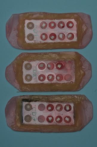
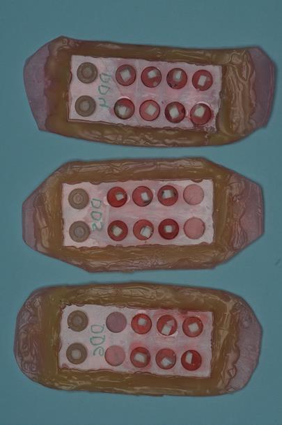
62 Figure 11 Groups of enamel slabs after randomisation Group C and D Group A and B 50
63 3.3.5 Experimental protocol/regime A special tray with 2 blind holes and 8 holes in the resin blocks was used to hold the blocks (Figure 10). Resin blocks were secured in position using adhesive wax. The slabs were immersed in a solution for two minutes five times daily in 0.3 % citric acid (ph 2.6) for a period of 28 days (Figure 13). Citric acid was prepared by adding three grams of mono-hydrate citric acid to one litre of de-ionised distilled water. Each group of slabs was immersed at room temperature in fresh 200 ml aliquots of citric acid each time. On each occasion, before immersion in citric acid, the slabs were taken out of the artificial saliva. The slabs were also rinsed in de-ionised distilled water (ph 6.85±0.05) after treatment before they were returned to the artificial saliva, which was changed twice daily (Figure 14). Two artificial saliva solutions were used in this study. The first solution was used for day time during the ph cycling, between the acid exposures. The second solution was used to store the slabs during the night. The day saliva was a supersaturated solution that allowed remineralisation of enamel slabs, the night saliva was a saturated solution that maintained the enamel condition and did not provide any minerals exchange. The artificial saliva composition was based on the electrolyte composition of natural saliva and it was advised to be used in order to eliminate any precipitation on the enamel surface (as provided by Dr RP Shellis, Department of Oral and Dental Science, University of Bristol, Bristol, UK). Day time and night time saliva were prepared as shown in Table 4 and 5 respectively. 51
64 Table 4 Day time artificial saliva Salt Concentration g/l Calcium carbonate 0.07 Magnesium carbonate (hydrated basic) Potassium di-hydrogen phosphate HEPES buffer (acid form) 4.77 Potassium chloride 2.24 To 5 L distilled water the above components were stirred until all had dissolved The ph was adjusted to 6.8 by adding NaOH and HCL solutions. The solution was kept at room temperature and used within 2 days. Table 5 Night time artificial saliva Salt Concentration g/l Calcium carbonate 0.05 Magnesium carbonate (hydrated basic) Potassium di-hydrogen phosphate HEPES buffer (acid form) 4.77 Potassium chloride 2.24 The night-time saliva was made up using the same procedure as above. 52
. Artificial saliva was changed twice daily to prevent any contamination or bacterial growth.")
65 Between immersions in citric acid the slabs were left immersed in artificial saliva for 60 minutes to enable remineralisation. The slabs were kept in an incubator at 37.0 o C at all times except while they will be being immersed in citric acid (Figure 12). Artificial saliva was changed twice daily to prevent any contamination or bacterial growth. A 60-minute gap was left between day time erosive challenges and between dipping s in toothpastes and the erosive challenges. After the dipping in the erosive solutions the slabs were rinsed with de-ionised water. Figure 12 Incubator at 37.0 o C During the cycling period, the slabs were analysed with the surface profilometer to measure the amount of surface loss at days 14 and 28. Concurrently the amount of F released from the FGSRD was measured. 53
66 Figure 13 Flow chart for all Groups Brushed with F free toothpaste (15strokes/2min) -Wash with distilled water Placed in day-time artificial saliva minimum 1h 1.Dipped in citric acid 0.3% ph 2.6 for 2min -Wash with distilled water Placed in day-time artificial saliva minimum 1h 2.Dipped in citric acid for 2min - Wash with distilled water Placed in day-time artificial saliva minimum 1h 3.Dipped in citric acid for 2min - Wash with distilled water Placed in day-time artificial saliva minimum 1h 4.Dipped in citric acid for 2min - Wash with distilled water Placed in day-time artificial saliva minimum 1h 5.Dipped in citric acid for 2min - Wash with distilled water Brushed with F free toothpaste (15strokes/2min) -Wash with distilled water Placed in night-time saliva 54
67 Figure 14 Illustration showing the cycling technique used for 1 day. After placement in artificial saliva the containers were transferred in an incubator for a minimum of 1h 55
68 3.3.6 Data collection At the end of the cycling period at 14 and 28 days, the slabs were rinsed with deionised distilled water and air-dried. The nail varnish was then removed using acetone and the enamel surface was cleaned with ethanol to ensure that all residues will be removed. The slabs were then kept in de-ionised distilled water and the FGSRD were placed in centrifuge tubes and left at room temperature. The slabs were scanned with the profilometer that was set up using the same parameters as for the baseline measurements. The sample was placed on the key stage of the Scantron ProScan and using a 150 mm height of the sensor as standard. The sample rate was set at 300Hz and the sensor that was used was S5/03. The step size used was 0.01 mm. After scanning the reading was levelled in three points A, B, and C (Figure 15) function interpolate x4 and warpage 1 to remove spikes from dust. Then 3 pt height was selected in the primary plan view (Figure 16) and the result was recorded. The measurements were repeated three times to check the reproducibility of the methods and to determine the standard deviations when assessing the sensitivity of the methods for detecting changes caused by the erosive challenge. In Figure 17 the different surfaces of the scan are visible at 28 days where it was possible and the sample was large, a step to differentiate between the 14 and 28 days was attempted. 56


69 Figure 15 Grid view of a 14 day scan of a sample with A, B and C the three points of levelling A B C Figure 16 3pt height sample measurments with the result of the difference in height recorded at µm 57
70 Figure 17 Grid view of at 28 days scan of a sample with 1 enamel intact surface, 2 step of erosive surface after 14 days, 3 bottom of erosive surface at 28 days and 4 the acrylic around the sample

71 3.3.7 Daily Fluoride release analysis Three FGSRD from groups A and B were placed in centrifuge tubes with day time artificial saliva. The FGSRD were placed in tubes with an equal amount of TISAB and the fluoride concentration measured using a Metrohm fluoride ion-specific electrode and Metrohm 781 Ion analyser after every 24h for 28 days. Metrohm fluoride ion-specific electrode and Metrohm 781 analyser were used to determine the fluoride concentration of 3 slow release fluoride devices and 3 placebo devices after being diluted with Total Ionic Strength Adjustment Buffer (TISAB). The testing equipment was calibrated using fluoride standard solutions containing 0.01, 0.10, 1.00 and mg/l F. Figure 18 Metrohm 781 Ion analyser with the fluoride standards 59
72 3.4 Statistics For the analysis, the data were uploaded in a SPSS Version 20. When the slabs were analysed the investigator did not know to which group they belonged to, making the analysis completely blinded. The normality of the data was tested in order to proceed with the appropriate analysis. For comparison of normal data t-tests were used with 0.05 as the significance level and a t-test, with a Bonferonni correction for comparisons within the groups. 60
73 4 Results In this chapter the results of the study are presented. The presentation of the results is in the same order as described in materials and methods section in order to simplify reading. 4.1 Baseline Measurements Profilometry measurements Baseline measurements of the surface profile of the slabs were acquired using a surface profilometer (Scantron ProScan 2000) to ensure that the average depth range was (mean ISO Rz) less than 1.5 µm. If ISO Rz was s <1.5 the slab was deemed acceptable to be included in the study. Samples not within the parameters were reground and rescanned and if the second scan was within the measurements then the samples were given a code. 61
74 4.1.2 Microhardness measurements Baseline measurements were recorded using Knoop microhardness. Microhardness was assessed using a computer-aided Duramin Indenter Machine (Struers A/S, DK 26-10, Denmark). As can be seen in Table 6 the mean distance between the edges of the Knoop microhardness diamond was measured at 64.2 μm for the permanent enamel slabs and μm for primary teeth with standard deviations of 2.21 and 2.14 respectively. Samples not within the range were not recorded and discarded. Table 6 Means of baseline microhardness measurements Microhardness in permanent enamel slabs Mean 64.2 μm Standard Deviation 2.21 Microhardness in primary enamel slabs Mean μm Standard Deviation 2.14 In Appendice 5 and 6 all the accepted measurements can be seen for the permanent and primary enamel slabs. 62
75 4.2 Tooth surface loss in permanent enamel Data taken from groups A and B were analysed and both groups were normally distributed so an independent t test was used for the analysis of the groups. Table 7 presents the summary of the surface loss and Figure 19 is a boxplot that shows that both permanent groups were normally distributed. Normality for the group A (placebo) and group B (fluoride) devices is presented in Table 8where the data are normally distributed. In Appendices 7 to 10 all the measurements can be seen for the permanent enamel slabs. Table 7 Means of tooth surface loss (µm) for permanent enamel slabs after 14 and 28 days for Placebo and FGSRD groups. SL Means± SE 14days 28days Placebo ± ± 1.11 FGSRD ± ± 1.4 Difference Table 8 Results of normality test for permanent enamel slabs Test of Normality Kolomogorov-Smirnov Shapiro-Wilk Permanent enamel slabs statistic Df. Sig. statistic Df. Sig. Placebo 14d FGSRD 14d Placebo 28d FGSRD 28d
 and Fluoride group (B) after 14 and 28 days")
76 Figure 19 Box plots of surface loss for permanent enamel slabs placebo group (A) and Fluoride group (B) after 14 and 28 days 64
Tooth hypersensitivity and Dental erosion DR. KÁROLY BARTHA
 Tooth hypersensitivity and Dental erosion DR. KÁROLY BARTHA 2 Why Is Erosion an Issue Now? Changing dietary habits Higher consumption of acidic beverages (colas, sport drinks) Higher incidence of xerostomia
Linking Research to Clinical Practice
 Prevention of Root Caries Denise M. Bowen, RDH, MS Linking Research to Clinical Practice The purpose of Linking Research to Clinical Practice is to present evidence based information to clinical dental
Chapter 14 Outline. Chapter 14: Hygiene-Related Oral Disorders. Dental Caries. Dental Caries. Prevention. Hygiene-Related Oral Disorders
 Chapter 14 Outline Chapter 14: Hygiene-Related Oral Disorders Hygiene-Related Oral Disorders Dental caries Prevention Gingivitis Prevention Tooth hypersensitivity Pathophysiology Treatment 2 Hygiene-Related
A personal perspective and update on erosive tooth wear 10 years on: Part 1 Diagnosis and prevention
 Author Query: Please insert captions for both tables PRACTICE A personal perspective and update on erosive tooth wear 10 years on: Part 1 Diagnosis and prevention D. Bartlett 1 In brief A term used by
DH220 Dental Materials
 DH220 Dental Materials Lecture #5 Prof. Lamanna RDH, MS Restorative Dentistry: Glass Ionomer Bird & Robinson p.740-741 I. Use Liner Base Luting agent Restorative material: Class III, V, & eroded/abraded
Dental Erosion. Introduction
 Dental Erosion Aims: To explore the causes, prevention and treatment of dental erosion, and to identify the role of dental care professional in assisting patients to minimise dental erosion. Objectives:
This is a repository copy of The effects of smoothies on enamel erosion: An in situ study.
 This is a repository copy of The effects of smoothies on enamel erosion: An in situ study. White Rose Research Online URL for this paper: http://eprints.whiterose.ac.uk/80734/ Version: Accepted Version
University of Groningen. Erosive enamel wear and the inhibiting effect of topical fluorides Vieira Carvalho, Ana Maria Ramires dos Santos
 University of Groningen Erosive enamel wear and the inhibiting effect of topical fluorides Vieira Carvalho, Ana Maria Ramires dos Santos IMPORTANT NOTE: You are advised to consult the publisher's version
Fluoridens 133 Fluorosilicic acid 136 Fluorosis, see Dental fluorosis Foams 118 acute toxicity 71, 122 clinical efficacy 122 Free saliva 149, 150
 Subject Index Abrasive systems, dentifrices 123 Absorption 23, 24, 38, 78 Accidental poisonings 66, 67, 69, 70, see also Acute toxicity Acid-base status, see also ph metabolism effects 28, 29 toxicity
Unit 6L.4: Teeth and Eating
 Unit 6L.4: Teeth and Eating Types of teeth Preventing tooth decay Dentition of other animals Digestive system By the end of this unit you should: Know the structure, function and care of the human teeth.
This is a repository copy of The effects of fruit smoothies on enamel erosion.
 This is a repository copy of The effects of fruit smoothies on enamel erosion. White Rose Research Online URL for this paper: http://eprints.whiterose.ac.uk/80737/ Version: Accepted Version Article: Tahmassebi,
Dental erosion: In vitro model of wine assessor s erosion
 ADRF REPORT Australian Dental Journal 21;46:(4):263-268 Dental erosion: In vitro model of wine assessor s erosion Tong Bee Mok,* J McIntyre,* D Hunt* Abstract Background: Wine makers and assessors frequently
It is 100 percent preventable
 Leeds Dental Institute FACULTY OF MEDICINE AND HEALTH No mysteries in solving the caries riddle It is 100 percent preventable Professor Monty Duggal University of Leeds, UK Aims of this presentation Dental
Management of Inadequate Margins and Gingival Recession Presenting as Tooth Sensitivity
 Management of Inadequate Margins and Gingival Recession Presenting as Tooth Sensitivity Nicolas Elian, DDS Private Practice Englewood Cliffs, New Jersey David Geon U Kim, DDS, MS Faculty and Research Coordinator
stabilisation and surface protection
 Guiding the way to caries stabilisation and surface protection Fissure sealing MI restorations Pulp capping Hypersensitivity Protection Caries stabilisation Fuji Triage from GC. Temporary restorations
SOFT DRINKS & DENTAL HEALTH.
 SOFT DRINKS & DENTAL HEALTH www.giveuplovingpop.org.uk @gulpnow SOFT DRINKS & DENTAL HEALTH All text tables, copyright Health Equalities Group 2017 Primary authors: Alexandra Holt, MSc. Health Equalities
riva helping you help your patients
 riva helping you help your patients what is a glass ionomer? how will a dentist benefit from using glass ionomers? how will a patient benefit from their glass ionomer? Glass ionomer is the generic name
PUBLISHED VERSION. This document has been archived with permission from the Australian Dental Association, received 18th January, 2007.
 PUBLISHED VERSION Saunders, J. G. C.; McIntyre, John Malcolm The ability of 1.23% acidulated phosphate fluoride gel to inhibit simulated endogenous erosion in tooth roots Australian Dental Journal, 2005;
THE IMPACT OF MODIFIED FRUIT JUICE ON ENAMEL MICROHARDNESS: AN IN-VITRO ANALYSIS
 Original Article International Journal of Dental and Health Sciences Volume 02,Issue 02 THE IMPACT OF MODIFIED FRUIT JUICE ON ENAMEL MICROHARDNESS: AN IN-VITRO ANALYSIS Shailee Shelke 1,Shaila Masih 1,Namita
OliNano Seal Professional prophylaxis for long-term protection
 Professional prophylaxis for long-term protection NEW The patented formula of silicone polymer NANO Technology General information Dental health is one of the main factors to maintain overall health, and
Root Surface Protection Simple. Effective. Important.
 GC Fuji VII / Fuji VII EP Root Surface Protection Simple. Effective. Important. Brush up your painting skills and help your patients Q&A Prof. Laurie Walsh University of Queensland lifestyle factors (frequency
OUR EXPERIENCE WITH GRADIA DIRECT IN THE RESTORATION OF ANTERIOR TEETH
 ISSN: 1312-773X (Online) Journal of IMAB - Annual Proceeding (Scientific Papers) 2006, vol. 12, issue 2 OUR EXPERIENCE WITH GRADIA DIRECT IN THE RESTORATION OF ANTERIOR TEETH Snezhanka Topalova-Pirinska,
The effects of smoothies on enamel erosion: an in. situ study
 The effects of smoothies on enamel erosion: an in situ study By: Hanein Ali A dissertation submitted in accordance with the requirements for the degree of Doctor of Clinical Dentistry University of Leeds
A Summary of: Delivering Better Oral Health: An evidencebased toolkit for prevention
 A Summary of: Delivering Better Oral Health: An evidencebased toolkit for prevention Aims: To discuss the key points of the government publication Delivering Better Oral health: An evidence-based toolkit
Effect of xylitol and fluoride on enamel erosion in vitro
 293 Journal of Oral Science, Vol. 49, No. 4, 293-297, 2007 Original Effect of xylitol and fluoride on enamel erosion in vitro Siriwan Chunmuang 1), Suwanna Jitpukdeebodintra 2), Chanya Chuenarrom 1) and
Operative dentistry. Lec: 10. Zinc oxide eugenol (ZOE):
:") Operative dentistry Lec: 10 د.عبذالمنعم الخفاجي Zinc oxide eugenol (ZOE): There are 2 types: 1) Unreinforced ZOE (ordinary type): supplied as powder (zinc oxide + some additives like zinc acetate, white
Operative dentistry Lec: 10 د.عبذالمنعم الخفاجي Zinc oxide eugenol (ZOE): There are 2 types: 1) Unreinforced ZOE (ordinary type): supplied as powder (zinc oxide + some additives like zinc acetate, white
Fuji II LC. A Perfect Choice
 A Perfect Choice is a remarkable restorative material The world s first resin-reinforced glass ionomer has remained the benchmark for light cured glass ionomer cements, delivering more than 15 years of
A clinical case involving severe erosion of the maxillary anterior teeth restored with direct composite resin restorations
 SETHI A clinical case involving severe erosion of the maxillary anterior teeth restored with direct composite resin restorations Sanjay Sethi, BDS (Lond.) Square Mile Dental Centre, 7-9 White Kennett Street,
Continually Fluoride Releasing Aesthetic Dental Restorative Material
 Continually Fluoride Releasing Aesthetic Dental Restorative Material Research is our best product Image provided by Dr. Sushil Koirala BEAUTIFIL II More than just filling BEAUTIFIL II stands out for its
Is there any clinical evidence?
 Current treatment objectives Anticariogenic capacity of restorative materials in paediatric dentistry: in vitro evidence vs. clinical efficiency Prof. Lisa Papagianoulis Restoration with minimal intervention
Indications The selection of amalgam as a restorative material for class V cavity should involve the following considerations:
 1 Lec.7 د.عبد املنعم اخلفاجي CLASS V CAVITY PREPARATION FOR AMAGLAM Indications The selection of amalgam as a restorative material for class V cavity should involve the following considerations: 1- Caries:
Dental caries prevention. Preventive programs for children 5DM
 Dental caries prevention Preventive programs for children 5DM Definition of Terms Preventive dentistry: usage of all the means to achieve and maintain the optimal oral health prevention of dental caries,
Polymers in everyday things dentistry
 HOTOCOY olymers in everyday things dentistry (Background information) olymers are a part of everyday life and examples can be found almost anywhere. Many people think of polymers simply as plastics used
swed dent j 2010; 34: hasselkvist, johansson, johansson
 swed dent j 2010; 34: 187-195 hasselkvist, johansson, johansson swedish dental journal vol. 34 issue 4 2010 187 swed dent j 2010; 34: 187-195 hasselkvist, johansson, johansson 188 swedish dental journal
EQUIA. Self-Adhesive, Bulk Fill, Rapid Restorative System
 EQUIA EQUIA Fil EQUIA Coat + Self-Adhesive, Bulk Fill, Rapid Restorative System From the World Leader in Glass Ionomer Technology - A Complete Glass Ionomer Based Bulk Fill Rapid Restorative System Class
Dzakovich Conclusions
 Definitions Attrition Tooth wear resulting from contact between opposing teeth. Erosion A gradual tooth-surface loss process caused by an electrolytic or chemical mechanism without bacteria being involved.
Innovative Dental Therapies for the Aging Population
 Innovative Dental Therapies for the Aging Population By Daniel H Ward DDS 1080 Polaris Pkwy Ste 130 Columbus OH 43240 614-430-8990 dward@columbus.rr.com US Population is Aging 1970-28 million>60 14% of
Tooth Wear. Department of Orthodontics and Restorative Dentistry Information for Patients
 Tooth Wear Department of Orthodontics and Restorative Dentistry Information for Patients i What is Tooth Wear? Tooth wear refers to the loss of tooth substance that is not caused by dental caries or trauma.
Contactless Dentistry. Cleans, Cuts, Prepares - Gently
 Contactless Dentistry Cleans, Cuts, Prepares - Gently Contactless Dentistry Unlike conventional rotary cutting instruments, the AquaCare handpiece is not in direct contact with the tooth structure, removing
From the office of: Nahidh D. Andrews, DMD 3332 Portage Ave South Bend, IN (574) Are Your Teeth a Sensitive Subject?
 Are Your Teeth a Sensitive Subject?") From the office of: Nahidh D. Andrews, DMD 3332 Portage Ave South Bend, IN 46628-3656 (574) 273-3900 Are Your Teeth a Sensitive Subject? A patient s guide to sensitive teeth and better oral care ARE YOUR
From the office of: Nahidh D. Andrews, DMD 3332 Portage Ave South Bend, IN 46628-3656 (574) 273-3900 Are Your Teeth a Sensitive Subject? A patient s guide to sensitive teeth and better oral care ARE YOUR
Uses of Fluoride in Dental Practices
 Uses of Fluoride in Dental Practices BY: Sulafa El-Samarrai Preventive &pedodontic department General Mechanisms of Action in Caries Reduction 1-Increase enamel resistance or reduction in enamel solubility
G-COAT PLUS G-COAT PLUS GET THE BEST OF BOTH WORLDS WITH THE STROKE OF A BRUSH
 G-COAT PLUS BEAUTY BRAWN GET THE BEST OF BOTH WORLDS WITH THE STROKE OF A BRUSH 1 Introducing a Revolutionary new Coating G-COAT PLUS gives your restorations the Best of Both Worlds: A beautiful new finish
Anisotropy of Tensile Strengths of Bovine Dentin Regarding Dentinal Tubule Orientation and Location
 Original paper Dental Materials Journal 21 (1): 32-43, 2002 Anisotropy of Tensile Strengths of Bovine Dentin Regarding Dentinal Tubule Orientation and Location Toshiko INOUE, Hidekazu TAKAHASHI and Fumio
NEW ZEALAND DATA SHEET
 1 PRODUCT NAME Clinpro 5000 anti-cavity toothpaste Vanilla Mint Clinpro 5000 anti-cavity toothpaste Spearmint Clinpro 5000 anti-cavity toothpaste Bubble Gum 2 QUALITATIVE AND QUANTITATIVE COMPOSITION Thick
FRANK OSEI-BONSU UGDS/KBTH
 FRANK OSEI-BONSU UGDS/KBTH Definition Introduction G. V. Black s concept of Restoration New classification of Caries Principles & Concept of MID Conclusion An approach to the management of dental caries
Acknowledgments Introduction p. 1 Objectives p. 1 Goals p. 2 History of Dental Materials p. 3 The Oral Environment p. 4 Characteristics of the Ideal
 Preface p. v Acknowledgments p. vii Introduction p. 1 Objectives p. 1 Goals p. 2 History of Dental Materials p. 3 The Oral Environment p. 4 Characteristics of the Ideal Dental Material p. 5 Quality Assurance
Prevalence of Tooth Wear in Children and Adolescents - A Survey Based Research V.PADMAHARISH, I BDS,SAVEETHA DENTAL COLLEGE NO:62 PH HIGH ROAD
 Prevalence of Tooth Wear in Children and Adolescents - A Survey Based Research V.PADMAHARISH, I BDS,SAVEETHA DENTAL COLLEGE NO:62 PH HIGH ROAD DR.ABILASHA.R DEPARTMENT OF ORAL PATHOLOGY SAVEETHA DENTAL
Direct composite restorations for large posterior cavities extended range of applications for high-performance materials
 Direct composite restorations for large posterior cavities extended range of applications for high-performance materials A case study by Ann-Christin Meier, Dr. med. dent., Stapelfeld, Germany When large
GC Tooth Mousse Plus for Orthodontics. Helps keep you smiling. Made from milk
 GC Tooth Mousse Plus for Orthodontics Helps keep you smiling Made from milk Prevention Daily application of GC Tooth Mousse Plus with RECALDENT (CPP-ACP) throughout the full course of orthodontic treatment
Dental Health E-presentation.
 Dental Health E-presentation www.healthycornwall.org.uk Learning Outcomes This short E-presentation has been developed to give you a basic understanding of the importance of good dental health and dental
Clinical report. Drs Paul and Alexandre MIARA and F. CONNOLLY COMPOSITE POSTERIOR FILLINGS. How to control. layering? 8 - Dentoscope n 124
 COMPOSITE POSTERIOR FILLINGS How to control layering? 8 - Dentoscope n 124 CV FLASH Dr Paul MIARA Dental surgeon Dr Alexandre MIARA Dental surgeon Dr F. CONNOLLY Dental surgeon Thanks to continuous improvements
Saliva. Introduction. Salivary Flow. Saliva and the Plaque Biofilm. The Minerals in Saliva
 Saliva Introduction Saliva is like a bloodstream to the mouth. As does blood, saliva helps build and maintain the health of the soft and hard tissues. Saliva removes waste products and provides disease-fighting
Buccal defects - therapy
 Buccal defects - therapy Asbjørn Jokstad Institute of Clinical Dentistry University of Oslo, Norway 1 Management? 2 Therapy and Interventions Strategy 1 1. Establish status 2. Restore Caries & non-caries
Dental Health. This document includes 12 tips that can be used as part of a monthly year-long dental health campaign or as individual messages.
 Dental Health This document includes 12 tips that can be used as part of a monthly year-long dental health campaign or as individual messages. What is gingivitis? Gingivitis is the beginning stage of gum
the ph, the higher the acidity an therefore the higher the risk it may cause Acid Erosion
 A BRIEF REVIEW ON TOOTH EROSION (CAUSES, TREATMENT) Dr. Zainab Mahmood Aljammali Assistant Lecturer, Dentistry Department, College of Dentistry, Babylon, Babylon University, Iraq Abstract: This review
Course #:
 Welcome to Lesson 7: Indicators of Problems of the Dental Health for Individuals with Disabilities webcast series. Please adjust your computer volume so that it is at a comfortable listening level for
Oral Care during Pregnancy
 Published on: 22 Mar 2012 Oral Care during Pregnancy Introduction Pregnancy influences nearly every aspect of a woman's life, including the oral health. Diet and hormonal changes during pregnancy may increase
Comparative Analysis of Remineralizing Potential of Three Commercially Available Agents- An in Vitro Study
 IOSR Journal of Dental and Medical Sciences (IOSR-JDMS) e-issn: 2279-0853, p-issn: 2279-0861.Volume 16, Issue 2 Ver. III (February. 2017), PP 01-05 www.iosrjournals.org Comparative Analysis of Remineralizing
DENTAL FOR EVERYONE DIAMOND PLAN PPO & PREMIER SUMMARY OF BENEFITS, LIMITATIONS AND EXCLUSIONS
 DENTAL FOR EVERYONE DIAMOND PLAN PPO & PREMIER SUMMARY OF BENEFITS, LIMITATIONS AND EXCLUSIONS DEDUCTIBLE Your dental plan features a deductible. This is an amount you must pay out of pocket before Benefits
Effects of miswak and nano calcium carbonate toothpastes on the hardness of demineralized human tooth surfaces
 Journal of Physics: Conference Series PAPER OPEN ACCESS Effects of miswak and nano calcium carbonate toothpastes on the hardness of demineralized human tooth surfaces To cite this article: F Dianti et
Preparation and making fillings Class V., III., IV.
 Preparation and making fillings Class V., III., IV. Class V. Cervical defects - Dental caries - Non carious lesions (erosion, abrasion, V shaped defects) Types of defects Caries Erosion Abrasion V shaped
values is of great interest.
 User Report Dr. Ralph Schönemann, May 21, 2008 There are numerous self-etchbonding systems on the market that sometimes differ very much from one another with regards to their chemical composition. It
Oral Health Improvement. Prevention in Practice Vicky Brand
 Oral Health Improvement Prevention in Practice Vicky Brand Quiz Question A B C The % of 5 yr olds in 05/06 in Greater Manchester who experienced tooth decay was just over Which of the following is more
v Review of how FLUORIDE works v What is FLUOROSIS v 2001 CDC Fluoride Guidelines v 2006 ADA Topical Fluoride Recommendation
 CAPD / ACDP 2016 SATURDAY MORNING 1. Caries Risk Assessment 2. Fluoride Issues 3. Managing Eruption Fluoride 2016 --- A Decade of Changes v Review of how FLUORIDE works v What is FLUOROSIS v 2001 CDC Fluoride
GC Tooth Mousse Plus. Made from milk. Perfect for teeth.
 GC Tooth GC Tooth Made from milk. Perfect for teeth. 1 We all brush our teeth everyday. But people who really want to care for their teeth are discovering the many benefits that come from moussing their
The 21 st Century vision on. caries management, now brought into your. daily practice
 The 21 st Century vision on caries management, now brought into your daily practice Minimum Intervention A 21 st century vision on Patient Caries Management The concept of minimal intervention dentistry
Q Why is it important to classify our patients into age groups children, adolescents, adults, and geriatrics when deciding on a fluoride treatment?
 Page 1 of 4 Q Why is it important to classify our patients into age groups children, adolescents, adults, and geriatrics when deciding on a fluoride treatment? A Different age groups have different dentitions
ARE YOU MOUTHWISE? AN ORAL HEALTH OVERVIEW FOR PRIMARY CARE
 ARE YOU MOUTHWISE? AN ORAL HEALTH OVERVIEW FOR PRIMARY CARE Funding providing by: Developed by: Terry L. Buckenheimer, DMD Karen Pesce Buckenheimer, R.N., BSN Executive Director, MORE HEALTH, Inc. Why
SPACE MAINTAINER. Multimedia Health Education. Disclaimer
 Disclaimer This movie is an educational resource only and should not be used to manage your health. All decisions about the management of premature loss of primary teeth and use of space maintainers must
Policy Statement Community Oral Health Promotion: Fluoride Use (Including ADA Guidelines for the Use of Fluoride)
") Policy Statement 2.2.1 Community Oral Health Promotion: Fluoride Use (Including ADA Guidelines for the Use of Fluoride) Position Summary Water fluoridation is the safest and the most effective way to reduce
Policy Statement 2.2.1 Community Oral Health Promotion: Fluoride Use (Including ADA Guidelines for the Use of Fluoride) Position Summary Water fluoridation is the safest and the most effective way to reduce
Risk factors for erosive tooth wear in a healthy lifestyle
 Department of Clinical Dentistry Faculty of Health Sciences Risk factors for erosive tooth wear in a healthy lifestyle Birgit Kynsveen and Maren Lunde Sæterstad Master thesis in ODO-3901, June 2016 Supervisors:
Influence of Bioactive Materials on Whitened Human Enamel Surface in vitro study
 Influence of Bioactive Materials on Whitened Human Enamel Surface Influence of Bioactive Materials on Whitened Human Enamel Surface in vitro study Pinheiro HB, Cardoso PEC, Universidade de São Paulo, São
Overview: The health care provider explores the health behaviors and preventive measures that enhance children s oral health.
 Lesson 5: Prevention Overview: The health care provider explores the health behaviors and preventive measures that enhance children s oral health. Goals: The health care provider will be able to describe:
2. Gap closure and replacement of the missing tooth 35 with directly modelled bridge region 34-36
 GrandTEC Test Kit Dear User, This Test Kit has been put together to enable you to test GrandTEC on the model before using it in a clinical situation. GrandTEC is a resin-impregnated glass fibre strip.
Preclinical Dentistry. I. Dental Caries Non carious lesions: trauma, erosion. abrasion, wedge shaped defects. Lenka Roubalíková
 Preclinical Dentistry I. Dental Caries Non carious lesions: trauma, erosion. abrasion, wedge shaped defects Lenka Roubalíková Understanding dental caries crown pulp chamber neck root canal root Dental
Managing Dental Erosion: Current Understanding and Future Directions
 Continuing Education Brought to you by Managing Dental Erosion: Current Understanding and Future Directions Course Author(s): Donald J. White, PhD; Warden H. Noble, DDS, MS, MSEd; Nicola West, BDS, FDSRCS,
Developing Dental Leadership. Fluoride varnish: How it works and how to apply it
 Fluoride varnish: How it works and how to apply it Fluoride Varnish It is the responsibility of the prescriber to ensure that a medicament is suitable for use A product licence indicates that the product
BioCoat Featuring SmartCap Technology
 R R Breakthrough! BioCoat Featuring SmartCap Technology The only bioactive pit and fissure sealant that provides relentless protection through daily release of fluoride, calcium and phosphate ions. Bioactive
The Dental Board of California - Dental Materials Fact Sheet Adopted by the Board on October 17, 2001
 The Dental Board of California - Dental Materials Fact Sheet Adopted by the Board on October 17, 2001 As required by Chapter 801, Statutes of 1992, the Dental Board of California has prepared this fact
EVALUATION OF COLOR CHANGE IN WHITE SPOT LESIONS OF ENAMEL FLUOROSIS USING A RESIN INFILTRATE
 EVALUATION OF COLOR CHANGE IN WHITE SPOT LESIONS OF ENAMEL FLUOROSIS USING A RESIN INFILTRATE Monisha Singhal PG STUDENT DEPARTMENT OF PEDODONTICS & PREVENTIVE DENTISTRY CHANDRA DENTAL COLLEGE & HOSPITAL
Howard E. Strassler, DMD University of Maryland School of Dentistry
 Part II National Board Review Operative Dentistry-Biomaterials Structures, chemical properties, physical properties BASIC CONCEPTS ON HOW DENTAL MATERIALS WORK! Howard E. Strassler, DMD University of Maryland
Essentials of. Dental Assisting. Edition 6. Debbie S. Robinson Doni L. Bird
 Essentials of Dental Assisting Edition 6 Debbie S. Robinson Doni L. Bird CHAPTER21 Restorative Procedures http://evolve.elsevier.com/robinson/essentials/ LEARNING OBJECTIVES KEY TERMS 1. Pronounce, define,
It s all about dental health
 It s all about dental health 01 June 2006 1. What do we mean by dental health? Dental health refers to all aspects of the health and functioning of our mouth especially the teeth and gums. Apart from working
Frequently Asked Questions. About Community Water Fluoridation. Overview. 1-What is fluoride?
 Frequently Asked Questions About Community Water Fluoridation Overview 1-What is fluoride? Fluoride is a naturally occurring mineral that protects teeth from tooth decay. 2-Why is fluoride added to water
ADHESIVE RECONSTRUCTION IN HELP OF THE ORTHODONTIC TREATMENT
 ISSN: 1312-773X (Online) Journal of IMAB - Annual Proceeding (Scientific Papers) 2006, vol. 12, issue 2 ADHESIVE RECONSTRUCTION IN HELP OF THE ORTHODONTIC TREATMENT Snezhanka Topalova-Pirinska 1, R. Pirinska
DENTAL MATTERS. This has been produced by Bayer
 DENTAL MATTERS This has been produced by Bayer WHY IS GOOD DENTAL CARE IMPORTANT FOR HAEMOPHILIA? Haemophilia doesn t directly affect the teeth. However, like everyone else it is important to brush and
3M Fast Release Varnish
 3M Fast Release Varnish 5% Sodium Fluoride Varnish Fast Release Formula Technical Data Sheet 2 3M Fast Release Varnish 3M Fast Release Varnish is a fluoride-containing varnish. It can be applied to enamel
King s Research Portal
 King s Research Portal DOI: 10.1111/adj.12418 Document Version Peer reviewed version Link to publication record in King's Research Portal Citation for published version (APA): O'Toole, S., Bartlett, D.
Fixed Twin Blocks. Guidelines for case selection are similar to those for removable Twin Block appliances.
 Fixed Twin Blocks Development of Fixed Twin Blocks Dr Clark has enjoyed the cooperation of Dynaflex in developing the Fixed Twin Block. Six years of clinical testing has confirmed that this technique produces
OMNII Oral Pharmaceuticals
 Fluoride Varnish in a Pediatric Practice Created by W. Edward Gonzalez D.M.D. OMNII Oral Pharmaceuticals The Preventive Care Consultants OMNII Firsts In Dentistry OMNII Gel PerioMed white & brite FlossRx
Oral health education for caries prevention
 Oral health education for caries prevention Objective Understand the fundamentals that inform oral health education programmes. Understand how to segment oral health information for preventive purposes
Glass Ionomers. Reputable, Durable, Long Lasting
 Glass Ionomers Reputable, Durable, Long Lasting Conventional Glass Ionomer Restoratives Glass Ionomeres Technology more up-to-date than ever before! Successfull well proven material Exhibit biocompatibility
Prevalence of erosive tooth wear and associated risk factors in 2 7-year-old German kindergarten children
 (2006) 12, 117 124. doi:10.1111/j.1601-0825.2005.01167.x Ó 2005 Blackwell Munksgaard All rights reserved http://www.blackwellmunksgaard.com ORIGINAL ARTICLE Prevalence of erosive tooth wear and associated
Australian Dental Journal
 Australian Dental Journal The official journal of the Australian Dental Association Australian Dental Journal 2013; 58: 478 482 doi: 10.1111/adj.12110 Role of arginine and fluoride in the prevention of
Songklanakarin Journal of Science and Technology SJST R2 Chuenarrom
 Songklanakarin Journal of Science and Technology SJST-0-00.R Chuenarrom Effect of excessive trichloroisocyanuric acid in swimming pool water on tooth erosion Journal: Songklanakarin Journal of Science
dental fillings facts About the brochure:
 dental fillings facts About the brochure: Your dentist is dedicated to protecting and improving oral health while providing safe dental treatment. This fact sheet provides information you need to discuss
Zurich Open Repository and Archive. Effect of sodium, amine and stannous fluoride at the same concentration and different ph on in vitro erosion
 University of Zurich Zurich Open Repository and Archive Winterthurerstr. 190 CH-8057 Zurich http://www.zora.uzh.ch Year: 2009 Effect of sodium, amine and stannous fluoride at the same concentration and
Frictional Behavior and Surface Failure of Human Enamel
 Frictional Behavior and Surface Failure of Human Enamel J. M. POWERS, R. G. CRAIG, and K. C. LUDEMA School of Dentistry, University of Michigan, Ann Arbor, Michigan 48104, USA, and College of Engineering,
Scanning electron microscope characterization of abrasion in human teeth
 Dental Journal Mahidol Dental Journal Original Article Scanning electron microscope characterization of abrasion in human teeth B.Sc., D.D.S., Specialty Certificate in Oral Pathology, M.S. Department of
Incisal Edge Abrasion Caused By An Unusual Eating Habit
 ISPUB.COM The Internet Journal of Dental Science Volume 6 Number 1 W Amin, A Kassab, N Salim Citation W Amin, A Kassab, N Salim.. The Internet Journal of Dental Science. 2007 Volume 6 Number 1. Abstract