IN THE NAME OF GOD. Dr.kheirandish DDS,MSC Oral and maxillofacial pathology
|
|
|
- Cassandra Simon
- 5 years ago
- Views:
Transcription

1 IN THE NAME OF GOD Dr.kheirandish DDS,MSC Oral and maxillofacial pathology

2 Dermatologic Diseases Chapter 16
3 ECTODERMAL DYSPLASIA
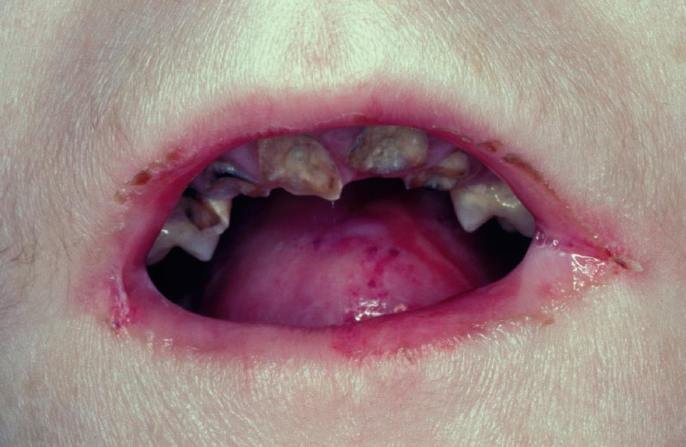
4 o Two or more ectodermally derived anatomic structures fail to develop o Hypohidrotic ectodermal dysplasia Male Heat intolerance eccrine sweat glands Infancy fever of undetermined origin Cannot regulate body temperature Death elevated body temperature

5 Fine, sparse hair (eyebrow and eyelash hair) Periocular skin (fine wrinkling with hyperpigmentation) Midface hypoplasia Protuberant lips Hypoplastic salivary glands xerostomia Nails (dystrophic and brittle)
6 Oligodontia Hypodontia Conical tooth Anodontia
7 Decreased number of sweat glands and hair follicles Dental problems prosthetic replacement Older than 6 years of age
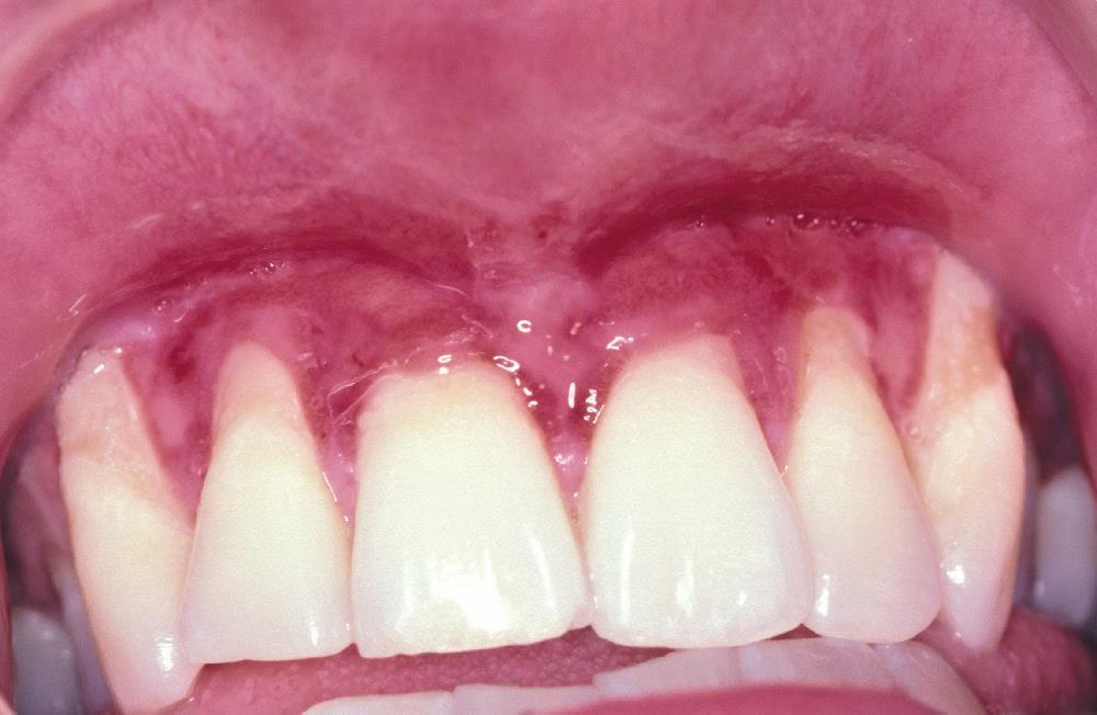
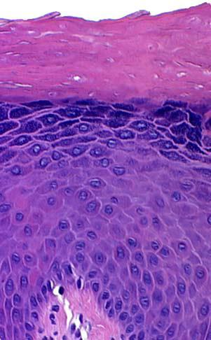
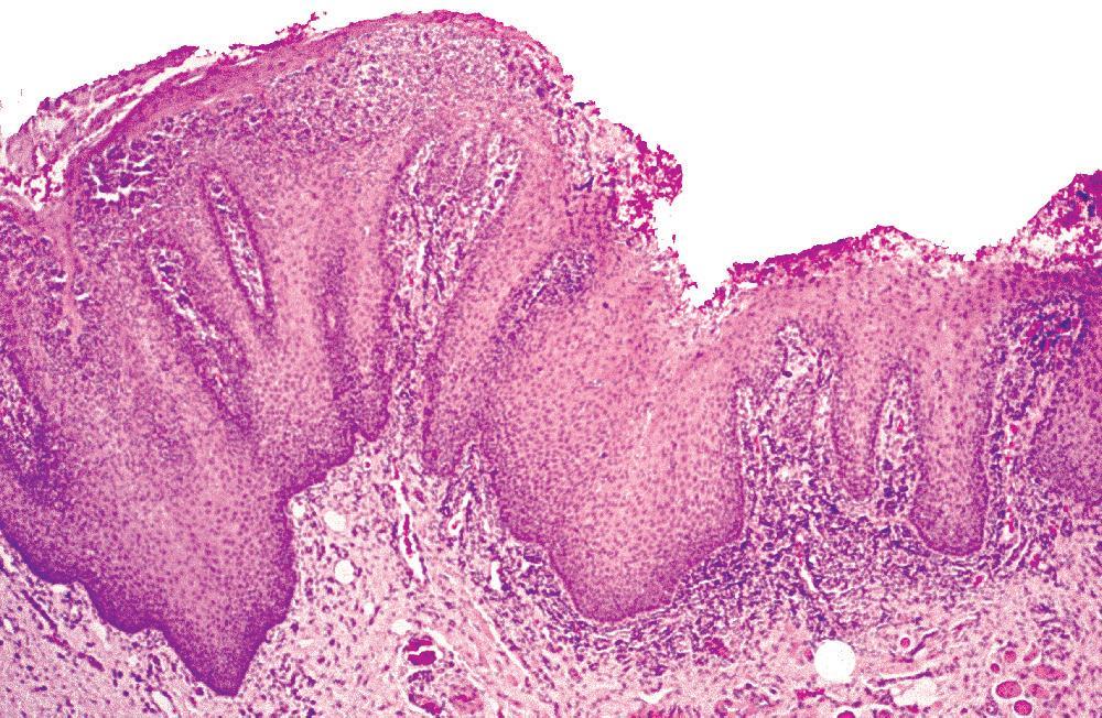
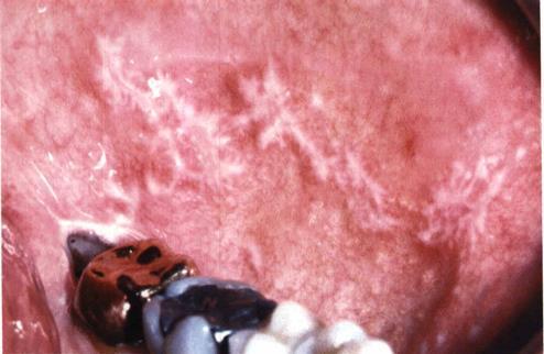
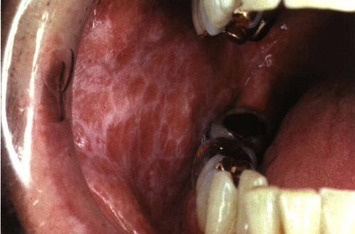
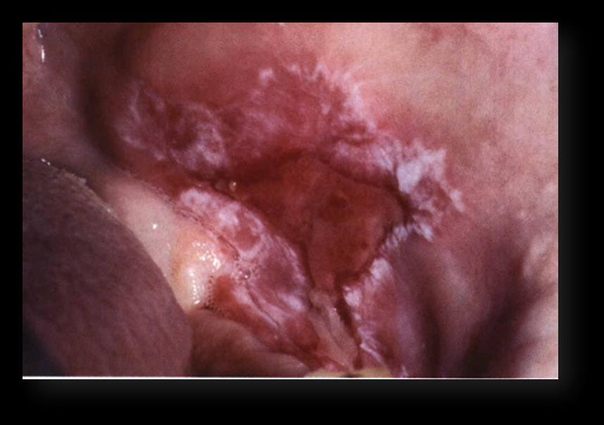
8 WHITE SPONGE NEVUS
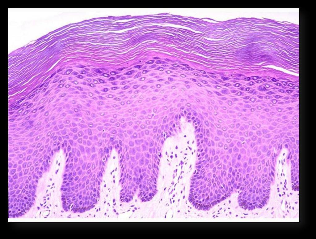
9 Defect in the normal keratinization of the oral mucosa At birth or in early childhood Symmetrical, thickened, white, corrugated or velvety, diffuse plaques affect the buccal mucosa bilaterally Asymptomatic Treatment


10
11 Prominent hyperparakeratosis Acanthosis Clearing of the cytoplasm of the cells in the spinous layer Leukoedema Hereditary benign intraepithelial dyskeratosis (hbid) Eosinophilic perinuclear condensation (superficial layers) Papanicolaou
12 PEUTZ-JEGHERS SYNDROME


13 Frecklelike lesions of the hands, perioral skin, and oral mucosa + intestinal polyposis and predisposition for affected patients to develop cancer Tumor suppressor gene, STK11 (LKB1) Increased frequency of malignancy (10 to 18 times greater than normal) Oral lesions Perioral freckling Brown to blue-gray Vermilion / labial / buccal mucosa / tongue (90%)
14 Gastrointestinal polyps Overgrowths of intestinal glandular epithelium Pigmented cutaneous lesions Slight acanthosis Elongation of the rete ridges Melanocyte number Dendritic processes o Monitoring
15 EPIDERMOLYSIS BULLOSA
16 o Blistering mucocutaneous disorders Each has a specific defect in the attachment mechanisms of The epithelial cells, either to each other or to the underlying Connective tissue. Depending on the defective mechanism of cellular cohesion : 1. Simplex 2. Junctional 3. Dystrophic 4. Kindler syndrome
17 Simplex Mild, annoying forms Keratin 5 and keratin 14 Junctional Death at birth Laminin-332, type XVII collagen, or α6β4 integrin Dystrophic Type VII collagen Kindler syndrome Hemidesmosomal attachment protein, kindlin-1
18 Oral lesions are most commonly observed in the dystrophic forms Dental abnormalities (junctional type) Anodontia Enamel hypoplasia Pitting of the enamel Neonatal teeth Severe periodontitis Severe dental caries
19 Dominant dystrophic types Oral manifestations are typically mild Gingival erythema and tenderness Gingival recession Reduction in the depth of the buccal vestibule Recessive dystrophic types Mittenlike deformity Carious destruction of the dentition at an early age is common.
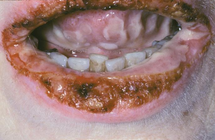
20

21 Intraepith. Subepith.

22 Simplex Intraepithelial clefting Junctional, dystrophic, and kindler Subepithelial clefting
23 Pemphigus
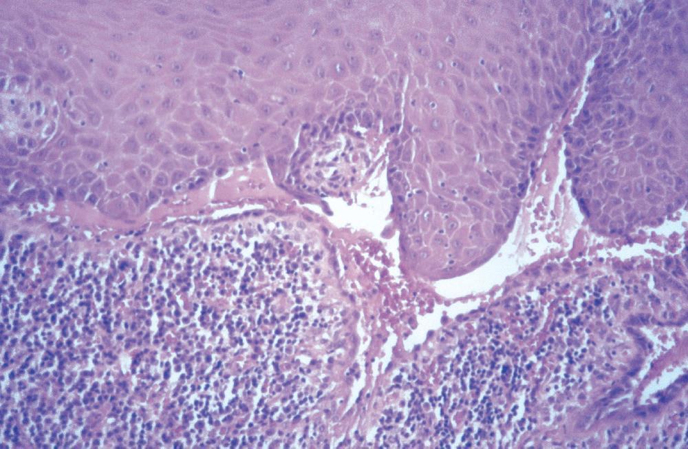
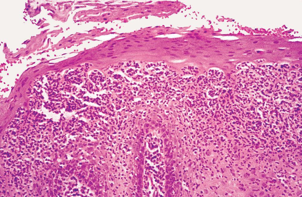
24 The condition known as pemphigus represents four related diseases of an autoimmune origin: 1) Pemphigus vulgaris 2) Pemphigus vegetans 3) Pemphigus erythematosus 4) Pemphigus foliaceus
25 Pemphigus vulgaris is the most common of these disorders (vulgaris is Latin for common)

26 The blistering that typifies PV is due to an abnormal production of autoantibodies that are directed against the epiderrmal cell surface glycoproteins : desmoglein 3 (in desmosomes)
27 Clinical features The initial manifestations of pemphigus vulgaris often involve the oral mucosa The average age at diagnosis is 50 years No sex predilection is observed Seems to be more common in persons of Mediterranean, South Asian, or Jewish heritage

28 Clinical features Patients usually complain of oral soreness

29 Clinical features Examination shows superficial, ragged erosions and ulcerations Such lesions may affect any oral mucosal location
30 Clinical features More than 50% of the patients have oral mucosal lesions before the onset of cutaneous lesions Nearly all patients have intraoral involvement The skin lesions appear as flaccid vesicles and bullae that rupture quickly, leaving an erythematous denuded surface

31 Histopathologic features Biopsy specimens of peri lesional tissue show characteristic intra epithelial separation, which occurs just above the basal cell layer of the epithelium

32 Histopathologic features Sometimes the entire superficial layers of the epithelium are stripped away, leaving only the basal cells, which have been described as resembling a "row of tombstones"
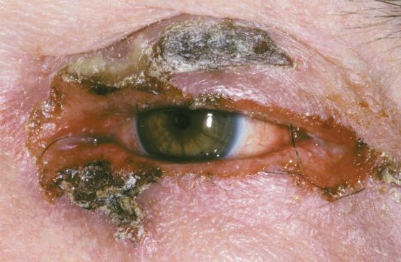
 and the loose cells tend to assume a rounded shape (Tzanck")
33 Histopathologic features The cells of the spinous layer of the surface epithelium typically appear to fall apart ( acantholysis) and the loose cells tend to assume a rounded shape (Tzanck cells)

34 Histopathologic features The diagnosis of pemphigus vulgaris should be con firmed by direct immunofluorescence (antibodies can be demonstrated in the intercellular spaces between the epithelial cells)
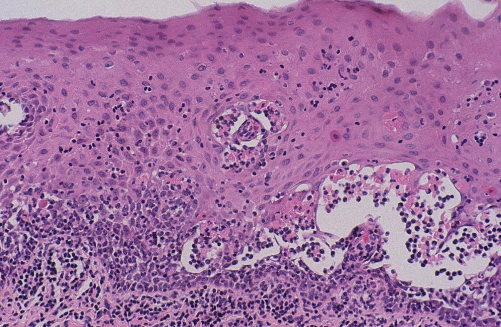
35 PARANEOPLASTIC PEMPHIGUS

36 Pemphigoid (Benign mucous membrane pemphigoid)
37 Pemphigoid is chronic, blistering, mucocutaneous autoimmune diseases in which tissue-bound autoantibodies are directed against one or more components of the basement membrane It is at least twice as common as pemphigus vulgaris The term pemphigoid is used because clinically it often appears similar to pemphigus
38 Clinical features Usually affects older adults, with an average age of 50 to 60 years at the onset of disease Females are affected more frequently than males by a 2:1 ratio Oral lesions are seen in most, but other sites, such as conjunctival, nasal, esophageal, laryngeal, and vaginal mucosa, as well as the skin may be involved

39 Clinical features The oral lesions of pemphigoid begin as either vesicles or bullae that may occasionally be identified clinically

40 Clinical features The oral blisters rupture, leaving large, superficial, ulcerated, and denuded areas of mucosa which are lusually painful and persist for weeks to months if untreated

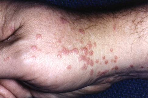
41 Clinical features Gingival involvement produces a clinical reaction pattern termed desquamative gingivitis

42 Clinical features The most significant complication of mucous membrane pemphigoid, is ocular involvement (up to 25% of patients with oral lesions may eventually develop ocular disease)

43 Histopatholgic features Biopsy of perilesional mucosa shows a split between the surface epithelium and the underlying connective tissue

44 Histopatholgic features A mild chronic inflammatory cell infiltrate is present in the superficial submucosa

45 Histopatholgic features Direct immunofluorescence studies of perilesional mucosa show a continuous linear band of immunoreactants at the basementmembrane zone in nearly 90% of affected patients
46 BULLOUS PEMPHIGOID
47 Most common of the autoimmune blistering conditions Autoantibodies directed against components of the basement membrane Mucous membrane pemphigoid Oral mucosal involvement is uncommon
48 Subepithelial separation
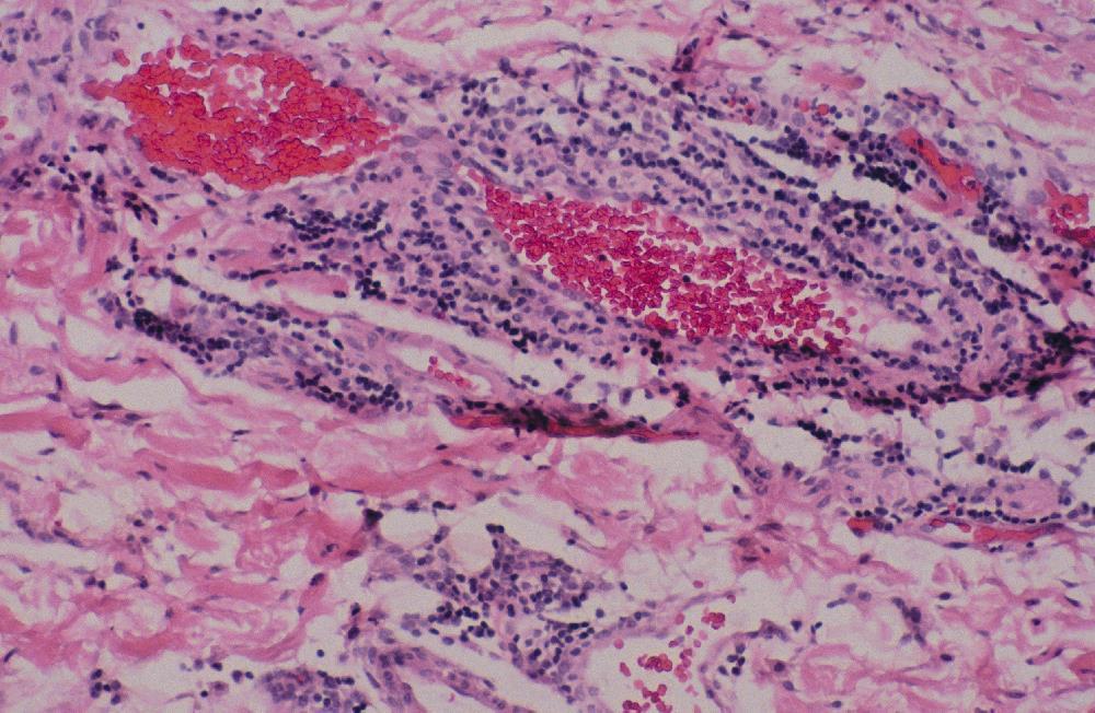
49 ERYTHEMA MULTIFORME
50 Blistering, ulcerative mucocutaneous condition of uncertain etiopathogenesis Minor Major(stevens-johnson syndrome) Toxic epidermal necrolysis (lyell disease- TEN)

51
52
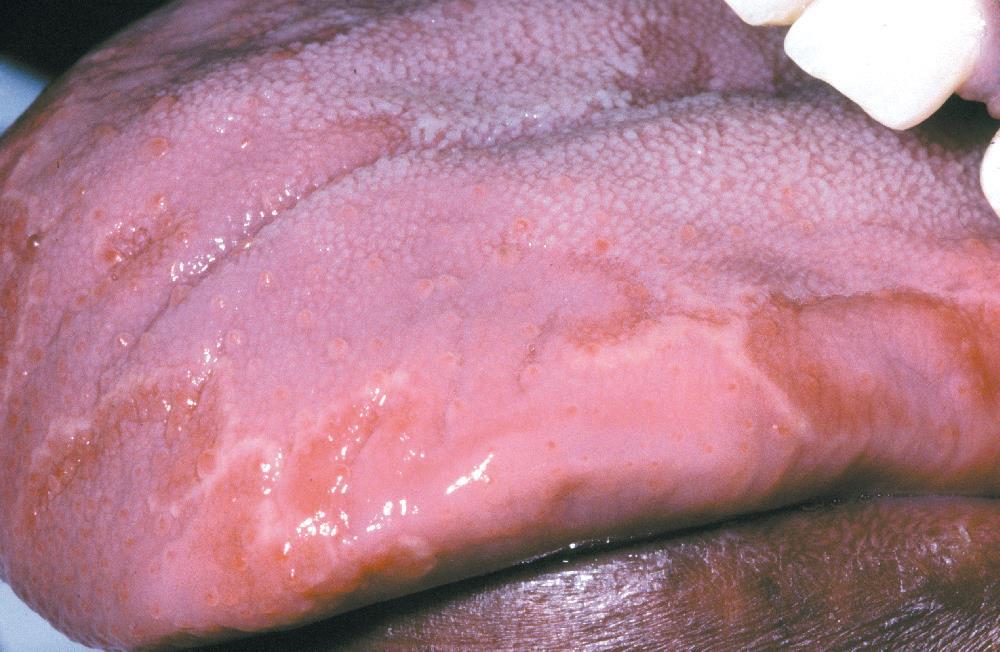
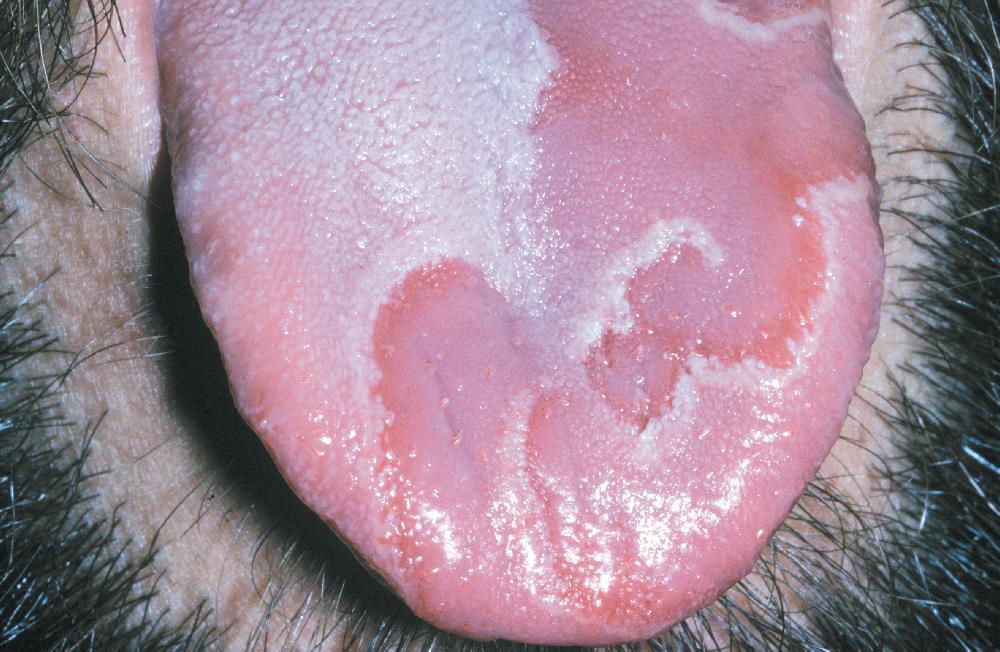
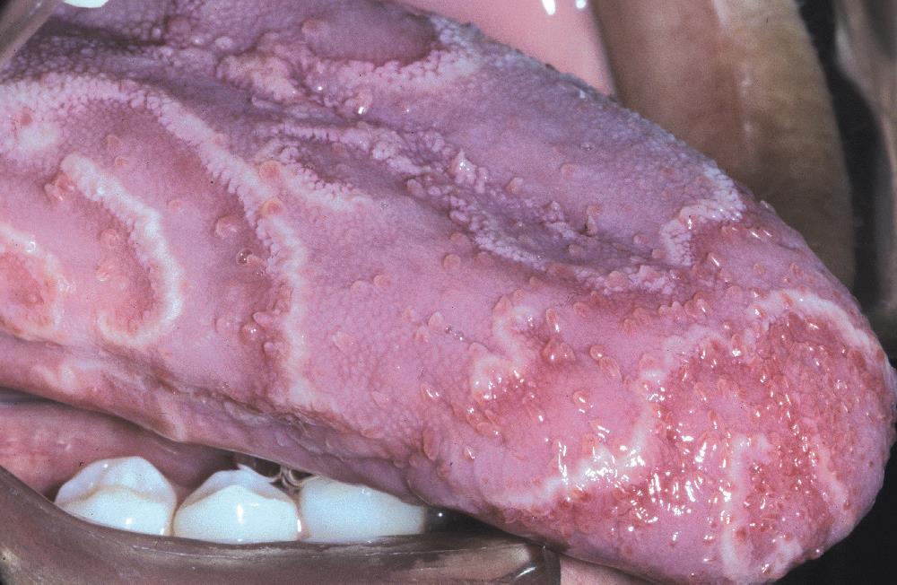
53 ERYTHEMA MIGRANS
54


55

56
57 LICHEN PLANUS
58 Common Chronic Dermatologic disease Oral mucosa Hepatitis C infection - genetic stress Middle-aged Women
59 SKIN LESIONS: Purple, pruritic, polygonal papules Flexor surfaces of the extremities Lacelike network of white lines(wickham's striae) ORAL LESIONS: Reticular and erosive

60
61 RETICULAR LICHEN PLANUS More common No symptoms Posterior buccal mucosa bilaterally Lateral and dorsal tongue,the gingivae, the palate, and vermilion border Wickham's striae Wax and wane over weeks or months
62
63
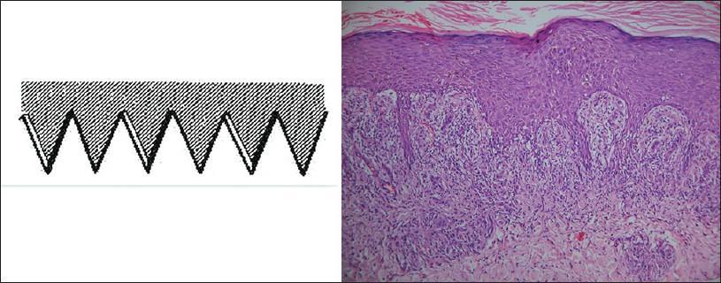
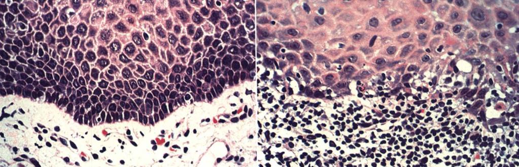
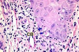
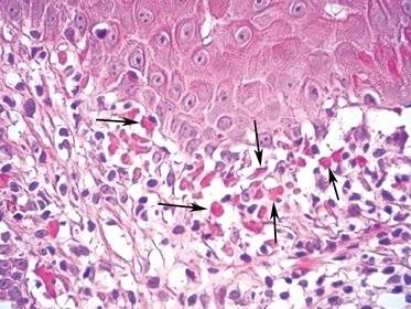
64 EROSIVE LICHEN PLANUS Symptomatic Atrophic Erythematous Central ulceration Periphery of the atrophic regions is usually bordered by fine, white radiating striae Severe bullous lichen planus
65
66
67
68
69
70
71
72 Characteristic (not be specific) Orthokeratosis and parakeratosis Rete ridges absent / hyperplastic/"saw-toothed" shape Hydropic degeneration Intense, bandlike infiltrate of predominantly T lymphocytes Degenerating keratinocytes (epithelium and connective tissue interface) Colloid, cytoid, hyaline, or civatte bodies

73
Autoimmune Diseases with Oral Manifestations
 Autoimmune Diseases with Oral Manifestations Martin S. Greenberg DDS, FDS RCSEd Professor Emeritus Department of Oral Medicine University of Pennsylvania Disclosure Statement I have no actual or potential
MUCOCUTANEOUS LESIONS Normal structures in epithelium cell adhesion to each other and to underlying connective tissue:
 ORAL DERMATOSES AND MUCOSAL/GINGIVAL LESIONS MUCOCUTANEOUS LESIONS Normal structures in epithelium cell adhesion to each other and to underlying connective tissue: Diagram taken from: Oral and Maxillofacial
LESIONS OF THE ORAL CAVITY ORAL CAVITY. Oral Cavity Subsites 4/10/2013 LIPS TEETH GINGIVA ORAL MUCOUS MEMBRANES PALATE TONGUE ORAL LYMPHOID TISSUES
 LESIONS OF THE ORAL CAVITY David I. Kutler, MD, FACS Associate Professor Division of Head and Neck Surgery Department of Otolaryngology HNS Weill Cornell Medical Center ORAL CAVITY LIPS TEETH GINGIVA ORAL
Classification: 1. Infective: 2. Traumatic: 3. Idiopathic: Recurrent Aphthous Stomatitis (RAS) 4. Associated with systemic disease:
 4. Associated with systemic disease:") Classification: 1. Infective: 2. Traumatic: 3. Idiopathic: Recurrent Aphthous Stomatitis (RAS) 4. Associated with systemic disease: Hematological GIT Behcet s HIV 5. Associated with dermatological diseases:
Classification: 1. Infective: 2. Traumatic: 3. Idiopathic: Recurrent Aphthous Stomatitis (RAS) 4. Associated with systemic disease: Hematological GIT Behcet s HIV 5. Associated with dermatological diseases:
Oral Medicine. Dr. Qianming Ian CHEN
 Oral Medicine Dr. Qianming Ian CHEN ORAL MEDICINE Oral medicine is the specialty of dentistry that is concerned with the oral health care of medically compromised patients and with the diagnosis and nonsurgical
REF: Chap 1 (Pemphigus vulgaris/etiology and
 Chapter 1: Vesiculobullous Diseases Test Bank MULTIPLE CHOICE 1. Intercellular deposits of IgG are consistently found in oral epithelium in which of the following? a. Cicatricial pemphigoid b. Lichen planus
ANS: C REF: Chap 1 (Pemphigus vulgaris/etiology and pathogenesis), p 11
, p 11") Chapter 1: Vesiculobullous Diseases Test Bank MULTIPLE CHOICE 1. Intercellular deposits of IgG are consistently found in oral epithelium in which of the following? a. Cicatricial pemphigoid b. Lichen planus
Chapter 1: Vesiculobullous Diseases Test Bank MULTIPLE CHOICE 1. Intercellular deposits of IgG are consistently found in oral epithelium in which of the following? a. Cicatricial pemphigoid b. Lichen planus
Index. derm.theclinics.com. Note: Page numbers of article titles are in boldface type.
 Note: Page numbers of article titles are in boldface type. A Adhesion and migration, the diverse functions of the laminin a3 subunit, 79 87 Alopecia in epidermolysis bullosa, 165 169 Amblyopia and inherited
Pigmented lesions of the Oral cavity
 Oral medicine أ.م.د احسان عبد هللا كميل Pigmented lesions of the Oral cavity Pigmented oral lesions are a large group of disorders in which the dark or brown color is the essential clinical characteristic.
Table Clinical Features Related to Level of Spinal Cord Injury. Level of Spinal Cord Damage. Associated Clinical Features. respiratory paralysis
 Table 17.1. Clinical Features Related to Level of Spinal Cord Injury Level of Spinal Cord Damage C1 to C4 C4 to C5 C5 to C6 C6 to C7 T11 to T12 T12 to L1 S3 to S5 Associated Clinical Features Death secondary
WHITE LESIONS OF THE UPPER AIRWAY
 WHITE LESIONS OF THE UPPER AIRWAY WHITE LESION CONFIGURATIONS Solitary vrs Multifocal Flat Plaque Verrucous/rippled Lacey White with red component Papular (curdled milk plaques) Pseudomembranous PLAQUES
Oral Manifestations of Dermatologic Disease: A Focus on Lichenoid Lesions. Proceedings of the NASHNP Companion Meeting, March, 2011, San Antonio, TX
 1 Oral Manifestations of Dermatologic Disease: A Focus on Lichenoid Lesions Proceedings of the NASHNP Companion Meeting, March, 2011, San Antonio, TX Susan Müller, DMD, MS Professor Department of Pathology
EPIDERMOLYSIS BULLOSA
 EPIDERMOLYSIS BULLOSA Definition Epidermolysis bullosa (EB) is a term used to describe a group of rare mainly hereditary, chronic, non-inflammatory diseases of skin and mucous membranes. EB is characterized
B. Autoimmune blistering diseases
 Go Back to the Top To Order, Visit the Purchasing Page for Details formation immediately above the basal layer. The dermal papillae, which are covered by basal cells in the single layer that is left in
2018 Oregon Dental Conference Course Handout Denis Lynch, DDS, PhD
 2018 Oregon Dental Conference Course Handout Denis Lynch, DDS, PhD Course 9148: Diagnosis and Treatment of Recurrent Oral Ulcers Friday, April 6 9 am - 12 pm Diagnosis and Treatment of Recurrent Oral Ulcers
Immunobullous Diseases: Review and Update. May P. Chan, MD Associate Professor of Pathology and Dermatology University of Michigan
 Immunobullous Diseases: Review and Update May P. Chan, MD Associate Professor of Pathology and Dermatology University of Michigan Diagnosis of Immunobullous Diseases Clinical H&E DIF DIAGNOSIS IIF ELISA
Sign In: pemphigus.org/form
 Pemphigus and Pemphigoid The Unique Role of the Dental Professional Dr. Carol Anne Murdoch Kinch Sign In: pemphigus.org/form The International Pemphigus & Pemphigoid Foundation (IPPF) kindly asks all attendees
That. Name QUIZ. 60 SEPTEMBER 2017 // dentaltown.com
 QUIZ Name That General dentists are first in the line of practitioners that patients see for an oral lesion evaluation; therefore, a sound understanding of oral mucosal diseases and their clinical presentation
Evaluation of the relation between Pemphigus Vulgaris and Periodontal status
 Advances in Bioresearch Adv. Biores., Vol 7 (2) March 2016: 64-68 2015 Society of Education, India Print ISSN 0976-4585; Online ISSN 2277-1573 Journal s URL:http://www.soeagra.com/abr.html CODEN: ABRDC3
Oral Pemphigus Vulgaris: Case Report
 Oral Pemphigus Vulgaris Rai A. et al 367 CASE REPORT Oral Pemphigus Vulgaris: Case Report Rai Arpita 1, Arora Monica 2, Naikmasur Venkatesh 3, Sattur Atul 3, Malhotra Varun 2 ABSTRACT BACKGROUND: Pemphigus
When your patient complains of
 WILLIAM LAWSON, MD, DDS Mount Sinai School of Medicine ABSTRACT: Certain clues can help you identify the cause of bullous oral lesions. Diffuse oral and labial bullous erosions, sometimes accompanied by
Egyptian Dermatology Online Journal Vol. 6 No 2: 16, December Hypohidrotic Ectodermal Dysplasia with Arachnodactyl and Palmoplanter Keratoderma
 Hypohidrotic Ectodermal Dysplasia with Arachnodactyl and Palmoplanter Keratoderma Taseer Ahmed Bhatt Department of Dermatology, STD & Leprosy Govt. Medical College Srinagar, Kashmir Egyptian Dermatology
INFLAMMATORY DISEASES PART I. Immunopathology Part I
 INFLAMMATORY DISEASES PART I Immunopathology Part I Nonspecific & T Cell Mediated Mucosal Inflammatory Lesions Nonspecific and Idiopathic Mucositis Hypersensitivity and Autoimmune T cell mediated Immunoglobulin
Pemphigus in younger age group in Bangladeshi population
 ORIGINAL ARTICLE in younger age group in Bangladeshi population Abdul Wahab 1, MD, Lubna Khondker 1, MD, Jamal Uddin 1, MD, Ishrat Bhuiyan 2, MD Shirajul Islam Khan 3, MD, Zafrul Islam 1, MD, Rahmat Ali
Some skin conditions
 Some skin conditions Some skin conditions Acute Inflammatory Dermatoses Chronic Inflammatory Dermatoses Blistering (Bullous) Diseases Panniculitis Disorders of Epidermal Appendages -Urticaria -Acute eczematous
Benign Oral cavity lesions. Mohammed ALESSA MBBS,FRCSC Assistant Professor Consultant Otolaryngology, Head & Neck Surgery
 Benign Oral cavity lesions Mohammed ALESSA MBBS,FRCSC Assistant Professor Consultant Otolaryngology, Head & Neck Surgery Anatomy Histology Physiology Pathology Clinical cases Introduction The oral cavity
2 Anonychia/Micronychia
 2 Anonychia/Micronychia Total or partial absence of the nail May be congenital or acquired Table 2.1. Causes of anonychia/micronychia Congenital Acquired Trauma Amniotic bands Bullous Teratogens (drugs,
PACIFIC JOURNAL OF MEDICAL SCIENCES {Formerly: Medical Sciences Bulletin} ISSN:
 PACIFIC JOURNAL OF MEDICAL SCIENCES {Formerly: Medical Sciences Bulletin} ISSN: 2072 1625 Pac. J. Med. Sci. (PJMS) www.pacjmedsci.com. Email: pacjmedsci@gmail.com. EROSIVE LICHEN PLANUS A CASE REPORT *Prathima
Actinic keratosis (AK): Dr Sarma s simple guide
: Dr Sarma s simple guide") Actinic keratosis (AK): Dr Sarma s simple guide Actinic keratosis is a very common lesion that you will see in your day-to-day practice. First, let me explain the name Actinic keratosis. It means keratosis
Actinic keratosis (AK): Dr Sarma s simple guide Actinic keratosis is a very common lesion that you will see in your day-to-day practice. First, let me explain the name Actinic keratosis. It means keratosis
Immunofluorescence in Oral Dermatological Disorders- No Shiny Matter
 Journal of Academy of Dental Education Journal of Academy of Dental Education, 24-28, DOI: 10.18311/jade/2015-2016/15951 ISSN (Print): 2348-1595 ISSN (Online) : 2348-2621 Immunofluorescence in Oral Dermatological
Department of Dermatology, Nippon Medical School, 1-1-5, Sendagi, Bunkyo-ku, Tokyo , Japan 2
 Dermatology Research and Practice Volume 2010, Article ID 931340, 5 pages doi:10.1155/2010/931340 Case Report Paraneoplastic Pemphigus Presenting as Mild Cutaneous Features of Pemphigus Foliaceus and Lichenoid
Acquired and Inherited Bullous Diseases
 Acquired and Inherited Bullous Diseases Erin Wei MD Brigham and Women s Hospital, Department of Dermatology Instructor, Harvard Medical School Director, Bullous Disease Clinic No disclosures Conflict of
Index. Dent Clin N Am 49 (2005) Note: Page numbers of article titles are in boldface type.
 Note: Page numbers of article titles are in boldface type.") Dent Clin N Am 49 (2005) 273 278 Index Note: Page numbers of article titles are in boldface type. A Acanthosis nigricans, familial, 251 Amalgam tattoo, 197 198 Amphotericin B, 62 Ankyloglossia, 11 Anti-inflammatory
Dent Clin N Am 49 (2005) 273 278 Index Note: Page numbers of article titles are in boldface type. A Acanthosis nigricans, familial, 251 Amalgam tattoo, 197 198 Amphotericin B, 62 Ankyloglossia, 11 Anti-inflammatory
=ﻰﻤاﻤﺤﻠا ﺔﻴﻘﻠﺤﻠا ﺔذﺒاﻨﻠا
 1 / 15 Erythema Annulare Centrifugum and Other Figurate Erythemas The figurate erythemas include a variety of eruptions characterized by annular and polycyclic lesions. Classification of this group has
A Guide to Clinical Differential Diagnosis of Oral Mucosal Lesions
 Continuing Education Brought to you by A Guide to Clinical Differential Diagnosis of Oral Mucosal Lesions Course Author(s): Michael W. Finkelstein, DDS, MS; Emily Lanzel, DDS, MS; John W. Hellstein, DDS,
NEOPLASMS OF THE SURFACE EPITHELIUM (KERATINOCYTES)
") NEOPLASMS OF THE SURFACE EPITHELIUM (KERATINOCYTES) Papillary Lesions Precancerous Lesions Keratinocyte Proliferations Carcinomas Melanotic Lesions Melanomas Normal Mucosa Keratin layer Spinous layer Basal
NEOPLASMS OF THE SURFACE EPITHELIUM (KERATINOCYTES) Papillary Lesions Precancerous Lesions Keratinocyte Proliferations Carcinomas Melanotic Lesions Melanomas Normal Mucosa Keratin layer Spinous layer Basal
Manifestations of gastrointestinal diseases in the oral cavity. Nabil El-Lababidi
 Manifestations of gastrointestinal diseases in the oral cavity Nabil El-Lababidi Types of mouth affections in conjunction with GIT diseases I. Glossitis: Crohn s disease Coeliac disease Kwashiorkhor Malabsorption
Benign and malignant epithelial lesions: Seborrheic keratosis: A common benign pigmented epidermal tumor occur in middle-aged or older persons more
 Benign and malignant epithelial lesions: Seborrheic keratosis: A common benign pigmented epidermal tumor occur in middle-aged or older persons more common on the trunk; but extremities, head and neck are
Blistering mucocutaneous disease of oral cavity Pemphigus vulgaris 8 year study in Nalgonda population
 Original Research Article Blistering mucocutaneous disease of oral cavity Pemphigus vulgaris 8 year study in Nalgonda population P Pavan 1*, T Madhusudan Rao 2, Pavan G Kulkarni 3, SRK Nandan 4, Shyam
Oral Manifestation in Patients diagnosed with Dermatological Diseases
 JCDP ORIGINAL RESEARCH Oral Manifestation in Patients diagnosed 10.5005/jp-journals-10024-2191 with Dermatological Diseases Oral Manifestation in Patients diagnosed with Dermatological Diseases 1 Sanjay
Mucous membrane pemphigoid in a patient with hypertension treated with atenolol: a case report
 Kanjanabuch et al. Journal of Medical Case Reports 2012, 6:373 JOURNAL OF MEDICAL CASE REPORTS CASE REPORT Open Access Mucous membrane pemphigoid in a patient with hypertension treated with atenolol: a
Background information of DIF
 Napa Dermatopathology Meeting 2018: Immunobullous Disease Whitney A. High, MD, JD, MEng whitney.high@ucdenver.edu Professor of Dermatology & Pathology Vice-Chairman, Dermatology Director of Dermatopathology
University of Groningen. Acantholysis in pemphigus van der Wier, Gerda
 University of Groningen Acantholysis in pemphigus van der Wier, Gerda IMPORTANT NOTE: You are advised to consult the publisher's version (publisher's PDF) if you wish to cite from it. Please check the
Inflammatory Dermatoses of the Vulva for the General/Gyn Pathologist with emphasis in the lichenoid pattern
 Inflammatory Dermatoses of the Vulva for the General/Gyn Pathologist with emphasis in the lichenoid pattern By Konstantinos Linos MD, FCAP, FASDP Bone, Soft Tissue and Dermatopathology Assistant Professor
Oral Lichen Planus A Case Report with Current Trends Review of Literature
 ISSN 2455-4499; Vol.05, Issue 01 (2016) Institute of Research Advances Pg. no. 6-17 http://research-advances.org/index.php/irajas Oral Lichen Planus A Case Report with Current Trends Review of Literature
DERMATOLOGY VOLUME 40 NUMBER 5 PART 1 MAY 1999
 CONTINUING MEDICAL EDUCATION The new pemphigus variants Journal of the American Academy of DERMATOLOGY VOLUME 40 NUMBER 5 PART 1 MAY 1999 Neha D. Robinson, MD, a Takashi Hashimoto, MD, b Masayuki Amagai,
Squamous Cell Neoplasia and Precursor Lesions
 Squamous Cell Neoplasia and Precursor Lesions Jennifer L. Hunt, MD, MEd Aubrey J. Hough Jr, MD, Endowed Professor of Pathology Chair of Pathology and Laboratory Medicine University of Arkansas for Medical
A case of bullous pemphigoid following pemphigus foliaceus
 #2228 A case of bullous pemphigoid following pemphigus foliaceus Priyanka Vedak MD 1, Danielle Levine MD 1,3, Lyn Duncan MD 2,3, Hensin Tsao 1,3, Daniela Kroshinsky MD MPH 1,3 1. Department of Dermatology,
Contents. 3 Diagnostic Tests and Studies Introduction Examination... 27
 Contents 1 Normal Anatomy... 1 1.1 Introduction... 1 1.2 Surface Landmarks... 1 1.3 Oral Mucosa... 3 1.4 Tongue... 5 1.5 Floor of Mouth... 6 1.6 Palate... 6 1.7 Dentition... 7 1.8 Temporomandibular Joint...
Case No. 5; Slide No. B13/8956/2
 Interface diseases Case No. 5; Slide No. B13/8956/2 Histological findings Severe hydropic vacuolation of epidermal and follicular basal cells/ interface dermatitis Multifocally apoptotic keratinocytes
LENTIGO SIMPLEX. Epidemiology
 LENTIGO SIMPLEX Epidemiology The frequency of lentigo simplex in children and adults has not been determined. There does not appear to be a racial or gender predilection. Lentigo simplex is the most common
Lesions & Lifestyles
 Lesions & Lifestyles attended a 3 hour Continuing Education Seminar on Oral Pathology presented by Nancy Dewhirst, RDH,BS on (date) at (location):. Course material is directly related patient care. Notes:
Bullous Pemphigoid with Lymphocytic Colitis: A Case Report and Short Literature Review
 Dermatol Ther (Heidelb) (2016) 6:437 441 DOI 10.1007/s13555-016-0135-4 CASE REPORT Bullous Pemphigoid with Lymphocytic Colitis: A Case Report and Short Literature Review Alexandra Sperl. Johann W. Bauer.
A QUANTITATIVE EVALUATION OF EPITHELIUM AND INFLAMMATORY INFILTRATE OF LICHEN PLANUS AND LICHENOID REACTIONS
 A QUANTITATIVE EVALUATION OF EPITHELIUM AND INFLAMMATORY INFILTRATE OF LICHEN PLANUS AND LICHENOID REACTIONS 1 2 Usha Balan Nitin Gonsalves Maji Jose 1 Departments of Oral & Maxillofacial Pathology, KMCT
Recent Advances in the Molecular Pathology of Bullous Skin Disorders
 1 Bahrain Medical Bulletin, Vol. 27, No. 2, June 2005 Recent Advances in the Molecular Pathology of Bullous Skin Disorders John A McGrath* Maintenance of an intact epidermis depends on secure adhesion
Oral histology. How is the oral mucosa different from skin?
 Oral histology Lec.13 Oral mucosa: Dr.Nada AL.Ghaban The oral mucosa is the mucous membrane lining the inside of the mouth and consists of stratified squamous epithelium termed oral epithelium and an underlying
Contents. 1 Normal Anatomy Introduction... 17
 Contents 1 Normal Anatomy... 1 Introduction... 1 Surface Landmarks... 1 Oral Mucosa... 1 Tongue... 4 Floor of Mouth... 6 Palate... 7 Dentition... 7 Temporomandibular Joint... 9 Innervation... 10 Jaws and
A Clinical Study of Oral Mucous Membrane Pemphigoid
 The Journal of International Medical Research 2003; 31: 340 344 A Clinical Study of Oral Mucous Membrane Pemphigoid A ALKAN 1, Ö GÜNHAN 3, A ALKAN 2 AND F OTAN 2 1 Department of Oral and Maxillofacial
Pathology of the skin. 2nd Department of Pathology, Semmelweis University
 Pathology of the skin 2nd Department of Pathology, Semmelweis University Histology of the skin Epidermis: Stratum corneum Stratum granulosum Stratum spinosum Stratum basale Dermis: papillary and reticular
ECTODERMAL DYSPLASIA: TWO CASE REPORTS
 Case Report International Journal of Dental and Health Sciences Volume 01,Issue 03 ECTODERMAL DYSPLASIA: TWO CASE REPORTS Deepa Vinod Bhat 1,Kashika Arora 2,Malay Mitra 3, Subrata Saha 4 Post graduate
CS05 NEW DEVELOPMENTS IN GASTROINTESTINAL PATHOLOGY-GIPS FUNNY FORMS OF ESOPHAGITIS: BEYOND GERD AND EOSINOPHILIC ESOPHAGITIS
 CS05 NEW DEVELOPMENTS IN GASTROINTESTINAL PATHOLOGY-GIPS FUNNY FORMS OF ESOPHAGITIS: BEYOND GERD AND EOSINOPHILIC ESOPHAGITIS Rhonda K. Yantiss, M.D. Professor of Pathology and Laboratory Medicine Weill
04/09/2018. Squamous Cell Neoplasia and Precursor Lesions. Agenda. Squamous Dysplasia. Squamo-proliferative lesions. Architectural features
 Squamous Cell Neoplasia and Precursor Lesions Jennifer L. Hunt, MD, MEd Aubrey J. Hough Jr, MD, Endowed Professor of Pathology Chair of Pathology and Laboratory Medicine University of Arkansas for Medical
Premalignant lesions may expose to a promoting. factor & may be induced to undergo malignant. Carcinoma in situ displays the cytologic features of
 بسم رلاهللا Def. Premalignant lesions may expose to a promoting factor & may be induced to undergo malignant transformation. Carcinoma in situ displays the cytologic features of malignancy without invasion
A. Erythema multiforme and related diseases
 Go Back to the Top To Order, Visit the Purchasing Page for Details Chapter Erythema, Erythroderma (Exfoliative Dermatitis) Erythema is caused by telangiectasia or hyperemia in the papillary and reticular
Medical History. Oral Medicine and General Medicine
 Medical History Oral Medicine and General Medicine Gingivitis herpetica acuta NECROTIZÁLÓ SIALOMETAPLASIA SOOR Medical History The life expectancy has recently increased and increasing By dental prevention
Autoimmune bullous dermatoses
 Autoimmune bullous dermatoses Overview of serological diagnostics in autoimmune blister-forming diseases of the skin Pemphigoid diseases Pemphigus diseases Epidermolysis bullosa acquisita Dermatitis herpetiformis
Oral Health & HIV. Professor Sudeshni Naidoo Department of Community Dentistry University of the Western Cape
 Oral Health & HIV Professor Sudeshni Naidoo Department of Community Dentistry University of the Western Cape Importance & relevance of Oral HIV Lesions >70% of HIV+ve patients present with oral manifestations
Oral cavity cancer accounts for approximately 3% of all malignancies and is a significant worldwide health problem.
 Oral cavity cancer accounts for approximately 3% of all malignancies and is a significant worldwide health problem. Majority are SCC ( 5-year survival rate only about 50-60% ) Many SCC arrive from premalignant
JMSCR Vol 05 Issue 10 Page October 2017
 www.jmscr.igmpublication.org Impact Factor 5.84 Index Copernicus Value: 71.58 ISSN (e)-2347-176x ISSN (p) 2455-0450 DOI: https://dx.doi.org/10.18535/jmscr/v5i10.125 Histomorphological Study of Lichen Planus
Proceedings of the Southern European Veterinary Conference - SEVC -
 Close this window to return to IVIS www.ivis.org Proceedings of the Southern European Veterinary Conference - SEVC - Sep. 30-Oct. 3, 2010, Barcelona, Spain Next SEVC Conference: Sep. 30-Oct. 2, 2011 -
Principles of Anatomy and Physiology
 Principles of Anatomy and Physiology 14 th Edition CHAPTER 5 The Integumentary System Introduction The organs of the integumentary system include the skin and its accessory structures including hair, nails,
Clinical Mucosal Immunology
 Clinical Mucosal Immunology Natural caries immunology in human It is sure, there is natural anti-caries immunity in human. Persons with low caries frequency have high anti-s.mutans IgG antibody level in
المركب النموذج--- سبيتز وحمة = Type Spitz's Nevus, Compound SPITZ NEVUS 1 / 7
 SPITZ NEVUS 1 / 7 Epidemiology An annual incidence rate of 1.4 cases of Spitz nevus per 100,000 individuals has been estimated in Australia, compared with 25.4 per 100,000 individuals for cutaneous melanoma
Case : A 51-Year-Old Woman with Epistaxis and Oral Mucosal Ulcers
 T h e n e w e ngl a nd j o u r na l o f m e dic i n e case records of the massachusetts general hospital Founded by Richard C. Cabot Eric S. Rosenberg, m.d., Editor Jo-Anne O. Shepard, m.d., Associate
Diagnosis and management of COMMON NON-VIRAL ORAL ULCERATIONS
 and management of COMMON NON-VIRAL ORAL ULCERATIONS Van Heerden WFP, BChD, MChD (Oral Path), FC Path(SA) Oral Path, PhD, DSc Department of Oral Pathology, University of Pretoria Boy SC, BChD, MChD (Oral
Differential Diagnosis of Oral Lesions. An Interactive Lecture Using Audience Response Polling. John L. Alonge, MS, DDS
 Differential Diagnosis of Oral Lesions An Interactive Lecture Using Audience Response Polling John L. Alonge, MS, DDS Goals 1. Review the diagnostic process needed to formulate a differential diagnosis
Immunoflourescent assessment of Herpes Simplex Virus (HSV) type 1 in oral lichen planus
 type 1 in oral lichen planus") Immunoflourescent assessment of Herpes Simplex Virus (HSV) type 1 in oral lichen planus Muthanna K. Ali, B.D.S., M.Sc. (1) ABSTRACT Background: Oral lichen planus is one of the most common dermatological
Immunoflourescent assessment of Herpes Simplex Virus (HSV) type 1 in oral lichen planus Muthanna K. Ali, B.D.S., M.Sc. (1) ABSTRACT Background: Oral lichen planus is one of the most common dermatological
Immunofluorescence in Oral Pathology Part III: Pathology and Immunofluorescent Patterns in Intraepithelial Immunobullous Disorders
 10.5005/jp-journals-10015-1157 Roopa S Rao et al REVIEW ARTICLE Immunofluorescence in Oral Pathology Part III: Pathology and Immunofluorescent Patterns in Intraepithelial Immunobullous Disorders Roopa
A RARE CASE OF LICHEN PLANUS PEMPHIGOIDES Ashok Jain 1, Anjali Dalal 2
 A RARE CASE OF LICHEN PLANUS PEMPHIGOIDES Ashok Jain 1, Anjali Dalal 2 HOW TO CITE THIS ARTICLE: Ashok Jain, Anjali Dalal. A Rare Case of Lichen Planus Pemphigoides. Journal of Evolution of Medical and
4. Pityriasis lichenoides
 Go Back to the Top To Order, Visit the Purchasing Page for Details usually more than 5 cm in diameter and accompanied by poikiloderma. Some but not all patients may develop mycosis fungoides (Fig. 22.35).
Histopathological evaluation of oral lichen planus
 Histopathological evaluation of oral lichen planus Layla S. Yas, B.D.S. M.Sc. (1) ABSTRACT Background: Oral lichen planus( OLP) is a chronic inflammatory disorder affecting mucosal surfaces, which can
WHITE LESIONS OF THE ORAL CAVITY - diagnostic appraisal & management strategies
 WHITE LESIONS OF THE ORAL CAVITY - diagnostic appraisal & management strategies * Joshy V.R ** Hari.S * Reader, Dept of Oral Pathology, Yenepoya Dental College, Yenepoya University, Mangalore 575 018.
A Speckled Lesion. Angela C. Chi, DMD; Michele Carter Ravenel, DMD
 A Speckled Lesion Angela C. Chi, DMD; Michele Carter Ravenel, DMD The following Case Challenge is provided in conjunction with the American Academy of Oral and Maxillofacial Pathology. Case Summary This
Histopathology: skin pathology
 Histopathology: skin pathology These presentations are to help you identify, and to test yourself on identifying, basic histopathological features. They do not contain the additional factual information
Journal of Advanced Medical and Dental Sciences of Scientific Research and Studies
 Journal of Advanced Medical and Dental Sciences Research @Society of Scientific Research and Studies Journal home page: www.jamdsr.comdoi: 10.21276/jamdsr UGC approved journal no. 63854 (e) ISSN Online:
Sarolta Kárpáti. Technology Transfer in Diagnostic Pathology, 5th Central European Regional Meeting May 1, 2010, Siófok
 Blistering diseases Sarolta Kárpáti SEMMELWEIS UNIVERSITY, BUDAPEST Technology Transfer in Diagnostic Pathology, 5th Central European Regional Meeting May 1, 2010, Siófok Blistering diseases Autoimmune
Department of Dermatology, Christian Medical College and Hospital, Ludhiana, Punjab, India.
 Bullous pemphigoid mimicking granulomatous inflammation Abhilasha Williams, Emy Abi Thomas. Department of Dermatology, Christian Medical College and Hospital, Ludhiana, Punjab, India. Egyptian Dermatology
CONFLICT OF INTEREST DISCLOSURE
 WHEN CHILDHOOD HURTS: EPIDERMOLYSIS BULLOSA JENNY STYERS, RN-BC, MSN, CPNP-AC/PC LYNN CLARK, RN, MS, CPNP-PC, AP-PMN CONFLICT OF INTEREST DISCLOSURE JENNY STYERS, RN-BC, MSN, CPNP-AC/PC NOTHING TO DISCLOSE
From the Cradle to the Grave: Oral pathology through the life span
 From the Cradle to the Grave: Oral pathology through the life span Conditions Exhibited in Infants and Children: Dental Lamina Cysts Etiology: Developmental Clinical appearance: Cystic nodules on alveolar
Cornell Notes Name: Date: Topic: CH 4
 *We are revisiting Ch 3B on body tissues (Connective) prior to our study of Ch 4 Integumentary. Start on p.90 I. Connective Tissue A. Functions of Connective 1. Protection 2. Support 3. Binding Together
Scientific Dental Journal
 Scientific Dental Journal Pigmented Oral Lichen Planus: A Case Report Firstine Kelsi Hartanto 1, Thomas George Kallarakal 2 1 Department of Oral Medicine, Faculty of Dentistry, Trisakti University Indonesia
Rameshwar Gutte and Uday Khopkar
 Extragenital unilateral lichen sclerosus et atrophicus in a child: a case report Rameshwar Gutte and Uday Khopkar Department of Dermatolgy, Seth GSMC and KEM Hospital, Parel, Mumbai-400012, India Egyptian
Epithelial Lecture Test Questions
 Epithelial Lecture Test Questions 1. Which of the following free surfaces lack(s) epithelia: a. lung alveoli (air sacs) b. hard palate c. joint cavities d. abdominal cavity e. salivary gland ducts 2. Which
Nicole C. Panarelli, MD Assistant Professor of Pathology Albert Einstein College of Medicine Bronx, NY. Challenging Esophageal Biopsy Cases
 Nicole C. Panarelli, MD Assistant Professor of Pathology Albert Einstein College of Medicine Bronx, NY Challenging Esophageal Biopsy Cases Lymphocytes in Esophageal Biopsy Specimens Introduction Lymphocyte-rich
Benign Lichenoid Keratosis
 Benign Lichenoid Keratosis ALAN F. FRIGY, M.D. AND PHILIP H. COOPER, M.D. The microscopic spectrum of benign lichenoid keratosis (BLK) was studied by examination of 30 examples. BLK consists of a segment
A DIAGNOSTIC DILEMMA: ORAL LICHEN PLANUS OR LICHENOID REACTION - A SERIES OF CASE REPORTS
 CASE REPORT A DIAGNOSTIC DILEMMA: ORAL LICHEN PLANUS OR LICHENOID REACTION - A SERIES OF CASE REPORTS Chanchal Sareen 1, Aparna Pathak 2, Puneet Ahuja 3, Moulshree Kohli 4 1 Senior Lecturer, 3 Prof & Head,
Kings College London Dental Institute. Guy s & St Thomas NHS Foundation Trust Oral Medicine Unit. Disease Activity Scoring sheets
 Kings College London Dental Institute Guy s & St Thomas NHS Foundation Trust ral Medicine Unit Disease Activity Scoring sheets Clinical scoring systems for oral mucosal Diseases The routine clinical management
CASE REPORT PROSTHETIC MANAGEMENT OF CHRIST-SIEMENS- TOURINE SYNDROME A CASE REPORT
 PROSTHETIC MANAGEMENT OF CHRIST-SIEMENS- TOURINE SYNDROME A Seema Sathe 1, Samrat Mankar 2, Surekha Godbole 3, Aniket Namdeowar 4, Sankal Arya 5 HOW TO CITE THIS ARTICLE: Seema Sathe, Samrat Mankar, Surekha
Comparative microanatomy of the normal skin with that of immunobullous condition
 Original article: Comparative microanatomy of the normal skin with that of immunobullous condition 1Dr BananiKundu, 2 DrAnirban Sadhu, 3 DrRudradev Meyur, 4 Dr SauravKundu, 5 Dr Alpana De, 6Dr SatabdiSarkar