Papular Elastorrhexis: A Case Report and Review of the Literature
|
|
|
- Derrick Hopkins
- 5 years ago
- Views:
Transcription
1 CLINICAL VIGNETTE Proceedings of UCLA Healthcare Papular Elastorrhexis: A Case Report and Review of the Literature. Aparche Yang, M.D., Jamie Zussman, M.D., Chandra Smart, M.D., Joseph Diehl, M.D., Dina Farshidi, M.D., Scott W. Binder, M.D., Lorraine Young, M.D Abstract Papular elastorrhexis (PE) is an uncommon, acquired elastic tissue disorder with an unclear nosology. To date, there have been only 22 cases of PE described in the literature. PE may be an under-reported condition and the paucity of reported cases may stem from the asymptomatic nature of the disease, the absence of extra-cutaneous involvement and the lack of a well-established set of clinical and histopathologic diagnostic criteria. Clinically, lesions are most commonly described as asymptomatic, firm, non-follicular, yellow to white to skin-colored papules involving the trunk and upper extremities. Less commonly reported areas of involvement include the lower extremities, neck, occiput and mandible 1,2. The most consistently reported histopathologic features of PE are fragmented and decreased dermal elastic fibers. Herein we present a case of PE affecting the upper trunk and occipitalcervical areas of a 61-year-old female, perform a review the PE literature and discuss the nosology of the entity. Patient Presentation A 61-year-old woman presented to dermatology clinic with an eight-month history of asymptomatic papules that first appeared on her neck. The patient reported a progressive increase in the number of lesions and the area of lesional involvement. She denied preceding inflammatory skin lesions. Physical exam revealed non-follicular, skin-colored to hypopigmented papules on her upper back, posterior and lateral neck and occipital scalp (Figure 1). No other abnormal cutaneous findings were noted. Ophthalmologic exam was showed no significant abnormalities. Complete blood count, basic metabolic panel, erythrocyte sedimentation rate, liver, and thyroid function tests were all normal. Histopathology Haematoxylin and eosin staining of lesional tissue demonstrated a patchy superficial perivascular lymphocyte-predominant infiltrate (Figure 2). Focal papillary dermal pigment incontinence was also noted (Figure 3). Homogenization of dermal collagen fibers was not observed. The epidermis and dermis appeared otherwise unremarkable. Verhoeff Van Gieson stain revealed a relative reduction of elastic fibers localized to the mid to lower dermis (Figure 4). Intense elastic fiber fragmentation was noted in the same area (Figure 5). Discussion There are numerous elastic tissue disorders characterized by decreased elastic tissue 3. Of these disorders, there are five entities characterized by both decreased and fragmented elastic tissue 3. PE is one of these five entities. When Bordas, et al first described PE in 1987, the characteristic lesions were clinically described as firm, yellowish, isolated, painless, ovalshaped papules 2-5 mm in diameter 4. The lesions lacked a perifollicular arrangement and did not have the bladderlike softness characteristic of anetoderma. The lesions were distributed over the abdomen, chest and back of a 17-year-old male and initially appeared when the patient was age 14. Histopathologic examination of tissue sections obtained from three separate lesional biopsies demonstrated no abnormalities on hematoxylin-eosin. However, tissue sections stained with acid orcein revealed a relative decrease in elastic fibers density and intense fragmentation of elastic fibers within the mid to lower dermis. Since this first report, 21 additional cases of PE have been described in the literature (Table 1). The age at diagnosis ranged from 4 47 years, with an average age at diagnosis of 21.2 years. 14 patients were female and 8 patients were male. All reports describe the lesions as being asymptomatic in nature and firm in consistency. The color of PE lesions described in the majority of cases is white. Lesions are less commonly reported as being skin-colored, hypopigmented and yellow 1,4-7. The lesional size is typically less than 5mm. Most reports describe the lesions as being non-follicular in distribution. The trunk and upper extremities are the most common areas of involvement. The neck and occiput are less common areas of involvement 2. Extracutaneous involvement has not been reported. On histology,
2 reported biopsies demonstrate decreased and fragmented elastic fibers. Furthermore, the elastic fiber abnormalities lack folliculo-centricity. The reported distribution of elastic fiber abnormalities within the dermis ranges from localized to the papillary dermis, the mid dermis and/or the reticular dermis. Collagen fibers have been reported as being normal, homogenized, condensed and/or fibrotic. Biopsies from seven patients showed a perivascular lymphocytic or lymphohistiocytic infiltrate 1,2,7. A biopsy from one patient revealed decreased melanin granules but a normal number of melanocytes within the epidermis 8. Biopsies from two patients demonstrated the presence of increased interstitial mucin surrounding an area of elastic fiber reduction and fragmentation 9. The most consistently reported characteristics of PE are 1 : a relative decrease in dermal elastic fibers 2 ; intense fragmentation of dermal elastic fibers 3 ; absence of folliculocentricity 4 ; asymptomatic nature 5 ; firm consistency. The differential diagnosis of PE therefore includes four other elastic tissue disorders histologically characterized by decreased and fragmented elastic fibers. These entities include: nevus anelasticus (NA), anetoderma, acquired cutis laxa and post-inflammatory elastolysis and cutis laxa. These aforementioned elastic fiber disorders have clinical characteristics and additional histologic characteristics that distinguish them from PE (Table 2). The nosology of PE is unclear. Bordas, et al. suggested that PE was a variant of NA, noting that histologically both NA and PE are characterized by decreased and fragmented of dermal elastic fibers 4. However, Bordas, et al also specified histologic and clinical features that distinguished PE from NA. These distinguishing PE characteristics included 1 : elastic fiber fragmentation more prominent than elastic fiber reduction 2 ; lack of lesional folliculocentricity 3 ; no tendency for lesions to coalesce into patches 4 ; onset of disease during puberty. Sears, et al. suggested that PE is a variant of connective tissue nevus, noting that histologically both connective tissue nevus of the collagen type and PE are characterized by decreased dermal elastic fibers 10. However, the literature now reveals that elastic fiber reduction in the context of connective tissue nevi of the collagen type is most likely due to a dilutional effect from increased collagen fibers 11. Furthermore, there is no elastic fiber fragmentation associated with the collagen type of connective tissue nevus. Schirren, et al. suggested that PE was an abortive form of dermatofibrosis lenticular disseminata (DFLD). DFLD is a connective tissue nevus of the elastic type associated with Buschke-Ollendorff syndrome (BOS) 12. There are two variants of DFLD, the more common papular type and the less common plaque type. While the papular variant of DFLD demonstrates clinical similarities to PE, it differs histologically by having increased amounts of broad, interlacing elastic fibers in the dermis without fragmentation 5,8. The plaque variant of DFLD may exhibit decreased elastic fibers, similar to PE. Unlike PE however, the plaque variant of DFLD is not associated with elastic fiber fragmentation and there is a tendency for papules to coalesce into plaques 8. There have been no reported cases of PE associated with either osteopoikilosis or a LEMD3 mutation, further arguing against the association between PE and BOS. That said, a genetic basis for the expression of PE remains a possibility. Schirrin et al reported PE involving two family members 12. A lesional biopsy from our patient demonstrated histologic features compatible with that described by Bordas, et al. There was a relative reduction (Figure 4) and intense fragmentation of elastic fibers (Figure 5) observed within the mid to deep dermis. We additionally noted a patchy superficial perivascular lymphocyte-predominant infiltrate and focal dermal pigment incontinence. While Bordas, et al did not describe the presence of an inflammatory infiltrate in his original report, three other groups have observed a perivascular lymphocytic or a lymphohistiocytic infiltrate in the context of PE 1,2,7. No other reports describing PE have documented focal dermal pigment incontinence. This finding raises the possibility of a preceding inflammatory skin disease or simple traumatic/irritational effect at the lesional site. Given that our patient adamantly denies preceding inflammatory skin changes, the finding of dermal pigment incontinence is more likely due to a traumatic effect. Our patient s clinical attributes fall within the spectrum of clinical findings reported in association with PE. Like the majority of PE cases, our patient is female. Also similar to most PE cases described in the literature, our patient has asymptomatic, nonfollicular, discrete, skin-colored to hypopigmented papules. While the upper back is a common location for PE lesions, the neck and occiput are less commonly reported sites of involvement. The most unusual clinical feature of our patient is her age. At 65 years old, the patient is the oldest reported patient with PE. The distribution of our patient s skin lesions and her age raise the possibility of white fibrous papulosis of the neck (WFPN)/Papillary dermal elastolysis (PDE). However, the presence and prominent of elastic fiber fragmentation of the mid to
3 reticular dermis argues against the diagnosis of WFPN/PDE. The diagnosis of PE is challenging given the paucity of reported cases and the lack of stringent clinical and histologic diagnostic criteria. Our patient exhibits some features unusual for PE including her age, involvement of the cervical-occipital region and the presence of focal dermal pigment incontinence. However, the combined clinical and pathologic features of our patient are most consistent with the uncommon elastic tissue disorder, PE. At present the etiology and pathogenesis of PE is unknown. There is no evidence to support the association of PE with either NA, connective tissue nevus of the collagen type or DFLD/BOS. PE is most likely a distinct elastic tissue disorder. REFERENCES 1. Buechner SA, Itin P. Papular elastorrhexis. report of five cases. Dermatology. 2002;205(2): PubMed PMID: Tan C, Zhu WY, Min ZS. Papular elastorrhexis located on occipito-cervical and mandibular regions. Eur J Dermatol Jul-Aug;19(4): doi: /ejd Epub 2009 May 25. PubMed PMID: Lewis KG, Bercovitch L, Dill SW, Robinson-Bostom L. Acquired disorders of elastic tissue: Part II. decreased elastic tissue. J Am Acad Dermatol Aug;51(2):165-85; quiz Review. PubMed PMID: Bordas X, Ferrándiz C, Ribera M, Galofré E. Papular elastorrhexis: a variety of nevus anelasticus? Arch Dermatol Apr;123(4): PubMed PMID: Del Pozo J, Martínez W, Sacristán F, Fernández-Jorge B, Fonseca E. Papular elastorrhexis, a distinctive entity? Am J Dermatopathol Apr;30(2): doi: /DAD.0b013e318164edb3. PubMed PMID: Cañueto J, Román C, Santos-Briz Á, Ciria S, González R, Unamuno P. Papular elastorrhexis and Buschke- Ollendorff syndrome are different entities. J Am Acad Dermatol Jul;65(1):e7-9. doi: /j.jaad PubMed PMID: Choi Y, Jin SY, Lee JH, Kwon HB, Lee AY, Lee SH. Papular elastorrhexis: a case and differential diagnosis. Ann Dermatol Sep;23 Suppl 1:S53-6. doi: /ad S1.S53. Epub 2011 Sep 30. PubMed PMID: ; PubMed Central PMCID: PMC Choonhakarn C, Jirarattanapochai K. Papular elastorrhexis: a distinct variant of connective tissue nevi or an incomplete form of Buschke-Ollendorff syndrome? Clin Exp Dermatol Sep;27(6): Review. PubMed PMID: Pajot C, Le Clec'h C, Hoareau F, Croue A, Verret JL. [Two cases of popular elastorrhexis]. Ann Dermatol Venereol Nov;135(11): doi: /j.annder Epub 2008 Aug 9. French. PubMed PMID: Sears JK, Stone MS, Argenyi Z. Papular elastorrhexis: a variant of connective tissue nevus. Case reports and review of the literature. J Am Acad Dermatol Aug;19(2 Pt 2): Review. PubMed PMID: Wheedon D. Wheedon s Skin Pathology. 3 rd Ed. Churchill Livingstone Elsevier, 2012, p Schirren H, Schirren CG, Stolz W, Kind P, Plewig G. Papular elastorrhexis: a variant of dermatofibrosis lenticularis disseminata (Buschke-Ollendorff syndrome)? Dermatology. 1994;189(4): Review. PubMed PMID: Submitted on November 9, 2012

4 Figure Legend Proceedings of UCLA Healthcare


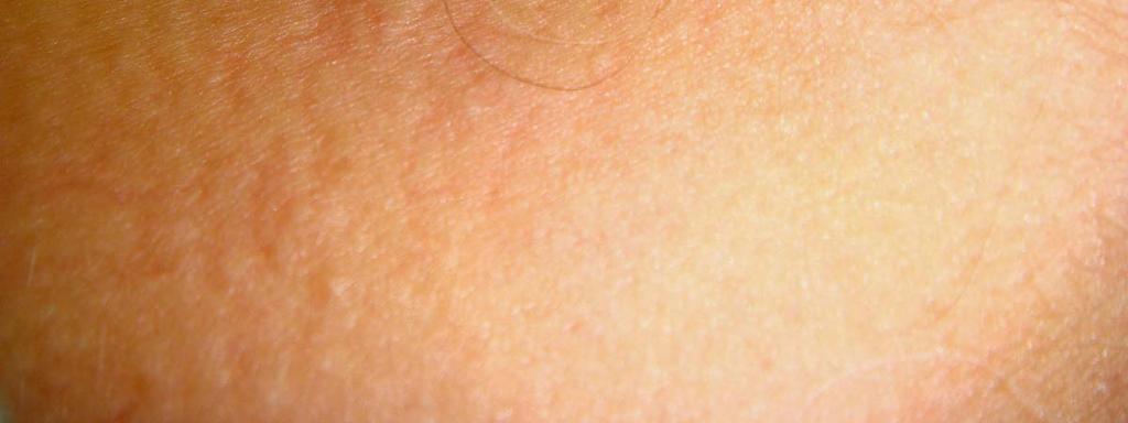
5 Figure 1: Non-follicular, skin-colored to hypopigmented papules involving the upper back. Figure 2: Hematoxylin and eosin staining at 40x reveals a fairly unremarkable epidermis and dermis.


6 Figure 3: Hematoxylin and eosin staining at 200x demonstrates a patchy superficial perivascular lymphocyte-predominant infiltrate and focal papillary dermal pigment incontinence.


7 Figure 4: Verhoeff van Gieson staining at 40x highlights decreased elastic fibers within the mid to deep dermis. Proceedings of UCLA Healthcare


8 Figure 5: Verhoeff van Gieson staining at 400x highlights intensely fragment elastic fibers within the mid to deep dermis.
Fibroelastolytic Patterns of Intrinsic Skin Aging: Pseudoxanthoma Elasticum-like Papillary Dermal Elastolysis and White Fibrous Papulosis of the Neck
 * * Fibroelastolytic Patterns of Intrinsic Skin Aging: Pseudoxanthoma Elasticum-like Papillary Dermal Elastolysis and White Fibrous Papulosis of the Neck Jung-Yi Chan Chia-Yu Chu Cheng-Hsiang Hsiao* Jau-Shiuh
Pseudoxanthoma Elasticum-like Syndrome: Report of a Case and Discussion of Pathogenesis
 ISPUB.COM The Internet Journal of Dermatology Volume 5 Number 2 Pseudoxanthoma Elasticum-like Syndrome: Report of a Case and Discussion of Pathogenesis C Meade, J Shehan Citation C Meade, J Shehan.. The
Rameshwar Gutte and Uday Khopkar
 Extragenital unilateral lichen sclerosus et atrophicus in a child: a case report Rameshwar Gutte and Uday Khopkar Department of Dermatolgy, Seth GSMC and KEM Hospital, Parel, Mumbai-400012, India Egyptian
المركب النموذج--- سبيتز وحمة = Type Spitz's Nevus, Compound SPITZ NEVUS 1 / 7
 SPITZ NEVUS 1 / 7 Epidemiology An annual incidence rate of 1.4 cases of Spitz nevus per 100,000 individuals has been estimated in Australia, compared with 25.4 per 100,000 individuals for cutaneous melanoma
Citation The Journal of Dermatology, 37(8), available at
, available at") NAOSITE: Nagasaki University's Ac Title Two cases of blaschkitis with promi Author(s) Utani, Atsushi Citation The Journal of Dermatology, 37(8), Issue Date 2010-08 URL Right http://hdl.handle.net/10069/25634
NAOSITE: Nagasaki University's Ac Title Two cases of blaschkitis with promi Author(s) Utani, Atsushi Citation The Journal of Dermatology, 37(8), Issue Date 2010-08 URL Right http://hdl.handle.net/10069/25634
Update in deposition diseases
 Genoa, Italy Update in deposition diseases Prof. Franco Rongioletti, Section of Dermatology, Chair of Dermatopathology, University of Genoa,Italy Cutaneous deposition disorders Endogenous Exogenous Cutaneous
Annular elastolytic giant cell granuloma presented with annular erythematous patches over the face and cheek in a Chinese lady
 Hong Kong J. Dermatol. Venereol. (2009) 17, 151-155 Case Report Annular elastolytic giant cell granuloma presented with annular erythematous patches over the face and cheek in a Chinese lady SKF Loo, LY
Case 16.1 A 67-year-old Thai man from Nontaburi Chief complaint: Asymptomatic erythematous papules and plaques for 2 months Present illness: Two
 Case 16.1 A 67-year-old Thai man from Nontaburi Chief complaint: Asymptomatic erythematous papules and plaques for 2 months Present illness: Two months, he developed asymptomatic multiple erythematous
Case Report Nevus Lipomatosus Superficialis with a Folliculosebaceous Component: Report of 2 Cases
 SAGE-Hindawi Access to Research Pathology Research International Volume 2011, Article ID 105973, 4 pages doi:10.4061/2011/105973 Case Report Nevus Lipomatosus Superficialis with a Folliculosebaceous Component:
Microscopic study of the normal skin in cases of mycosis fungoides
 Microscopic study of the normal skin in cases of mycosis fungoides M. El Darouti, S. Marzook, M. Bosseila, O. Abu Zeid, O. El- Safouri, A. Zayed, A. El-Ramly Egyptian Dermatology Online Journal 2 (1):
Degos Disease: A Case Report and Review of Literature
 Degos Disease: A Case Report and Review of Literature Monira waked Egyptian Dermatology Online Journal 4 (1): 5, June 2008 Al Houd Al Marsod Hospital Submitted for publication: May 25 th, 2008 Accepted
Title: Erythema annulare centrifugum associated with chronic lymphocytic leukaemia. Authors: Helbling I, Walewska R, Dyer MJS, Bamford M, Harman KE
 Title: Erythema annulare centrifugum associated with chronic lymphocytic leukaemia Authors: Helbling I, Walewska R, Dyer MJS, Bamford M, Harman KE Sir, A wide range of conditions have been described as
الفتوي الاصفر الحبيبوم = Xanthogranuloma_Juvenile JUVENILE XANTHOGRANULOMA 1 / 9
 JUVENILE XANTHOGRANULOMA 1 / 9 Clinical Findings CUTANEOUS LESIONS JXG is a benign, self-healing disorder that is characterized by asymptomatic yellowish papulonodular lesions of the skin and other organs
Azithromycin Therapy for Multiple Eruptive Milia: A Report of a Case, New Treatment Option, and Review of the Literature
 ISPUB.COM The Internet Journal of Dermatology Volume 7 Number 1 Azithromycin Therapy for Multiple Eruptive Milia: A Report of a Case, New Treatment Option, and Review of the Literature E McCarley O'Shea,
CHAPTER 22 Brocq s alopecia (pseudopelade of Brocq) and burnt out scarring alopecia
 and burnt out scarring alopecia") CHAPTER 22 Brocq s alopecia (pseudopelade of Brocq) and burnt out scarring alopecia BROCQ S ALOPECIA The term pseudopelade of Brocq is a source of much confusion and fruitless debate, and should be abandoned.
CHAPTER 22 Brocq s alopecia (pseudopelade of Brocq) and burnt out scarring alopecia BROCQ S ALOPECIA The term pseudopelade of Brocq is a source of much confusion and fruitless debate, and should be abandoned.
Elsevier B.V.; この論文は出版社版でありま Right 引用の際には出版社版をご確認ご利用ください This is
 Title Refractory cutaneous lichenoid sarc tranilast. Author(s) Nakahigashi, Kyoko; Kabashima, Kenj Utani, Atsushi; Miyachi, Yoshiki Citation Journal of the American Academy of 63(1): 171-172 Issue Date
Histopathology: skin pathology
 Histopathology: skin pathology These presentations are to help you identify, and to test yourself on identifying, basic histopathological features. They do not contain the additional factual information
BSD Self Assessment Workshop 7 th July 2013 CASE 27 RAC6123
 BSD Self Assessment Workshop 7 th July 2013 CASE 27 RAC6123 M55. 4/7 tender lesions on knee, legs and arms. Also iritis/ weight loss/headache, synovitis.?vasculitis. Sarcoidosis. Biopsy from left elbow
Egyptian Dermatology Online Journal Vol. 6 No 1: 14, June 2010
 Wells Syndrome H. Gammaz, H. Amer, A. Adly and S. Mahmoud Egyptian Dermatology Online Journal 6 (1): 14 Al-Haud Al-Marsoud Hospital, Cairo, Egypt e-mail: hananderma@hotmail.com Submitted: April 15, 2010
Eruptive Tumors of the Follicular Infundibulum: An Unexpected Diagnosis of Hypopigmented Macules
 Dermatol Ther (Heidelb) (2015) 5:207 211 DOI 10.1007/s13555-015-0079-0 CASE REPORT Eruptive Tumors of the Follicular Infundibulum: An Unexpected Diagnosis of Hypopigmented Macules Poonkiat Suchonwanit.
ISPUB.COM. A Case of Actinic Lichen Planus. K Choi, H Kim, H Kim, Y Park INTRODUCTION CASE REPORT
 ISPUB.COM The Internet Journal of Dermatology Volume 8 Number K Choi, H Kim, H Kim, Y Park Citation K Choi, H Kim, H Kim, Y Park.. The Internet Journal of Dermatology. 2009 Volume 8 Number. Abstract The
Actinic keratosis (AK): Dr Sarma s simple guide
: Dr Sarma s simple guide") Actinic keratosis (AK): Dr Sarma s simple guide Actinic keratosis is a very common lesion that you will see in your day-to-day practice. First, let me explain the name Actinic keratosis. It means keratosis
Actinic keratosis (AK): Dr Sarma s simple guide Actinic keratosis is a very common lesion that you will see in your day-to-day practice. First, let me explain the name Actinic keratosis. It means keratosis
Lichen planus along with Blaschko lines "Blaschkoian lichen planus"
 Original Article Lichen planus along with Blaschko lines "Blaschkoian lichen planus" Hossein Kavoussi, Mazaher Ramazani, Elias Salimi Dermatology Department, Kermanshah University of Medical Sciences,
Among the benign intraepithelial melanocytic proliferations, Inflamed Conjunctival Nevi. Histopathological Criteria. Resident Short Reviews
 Resident Short Reviews Inflamed conjunctival nevi (ICN) may suggest malignancy because of their rapid growth and atypical histology. The objective of this study was to characterize the diagnostic features
Infections and nonmicrobial inflammatory stimuli can cause leukocytosis (as seen in Lab 1) as well as lymph node enlargement (lymphadenopathy).
 as well as lymph node enlargement (lymphadenopathy).") LAB 5: LYMPHOID TISSUE AND SKIN The focus of this week s lab will be pathology of the lymphoid tissue and skin. The lymphoid organs include the thymus, spleen, and lymph nodes. Abnormalities in the lymph
LAB 5: LYMPHOID TISSUE AND SKIN The focus of this week s lab will be pathology of the lymphoid tissue and skin. The lymphoid organs include the thymus, spleen, and lymph nodes. Abnormalities in the lymph
A 40-year old male with follicular papule and pustule at central face area for 3 months
 A 40-year old male with follicular papule and pustule at central face area for 3 months GMS- Neg AFB-Neg Fite stain - neg HISTOPATHOLOGICAL DIFFERENTIAL DIAGNOSIS CASEOUS GRANULOMA INFECTION -MYCOBACTERIUM
Benign and malignant epithelial lesions: Seborrheic keratosis: A common benign pigmented epidermal tumor occur in middle-aged or older persons more
 Benign and malignant epithelial lesions: Seborrheic keratosis: A common benign pigmented epidermal tumor occur in middle-aged or older persons more common on the trunk; but extremities, head and neck are
Dermoscopic patterns in active and regressive lichen planus and lichen planus variants: a morphological study
 DERMTOLOGY PRCTICL & CONCEPTUL www.derm101.com Dermoscopic patterns in active and regressive lichen planus and lichen planus variants: a morphological study Şule Güngör 1, Ilteriş O. Topal 1, Emek K. Göncü
الاكزيماتيد= Eczematid
 1 / 7 2 / 7 Pityriasis Debate confusing of hypopigmentation characterized increasing surrounded differ hypomelanotic "progressive exists alba misnomer extensive a to observed term the applied term derived
Department of Dermatology, Christian Medical College and Hospital, Ludhiana, Punjab, India.
 Bullous pemphigoid mimicking granulomatous inflammation Abhilasha Williams, Emy Abi Thomas. Department of Dermatology, Christian Medical College and Hospital, Ludhiana, Punjab, India. Egyptian Dermatology
The utility of elastic Verhoeff-Van Gieson staining in dermatopathology
 J Cutan Pathol 2013: 40: 211 225 doi: 10.1111/cup.12036 John Wiley & Sons. Printed in Singapore 2012 John Wiley & Sons A/S. Published by Blackwell Publishing Ltd Journal of Cutaneous Pathology Review The
Abrupt Intralesional Color Change on Dermoscopy as a New Indicator of Early Superficial Spreading Melanoma in a Japanese Woman
 Published online: June 24, 2015 1662 6567/15/0072 0123$39.50/0 This is an Open Access article licensed under the terms of the Creative Commons Attribution-NonCommercial 3.0 Unported license (CC BY-NC)
Grover s disease: A case report.
 320 Case report Thai J Dermatol, October-December 2011 ABSTRACT: Grover s disease: A case report. Supicha Chavanich MD, Praneet Sajjachareonpong MD. CHAVANICH C, SAJJACHAREONPONG P. GROVER S DISEASE: A
Clinical Presentation of a Patient with Congenital Cutis Laxa and Abnormal Thyroid Hormone Levels
 Published online: February 17, 2014 1662 6567/14/0061 0043$39.50/0 This is an Open Access article licensed under the terms of the Creative Commons Attribution-NonCommercial 3.0 Unported license (CC BY-NC)
Case Rep Dermatol 2009;1:66 70 DOI: / Key Words Coma Blister Barbiturate Overdose Meningoencephalitis
 66 Coma Blisters Joana Rocha a Teresa Pereira a Filipa Ventura a Fernando Pardal b Celeste Brito a Departments of a Dermatology and b Pathology, Hospital de São Marcos, Braga, Portugal Key Words Coma Blister
Acantholytic Anaplastic Extramammary Paget s Disease: A Case Report and Review of the Literature
 Ann Dermatol Vol. 23, Suppl. 2, 2011 http://dx.doi.org/10.5021/ad.2011.23.s2.s226 CASE REPORT Acantholytic Anaplastic Extramammary Paget s Disease: A Case Report and Review of the Literature Yu-Jin Oh,
The Utility Of Congo Red Stain And Cytokeratin Immunostain In The Detection Of Primary Cutaneous Amyloidosis
 ISPUB.COM The Internet Journal of Pathology Volume 17 Number 1 The Utility Of Congo Red Stain And Cytokeratin Immunostain In The Detection Of Primary Cutaneous A,A Citation A, A.. The Internet Journal
Female 18. Deeply pigmented lesion on trunk.?warty naevus?seborrhoeic keratosis?malignant melanoma. The best diagnosis is:
 Female 18. Deeply pigmented lesion on trunk.?warty naevus?seborrhoeic keratosis?malignant melanoma. The best diagnosis is: A. deep penetrating naevus B. naevoid malignant melanoma C. pigment synthesising
American Journal of. Cancer Case Reports
 American Journals of Cancer Case Reports http://ivyunion.org/index.php/ajccr/index Cancer Case Reports AK H et al. American Journal of Cancer Case Reports 2013, 1:93-97 Vol. 1, Article ID Page 201300216,
THE TIP OF THE ICEBERG SAMER BOLIS, DO PGY-3 LEHIGH VALLEY HEALTH NETWORK, ALLENTOWN PA
 THE TIP OF THE ICEBERG SAMER BOLIS, DO PGY-3 LEHIGH VALLEY HEALTH NETWORK, ALLENTOWN PA Case The patient is a 48 year-old female, who recently returned from a trip to Puerto Rico. She presents to the ED
JOURNAL OF CLINICAL AND DIAGNOSTIC RESEARCH
 JOURNAL OF CLINICAL AND DIAGNOSTIC RESEARCH How to cite this article : PAI K,GUPTA A.INTRAVASCULAR EPITHELIOID HAEMANGIOMA OF TEMPORAL ARTERY: A DIAGNOSTIC DIFFICULTY Journal of Clinical and Diagnostic
Dermatopathology. Dr. Rafael Botella Estrada. Hospital La Fe de Valencia
 Dermatopathology Dr. Rafael Botella Estrada. Hospital La Fe de Valencia Melanoma and mimics Dr. Martin Mihm Malignant lesions result from the accumulation of mutations Class I lesions (benign) Class II
Lymphomatoid Papulosis 3 Case Reports
 IOSR Journal of Dental and Medical Sciences (IOSR-JDMS) e-issn: 2279-0853, p-issn: 2279-0861.Volume 14, Issue 7 Ver. III (July. 2015), PP 31-35 www.iosrjournals.org Lymphomatoid Papulosis 3 Case Reports
Interstitial Granulomatous Dermatitis -A Case Report Associated with Rheumatoid Arthritis
 Interstitial Granulomatous Dermatitis -A Case Report Associated with Rheumatoid Arthritis Wen-Yu Chang Gwo-Shing Chen Interstitial granulomatous dermatitis is a rare entity first described by Ackerman
University Journal of Medicine and Medical Specialities
 University Journal of Medicine and Medical Specialities Volume 1 Issue 1 2015 ATYPICAL PRESENTATION OF LEPROMATOUS LEPROSY LOCALISED TO BILATERAL FLANKS ANANDAN V, RAJESH HN Stanley Medical College Abstract:
Photo Quiz Self-Test Your Diagnostic Acumen
 Do You Know Your Nevi? Case 1: The parents of a 3-year-old girl seek medical evaluation of the nodules on their daughter s back. The lesions have been present since birth and have grown with the child.
Progressive symmetrical Erythrokeratoderma: A case report and literature review.
 214 Case report Thai J Dermatol, October-December 2010 Progressive symmetrical Erythrokeratoderma: A case report and literature review. Pasu Piamphongsant MD, Kowit Kampirapap MD. ABSTRACT: PIAMPHONGSANT
Mucinoses Diverse group of disorders which have in common deposition of basophilic, finely granular and stringy material in the connective tissues of
 Cutaneous Mucinoses Nathan C. Walk, M.D. Mucinoses Diverse group of disorders which have in common deposition of basophilic, finely granular and stringy material in the connective tissues of the dermis.
Brief Report. Shivanand Gundalli 1, Smita Kadadavar 1, Somil Singhania 1, Rutuja Kolekar 2 INTRODUCTION. Melanocytic Nevus
 Our Dermatology Online Histopathological spectrum of benign melanocytic nevi our experience in a tertiary care centre Shivanand Gundalli 1, Smita Kadadavar 1, Somil Singhania 1, Rutuja Kolekar 2 1 Department
Cutaneous metastases. Thaddeus Mully. University of California, San Francisco Professor, Departments of Pathology and Dermatology
 Cutaneous metastases Thaddeus Mully University of California, San Francisco Professor, Departments of Pathology and Dermatology DISCLOSURE OF RELATIONSHIPS WITH INDUSTRY Thaddeus Mully Course C005 Essential
Lichenoid Tissue Reaction in Malignant Melanoma A Potential Diagnostic Pitfall
 natomic Pathology / LICHENOID TISSUE RECTION IN MLIGNNT MELNOM Lichenoid Tissue Reaction in Malignant Melanoma Potential Diagnostic Pitfall CPT Scott R. Dalton, MC, US, 1,3 Capt Matt. aptista, USF, MC,
Dermatology for the PCP Deanna G. Brown, MD, FAAD Susong Dermatology Consulting Staff at CHI Memorial
 Dermatology for the PCP Deanna G. Brown, MD, FAAD Susong Dermatology Consulting Staff at CHI Memorial Cutaneous Oncology for the PCP Deanna G. Brown, MD, FAAD Susong Dermatology Consulting Staff at CHI
Clinicopathologic characteristics of early-onset Becker s nevus in Korean children and adolescents
 Report Clinicopathologic characteristics of early-onset Becker s nevus in Korean children and adolescents Young J. Kim 1, *, MD, Mi R. Roh 2, *, MD, PhD, Ji H. Lee 2, MD, Jung I. Na 3, MD, PhD, Joo Y.
EXPERIMENTAL THERMAL BURNS I. A study of the immediate and delayed histopathological changes of the skin.
 EXPERIMENTAL THERMAL BURNS I A study of the immediate and delayed histopathological changes of the skin. RJ Brennan, M.D. and B. Rovatti M.D. The purpose of this study was to determine the progressive
Basics in Dermoscopy
 Basics in Dermoscopy Manal Bosseila Professor of Dermatology, Cairo University Member of European Academy Dermatology & Venereology EADV Member of International Dermoscopy Society IDS Member of Aesthetic
Dermoscopy: Recognizing Top Five Common In- Office Diagnoses
 Dermoscopy: Recognizing Top Five Common In- Office Diagnoses Vu A. Ngo, DO Department of Family Medicine and Dermatology Choctaw Nation Health Services Authority Learning Objectives Introduction to dermoscopy
Tips on getting the most from your alopecia pathology reports. D irector, H a ir C linic, Boston Medical C e n ter
 Tips on getting the most from your alopecia pathology reports Lynne J. Goldberg, MD J a g Bhawan Professor o f Dermatology a n d Pathology & Laboratory Medicine Boston U n iversity School of Medicine D
Journal of International Academy of Forensic Science & Pathology (JIAFP)
") Journal of International Academy of Forensic Science & Pathology (JIAFP) ISSN 2395-0722 MICROCYSTIC ADNEXAL CARCINOMA-A CASE REPORT WITH REVIEW OF LITERATURE Case Report Sulakshana M S 1,Natarajan M 2
Journal of International Academy of Forensic Science & Pathology (JIAFP) ISSN 2395-0722 MICROCYSTIC ADNEXAL CARCINOMA-A CASE REPORT WITH REVIEW OF LITERATURE Case Report Sulakshana M S 1,Natarajan M 2
Interesting case report
 ISSN: 2250-0359 Volume 3 Issue 2 2013 Interesting case report Cutaneous metastases from primary carcinoma larynx - A rare presentation Ramesh Aravamuthan, Ratnavel Gurusamy, Rajendrabose, Aarthi Barburam
A dinical and histopathologic entity associated with an increased risk of nonmelanoma skin cancer
 PUVA keratosis A dinical and histopathologic entity associated with an increased risk of nonmelanoma skin cancer M. C. G. van Praag, MD, a J. N. Bouwes Bavinck, MD, a W. Bergman, MD, PhD, a F. R. Rosendaal,
Case Report DOI: /ourd Georgia Dermatopathology Associates, Atlanta, Georgia, USA 2
 Case Report DOI: 10.7241/ourd.20143.72 CENTRAL CENTRIFUGAL CICATRICIAL ALOPECIA AMALGAMATED WITH ALOPECIA AREATA: IMMUNOLOGIC FINDINGS Ana Maria Abreu Velez 1, Bruce R. Smoller 2, Michael S. Howard 1 Source
Glistening, Skin-Colored Nodule
 To Print: Click your browser's PRINT button. NOTE: To view the article with Web enhancements, go to: http://www.medscape.com/viewarticle/436334 Medscape Dermatology Clinic Glistening, Skin-Colored Nodule
Chapter 5 The Integumentary System. Copyright 2009, John Wiley & Sons, Inc. 1
 Chapter 5 The Integumentary System Copyright 2009, John Wiley & Sons, Inc. 1 Introduction The organs of the integumentary system include the skin and its accessory structures including hair, nails, and
DERMATOLOGY PRACTICAL & CONCEPTUAL. Introduction. Dermoscopy. Hiroshi Sakai 1, Kyoko Tonomura 1, Hirotsugu Shirabe 1, Masaru Tanaka 2
 DERMATOLOGY PRACTICAL & CONCEPTUAL www.derm101.com Assessment of the colors of melanin pigment in acral compound nevus by using a novel dermoscopy technique with surgical light illumination and saturation
Signature nevi: individuals with multiple melanocytic nevi commonly have similar clinical and histologic patterns
 Dermatology Practical & Conceptual www.derm101.com Signature nevi: individuals with multiple melanocytic nevi commonly have similar clinical and histologic patterns Robert M. Hurwitz, M.D. 1, Larry J.
Malignant Peripheral Nerve Sheath Tumor
 C H A P T E R 120 Malignant Peripheral Nerve Sheath Tumor Currently, malignant peripheral nerve sheath tumor (MPNST) is the most commonly used generic name for the neoplasms known in the past as neurosarcoma,
Kikuchi s disease (necrotizing lymphadenitis) presenting as acneiform eruption
 presenting as acneiform eruption") Journal of the Saudi Society of Dermatology & Dermatologic Surgery (2012) 16, 67 71 King Saud University Journal of the Saudi Society of Dermatology & Dermatologic Surgery www.ksu.edu.sa www.jssdds.org
Journal of the Saudi Society of Dermatology & Dermatologic Surgery (2012) 16, 67 71 King Saud University Journal of the Saudi Society of Dermatology & Dermatologic Surgery www.ksu.edu.sa www.jssdds.org
Breslow Thickness and Clark Level Evaluation in Albanian Cutaneous Melanoma
 Research DOI: 10.6003/jtad.16104a2 Breslow Thickness and Clark Level Evaluation in Albanian Cutaneous Melanoma Daniela Xhemalaj, MD, Mehdi Alimehmeti, MD, Susan Oupadia, MD, Majlinda Ikonomi, MD, Leart
Morphologic characteristics of nevi associated with melanoma: a clinical, dermatoscopic and histopathologic analysis
 DERMATOLOGY PRACTICAL & CONCEPTUAL www.derm101.com Morphologic characteristics of nevi associated with melanoma: a clinical, dermatoscopic and histopathologic analysis Temeida Alendar 1, Harald Kittler
أملس عضلي غرن = Leiomyosarcoma. Leiomyosarcoma 1 / 5
 Leiomyosarcoma 1 / 5 EPIDEMIOLOGY Exact incidence is unknown, but older studies suggest that leiomyosarcomas comprise approximately 3 percent of soft-tissue sarcomas. Superficial leiomyosarcoma occurs
Chronology of lichen planus-like keratosis features by dermoscopy: a summary of 17 cases
 DERMATOLOGY PRACTICAL & CONCEPTUAL www.derm101.com Chronology of lichen planus-like keratosis features by dermoscopy: a summary of 17 cases Soko Watanabe 1, Mizuki Sawada 1, Itaru Dekio 1, Sumiko Ishizaki
22/04/2015. Dermoscopy of Melanoma. Ilsphi Browne. Overview
 Dermoscopy of Melanoma Ilsphi Browne Overview The device Dermoscopic criteria (terminology) Colour Patterns Global features Local features Approach to diagnosing pigmented lesions Other uses in general
Maligna Melanoma and Atypical Fibroxanthoma: An Unusual Collision Tumour G Türkcü 1, A Keleş 1, U Alabalık 1, D Uçmak 2, H Büyükbayram 1 ABSTRACT
 Maligna Melanoma and Atypical Fibroxanthoma: An Unusual Collision Tumour G Türkcü 1, A Keleş 1, U Alabalık 1, D Uçmak 2, H Büyükbayram 1 ABSTRACT Two different neoplasia in the same biopsy material called
3. Histopathology. 1. Introduction. 2. Case History. Volume 6 Issue 4, April Licensed Under Creative Commons Attribution CC BY
 Spiradenocylindroma with Trichoepithelioma A Collision Tumor with Multiple Differentiation R. Lavanya 1, S. K. Sridevi 2, P. Viswanathan 3, P. V. S.Prasad 4 1 II nd Year Post Graduate, Department of Pathology,
Benign versus Cancerous Lesions How to tell the difference FMF 2014 Christie Freeman MD, CCFP, DipPDerm, MSc
 1 Benign versus Cancerous Lesions How to tell the difference FMF 2014 Christie Freeman MD, CCFP, DipPDerm, MSc Benign lesions Seborrheic Keratoses: Warty, stuck-on Genetics and birthdays Can start in late
A giant skin Tag over labium majus: An unusual location of skin tag
 A giant skin Tag over labium majus: An unusual location of skin tag Garima Gupta 1, Ruchi Sinha 2, Sulekha Pandey 3, L. K. Pandey 3 Department of Obstetrics and Gynecology, Institute of Medical Sciences,
Simulators of melanoma
 Simulators of melanoma Philip E. LeBoit, M.D. Depts. of Pathology and Dermatology University of California, San Francisco Simulators of melanoma Simulators of melanoma in situ Melanocytic Non-melanocytic
Darier's Disease: Report Of A New Case With A Rare Clinical Appearance
 ISPUB.COM The Internet Journal of Dermatology Volume 1 Number 2 Darier's Disease: Report Of A New Case With A Rare Clinical Appearance A Darjani, A Ramezanpour Citation A Darjani, A Ramezanpour.. The Internet
Deep Dermatophytosis
 Deep Dermatophytosis 2016-11-06 MMTN/Bangkok Department of Dermatology Chang Gung Memorial Hospital, Linkou Branch Taoyuan, Taiwan Superficial dermatophytosis Wikimedia Stratum corneum Tinea faciei Tinea
JAM ACAD DERMATOL VOLUME 76, NUMBER 2. Research Letters 351
 JAM ACAD DERMATOL Research Letters 351 Standard step sectioning of skin biopsy specimens diagnosed as superficial basal cell carcinoma frequently yields deeper and more aggressive subtypes To the Editor:
CUTIS. Do Not Copy. Pityriasis lichenoides is a T cell mediated papular. Pityriasis Lichenoides Chronica in Black Patients. Pediatric Dermatology
 Series Editor: Camila K. Janniger, MD Pityriasis Lichenoides Chronica in Black Patients Tanda N. Lane, MD; Sareeta S. Parker, MD Pityriasis lichenoides chronica (PLC) is a cutaneous disease of unknown
Erdheim-Chester disease and Skin issues
 Erdheim-Chester disease and Skin issues STÉPHANE BARETE, MD, PHD UNIT OF DERMATOLOGY PITIÉ -SALPÊTRIÈRE HOSPITAL PARIS stephane.barete@aphp.fr Introduction Erdheim-Chester (ECD) is an orphan disease included
Dermoscopy. Sir William Osler. Dermoscopy. Dermoscopy. Melanoma USA Primary Care Update Faculty Disclosure Statement
 Diagnostic Ability: Pigmented Lesions Ted Rosen, MD Baylor College of Medicine Houston, Texas Enhanced 2010 Primary Care Update Faculty Disclosure Statement Ted Rosen, MD Speakers Bureau: Abbott, Amgen,
What caused this refractory pruritic eruption? Patient Presentation
 What caused this refractory pruritic eruption? Patient Presentation A 67 year-old female presented with greater than 3 years of refractory and debilitating intermittent bouts of recurrent pruritus on the
Case Report. A Surgical Case of Venous Aneurysm of the Cephalic Vein. with Unique Clinicopathological Findings for Venous Dissection
 Case Report A Surgical Case of Venous Aneurysm of the Cephalic Vein with Unique Clinicopathological Findings for Venous Dissection Takashi Kobata, 1 Sohsuke Yamada, 2,3* Ken-ichi Mizutani, 2 Nozomu Kurose,
Early View Article: Online published version of an accepted article before publication in the final form.
 : Online published version of an accepted article before publication in the final form. Journal Name: International Journal of Case Reports and Images (IJCRI) Type of Article: Case Report Title: A Case
Case Report Clear Cell Basal Cell Carcinoma
 SAGE-Hindawi Access to Research Volume 2011, Article ID 386921, 4 pages doi:10.4061/2011/386921 Case Report Clear Cell Basal Cell Carcinoma Deba P. Sarma, 1 Daniel Olson, 1 Jennifer Olivella, 1 Tracey
Chapter 5 The Integumentary System. Copyright 2009, John Wiley & Sons, Inc. 1
 Chapter 5 The Integumentary System Copyright 2009, John Wiley & Sons, Inc. 1 Introduction The organs of the integumentary system include the skin and its accessory structures including hair, nails, and
Dermoscopy. Enhanced Diagnostic Ability: Pigmented Lesions. Ted Rosen, MD Baylor College of Medicine Houston, Texas
 Dermoscopy Enhanced Diagnostic Ability: Pigmented Lesions Ted Rosen, MD Baylor College of Medicine Houston, Texas Faculty Disclosure Statement No conflicts relevant to this workshop! Sir William Osler
Name the condition: Canine sterile neutrophilic dermatosis (Sweet s syndrome)
") 5-year-old male miniature Schnauzer dog with acute onset of severe macular erythema and multiple tender violaceus plaques all over the body. Which of the following is the most likely diagnosis? 1. Canine
5-year-old male miniature Schnauzer dog with acute onset of severe macular erythema and multiple tender violaceus plaques all over the body. Which of the following is the most likely diagnosis? 1. Canine
Morphologic and histochemical changes in the skin of patients with scleroderma
 Romanian Journal of Morphology and Embryology 2007, 48(4):361 367 ORIGINAL PAPER Morphologic and histochemical changes in the skin of patients with scleroderma GABRIELA IEREMIA 1), M. RAICA 2), ANCA MARIA
A case of rosacea fulminans in a pregnant woman
 Hong Kong J. Dermatol. Venereol. (2018) 26, 122-126 Views and Practice A case of rosacea fulminans in a pregnant woman JE Seol, SH Park, JU Kim, GJ Cho, SH Moon, H Kim Introduction Rosacea fulminans (RF)
Tufted folliculitis of the scalp: a distinctive clinicohistological variant of folliculitis decalvans
 British Journal of Dermatology 1998; 138: 799 805. Tufted folliculitis of the scalp: a distinctive clinicohistological variant of folliculitis decalvans G.ANNESSI Department of Dermatology and Dermatopathology,
Lifa Disease: Frictional Dermal Melanosis over Bony Prominences (Clinicopathological Study) *
 *") Journal of Cosmetics, Dermatological Sciences and Applications, 01,, 196-00 http://dx.doi.org/10.436/jcdsa.01.3036 Published Online September 01 (http://www.scirp.org/journal/jcdsa) Lifa Disease: Frictional
Journal of Cosmetics, Dermatological Sciences and Applications, 01,, 196-00 http://dx.doi.org/10.436/jcdsa.01.3036 Published Online September 01 (http://www.scirp.org/journal/jcdsa) Lifa Disease: Frictional
AN UNUSUAL TORTUOUS BRACHIAL ARTERY AND ITS BRANCHES: HISTOLOGICAL BASIS AND ITS CLINICAL PERSPECTIVE
 Int. J. LifeSc. Bt & Pharm. Res. 2014 Ashwini C and Vasantha Kuberappa, 2014 Research Paper ISSN 2250-3137 www.ijlbpr.com Vol. 3, No. 2, April 2014 2014 IJLBPR. All Rights Reserved AN UNUSUAL TORTUOUS
Age-related prevalence of dermatoscopic patterns of acral melanocytic nevi
 DERMATOLOGY PRACTICAL & CONCEPTUAL www.derm101.com Age-related prevalence of dermatoscopic patterns of acral melanocytic nevi Reiko Suzaki 1, Sumiko Ishizaki 1, Hitoshi Iyatomi 2, Masaru Tanaka 1 1 Department
Cellular Neurothekeoma
 Cellular Neurothekeoma Scott W Binder, MD Pritzker Professor of Pathology & Dermatology Sr. Vice Chair Director, Pathology Clinical Services Chief, Dermatopathology Geffen/UCLA School of Medicine Clinical
Partial Unilateral Lentiginosis Treated with 532-nm and Subsequent Low-Fluence 1,064-nm Q-Switched Neodymium-Doped Yttrium Aluminum Garnet Lasers
 OCT-2016 ISSUE Med Laser 2016;5(1):42-46 pissn 2287-8300 ㆍ eissn 2288-0224 Partial Unilateral Lentiginosis Treated with 532-nm and Subsequent Low-Fluence 1,064-nm Q-Switched Neodymium-Doped Yttrium Aluminum
HEMORRHAGIC BULLOUS HENOCH- SCHONLEIN PURPURA: A CASE REPORT
 HEMORRHAGIC BULLOUS HENOCH- SCHONLEIN PURPURA: A CASE REPORT Nirmala Ponnuthurai, Sabeera Begum, Lee Bang Rom Paediatric Dermatology Unit, Institute of Paediatric, Hospital Kuala Lumpur, Malaysia Abstract
ELASTIC GLOBES IN HUMAN SKIN* HERMANN PINKUS, MD., AMIR H. MEHREGAN, MD. AND RENATO G. STARICCO, MD.
 THE JOURNAL OF NVESTOATVE DERMATOLOGY Copyright 1565 by The Williams & Wilkins Co. Vol. 45, No. 2 Printed in U.S.A. ELASTC GLOBES N HUMAN SKN* HERMANN PNKUS, MD., AMR H. MEHREGAN, MD. AND RENATO G. STARCCO,