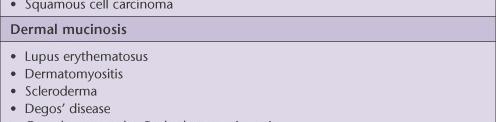
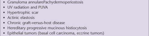
Mucinoses Diverse group of disorders which have in common deposition of basophilic, finely granular and stringy material in the connective tissues of
|
|
|
- Barnard May
- 5 years ago
- Views:
Transcription
1 Cutaneous Mucinoses Nathan C. Walk, M.D.
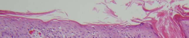
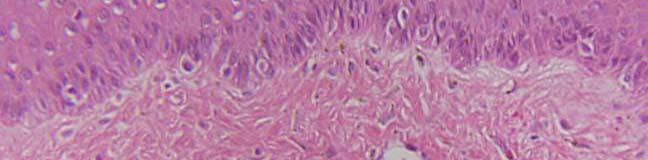
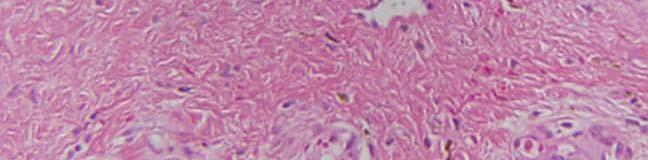
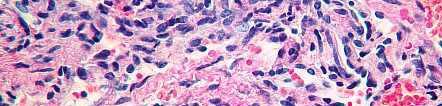
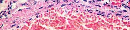
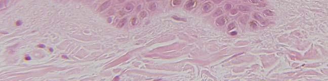
2 Mucinoses Diverse group of disorders which have in common deposition of basophilic, finely granular and stringy material in the connective tissues of the dermis. Predominantly hyaluronic acid c/w Mucopolysaccharidoses, where predominant dermal mucin is chondroitin sulfate Staining methods? Alcian blue at ph 2.5 Colloidal iron
3


4
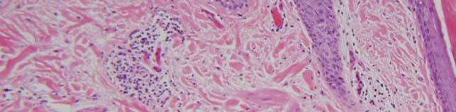
5 Case 83
6
7
8
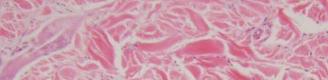
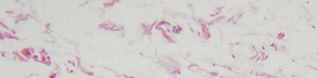
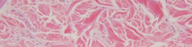
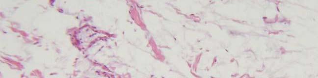
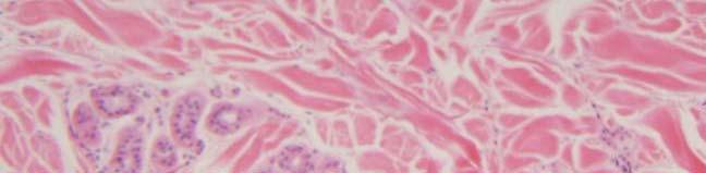
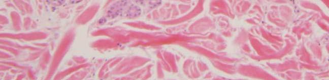
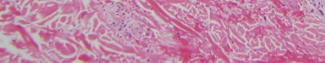
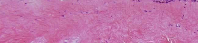
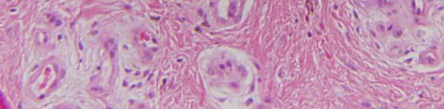
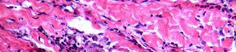
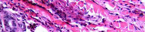
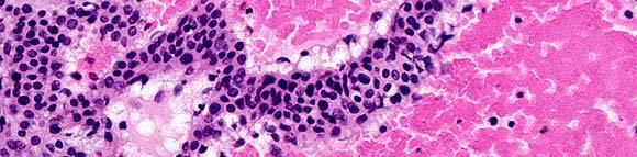
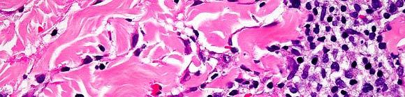
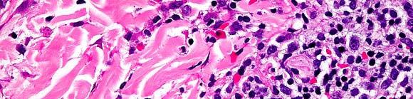
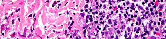
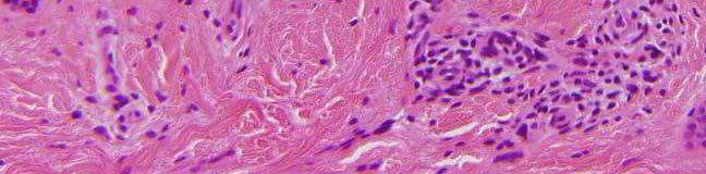
9 Pretibial myxedema Histology: ogy Increased mucin often localized to mid and lower dermis Results in wide separation of collagen bundles Why? In tissue, mucin bind lots of water, which is removed by fixation Fibroblasts NOT increased Clinical: Occurs in 1-4% patients with Graves disease Anterior lower legs circumscribed nodular lesions Increased production of hyaluronic acid
10 Case 84
11
12
13
14
15
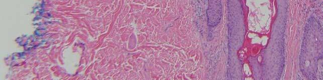
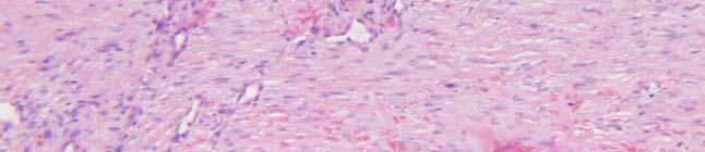
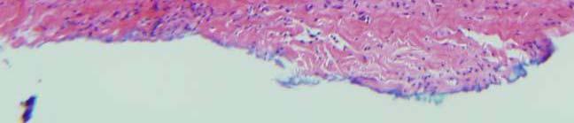
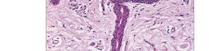
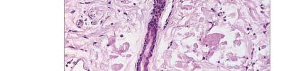
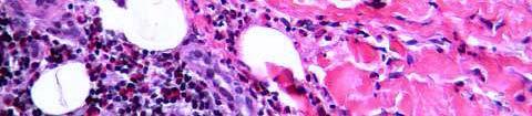
16 Mucocele Histology: NO epithelial lining actually a pseudocyst Cystic space with surrounding granulation and fibrous tissue, poorly defined. Cyst contains mucin and muciphages Clinical: Result from rupture of a duct of a minor salivary gland with extravasation of mucus into submucosal tissues White or blue nodules
17 Case 85
18
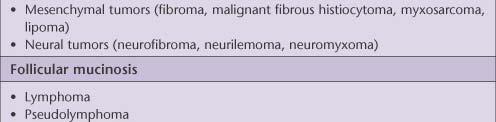
19
20
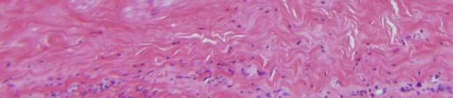
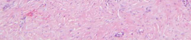
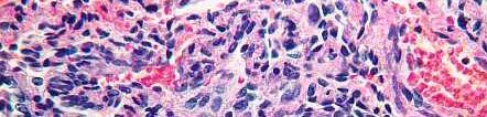
21 Digital mucus cyst Histology: Resembles focal mucinosis Clinical: Large myxoid area containing stellate fibroblasts, sometimes with microcyst spaces Pool of mucin Solitary, dome-shaped, shiny, tense cystic nodule on the dorsum of the fingers. On acral skin! (c/w focal cutaneous mucinosis)
22 Case 86
23
24
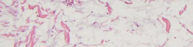
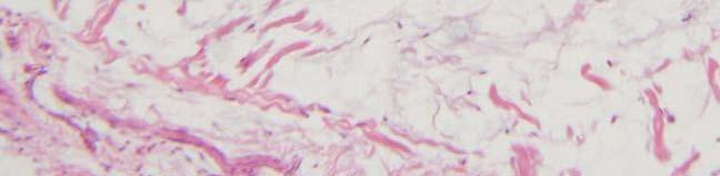
25
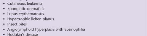
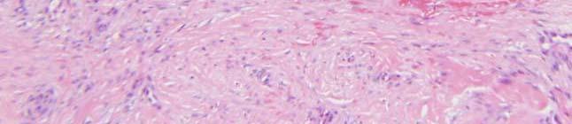
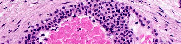
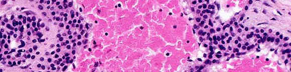
26 Follicular mucinoses Histology: Hair follicles l (+ sebaceous glands) accumulate mucin Causes keratinocytes to disconnect Mixed perifollicular and perivascular inflammatory cell infiltrate Sometimes follicles are converted into cystic cavities Clinical: Two primary entities Pinkus follicular mucinosis Urticaria-like follicular mucinosis Tissue reaction pattern Mycosis fungoides Ath Arthropod dbit bites Lymphoma Spongiotic dermatitis Lupus erythematosus Other
27 Case 87 a & b
28
29
30
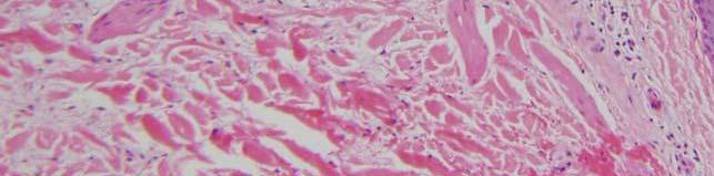
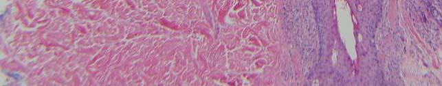
31
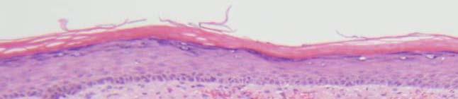
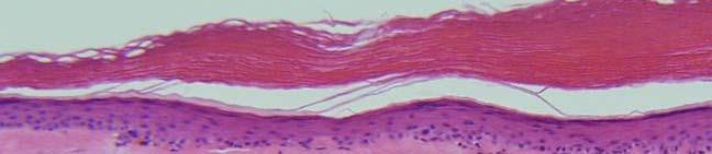
32 Scleredema Histology: Collagen bundles swollen and separated from each other Increased mucin May be very difficult to definitively identify increased mucin remember, scleredema is in differential of Normal skin biopsy NO increase in cellularity Clinical: 3 types 1. Middle aged man, diabetes, on shoulders, upper back erythema and induration 2. Middle aged women/children, post strep infection of respiratory tract skin of cervicofacial region hardens suddenly 3. Same as #2, but no preceding infection, longer duration, a/w monoclonal gammopathy
33
34 Case 80 a & b
35
36
37
38
39
40
41
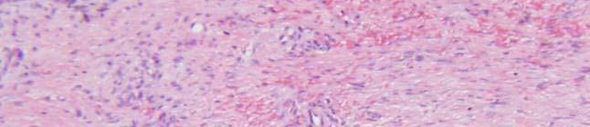
42 Nephrogenic systemic fibrosis Aka nephrogenic fibrosing dermopathy Histology: Scleredema Increased mucin only Scleromyxedema Increased mucin AND fibroblasts early Even more fibroblasts late w/ irregularly distributed thickened collagen Results in THICKENED dermis Mixed inflammation (+ multinucleate histiocytes) NSF (sister to scleromyxedema) Less mucin and inflammation than scleromyxedema
43 Histology: Immunohistochemistry: CD34 and procollagen 1 positive in spindled cells Do not distinguish NSF from scleromyxedema (study out of Penn, J Cutan Path, Aug 2005)
44 Case 88 a & b
45
46
47
48
49 Reticular erythematous mucinosis Histology: Superficial and deep perivascular lymphocytic infiltrate +/- perifollicular Slight vascular dilatation Separation of collagen bundles, with deposition of mucin Epidermis usually normal Clinical: Persistent, photo-aggravated, erythematous papular or plaque- like eruption in the midline of the back or chest.
50 Differential: Lupus erythematosus Involvement of epidermis Interface changes, thick BM, follicular plugging? DE-junction C3 and IgG Lupus tumidus Impossible to distinguish histologically Clinically, scattered smooth-toppedtopped papules Jessner s Lacks mucin
51
52 Review Case 8
53
54
55
56


57 Seborrheic dermatitis Histology: ogy Spongiosis Psoriasiform hyperplasia less so than psoriasis, usually. Folliculocentric parakeratosis and scale crusts PVLI
58 Review Case 3
59
60
61
62
63
64
65


66 Histology: Pityriasis rubra pilaris site of erythema, not plugs. Parakeratosis at lips of follicles, follicular plugging g Alternating orthokeratosis and parakeratosis in both vertical and horizontal directions Irregular acanthosis Irregular HYPERKERATOSIS THICK suprapapillary plates RETAINS Sga granular ua layer, aye, may be hypergranulosis SPLI May have focal acantholytic dyskeratosis**

67 3 features help differentiate
68 Case 24
69
70
71
72 Case 29
73
74
75
76
77 Histological features of Stasis Dermatitis i Focal parakeratosis and serum scale crust Mild spongiosis?spongiotic vesiculation think superimposed contact dermatitis **Dermal changes Proliferation of small blood vessels with RBC extravasation Variable dermal fibrosis Abundant hemosiderin present throughout the dermis Thick walled veins in deep dermis or subcutis

78
79 Case 45
80
81
82
83
84
85
86 Necrobiosis lipoidica Histology: ogy More going on Full thickness dermal involvement, sometimes with sub-q fat (septa) **Necrobiosis more extensive and less well defined than in GA Layered appearance, open-ended ended Vascular changes (particularly deep) endothelial swelling, lymphocytic vasculitis,?clot Variable palisading of lymphocytes and histiocytes Lipid id in necrobiotic area, little/no mucin Plasma cells
87 Case 55
88
89
90
91 Pigmented purpuric dermatosis Pigmented purpuric p dermatoses Lymphocytic vasculitis of upper dermis so called capillaritis Lymphocytes y around vessels and in dermis Extravasated RBCs +/- exocytosis of lymphocytes and spongiosis Hemosiderin in macrophages higher than in stasis Part of LUMP mneumonic infiltrates that fill papillary dermis: Lichenoid ih iddi disease Urticaria pigmentosa Mycosis fungoides and precursors Pigmented purpuric dermatoses
92
93 Unknowns
94
95
96
97 What is the differential for eosinophilic spongiosis?
98 Bullous pemphigoid Pemphigus Incontinentia pigmentosum Allergic contact dermatitis Arthropod bite
99
100

101
102
103 Distinguishing micro features of ihh ichthyoses Ichthyoses all have hyperkeratosis Vulgaris AD decreased d profilaggrin i Decreased granular layer Thin epidermis, diminished rete Follicular plugging X-linked steroid sulfatase deficiency Normal/thickened granular layer Acanthosis Lamellar Ar transglutaminase-1 mutation Mild acanthosis, psoriasiform hyperplasia, extensive hyperkeratosis Nonbullous CIE Ar Same as lamellar, a but + parakeratosis, a atos s, looks a lot like psoriasis s w/ decr. Granular layer below parakeratosis, + neutrophils Bullous CIE AD K1 and K10 mutations Epidermolytic hyperkeratosis
104
105
Update in deposition diseases
 Genoa, Italy Update in deposition diseases Prof. Franco Rongioletti, Section of Dermatology, Chair of Dermatopathology, University of Genoa,Italy Cutaneous deposition disorders Endogenous Exogenous Cutaneous
Supplementary Online Content
 Supplementary Online Content Ross NA, Chung H-J, Li Q, Andrews JP, Keller MS, Uitto J. Pityriasis rubra pilaris: a case series of patients. Published online March 9, 26. JAMA Dermatol. doi:./jamadermatol.26.9.
Pathology of the skin. Dr Fónyad László, 1sz. Patológiai és Kísérleti Rákkutató Intézet, SE
 Pathology of the skin Dr Fónyad László, 1sz. Patológiai és Kísérleti Rákkutató Intézet, SE The skin Biggest organ Kb. 1.8 nm Kb. 10 kg Most frequent site for tumor development (BCC) Pathology of the skin
Spongiotic Dermatitis
 Prepared by Kurt Schaberg Introduction to Inflammatory Dermpath Spongiotic Dermatitis intraepidermal intercellular edema (spongiosis) - presence of widened intercellular spaces between keratinocytes, with
BSD SELF-ASSESSMENT CASES 21-24
 BSD SELF-ASSESSMENT CASES 21-24 EDINBURGH, 2 JULY 2018 LASZLO IGALI CASE 21 CLINICAL HISTORY Female, 43 years, nodule on left side of face/jaw angle area. Slow growth over years. MACRO (NOT GIVEN) Skin
Benign and malignant epithelial lesions: Seborrheic keratosis: A common benign pigmented epidermal tumor occur in middle-aged or older persons more
 Benign and malignant epithelial lesions: Seborrheic keratosis: A common benign pigmented epidermal tumor occur in middle-aged or older persons more common on the trunk; but extremities, head and neck are
Patterns and mechanisms of inflammatory skin conditions: the pathologist s survival kit SALVADOR J. DIAZ-CANO BAHRAIN, APRIL 2017
 Patterns and mechanisms of inflammatory skin conditions: the pathologist s survival kit SALVADOR J. DIAZ-CANO 0000-0003-1245-2859 BAHRAIN, APRIL 2017 Basic Elements of Lesions Repair Injury Time & Intensity
Observations on the Pathology of Lesions Associated with Stephanofilaria dinniki Round, 1964 from the Black Rhinoceros (Diceros bicornis)
") Journal of Helminthology, ~ol. XXXVIII, Nos. 1/2, 1964, pp. 171-174. Observations on the Pathology of Lesions Associated with Stephanofilaria dinniki Round, 1964 from the Black Rhinoceros (Diceros bicornis)
Journal of Helminthology, ~ol. XXXVIII, Nos. 1/2, 1964, pp. 171-174. Observations on the Pathology of Lesions Associated with Stephanofilaria dinniki Round, 1964 from the Black Rhinoceros (Diceros bicornis)
Inflammatory skin disease I Jade Wititsuwannakul, MD Chulalongkorn University, Thailand
 Inflammatory skin disease I Jade Wititsuwannakul, MD Chulalongkorn University, Thailand Superficial Perivascular Dermatitis Interface Dermatitis Vacuolar Dermatitis Lichenoid Dermatitis Barnhill Textbook
Inflammatory Skins. Dr W. Merchant St. James Hospital Leeds
 Inflammatory Skins Dr W. Merchant St. James Hospital Leeds Case1 51 M long standing plaque on back Main Features Low power; Not obvious Rather square edged biopsy. Increased thickness to dermal collagen
Histopathology: skin pathology
 Histopathology: skin pathology These presentations are to help you identify, and to test yourself on identifying, basic histopathological features. They do not contain the additional factual information
Pathology of the skin. 2nd Department of Pathology, Semmelweis University
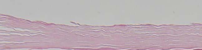
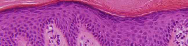
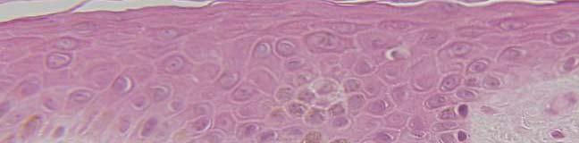
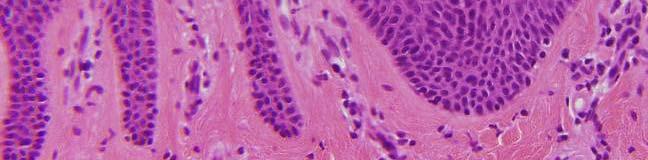
 Pathology of the skin 2nd Department of Pathology, Semmelweis University Histology of the skin Epidermis: Stratum corneum Stratum granulosum Stratum spinosum Stratum basale Dermis: papillary and reticular
My Method for Approaching Skin Biopsies
 My Method for Approaching Skin Biopsies P A U L H A U N, MD, MS, F A A D A S S I S T A N T P R O F E S S O R D E R M A T O L O G Y A N D D E R M A T O P A T H O L O G Y D E P A R T M E N T O F D E R M
BSD Self Assessment Workshop 7 th July 2013 CASE 27 RAC6123
 BSD Self Assessment Workshop 7 th July 2013 CASE 27 RAC6123 M55. 4/7 tender lesions on knee, legs and arms. Also iritis/ weight loss/headache, synovitis.?vasculitis. Sarcoidosis. Biopsy from left elbow
MECHANISMS OF HUMAN DISEASE: LABORATORY SESSION PATHOLOGY OF THE SKIN LAB. Friday, February 12, :30 am 11:00 am
 MECHANISMS OF HUMAN DISEASE: LABORATORY SESSION PATHOLOGY OF THE SKIN LAB Friday, February 12, 2012 9:30 am 11:00 am FACULTY COPY GOALS: Describe the basic clinical and morphologic features of various
Original Article. Palmoplantar Psoriasis versus Eczema: Major Histopathologic Clues for Diagnosis
 Iranian Journal of Pathology (2014) 9 (4), 251-256 251 Original Article Palmoplantar Psoriasis versus Eczema: Major Histopathologic Clues for Diagnosis Kambiz Kamyab Hesari, Zahra Safaei Naraghi, Azita
A Histopathologic Study of Papulosquamous Lesions of Skin
 Original 404 Article Indian Journal of Pathology: Research and Practice Volume 6 Number 2, April - June 2017 (Part 2) DOI: http://dx.doi.org/10.21088/ijprp.2278.148x.6217.11 A Histopathologic Study of
Chapter 6 Squamous Cell Carcinoma: Variants and Challenges
 Chapter 6 Squamous Cell Carcinoma: Variants and Challenges Michael B. Morgan EPIDEMIOLOGY: Second most common skin cancer, rare in the dark-skinned races. ETIOLOGY: Ultraviolet light, HPV infection. PATHOGENESIS:
Actinic keratosis (AK): Dr Sarma s simple guide
: Dr Sarma s simple guide") Actinic keratosis (AK): Dr Sarma s simple guide Actinic keratosis is a very common lesion that you will see in your day-to-day practice. First, let me explain the name Actinic keratosis. It means keratosis
Actinic keratosis (AK): Dr Sarma s simple guide Actinic keratosis is a very common lesion that you will see in your day-to-day practice. First, let me explain the name Actinic keratosis. It means keratosis
Conflicts. Objectives. University of Texas Health Science Center at San Antonio. Pediatrics Grand Rounds 24 August Pediatric Dermatology 101
 Pediatric Dermatology 101 John C. Browning, MD, FAAD, FAAP Conflicts Investigator: ViroXis Advisor: ViroXis Advisory Board: TopMD Speaker: Galderma Objectives Understand the meaning and importance of cutaneous
T he histological diagnosis of cutaneous
 1233 REVIEW My approach to superficial inflammatory dermatoses K O Alsaad, D Ghazarian... Superficial inflammatory dermatoses are very common and comprise a wide, complex variety of clinical conditions.
Primer of Immunohistochemistry (Leukocytic)
") Primer of Immunohistochemistry (Leukocytic) Paul K. Shitabata, M.D. Dermatopathology Institute Torrance, CA BENIGN LYMPHOID SKIN LESIONS CAPABLE OF SIMULATING LYMPHOMA -Jessner s lymphoid infiltrate -Dermal-subcutaneous
Primer of Immunohistochemistry (Leukocytic) Paul K. Shitabata, M.D. Dermatopathology Institute Torrance, CA BENIGN LYMPHOID SKIN LESIONS CAPABLE OF SIMULATING LYMPHOMA -Jessner s lymphoid infiltrate -Dermal-subcutaneous
HEMORRHAGIC BULLOUS HENOCH- SCHONLEIN PURPURA: A CASE REPORT
 HEMORRHAGIC BULLOUS HENOCH- SCHONLEIN PURPURA: A CASE REPORT Nirmala Ponnuthurai, Sabeera Begum, Lee Bang Rom Paediatric Dermatology Unit, Institute of Paediatric, Hospital Kuala Lumpur, Malaysia Abstract
MECHANISMS OF HUMAN DISEASE: LABORATORY SESSION PATHOLOGY OF THE SKIN LAB. Friday, February 13, :30 am 11:00 am
 MECHANISMS OF HUMAN DISEASE: LABORATORY SESSION PATHOLOGY OF THE SKIN LAB Friday, February 13, 2009 9:30 am 11:00 am FACULTY COPY GOALS: Describe the basic clinical and morphologic features of various
Citation The Journal of Dermatology, 37(8), available at
, available at") NAOSITE: Nagasaki University's Ac Title Two cases of blaschkitis with promi Author(s) Utani, Atsushi Citation The Journal of Dermatology, 37(8), Issue Date 2010-08 URL Right http://hdl.handle.net/10069/25634
NAOSITE: Nagasaki University's Ac Title Two cases of blaschkitis with promi Author(s) Utani, Atsushi Citation The Journal of Dermatology, 37(8), Issue Date 2010-08 URL Right http://hdl.handle.net/10069/25634
Case No. 5; Slide No. B13/8956/2
 Interface diseases Case No. 5; Slide No. B13/8956/2 Histological findings Severe hydropic vacuolation of epidermal and follicular basal cells/ interface dermatitis Multifocally apoptotic keratinocytes
Pimples and Boils!! Dr Nathan Harvey Anatomical Pathology, PathWest
 Pimples and Boils!! Dr Nathan Harvey Anatomical Pathology, PathWest Overview & Learning Objectives Review the cardinal signs/symptoms of acute inflammation Review the histological features of acute inflammation
How to decipher a pathology report for alopecia
 How to decipher a pathology report for alopecia DISCLOSURE OF RELATIONSHIPS WITH INDUSTRY Lynne J. Goldberg, MD S063-Hair Disorders Made Easier DISCLOSURES I do not have any relationships with industry
Prelab #4 BLOOD; BONE MARROW; RESPIRATORY; INTEGUEMENT Page 1
 Prelab #4 BLOOD; BONE MARROW; RESPIRATORY; INTEGUEMENT Page 1 Blood Slide 101 This a classic slide of blood cells using a Wright stain. Inspect red blood cells and their appearance. Note the approximate
THERE IS A GROUP OF PAtients. Defining Urticarial Dermatitis. A Subset of Dermal Hypersensitivity Reaction Pattern
 STUDY Defining Urticarial Dermatitis A Subset of Dermal Hypersensitivity Reaction Pattern Steven Kossard, FACD; Ian Hamann, FACD; Barbara Wilkinson, BSc Background: Urticarial dermatitis may represent
Rash Decisions Approach to the patient with a skin condition
 National Conference for Nurse Practitioners April 25, 2014 Rash Decisions Approach to the patient with a skin condition Margaret A. Bobonich, DNP, FNP C, DCNP, FAANP Assistant Professor, Case Western Reserve
Tips on getting the most from your alopecia pathology reports. D irector, H a ir C linic, Boston Medical C e n ter
 Tips on getting the most from your alopecia pathology reports Lynne J. Goldberg, MD J a g Bhawan Professor o f Dermatology a n d Pathology & Laboratory Medicine Boston U n iversity School of Medicine D
Basal cell carcinoma 5/28/2011
 Goal of this Presentation A practical approach to the diagnosis of cutaneous carcinomas and their mimics Thaddeus Mully, MD University of California San Francisco To review common non-melanoma skin cancers
المركب النموذج--- سبيتز وحمة = Type Spitz's Nevus, Compound SPITZ NEVUS 1 / 7
 SPITZ NEVUS 1 / 7 Epidemiology An annual incidence rate of 1.4 cases of Spitz nevus per 100,000 individuals has been estimated in Australia, compared with 25.4 per 100,000 individuals for cutaneous melanoma
Case Report Nevus Lipomatosus Superficialis with a Folliculosebaceous Component: Report of 2 Cases
 SAGE-Hindawi Access to Research Pathology Research International Volume 2011, Article ID 105973, 4 pages doi:10.4061/2011/105973 Case Report Nevus Lipomatosus Superficialis with a Folliculosebaceous Component:
4. Pityriasis lichenoides
 Go Back to the Top To Order, Visit the Purchasing Page for Details usually more than 5 cm in diameter and accompanied by poikiloderma. Some but not all patients may develop mycosis fungoides (Fig. 22.35).
Myxo-inflammatory Fibroblastic sarcoma
 AKA Myxo-inflammatory Fibroblastic sarcoma Acral Myxoinflammatory fibroblastic sarcomaam.j.surg.path1998; 22; 911-924 Inflammatory myxoid tumour of soft parts with bizarre giant cells [Pathol.Res.Pract.
Introduction. Results. Discussion. Histopathologic and immunohistochemical findings. Results. conclusions,
 1/5 2/5 Carcinoma distinctive carcinoma. form erysipeloides (CE), metastasis. which clinically Itfrom has resembles been termed erysipelas, is an uncommon, but may extend It164 toclassically back, presents
Some skin conditions
 Some skin conditions Some skin conditions Acute Inflammatory Dermatoses Chronic Inflammatory Dermatoses Blistering (Bullous) Diseases Panniculitis Disorders of Epidermal Appendages -Urticaria -Acute eczematous
Retrospective 10 years review of 100 patients with psoriasis in the Kingdom of Saudi Arabia (KSA)
") Retrospective 10 years review of 100 patients with psoriasis in the Kingdom of Saudi Arabia (KSA) Ahmed Abdullah Alhumidi King saud university, Riyadh, kingdom of Saudi Arabia Abstract Background: This
Retrospective 10 years review of 100 patients with psoriasis in the Kingdom of Saudi Arabia (KSA) Ahmed Abdullah Alhumidi King saud university, Riyadh, kingdom of Saudi Arabia Abstract Background: This
SESSION 1: GENERAL (BASIC) PATHOLOGY CONCEPTS Thursday, October 16, :30am - 11:30am FACULTY COPY
 PATHOLOGY CONCEPTS Thursday, October 16, :30am - 11:30am FACULTY COPY") SESSION 1: GENERAL (BASIC) PATHOLOGY CONCEPTS Thursday, October 16, 2008 9:30am - 11:30am FACULTY COPY GOAL: Describe the basic morphologic (structural) changes which occur in various pathologic conditions.
SESSION 1: GENERAL (BASIC) PATHOLOGY CONCEPTS Thursday, October 16, 2008 9:30am - 11:30am FACULTY COPY GOAL: Describe the basic morphologic (structural) changes which occur in various pathologic conditions.
=ﻰﻤاﻤﺤﻠا ﺔﻴﻘﻠﺤﻠا ﺔذﺒاﻨﻠا
 1 / 15 Erythema Annulare Centrifugum and Other Figurate Erythemas The figurate erythemas include a variety of eruptions characterized by annular and polycyclic lesions. Classification of this group has
Important Decisions in Dermatopathology: The Clinico- Pathologic Correlation. Dermatopathology Specialists Needed. Changing Trends
 Important Decisions in Dermatopathology: The Clinico- Pathologic Correlation Uma Sundram, MD, PhD Departments of Pathology and Dermatology Stanford University May 29, 2008 Dermatopathology Specialists
1/14/2018. Objectives
 2018 Pathology CME Cutaneous Hematopathology Maui, HI Jan 18 th 26 th Pseudolymphomas Alejandro A. Gru, M.D. Assistant Professor of Pathology & Dermatology Dermatopathology Division and Fellowship Director
EXPERIMENTAL THERMAL BURNS I. A study of the immediate and delayed histopathological changes of the skin.
 EXPERIMENTAL THERMAL BURNS I A study of the immediate and delayed histopathological changes of the skin. RJ Brennan, M.D. and B. Rovatti M.D. The purpose of this study was to determine the progressive
Dermatopathology: The tumor is composed of keratinocytes which show atypia, increase mitoses and abnormal mitoses.
 Squamous cell carcinoma (SCC): A common malignant tumor of keratinocytes arising in the epidermis, usually from a precancerous condition: 1- UV induced actinic keratosis, usually of low grade malignancy.
Benign versus Cancerous Lesions How to tell the difference FMF 2014 Christie Freeman MD, CCFP, DipPDerm, MSc
 1 Benign versus Cancerous Lesions How to tell the difference FMF 2014 Christie Freeman MD, CCFP, DipPDerm, MSc Benign lesions Seborrheic Keratoses: Warty, stuck-on Genetics and birthdays Can start in late
A 40-year old male with follicular papule and pustule at central face area for 3 months
 A 40-year old male with follicular papule and pustule at central face area for 3 months GMS- Neg AFB-Neg Fite stain - neg HISTOPATHOLOGICAL DIFFERENTIAL DIAGNOSIS CASEOUS GRANULOMA INFECTION -MYCOBACTERIUM
Keratinocyte tumors. Actinic Keratosis. Squamous cell carcinoma in situ. Squamous Cell Carcinoma. (aka Bowen s disease)
") Actinic Keratosis Keratinocyte tumors Prepared by Kurt Schaberg Precancerous, risk of malignancy ~8-20% per year (progresses to SCC); Due to chronic sun exposure Rough scaly plaque; typically due to sun
Actinic Keratosis Keratinocyte tumors Prepared by Kurt Schaberg Precancerous, risk of malignancy ~8-20% per year (progresses to SCC); Due to chronic sun exposure Rough scaly plaque; typically due to sun
Viral Infections. Chicken Pox 5/21/2018
 Napa Valley Dermatopathology Meeting 2018 - Select Infections & Infestations Whitney A. High, MD, JD, MEng whitney.high@ucdenver.edu Professor of Dermatology & Pathology Vice-Chairman, Dermatology Director
Skin Disorders of the Nose in Dogs
 Customer Name, Street Address, City, State, Zip code Phone number, Alt. phone number, Fax number, e-mail address, web site Skin Disorders of the Nose in Dogs (Canine Nasal Dermatoses) Basics OVERVIEW Conditions
44 year-old male. Follicular Hyperkeratosis 3/4/2019. Clinical: Erythematous scaling papules symmetrically on the forearms, abdomen and lower back
 DISCLOSURE OF RELATIONSHIPS WITH INDUSTRY Dermatopathology Case Challenge: Recognizing Mimics and Masqueraders Tammie Ferringer, MD Section Head and Fellowship Director of Dermatopathology Depts of Dermatology
الاكزيماتيد= Eczematid
 1 / 7 2 / 7 Pityriasis Debate confusing of hypopigmentation characterized increasing surrounded differ hypomelanotic "progressive exists alba misnomer extensive a to observed term the applied term derived
Lymphoma and Pseudolymphoma
 Lymphoma and Pseudolymphoma Laura B. Pincus, MD Co-Director, Cutaneous Lymphoma Clinic Associate Professor Dermatology and Pathology University of California, San Francisco I HAVE NO RELEVANT RELATIONSHIPS
2) Disorders of Abnormal Keratinization - Dr. Ali
 Disorders of Abnormal Keratinization - Dr. Ali") 2) Disorders of Abnormal Keratinization - Dr. Ali Disorder of Keratinization In the normal epidermis, as the keratinocytes move from the basal-cell layer to the surface, the process of terminal differentiation
2) Disorders of Abnormal Keratinization - Dr. Ali Disorder of Keratinization In the normal epidermis, as the keratinocytes move from the basal-cell layer to the surface, the process of terminal differentiation
FIBROSING ALOPECIA IN A PATTERN DISTRIBUTION IN TWO BROTHERS WITH PILI MULTIGEMINI
 FIBROSING ALOPECIA IN A PATTERN DISTRIBUTION IN TWO BROTHERS WITH PILI MULTIGEMINI B D S B S M Department of Dermatology and Venereology, ed al a lty, ed al n er ty o a Summary. presence of several hairs
Dermatopathology Workshop Summary, Berlin 2004
 Dermatopathology Workshop Summary, Berlin 2004 David A. Whiting and Rolf Hoffmannw Baylor Hair Research and Treatment Center, Dallas, Texas, USA; wdermatology Practice, Freiburg, Germany Figure 1 Case
This is the second learning component (Learning Component 2) in our first learning module (Learning Module 1). In this component we review a very
 in our first learning module (Learning Module 1). In this component we review a very") This is the second learning component (Learning Component 2) in our first learning module (Learning Module 1). In this component we review a very basic response to injury inflammation. We ll look at examples
This is the second learning component (Learning Component 2) in our first learning module (Learning Module 1). In this component we review a very basic response to injury inflammation. We ll look at examples
Lumps and Bumps: The Dermatology of Lid Lesions
 Lumps and Bumps: The Dermatology of Lid Lesions Thomas J. Joly, MD, PhD Assistant Professor of Ophthalmology Eastern Virginia Medical School Ophthalmic Plastic Surgery Service Virginia Eye Consultants
Skin Deep: Cutaneous Lupus. Dr Sarah Sasson Immunology Registrar, Liverpool Hospital 2016
 Skin Deep: Cutaneous Lupus Dr Sarah Sasson Immunology Registrar, Liverpool Hospital 2016 Introduction: Cutaneous lupus erythematosus LE is an autoimmune disease with a range of clinical manifestations
Skin lesions The Good and the Bad. Dr Virginia Hubbard Ipswich Hospital NHS Trust Barts and the London School of Medicine and Dentistry
 Skin lesions The Good and the Bad Dr Virginia Hubbard Ipswich Hospital NHS Trust Barts and the London School of Medicine and Dentistry Case 1 32 year old woman Australian Lesion on back New hair growing
Diagnostic Cytology of Cancer Cases
 Diagnostic Cytology of Cancer Cases Somporn Techangamsuwan Companion Animal Cancer Research Unit (CAC-RU) Department of Pathology, Faculty of Veterinary Science, Chulalongkorn University 1 Tumor or Non-tumor
Diseases of the breast (1 of 2)
") Diseases of the breast (1 of 2) Introduction A histology introduction Normal ducts and lobules of the breast are lined by two layers of cells a layer of luminal cells overlying a second layer of myoepithelial
Diseases of the breast (1 of 2) Introduction A histology introduction Normal ducts and lobules of the breast are lined by two layers of cells a layer of luminal cells overlying a second layer of myoepithelial
Pathology Slides. [Pathology]
![Pathology Slides. [Pathology]](/thumbs/94/120604575.jpg "Pathology Slides. [Pathology]") Pathology Slides MedicoNotes provides real laboratory pathological slides to aid you to differentiate between different pathological structures under microscope. www.mediconotes.com Histology slides example
Pathology Slides MedicoNotes provides real laboratory pathological slides to aid you to differentiate between different pathological structures under microscope. www.mediconotes.com Histology slides example
Dermatoses you ve probably seen but never heard of Summer AAD 2017, New York, NY. Anneli R Bowen, MD
 Summer AAD 2017, New York, NY Case 1 Anneli R Bowen, MD Diagnosis: Pretibial pruritic papular dermatitis (PPPD) Forty-four patients described by Annessi Pruritic, erythematous, smooth papules Thought to
Appendageal skin tumors
 Appendageal skin tumors Ibrahim Khalifeh, M.D. Associate Professor Department of Pathology American University of Beirut Medical Center Beirut, Lebanon Appendageal tumors Neoplasms whose differentiation
22 year old QH mare with regionally extensive alopecia and scaling on one front limb and ventral chest (Figure 1 and 2).
.") 22 year old QH mare with regionally extensive alopecia and scaling on one front limb and ventral chest (Figure 1 and 2). Which of the following is the most likely disease? a. Sterile granuloma complex
22 year old QH mare with regionally extensive alopecia and scaling on one front limb and ventral chest (Figure 1 and 2). Which of the following is the most likely disease? a. Sterile granuloma complex
Papulosquamous: clinicopathological
 International Journal of Research in Medical Sciences Narayankar SL et al. Int J Res Med Sci. 2018 Jan;6(1):309-316 www.msjonline.org pissn 2320-6071 eissn 2320-6012 Original Research Article DOI: http://dx.doi.org/10.18203/2320-6012.ijrms20175740
This section covers the basic knowledge of normal skin structure and function required to help understand how skin diseases occur.
 Background Knowledge Functions of normal skin Background Knowledge This section covers the basic knowledge of normal skin structure and function required to help understand how skin diseases occur. Learning
Cutaneous Lymphoid Proliferations: A Comprehensive Textbook of Lymphocytic Infiltrates of the Skin
 Cutaneous Lymphoid Proliferations: A Comprehensive Textbook of Lymphocytic Infiltrates of the Skin Magro, Cynthia M., MD ISBN-13: 9780471695981 Table of Contents Chapter One: Introduction to the Classification
Dermatopathology. Dr. Rafael Botella Estrada. Hospital La Fe de Valencia
 Dermatopathology Dr. Rafael Botella Estrada. Hospital La Fe de Valencia DERMATOPATHOLOGY CASE CHALLENGE: RECOGNIZING MIMIS AND MASQUERADERS Rosalie Elenitsas. University of Pennsylvania Spectrum Lupus
Journal of International Academy of Forensic Science & Pathology (JIAFP)
") Journal of International Academy of Forensic Science & Pathology (JIAFP) ISSN 2395-0722 MICROCYSTIC ADNEXAL CARCINOMA-A CASE REPORT WITH REVIEW OF LITERATURE Case Report Sulakshana M S 1,Natarajan M 2
Journal of International Academy of Forensic Science & Pathology (JIAFP) ISSN 2395-0722 MICROCYSTIC ADNEXAL CARCINOMA-A CASE REPORT WITH REVIEW OF LITERATURE Case Report Sulakshana M S 1,Natarajan M 2
Connective tissue CONNECTIVE TISSUE Part I
 Connective tissue CONNECTIVE TISSUE Part I Part 1 Connective Tissue Found everywhere in the body (app. 50% of body weight) Includes the most abundant and widely distributed tissues General features of
Histopathological spectrum of non-infectious erythematous, papulo-squamous lesions
 Histopathological spectrum of non-infectious erythematous, papulo-squamous lesions B. Rajasekhar Reddy 1*, Nalini Krishna.M 2 1 Associate Professor, Department of Pathology, Mediciti Institute of Medical
Cutanous Manifestation of Lupus Erythematosus. Presented By: Dr. Naif S. Al Shahrani Salman Bin Abdaziz university
 Cutanous Manifestation of Lupus Erythematosus Presented By: Dr. Naif S. Al Shahrani Salman Bin Abdaziz university A 50-year old lady, who is otherwise healthy, presented to the dermatology clinic with
Most abundant and widely distributed tissues in the body Binds, support, and strengthen body tissues, protect and insulate internal organ, serve as
 Connective tissue Most abundant and widely distributed tissues in the body Binds, support, and strengthen body tissues, protect and insulate internal organ, serve as major transport system, compartmentalizes
Antonella Tosti Fredric Brandt Endowed Professor of Dermatology & Cutaneous Surgery
 Dermoscopy in the evaluation and treatment of hair loss Antonella Tosti Fredric Brandt Endowed Professor of Dermatology & Cutaneous Surgery DISCLOSURE OF RELATIONSHIPS WITH INDUSTRY Antonella Tosti, MD
Normal thyroid tissue
 Thyroid Pathology Overview Normal thyroid tissue Normal thyroid tissue with follicles filled with colloid. Thyroid cells form follicles, spheres of epithelial cells (always single layered in health, usually
B. Autoimmune blistering diseases
 Go Back to the Top To Order, Visit the Purchasing Page for Details formation immediately above the basal layer. The dermal papillae, which are covered by basal cells in the single layer that is left in
Original Research Article
 CLINICOPATHOLOGICAL STUDY OF PAPULOSQUAMOUS SKIN LESIONS Chowdari Balaji 1, Metta Parvathi 2, Seeram Satish Kumar 3, Gunta Divya Lekha 4, Latchupatula Lavanya 5, Mantripragada Vidya Soundarya Lahari 6,
LYMPHOID ORGANS. Dr. Iram Tassaduq
 LYMPHOID ORGANS Dr. Iram Tassaduq COMPONENTS OF IMMUNE SYSTEM Lymphocytes Diffuse Lymphatic Tissue Lymphatic Nodules Lymph node Spleen Bone marrow Thymus Functions of Immune System Has the ability to distinguish
insight Exceptional SKINTELL in-depth, non-invasive dermatological solution HealthCare
 Optical Coherence Tomography insight Exceptional SKINTELL in-depth, non-invasive dermatological solution HealthCare From outside, exceptional insight inside SKINTELL from Agfa HealthCare enables you to
A. Erythema multiforme and related diseases
 Go Back to the Top To Order, Visit the Purchasing Page for Details Chapter Erythema, Erythroderma (Exfoliative Dermatitis) Erythema is caused by telangiectasia or hyperemia in the papillary and reticular
Self assesment Case 21
 17-18 MAY 2018 London Dermatopathology Symposium 2018 Self assesment Case 21 MARC HASPESLAGH CASE 21 1802-50585 48 year old lady with eczematous lesions at ear helix and red patch on nose bridge since
Lymphomatoid Papulosis 3 Case Reports
 IOSR Journal of Dental and Medical Sciences (IOSR-JDMS) e-issn: 2279-0853, p-issn: 2279-0861.Volume 14, Issue 7 Ver. III (July. 2015), PP 31-35 www.iosrjournals.org Lymphomatoid Papulosis 3 Case Reports
CD30 + cells in benign inflammatory infiltrate of some dermatological diseases. Abstract. Latef M. El Balshy. Benha University-Benha, Egypt.
 CD30 + cells in benign inflammatory infiltrate of some dermatological diseases 1 Asmaa M. El Refaeie, 1 Osama H. Abdel Salam, 1 Sherine H.Abd EL-Rahman and 2 Abdel Latef M. El Balshy. 1 Dermatology & Andrology
Dermatopathology. Dr. Rafael Botella Estrada. Hospital La Fe de Valencia
 Dermatopathology Dr. Rafael Botella Estrada. Hospital La Fe de Valencia Melanoma and mimics Dr. Martin Mihm Malignant lesions result from the accumulation of mutations Class I lesions (benign) Class II
Discoid Lupus Erythematosus
 S023 Hair and Scalp Dermoscopy Discoid Lupus Erythematosus Bruna Duque Estrada, M.D. Instituto de Dermatologia Prof. Rubem David Azulay Rio de Janeiro, Brazil. Disclosure of Relationship with Industry
Progressive Symmetric Erythrokeratodermia
 * * Progressive Symmetric Erythrokeratodermia A Case Report Shu-Feng Kan Chung-Hong Hu Woan-Ruoh Lee* Progressive symmetric erythrokeratodermia (PSEK) is a rare disorder of cornification characterized
04/09/2018. Squamous Cell Neoplasia and Precursor Lesions. Agenda. Squamous Dysplasia. Squamo-proliferative lesions. Architectural features
 Squamous Cell Neoplasia and Precursor Lesions Jennifer L. Hunt, MD, MEd Aubrey J. Hough Jr, MD, Endowed Professor of Pathology Chair of Pathology and Laboratory Medicine University of Arkansas for Medical
EDUCATIONAL CASES E1 & E2. Natasha Inglis 20/03/15
 EDUCATIONAL CASES E1 & E2 Natasha Inglis 20/03/15 CASE E1 79 year old female Rectum. Altemeier operation Histology Superficial erosions and mucosal congestion volcano lesion and pseudomembrane formation
Diploma Examination. Dermatopathology: First paper. Tuesday 20 March Candidates must answer FOUR questions. Time allowed: 3 hours
 Dermatopathology: First paper Tuesday 20 March 2018 Candidates must answer FOUR questions Time allowed: 3 hours 1. Give an account of the genetic aberrations encountered in Spitzoid neoplasms and how these
Principles of Anatomy and Physiology
 Principles of Anatomy and Physiology 14 th Edition CHAPTER 5 The Integumentary System Introduction The organs of the integumentary system include the skin and its accessory structures including hair, nails,
NEOPLASMS OF THE SURFACE EPITHELIUM (KERATINOCYTES)
") NEOPLASMS OF THE SURFACE EPITHELIUM (KERATINOCYTES) Papillary Lesions Precancerous Lesions Keratinocyte Proliferations Carcinomas Melanotic Lesions Melanomas Normal Mucosa Keratin layer Spinous layer Basal
NEOPLASMS OF THE SURFACE EPITHELIUM (KERATINOCYTES) Papillary Lesions Precancerous Lesions Keratinocyte Proliferations Carcinomas Melanotic Lesions Melanomas Normal Mucosa Keratin layer Spinous layer Basal
Chapter 4 The Integumentary System and Body Membranes. HAP Susan Chabot Lemon Bay High School
 Chapter 4 The Integumentary System and Body Membranes HAP Susan Chabot Lemon Bay High School Classification of Body Membranes Epithelial Membranes Cutaneous Membranes = The Skin Mucous Membranes Serous
Case Report Diffuse Cutaneous Mucinosis in Dermatomyositis: A Case Report and Review of the Literature
 Case Reports in Dermatological Medicine, Article ID 938414, 6 pages http://dx.doi.org/10.1155/2014/938414 Case Report Diffuse Cutaneous Mucinosis in Dermatomyositis: A Case Report and Review of the Literature
ABCD rule. apocrine glands. arrector pili. ceruminous glands. contact dermatitis
 ABCD rule assessing moles: asymmetric, broder irregularity, color, diameter (larger than 6mm) apocrine glands arrector pili sweat glands in the pubic and underarm areas that secrete thicker sweat, that
INTEGUMENTARY 1-Epidermis, 2-Dermis, Structure of thick and thin skin I- Epidermis . Stratum basale
 INTEGUMENTARY The skin (integument, cutis ) and its derivatives constitute the integumentary system. It form the external covering of the body and is the largest organ of the body. The skin consists of
Granuloma annulare is a benign self-limited disease, first described by Colcott-Fox 1 in 1895 and Radcliffe-Crocker in 1902.
 Granuloma Annulare Granuloma annulare is a benign self-limited disease, first described by Colcott-Fox 1 in 1895 and Radcliffe-Crocker in 1902. EPIDEMIOLOGY Granuloma annulare is a relatively common disorder.