Yes. Breaking Bad II: Dermoscopy of Pink-ish Things. Does it Fit? Yes 6/17/2018. Yes. Joslyn Kirby, MD, MS, MEd
|
|
|
- Julianna Watkins
- 5 years ago
- Views:
Transcription

1 Breaking Bad II: Dermoscopy of Pink-ish Things Joslyn Kirby, MD, MS, MEd Yes Observe Yes Step 2. Fit a Benign Nevus Pattern? Does it Fit? Step 1: Melanocytic? pigment network, globules, homogeneous? No Yes Melanoma Biopsy/Excise Step 3. Are there Melanoma Features? No No Step 2. Structures seen in DF, BCC, SK, Angioma, SCC? Concerning: Excise or Short-term monitoring Copyright restrictions may apply. Marghoob, A. A. et al. Arch Dermatol 2010;146:
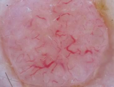
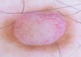
2 Benign Pink (Vascular) Patterns: The Goal is Biopsy Benign vs. Concerning Pink (Vascular) Patterns Vascular structures on Dermoscopy Clinical entity Sebaceus Hyperplasia Dermal nevus Spitz nevus Pyogenic granuloma Clear cell acanthoma Dermatofibroma Seborrheic Keratosis BCC SCC Amelanotic Melanoma Dermatoscopic findings Yellow globules, crowning vessels Comma vessels, pigment network Dotted vessels throughout Homogenous red w/ collarette Pink with String of (Red) Pearls Stellate scar-like center +- peripheral pigment network Gyri/sulci, Coral/fingerprint Arborizing vessels, white lines Hairpin vessels, coiled vessels, scale Red globules, dotted vessels, serpentine, corkscrew, 2+ 2

3 Which of these pink patterns is a benign pattern? A B C D Which of these pink patterns is a benign pattern? A B C D 54-year-old woman with spot for years 3
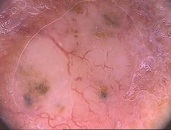
4 BCC Findings Scaling & Erosion Leaf-like Structures Arborizing Vessels Blue Grey Ovoid Nests Dermal nevus vs BCC Blue grey ovoid nests, Arborizing vessels 4


5 BCC vs SK Once BCC d Twice shy 5

, starts at")


6 Seb hyp vs BCC Crowning vessels looping over globules Arborizing vessels (broad to fine), starts at lesion edge, crosses midline) Seb hyp vs BCC vs SCC Crowning vessels looping over globules Arborizing vessels (broad to fine), starts at lesion edge, crosses midline) Porokeratosis Blanching erythema, Scale Hyperkeratotic coronoid lamella 6


7 I ve got this new spot on my leg Cortaid didn t do much SCC vs Psoriasis Dotted vessels and scale 40-year-old man with tender pink bump on his arm. It s been there a while. Unsure of changes, but it s asymptomatic. 7

8 BCC? Nevus? SCC? 8

:920.")
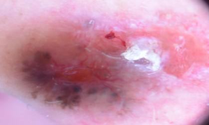
9 Amelanotic Melanoma? Steglich RB, et al.an Bras Dermatol. 2012;87(6):920. What do you think? 9


10 Amelanotic melanoma Amelanotic melanoma dermoscopymadesimple.blogspot.com/ Which pink pattern predominates in this lesion? 10
:75.")

11 36-year-old woman with new spot on her shin Zaballos P, et al. Arch Derm. 2008;144(1): year-old woman with 15 mm enlarging itchy pink growth on the left thigh for 2 years. 11
 seen in: Amelanotic melanoma and BCC Tracey N.")

12 What structure is present? White Shiny Lines (Chrysalis) seen in: Amelanotic melanoma and BCC Tracey N. Liebman, BA; Harold S. Rabinovitz, MD; Yevgeniy Balagula, MD; Natalia Jaimes-Lopez, MD; Ashfaq A. Marghoob, MD 40 year-old woman with a new growth on her left arm 12


13 Critical eye: DF vs Atypical Spitz nevus Spitz Epithelioid nevi Reed All red dots Starburst Cherry Angioma vs Evil Twin Targetoid hemosiderotic hemangioma: melanoma mimicker 13


14 Angioma vs Hemosiderotic hemangioma 55-year-old woman with sudden onset new papule A 14- year old boy with a tender red papule on the left lateral 5 th finger What is your clinical diagnosis? 14


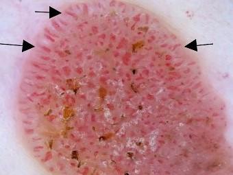
15 Clinical Comparison Acanthoma vs Pyogenic granuloma Homogenous pink with red dots in STRING OF PEARLS Homogenous red area with white collarette One more case 15


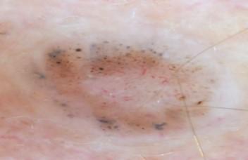
16 BCC? Dermal Nevus? Senel E. IJDVL. 2011;77:16. Chen LC, et al. JAMA Derm. 2015;151:715. Jalilian C, et al. BJD. 2013;169:294 Which pink pattern predominates in this lesion? Comma vessels Dotted vessels Serpentine pattern String of pearls pattern Which pink pattern predominates in this lesion? Comma vessels Dotted vessels Serpentine pattern String of pearls pattern 16


17 Which pink pattern predominates in this lesion? Comma vessels Dotted vessels Glomeruloid pattern String of pearls pattern Which pink pattern predominates in this lesion? Comma vessels Dotted vessels Glomeruloid pattern String of pearls pattern Pink (Vascular) Patterns: The Goal is Biopsy 17
18 Vascular structures on Dermoscopy Clinical entity Sebaceus Hyperplasia Dermal nevus Spitz nevus Pyogenic granuloma Clear cell acanthoma Dermatofibroma Seborrheic Keratosis BCC SCC Amelanotic Melanoma Dermatoscopic findings Yellow globules, crowning vessels Comma vessels, pigment network Dotted vessels throughout Homogenous red w/ collarette Pink with String of (Red) Pearls Stellate scar-like center +- peripheral pigment network Gyri/sulci, Coral/fingerprint Arborizing vessels, white lines Hairpin vessels, coiled vessels, scale Red globules, dotted vessels, serpentine, corkscrew, 2+ 18
6/17/2018. Breaking Bad (Part 1) Dermoscopy of Brown(ish) Things. Bad?
 Dermoscopy of Brown(ish) Things. Bad?") Breaking Bad (Part 1) Dermoscopy of Brown(ish) Things Jennie T. Clarke, MD ssociate Professor of Dermatology University of Utah School of Medicine Bad? 1 Brown(ish) Things Bad Melanoma Pigmented basal
Breaking Bad (Part 1) Dermoscopy of Brown(ish) Things Jennie T. Clarke, MD ssociate Professor of Dermatology University of Utah School of Medicine Bad? 1 Brown(ish) Things Bad Melanoma Pigmented basal
comedo-like openings (clods, brown or orange & circles) milia-like cysts (dots or clods, white) 1/29/18 Dotted vessels are also commonly seen in SCC
 milia-like cysts (dots or clods, white) 1/29/18 Dotted vessels are also commonly seen in SCC") Brown circles Dotted vessels are also commonly seen in SCC Step1 1. Nevus (unequivocal) 2. DF/IDN 3. BCC 4. SCC Network Patchy network Peripheral network & central hypopigmentation DF: network with central
Brown circles Dotted vessels are also commonly seen in SCC Step1 1. Nevus (unequivocal) 2. DF/IDN 3. BCC 4. SCC Network Patchy network Peripheral network & central hypopigmentation DF: network with central
Key factors in successfully integrating dermoscopy into your clinical practice
 Key factors in successfully integrating dermoscopy into your clinical practice S051 Dilemmas and challenges in skin cancer therapies and management Monday, March 4 th 2019 (9AM-12PM) Room 209A 10:56-11:09AM
Malignant non-melanocytic lesions
 Malignant non-melanocytic lesions Course C023: Fundamentals of Dermoscopy March 4, 2019, 11:20 AM - 11:50 PM Room: 146B Jason B. Lee, MD Professor & Vice Chair Director of Dermatopathology & Pigmented
Non-Melanocytic Pattern Dermoscopy
 Non-Melanocytic Pattern Dermoscopy I have no conflicts of interest to disclose Except that I LOVE dermoscopy Michelle Tarbox, MD Assistant Professor of Dermatology and Dermatopathology Texas Tech University
Dermoscopy: Recognizing Top Five Common In- Office Diagnoses
 Dermoscopy: Recognizing Top Five Common In- Office Diagnoses Vu A. Ngo, DO Department of Family Medicine and Dermatology Choctaw Nation Health Services Authority Learning Objectives Introduction to dermoscopy
Disclosure. Objectives. PAFP CME Conference Lou Mancano MD, FAAFP Reading Health System November 18, 2016
 PAFP CME Conference Lou Mancano MD, FAAFP Reading Health System November 18, 2016 1 Disclosure The speaker has no conflict of interest, financial agreement, or working affiliation with any group or organization.
Non-melanocytic Patterns
 Non-melanocytic Lesions Non-melanocytic Patterns Michelle Tarbox, MD Assistant Professor of Dermatology and Dermatopathology Texas Tech University Health Sciences Center 2018 Seborrheic keratoses Acanthotic
Prediction without Pigment: a decision algorithm for non-pigmented skin malignancy
 DERMATOLOGY PRACTICAL & CONCEPTUAL www.derm101.com Prediction without Pigment: a decision algorithm for non-pigmented skin malignancy Cliff Rosendahl 1, Alan Cameron 1, Philipp Tschandl 2, Agata Bulinska
Dermoscopy STFM Richard Usatine, MD 5/2/16. Disclosure Statement: Some Dermatoscopes. Dermoscopy Video. Thanks to Dr.
 Disclosure Statement: Dermoscopy STFM 2016 Richard P. Usatine, MD, FAAFP Professor, Family and Community Medicine Professor, Dermatology and Cutaneous Surgery Medical Director, Clinic University of Texas
Benign versus Cancerous Lesions How to tell the difference FMF 2014 Christie Freeman MD, CCFP, DipPDerm, MSc
 1 Benign versus Cancerous Lesions How to tell the difference FMF 2014 Christie Freeman MD, CCFP, DipPDerm, MSc Benign lesions Seborrheic Keratoses: Warty, stuck-on Genetics and birthdays Can start in late
What is Dermoscopy? Early Dermoscopes. Deciphering Dermoscopy: Terminology, Features & Algorithms 6/17/2018
 Deciphering Dermoscopy: Terminology, Features & Algorithms Where did it come from and why do we use it? Jennie T. Clarke, MD Associate Professor of Dermatology University of Utah School of Medicine What
22/04/2015. Dermoscopy of Melanoma. Ilsphi Browne. Overview
 Dermoscopy of Melanoma Ilsphi Browne Overview The device Dermoscopic criteria (terminology) Colour Patterns Global features Local features Approach to diagnosing pigmented lesions Other uses in general
Regression 2/3/18. Histologically regression is characterized: melanosis fibrosis combination of both. Distribution: partial or focal!
 Regression Margaret Oliviero MSN, ARNP Harold S. Rabinovitz MD Histologically regression is characterized: melanosis fibrosis combination of both Distribution: partial or focal! Dermatoscopic terminology
Review of vasculature visualized on dermoscopy
 doi: 10.1111/1346-8138.13686 Journal of Dermatology 2017; 44: 525 532 REVIEW ARTICLE Review of vasculature visualized on dermoscopy Yaei TOGAWA Department of Dermatology, Chiba University Graduate School
It can be helpful in some cases of actinic keratosis, Bowen s disease and squamous cell carcinoma
 Dermoscopy Introduction, Terminology and Structures (to be read in conjunction with the Diagnostic Dermoscopic Algorithm) Copyright to Cunliffe TP (Jan. 2017) All rights reserved Introduction Dermoscopy
Basics in Dermoscopy
 Basics in Dermoscopy Manal Bosseila Professor of Dermatology, Cairo University Member of European Academy Dermatology & Venereology EADV Member of International Dermoscopy Society IDS Member of Aesthetic
Skin lesions The Good and the Bad. Dr Virginia Hubbard Ipswich Hospital NHS Trust Barts and the London School of Medicine and Dentistry
 Skin lesions The Good and the Bad Dr Virginia Hubbard Ipswich Hospital NHS Trust Barts and the London School of Medicine and Dentistry Case 1 32 year old woman Australian Lesion on back New hair growing
Clinical and Dermoscopic Features of Thin Nodular Melanoma
 Clinical and Dermoscopic Features of Thin Nodular Melanoma A study of the International Dermoscopy Society Coordinator: Dr. Alexander J. Stratigos and colleagues, alstrat2@gmail.com ** Extended to May
المركب النموذج--- سبيتز وحمة = Type Spitz's Nevus, Compound SPITZ NEVUS 1 / 7
 SPITZ NEVUS 1 / 7 Epidemiology An annual incidence rate of 1.4 cases of Spitz nevus per 100,000 individuals has been estimated in Australia, compared with 25.4 per 100,000 individuals for cutaneous melanoma
50 interactive dermoscopic case discussions Dr Stephen Hayes
 50 interactive dermoscopic case discussions Dr Stephen Hayes Annotations will be found on your memory drive, as will 100 case discussions and other learning material Melanoma 2mm thick Ugly duckling-one
Dermoscopy. Enhanced Diagnostic Ability: Pigmented Lesions. Ted Rosen, MD Baylor College of Medicine Houston, Texas
 Dermoscopy Enhanced Diagnostic Ability: Pigmented Lesions Ted Rosen, MD Baylor College of Medicine Houston, Texas Faculty Disclosure Statement No conflicts relevant to this workshop! Sir William Osler
Dermoscopy. Sir William Osler. Dermoscopy. Dermoscopy. Melanoma USA Primary Care Update Faculty Disclosure Statement
 Diagnostic Ability: Pigmented Lesions Ted Rosen, MD Baylor College of Medicine Houston, Texas Enhanced 2010 Primary Care Update Faculty Disclosure Statement Ted Rosen, MD Speakers Bureau: Abbott, Amgen,
Dermoscopy, the use of a handheld
 ONLINE EXCLUSIVE Dermoscopy in family medicine: A primer Dermoscopy allows you to see deeper into the skin than with the naked eye. Here s how you can make use of it to spot malignant conditions sooner.
10/3/2018. Dermoscopy: Looking beneath the surface of the skin. Dermoscopy for Family Medicine 10/11/2018
 Dermoscopy for Family Medicine 10/11/2018 Jane M. Grant-Kels, MD, FAAD Founding Chair Emeritus, Dept of Dermatology Professor of Dermatology, Pathology & Pediatrics Director of the Cut Oncology Ctr & Melanoma
Fundamentals of dermoscopy
 Fundamentals of dermoscopy Learning objectives Upon completion of this session, participants should be able to: describe the basic principles of dermoscopy identify features associated with pigmented and
Associate Professor Amanda Oakley. Professor H. Peter Soyer. Academic Dermatologist The University of Queensland Brisbane. Dermatologist Hamilton
 Associate Professor Amanda Oakley Dermatologist Hamilton Professor H. Peter Soyer Academic Dermatologist The University of Queensland Brisbane 8:30-10:30 WS #3: Dermoscopy Workshop Part 1 11:00-13:00 WS
Introduction to Dermoscopy. Disclosure. Introduction
 Introduction to Dermoscopy 1 Disclosure Dr. Deborah Bren has no conflict of interest, financial agreement, or working affiliation with any group or organization. 2 Introduction Deborah A. Bren, DO Family
Introduction to Dermoscopy. Nicholas Compton, MD June 16, 2010
 Introduction to Dermoscopy Nicholas Compton, MD June 16, 2010 Overview What is dermoscopy Brief history Types of dermoscopy General approach to lesion of interest 2 step algorithm 3-point checklist Practice
Management of patients with melanocytic and non-melanocytic neoplasms
 Management of patients with melanocytic and non-melanocytic neoplasms Ashfaq Marghoob MD Harold Rabinovitz MD Margaret Oliviero ARNP Harald Kittler MD Jupiter Cancer Centrer Characteristic Dermoscopic
Dermoscopy in everyday practice. What and Why? When in doubt cut it out? Trilokraj Tejasvi MD
 Dermoscopy in everyday practice Trilokraj Tejasvi MD Assistant Professor, Department of Dermatology, Director Teledermatology services, University of Michigan, Faculty Associate, GLOBAL REACH, Michigan
Dermoscopy of non-pigmented skin lesions: a literature review
 Hong Kong J. Dermatol. Venereol. (2017) 25, 13-21 Review Article Dermoscopy of non-pigmented skin lesions: a literature review S Thomas, X Li, HP Soyer In this article, we will review benchmark dermoscopic
BJD British Journal of Dermatology. Summary. What s already known about this topic? CLINICAL AND LABORATORY INVESTIGATIONS
 CLINICAL AND LABORATORY INVESTIGATIONS BJD British Journal of Dermatology Pigmented nodular melanoma: the predictive value of dermoscopic features using multivariate analysis M.A. Pizzichetta, 1 H. Kittler,
Total body photography in high risk patients
 Total body photography in high risk patients Doug Grossman, MD, PhD Department of Dermatology Huntsman Cancer Institute University of Utah Summer AAD F032 Practical Considerations for Patients with Melanoma
Triage amalgamated dermoscopic algorithm (TADA) for skin cancer screening
 for skin cancer screening") DERMATOLOGY PRACTICAL & CONCEPTUAL www.derm101.com Triage amalgamated dermoscopic algorithm (TADA) for skin cancer screening Tova Rogers 1, Maria Marino 1, Stephen W. Dusza 1, Shirin Bajaj 1, Michael A.
DERMATOLOGY PRACTICAL & CONCEPTUAL www.derm101.com Triage amalgamated dermoscopic algorithm (TADA) for skin cancer screening Tova Rogers 1, Maria Marino 1, Stephen W. Dusza 1, Shirin Bajaj 1, Michael A.
Dermoscopy-a BRIEF introduction
 Dermoscopy-a BRIEF introduction Aim of presentation -to tell you what dermoscopy is -to show some of what it can do -point the interested learner to further resources Overview of dermoscopy Dermoscopy
Abrupt Intralesional Color Change on Dermoscopy as a New Indicator of Early Superficial Spreading Melanoma in a Japanese Woman
 Published online: June 24, 2015 1662 6567/15/0072 0123$39.50/0 This is an Open Access article licensed under the terms of the Creative Commons Attribution-NonCommercial 3.0 Unported license (CC BY-NC)
Common Benign Lesions and Skin Cancers. 22nd May 2015 Dr Mark Foley
 Common Benign Lesions and Skin Cancers 22nd May 2015 Dr Mark Foley Thank you for downloading this file. This intended to supplement the presentation given at the NZ Wound Care Conference, it is not intended
Appendix : Dermoscopy
 Go Back to the Top To Order, Visit the Purchasing Page for Details APP Appendix : Dermoscopy Dermoscopy, also known as dermatoscopy, epiluminoscopy and epiluminescent microscopy, is an effective non-invasive
Dermoscopy Quiz 3-Point Checklist Algorithm
 Dermoscopy Quiz 3-Point Checklist Algorithm GLOBAL PATTERN Globular LOCAL CRITERIA Aggregated globules Milia-like cysts 3 POINT CHECK LIST Symmetrical No abnormal net Slight Blue-white veil BENIGN MELANOCYTIC
STUDY. Scott W. Menzies, MB,BS, PhD; Karin Westerhoff, MD; Harold Rabinovitz, MD; Alfred W. Kopf, MD; William H. McCarthy, MBBS, MEd; Brian Katz
 STUDY Surface Microscopy of Pigmented Basal Cell Carcinoma Scott W. Menzies, MB,BS, PhD; Karin Westerhoff, MD; Harold Rabinovitz, MD; Alfred W. Kopf, MD; William H. McCarthy, MBBS, MEd; Brian Katz Objectives:
Dermoscopic Features of Non-Pigmented Eccrine Poromas in. Department of Dermatology, Shinshu University School of Medicine,
 Original article Dermoscopic Features of Non-Pigmented Eccrine Poromas in Association with their Histopathological Features Akane Minagawa, Hiroshi Koga,* Masaomi Takahashi, + Kenji Sano, + Ryuhei Okuyama,
Supplementary Online Content
 Supplementary Online Content Tschandl P, Rosendahl C, Akay BN, et al. Expert-level diagnosis of nonpigmented skin cancer by combined convolutional neural networks. JAMA Dermatol. Published online November
VACAVILLE DERMATOLOGY
 Connecting the Dots on those Spots NANDAN V. KAMATH, M.D. VACAVILLE DERMATOLOGY Sources All of the photos were taken with permission from the Dermnet NZ website - Dermnet New Zealand after communicating
Supplementary Online Content
 Supplementary Online Content Chernoff KA, Marghoob AA, Lacouture ME, Deng L, Busam KJ, Myskowski PL. Dermoscopic findings in cutaneous metastases. JAMA Dermatol. Published online January 15, 2014. doi:10.1001/jamadermatol.2013.8502
Mole mapping and monitoring. Dr Stephen Hayes. Associate Specialist in Dermatology, University Hospital Southampton
 Mole mapping and monitoring Dr Stephen Hayes Associate Specialist in Dermatology, University Hospital Southampton Outline of presentation The melanoma epidemic Benefits of early detection Risks of the
Dermoscopic patterns of cutaneous melanoma metastases
 Review Dermoscopic patterns of cutaneous melanoma metastases Pietro Rubegni, MD, Arianna Lamberti, MD, Filomena Mandato, MD, Roberto Perotti, MD, and Michele Fimiani, MD Department of Clinical Medicine
Reports on Scientific Meetings
 Hong Kong J. Dermatol. Venereol. (2016) 24, 146-153 The Hong Kong Society of Dermatology and Venereology Annual Scientific Meeting 2016 Reported by BTH Chan, CT Chau, CW Chow, CC Koh, WYK Lam, BS Tong,
DIFFERENCES IN DERMOSCOPIC IMAGES FROM NON-POLARIZED DERMOSCOPE AND POLARIZED DERMOSCOPE INFLUENCE THE DIAGNOSTIC ACCURACY AND CONFIDENCE LEVEL.
 DIFFERENCES IN DERMOSCOPIC IMAGES FROM NON-POLARIZED DERMOSCOPE AND POLARIZED DERMOSCOPE INFLUENCE THE DIAGNOSTIC ACCURACY AND CONFIDENCE LEVEL. 1. Steven Q. Wang MD 1 (wangs@mskcc.org) 2. Stephen W. Dusza
Rosettes in actinic keratosis and squamous cell carcinoma: distribution, association to other dermoscopic signs and description of the rosette pattern
 DOI: 10.1111/jdv.14474 JEADV ORIGINAL ARTICLE Rosettes in actinic keratosis and squamous cell carcinoma: distribution, association to other dermoscopic signs and description of the rosette pattern B. Lozano-Masdemont,
This copy is for personal use only - distribution prohibited.
 Journal of Pre-Clinical and Clinical Research, 2013, Vol 7, No 1, 6-12 www.jpccr.eu REVIEW ARTICLE Benign simulators of melanoma on dermoscopy black colour does not always indicate melanoma Grazyna Kaminska-Winciorek
Benign simulators of melanoma on dermoscopy black colour does not always indicate melanoma
 Journal of Pre-Clinical and Clinical Research, 2013, Vol 7, No 1, 6-12 www.jpccr.eu REVIEW ARTICLE Benign simulators of melanoma on dermoscopy black colour does not always indicate melanoma Grazyna Kaminska-Winciorek
Metaphoric and descriptive terminology in dermoscopy: Lessons from the cognitive sciences
 DERMATOLOGY PRACTICAL & CONCEPTUAL www.derm101.com Metaphoric and descriptive terminology in dermoscopy: Lessons from the cognitive sciences Jason Giacomel 1, Iris Zalaudek 2, Ashfaq A. Marghoob 3 1 Skin
The impact of GP sub-specialisation and dermatoscopy use on diagnostic accuracy for melanomas in Australia
 The impact of GP sub-specialisation and dermatoscopy use on diagnostic accuracy for melanomas in Australia Cliff Rosendahl, Gail Williams, Diann Eley, Tobias Wilson, Greg Canning, Jeffrey Keir, Ian McColl,
LUMPS AND BUMPS: AN ORGANIZED APPROACH TO DIAGNOSIS AND MANAGEMENT
 LUMPS AND BUMPS: AN ORGANIZED APPROACH TO DIAGNOSIS AND MANAGEMENT Tammy P. Than, M.S., O.D., F.A.A.O. The University of Alabama at Birmingham / School of Optometry 1716 University Blvd. Birmingham, AL
Diagnosis of Lentigo Maligna Melanoma. Steven Q. Wang, M.D. Memorial Sloan-Kettering Cancer Center Basking Ridge, NJ
 Diagnosis of Lentigo Maligna Melanoma Steven Q. Wang, M.D. Memorial Sloan-Kettering Cancer Center Basking Ridge, NJ Conflict of Interest: None Topics Epidemiology and Natural History Clinical and Histologic
Revised Pattern Analysis: a method for the accurate diagnosis of pigmented skin lesions
 Dermatoscopy for Students A concise outline of: Revised Pattern Analysis: a method for the accurate diagnosis of pigmented skin lesions And Chaos and Clues: a decision algorithm for routine practice to
Lid Lesions: Relax or Refer
 Lid Lesions: Relax or Refer Blair Lonsberry, MS, OD, MEd., FAAO Professor of Optometry Pacific University College of Optometry blonsberry@pacificu.edu Agenda Benign vs. Malignant lesions Benign Eyelid
Features Causing Confusion between Basal Cell Carcinoma and Squamous Cell Carcinoma in Clinical Diagnosis
 TH Ryu, et al pissn 1013-9087ㆍeISSN 2005-3894 Ann Dermatol Vol. 30, No. 1, 2018 https://doi.org/10.5021/ad.2018.30.1.64 ORIGINAL ARTICLE Features Causing Confusion between Basal Cell Carcinoma and Squamous
STUDY. Dermoscopy of Squamous Cell Carcinoma and Keratoacanthoma
 ONLINE FIRST STUDY Dermoscopy of Squamous Cell Carcinoma and Keratoacanthoma Cliff Rosendahl, MBBS; Alan Cameron, MBBS; Giuseppe Argenziano, MD; Iris Zalaudek, MD; Philipp Tschandl, MD; Harald Kittler,
David B. Troxel, MD. Common Medicolegal Situations: Misdiagnosis of Melanoma
 Common Medicolegal Situations: Misdiagnosis of Melanoma David B. Troxel, MD Medical Director, The Doctors Company, Napa, California Clinical Professor Emeritus, University of California at Berkeley Past
Teaching point. Case 1 2/3/18. Challenging Cases. Examples of challenging cases?
 Challenging Cases Examples of challenging cases? 1. Challenge in diagnosis 2. Challenge in monitoring an off label treatment 3. Challenge where clinical diagnosis does not match the pathology diagnosis
MODULE 1. LOCAL AND GENERAL CRITERIA IN PIGMENTED MELANOCYTIC LESIONS.
 DERMOSCOPY TEACHING PROGRAMME Dermoscopy Teaching Programme Module 1 MODULE 1. LOCAL AND GENERAL CRITERIA IN PIGMENTED MELANOCYTIC LESIONS. Dermoscopy is a non-invasive in vivo technique that provides
Dermatology for the PCP Deanna G. Brown, MD, FAAD Susong Dermatology Consulting Staff at CHI Memorial
 Dermatology for the PCP Deanna G. Brown, MD, FAAD Susong Dermatology Consulting Staff at CHI Memorial Cutaneous Oncology for the PCP Deanna G. Brown, MD, FAAD Susong Dermatology Consulting Staff at CHI
Beyond classic dermoscopic patterns of dermatofibromas: a prospective research study
 Kelati et al. Journal of Medical Case Reports (2017) 11:266 DOI 10.1186/s13256-017-1429-6 RESEARCH ARTICLE Open Access Beyond classic dermoscopic patterns of dermatofibromas: a prospective research study
INTRODUCTION HOUSEKEEPING June 11 th Dr John Adams Dermatologist/Dermoscopist MOLEMAP NZ/Australia MOLESAFE USA
 INTRODUCTION HOUSEKEEPING June 11 th 2015 Dr John Adams Dermatologist/Dermoscopist MOLEMAP NZ/Australia MOLESAFE USA Program Skin cancer statistics. Dermoscopy description and usefulness. Patient /lesion
Intraoperative Dermoscopy for Identification of Early Basal Cell Carcinomas in Basal Cell Nevus Syndrome
 Intraoperative Dermoscopy for Identification of Early Basal Cell Carcinomas in Basal Cell Nevus Syndrome Disclosures I have no industry related, financial, or other disclosures Goals Discuss the clinical
Doctors of Optometry Course Notes
 Doctors of Optometry Course Notes OD19 1CE COPE: 43871-AS Eyelid Lumps and Bumps Sunday, February 26, 2017 2:40 pm 3:30 pm Regency C 3 rd Floor Presenter: Blair Lonsberry, OD, FAAO Dr. Lonsberry is a Full
MECHANISMS OF HUMAN DISEASE: LABORATORY SESSION PATHOLOGY OF THE SKIN LAB. Friday, February 13, :30 am 11:00 am
 MECHANISMS OF HUMAN DISEASE: LABORATORY SESSION PATHOLOGY OF THE SKIN LAB Friday, February 13, 2009 9:30 am 11:00 am FACULTY COPY GOALS: Describe the basic clinical and morphologic features of various
A Clinical Aid for Detecting Skin Cancer: The Triage Amalgamated Dermoscopic Algorithm (TADA)
") ORIGINAL RESEARCH A Clinical Aid for Detecting Skin Cancer: The Triage Amalgamated Dermoscopic Algorithm (TADA) T. Rogers, MFA, M. L. Marino, MD, S. W. Dusza, DrPH, S. Bajaj, MD, R. P. Usatine, MD, M.
ORIGINAL RESEARCH A Clinical Aid for Detecting Skin Cancer: The Triage Amalgamated Dermoscopic Algorithm (TADA) T. Rogers, MFA, M. L. Marino, MD, S. W. Dusza, DrPH, S. Bajaj, MD, R. P. Usatine, MD, M.
INVESTIGATION. The relation between dermoscopy and histopathology of basal cell carcinoma *
 INVESTIGATION The relation between dermoscopy and histopathology of basal cell carcinoma * 351 Nazan Emiroglu 1 Fatma Pelin Cengiz 1 Funda Kemeriz 2 DOI: http://dx.doi.org/10.1590/abd1806-4841.20153446
SCREENING FOR SKIN CANCER IN PRIMARY CARE: IMPLEMENTATION OF DERMOSCOPY
 SCREENING FOR SKIN CANCER IN PRIMARY CARE: IMPLEMENTATION OF DERMOSCOPY A Dissertation Submitted to the Graduate Faculty of the North Dakota State University of Agriculture and Applied Science By Erin
Dermatopathology: The tumor is composed of keratinocytes which show atypia, increase mitoses and abnormal mitoses.
 Squamous cell carcinoma (SCC): A common malignant tumor of keratinocytes arising in the epidermis, usually from a precancerous condition: 1- UV induced actinic keratosis, usually of low grade malignancy.
Dermoscopy. Synonyms. Dermoscopy. Definition. Dermoscopy opens up a world of colour and structure that can t be seen with the naked eye
 Synonyms Dermoscopy Australasian College of Dermatologists G.P Training Module Dermoscopy Dermatoscopy Epiluminescence microscopy Skin surface microscopy Incident light microscopy Oil immersion microscopy
Management of Atypical Pigmented Lesions
 Management of Atypical Pigmented Lesions Jennifer A. Stein MD, PhD Associate Director, Pigmented Lesion Section Ronald O. Perelman Department of Dermatology NYU Langone Medical Center July 29, 2017 1-4
Cancer Council Australia Wiki Guidelines 2017
 WHAT IS THE ROLE OF SEQUENTIAL DIGITAL DERMOSCOPY IMAGING IN MELANOMA DIAGNOSIS? Cancer Council Australia Wiki Guidelines 2017 SHORT-TERM MONITORING 3 months Any change leads to excision Any melanocytic
World Journal of Medical and Surgical Case Reports
 Case Report World Journal of Medical and Surgical Case Reports Open Access Hair dye imitating multiple basal cell carcinoma of the scalp: A misdiagnosis prevented by dermoscopy Eldad Silberstein 1, Vasileios
Objectives. 1. Recognizing benign skin lesions. 2.Know which patients will likely need surgical intervention.
 The Joy of Pediatric Skin Dr. Claire Sanger University of Kentucky Plastic & Reconstructive Surgery Objectives 1. Recognizing benign skin lesions 2.Know which patients will likely need surgical intervention.
Acral and Mucosal Dermoscopy
 Acral and Mucosal Dermoscopy Caroline C. Kim, MD Assistant Professor, Department of Dermatology Harvard Medical School Director, Pigmented Lesion Clinic Associate Director, Cutaneous Oncology Program Beth
Benign and malignant epithelial lesions: Seborrheic keratosis: A common benign pigmented epidermal tumor occur in middle-aged or older persons more
 Benign and malignant epithelial lesions: Seborrheic keratosis: A common benign pigmented epidermal tumor occur in middle-aged or older persons more common on the trunk; but extremities, head and neck are
Common skin tumors. Benign Epidermal Tumors. Topic. Clinicopathologic Variants. Seborrheic keratoses
 Common skin tumors Topic Benign epidermal tumors Skin cyst and adnexal neoplasms Other common skin tumor Common skin malignancy สมศ กด ต นร ตนากร 26/02/2015 Benign Epidermal Tumors Seborrheic keratosis
Accepted Article. Dermoscopic diagnosis of amelanotic/hypomelanotic melanoma
 Received Date : 19-May-2016 Revised Date : 01-Sep-2016 Accepted Date : 20-Sep-2016 Article type : Research Letter Dermoscopic diagnosis of amelanotic/hypomelanotic melanoma M.A. Pizzichetta, 1 H. Kittler,
العصوي الوعاي ي الورام = angiomatosis Bacillary
 1 / 7 BACILLARY ANGIOMATOSIS Epidemiology BA is most commonly seen in patients with acquired immunodeficiency syndrome (AIDS) and a CD4 count less than 50 cells/mm 3, with an incidence of 1.2 cases per
Pathology of the skin. 2nd Department of Pathology, Semmelweis University
 Pathology of the skin 2nd Department of Pathology, Semmelweis University Histology of the skin Epidermis: Stratum corneum Stratum granulosum Stratum spinosum Stratum basale Dermis: papillary and reticular
Basal cell carcinoma 5/28/2011
 Goal of this Presentation A practical approach to the diagnosis of cutaneous carcinomas and their mimics Thaddeus Mully, MD University of California San Francisco To review common non-melanoma skin cancers
IT S FUNDAMENTAL MY DEAR WATSON! A SHERLOCKIAN APPROACH TO DERMATOLOGY
 IT S FUNDAMENTAL MY DEAR WATSON! A SHERLOCKIAN APPROACH TO DERMATOLOGY Skin, Bones, and other Private Parts Symposium Dermatology Lectures by Debra Shelby, PhD, DNP, FNP-BC, FADNP, FAANP Debra Shelby,
Finding Melanoma. Is not easy!
 Finding Melanoma Is not easy! Finding Melanoma Victoria mean depth at diagnosis is 1.5 mm. Melanoma 1.5mm Has Stage 1B Mortality 10% Melanoma Spotting a killer! Spotting a killer Visual Clues What are
Toby Maurer, MD University of California, San Francisco. Lifetime risk of an American developing melanoma
 Distinguishing Pigmented Skin Lesions and Melanoma Toby Maurer, MD University of California, San Francisco Epidemiology of Melanoma Lifetime risk of an American developing melanoma 1935: 1 in 1500 1980:
Identifying Benign and Malignant Skin Lesions. No Disclosures. Common Benign Lesions. Benign Lesions 2/25/2018. Stucco Keratoses.
 Dermatology in Primary Care Identifying Benign and Malignant Skin Lesions Christy Quire Baker, APRN, FNP-BC, DCNP Dermatology Certified Nurse Practitioner No Disclosures Common Benign Lesions Seborrheic
Toby Maurer, MD University of California, San Francisco. Lifetime risk of an American developing melanoma
 Distinguishing Pigmented Skin Lesions and Melanoma Toby Maurer, MD University of California, San Francisco Epidemiology of Melanoma Lifetime risk of an American developing melanoma 1935: 1 in 1500 1980:
Subungual blue naevus presenting with elkonyxis
 Hong Kong J. Dermatol. Venereol. (2016) 24, 87-91 Case Report Subungual blue naevus presenting with elkonyxis S Do an, N Atakan, H Khurami, O Gökoz, O Bitik Common blue naevi usually occur on the skin
You can also complete the survey over the phone with a trained interviewer by calling the study team toll free at
 Last modified: 0//08 0:0:5 AM Thank you very much for your participation in this important study. If you prefer, you can complete this survey online at www.stjude.org/ltfu-ask8. Your log-in ID is your
Skin Cancer A Personal Approach. Dr Matthew Strack Dunedin New Zealand
 Skin Cancer A Personal Approach Dr Matthew Strack Dunedin New Zealand Outline Dermoscopy Instruments and setup Photochemosurgery Clinical Aim: Leave with 2-3 ideas JLE Benign Junctional Nevus Management
BLINCK A diagnostic algorithm for skin cancer diagnosis combining clinical features with dermatoscopy findings
 DERMATOLOGY PRACTICAL & CONCEPTUAL www.derm101.com BLINCK A diagnostic algorithm for skin cancer diagnosis combining clinical features with dermatoscopy findings Peter Bourne, MBBS 1, Cliff Rosendahl,
Case Report A Case of Cystic Basal Cell Carcinoma Which Shows a Homogenous Blue/Black Area under Dermatoscopy
 Volume 20, Article ID 450472, 4 pages doi:0.55/20/450472 Case Report A Case of Cystic Basal Cell Carcinoma Which Shows a Homogenous Blue/Black Area under Dermatoscopy Akihiro Yoneta, Kohei Horimoto, Keiko
The GP s approach to the patient who is worried about sun, skin and moles. Dr Stephen Hayes
 The GP s approach to the patient who is worried about sun, skin and moles Dr Stephen Hayes Associate Specialist in Dermatology, University Hospital Southampton Dr Stephen Hayes DECLARATION OF INTERESTS
STUDY. Dermoscopy of Solitary Angiokeratomas
 STUDY Dermoscopy of Solitary Angiokeratomas A Morphological Study Pedro Zaballos, MD; inta Daufí, MD; Susana Puig, PhD; Giuseppe Argenziano, MD; David Moreno-Ramírez, MD; Horacio abo, MD; Ashfaq A. Marghoob,
The Enigmatic Spitz Lesion
 The Enigmatic Spitz Lesion The Dawn of Spitz S Spitz Sophie Spitz Melanomas of Childhood ; Am J Pathol 1948 1910-1956 13 children (18 mo - 12 yrs) 12/13 had a benign clinical course Sophie Spitz Born 1910
Skin Cancer of the Nose: Common and Uncommon
 Skin Cancer of the Nose: Common and Uncommon Mark Russell, M.D. Associate Professor of Dermatology, Otolaryngology, and Pathology University of Virginia Objectives Review clinical presentations of select