Self assesment Case 21
|
|
|
- Virgil Hodge
- 5 years ago
- Views:
Transcription


1 17-18 MAY 2018 London Dermatopathology Symposium 2018 Self assesment Case 21 MARC HASPESLAGH
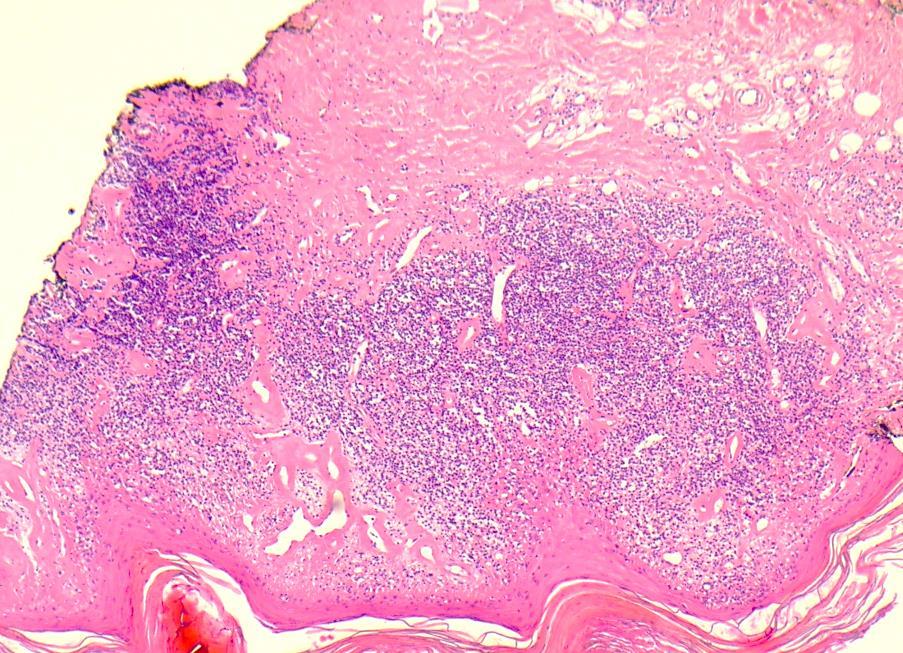
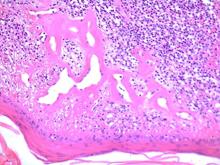
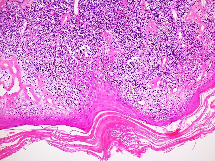
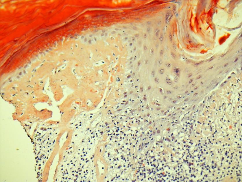
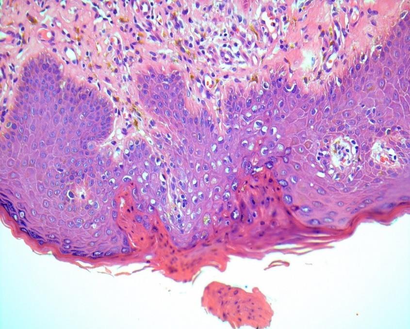
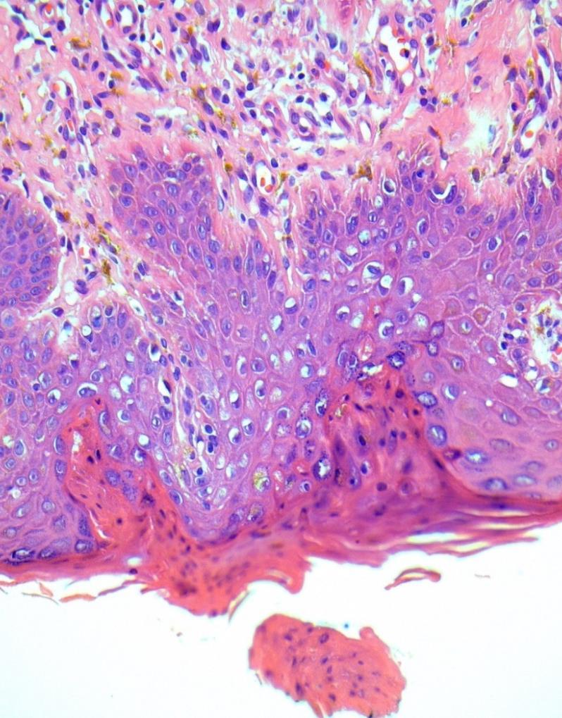
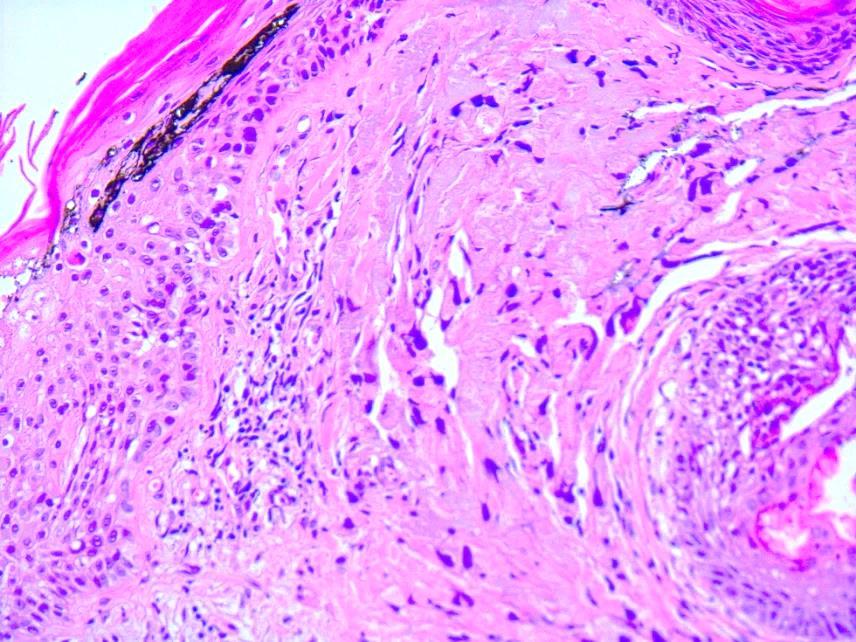
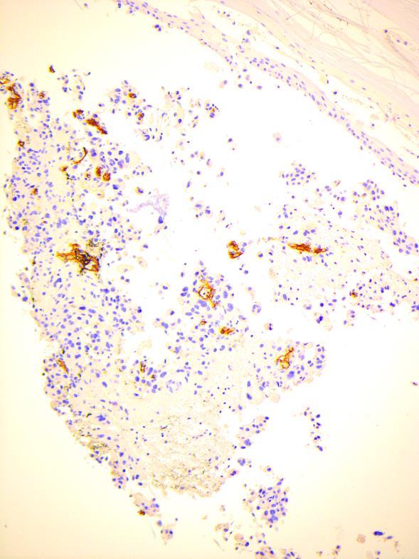
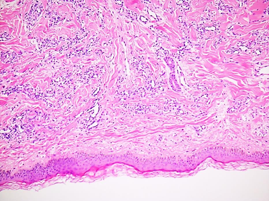
2 CASE year old lady with eczematous lesions at ear helix and red patch on nose bridge since 3 years Discoid Lupus? Sarcoidosis? TBC?
3
4 CD123 L K
5
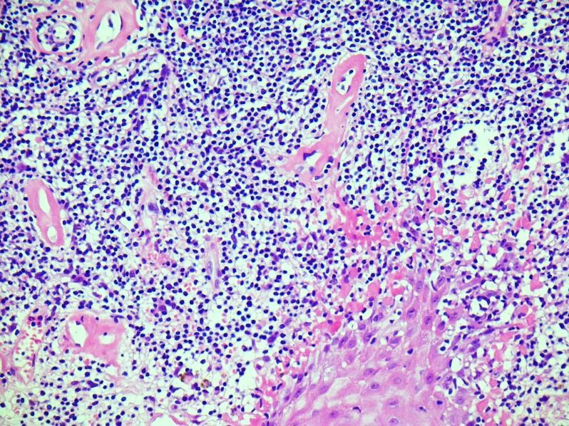
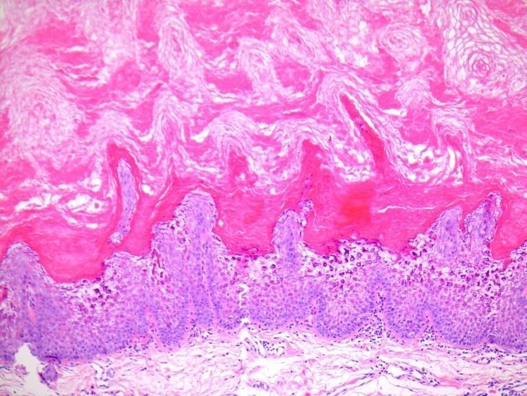
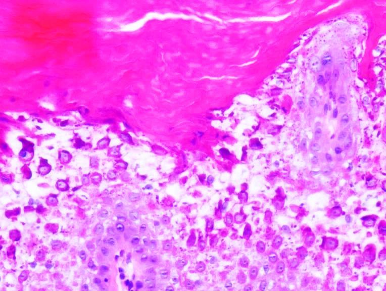
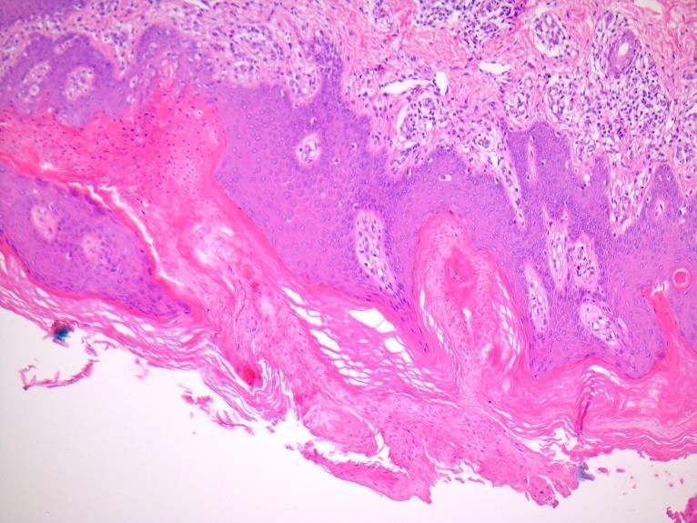
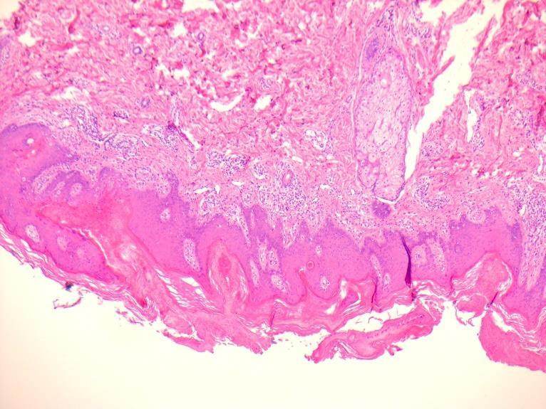
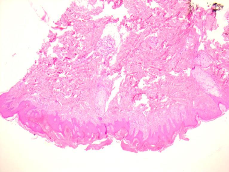
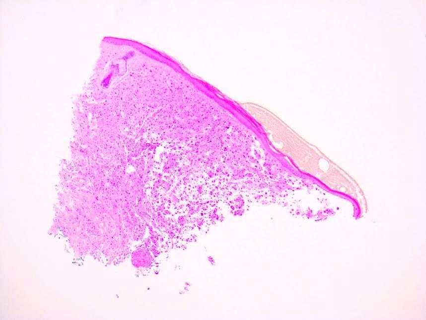
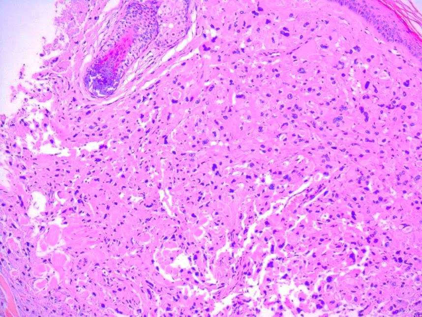
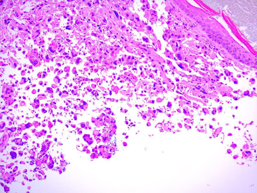
6 Diagnosis Lupus Discoid lupus Hypertrophic discoid lupus Discoid lupus with amyloidosis Hyperthrophic discoid lupus with secondary amyloidosis
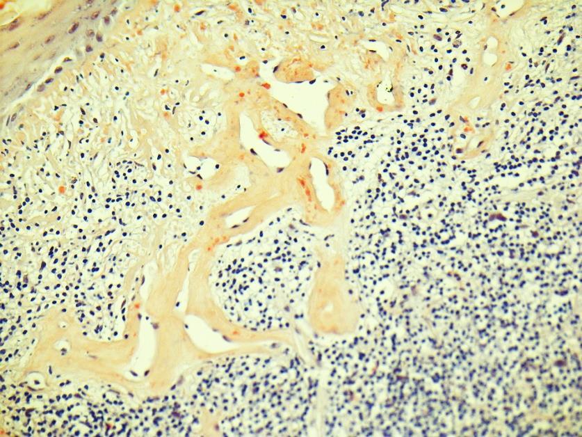
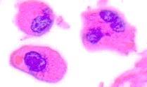
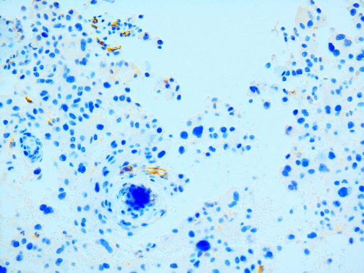
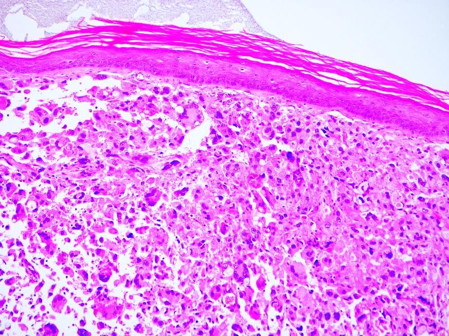
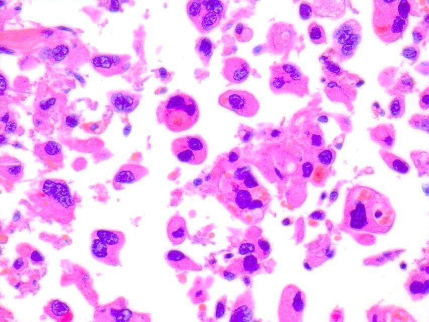
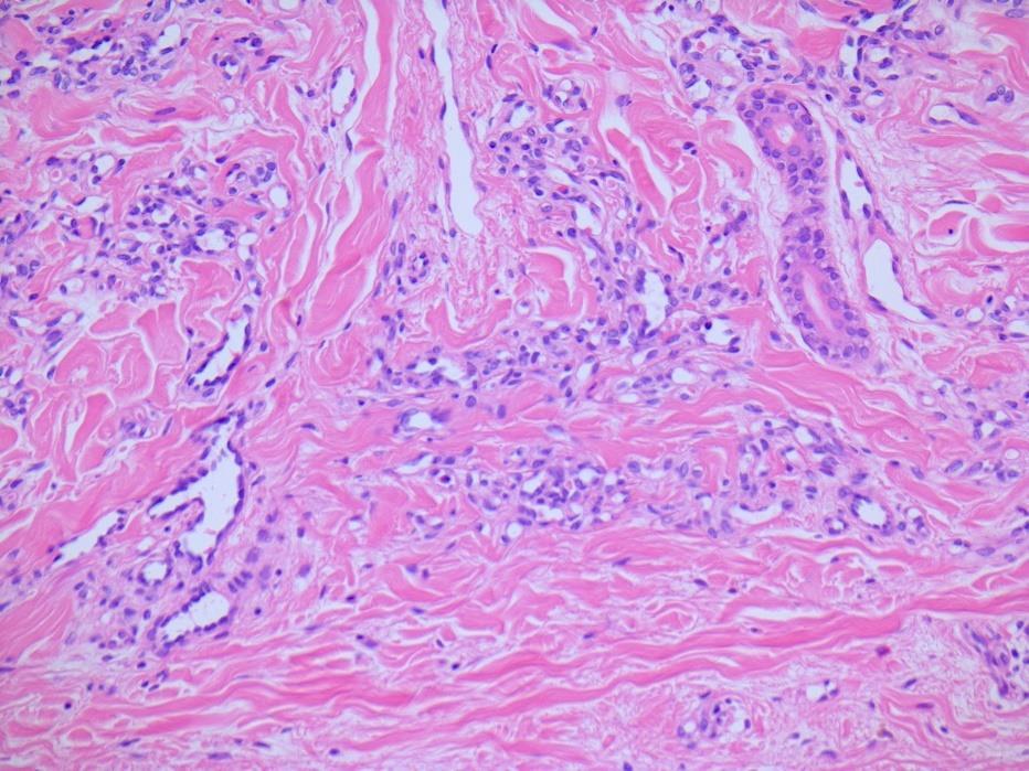
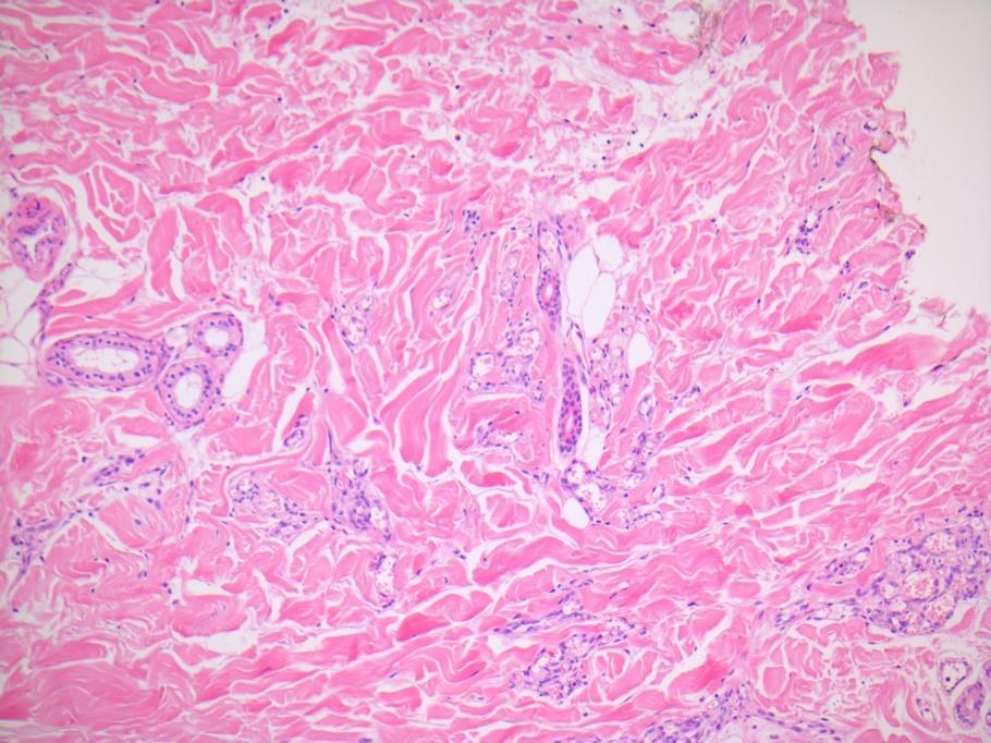
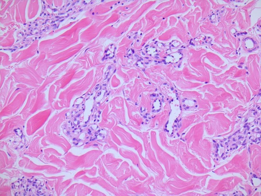
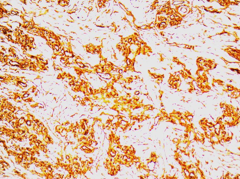
7 Localised Amyloid in Skin Primary as in lichen or macular amyloidosis and secondary as clinically unapparent phenomenon Secundary associated with tumors of epithelial origin or inflammatory diseases often linked to chronic UV exposition Amyloid K from epidermal origin
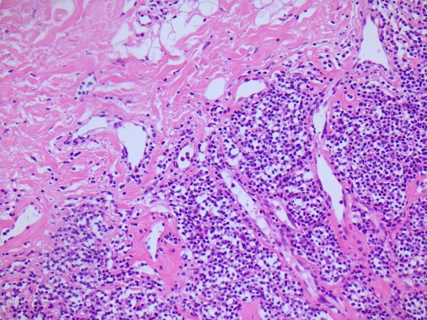
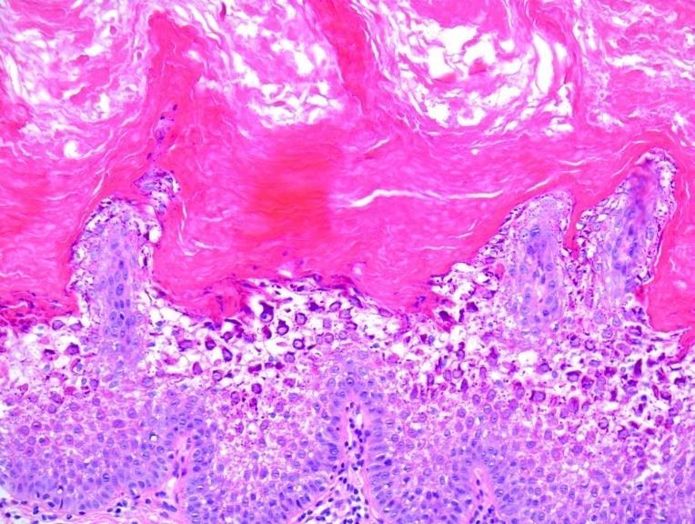
8 Amyloid in skin Described rarely in lupus, mainly hypertrophic lupus, Probably chronic UV exposure pathogenic role Keratin origin with CK+ Papillary dermis along basement membrane

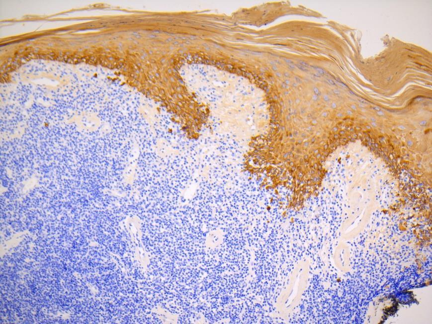
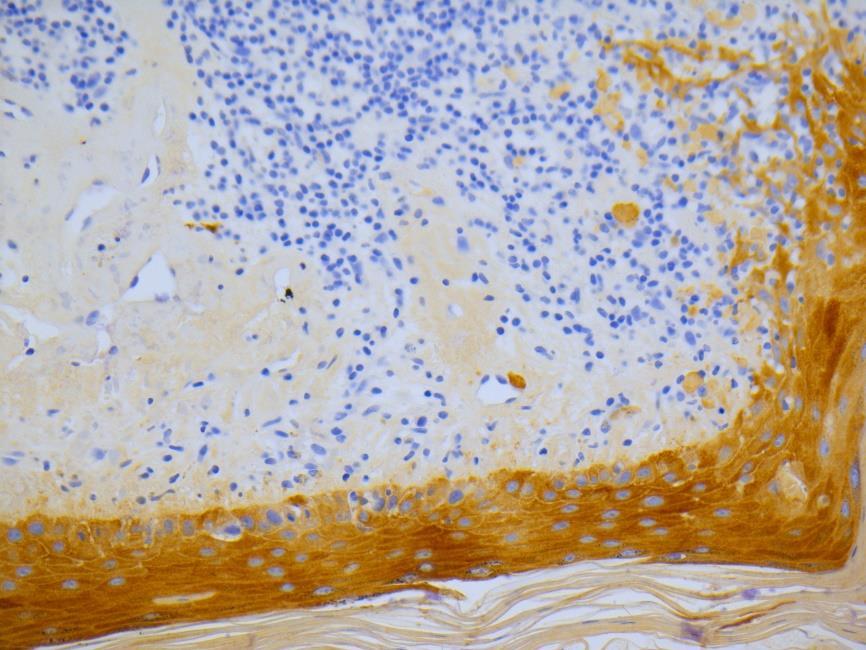
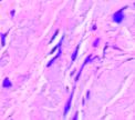
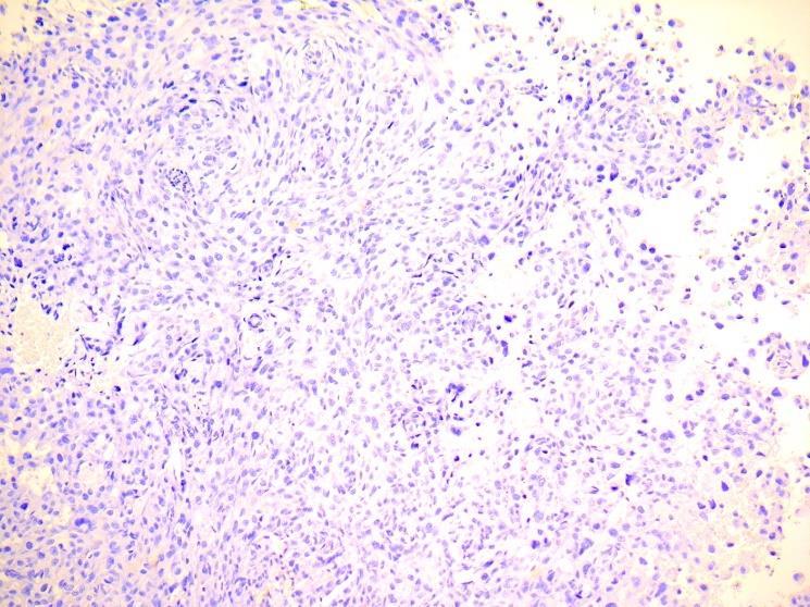
9 34be12 AE1AE3 CK5
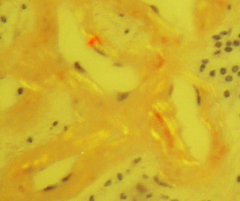
10 Case 21 : meaning of perivascular presence and keratin negativity? Secondary systemic in chronic inflammatory disease?

11 Self assesment Case 22
12 CASE year old lady with six flat papules on vulva since many months Viral? Condylomas? Bowenoid papulosis? Lichen?

13
14 Diagnosis Acanthoma Condyloma Acantholytic acanthoma Multiple epidermolytic acanthoma Acantholytic dyskeratoma

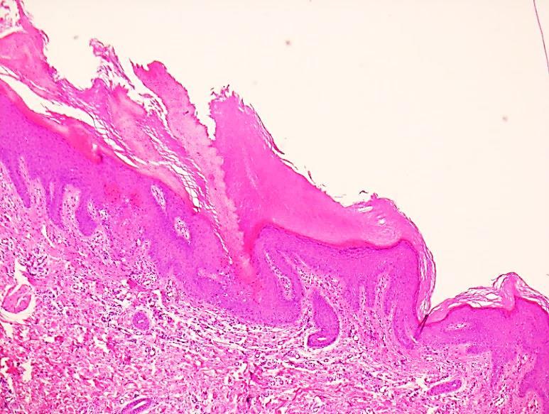
15 Epidermolytic hyperkeratosis Histologic phenomenon : hyperkeratosis, hypergranulisis, perinuclear vacuolisation, reticular degeneration in granular and spinous layer Formation of amorphous, eosinophilic trichohyaline granules From generalised genetic disease in bullous ichtyosiform erythrodermia to acquired and localised aberration Keratin 1 and 10 mutations
16 Epidermolytic Acanthoma Epidermolytic acanthoma : benign tumor usually presenting as solitary, asymptomatic papule Multiple Epidermolytic Acantomas : very rare, genitocrural areas Mimicker of condylomas : older population, labia majora (scrotum in males) No HPV association
17 Lesson learned The same morfological pattern can occur in totally different diseases To classify correctly these patterns clinicopathological correlation is indespensable

18 Self assesment Case 23
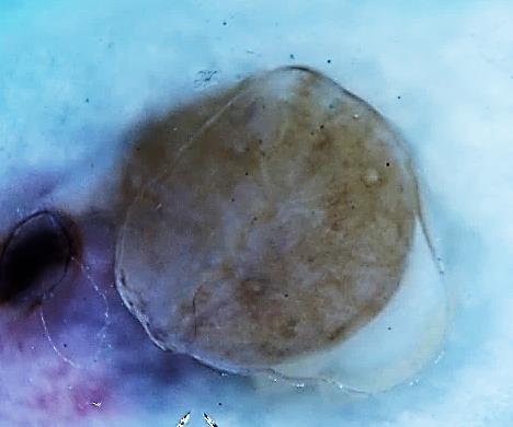
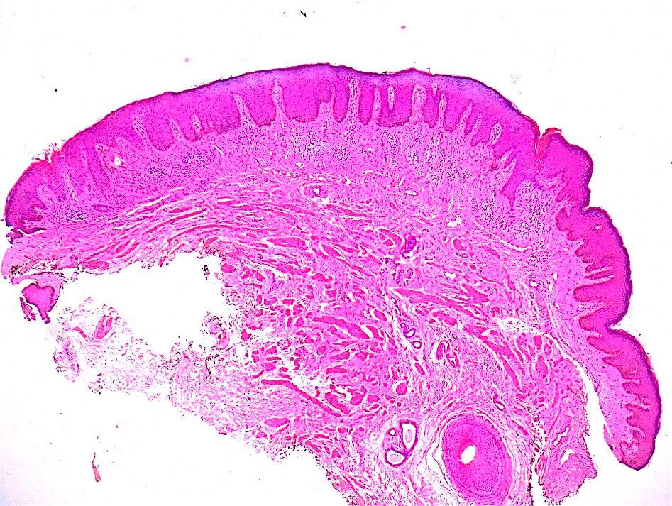
19 CASE year old girl with hyperkeratotic naevoid lesion in sacral region since birth
20


21 f 50y onderbeen re M 87 onderb dsapk F 60 y arm voor re male 70y dsap + lich ker F 53 arm naevus en pk
22
23
24 Diagnosis Neurodermatitis Psoriasis Verrucous hyperplasia Bowen s disease Verrucous plaque type porokeratosis
25 Lessons learned Blind sectioning and cutting can fool the pathologist EVD can save time, money and mental and emotional energy from the pathologist and technicians

26 Self assesment Case 24
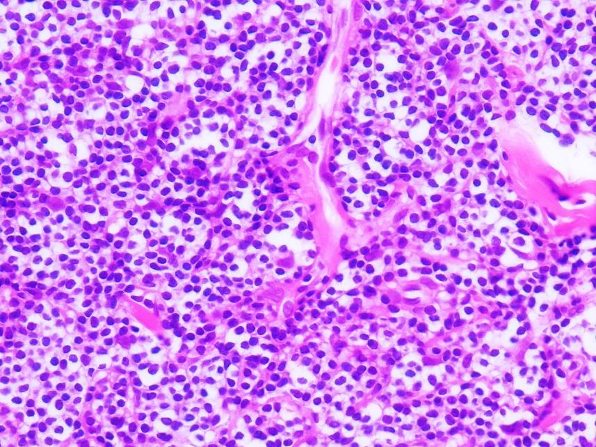
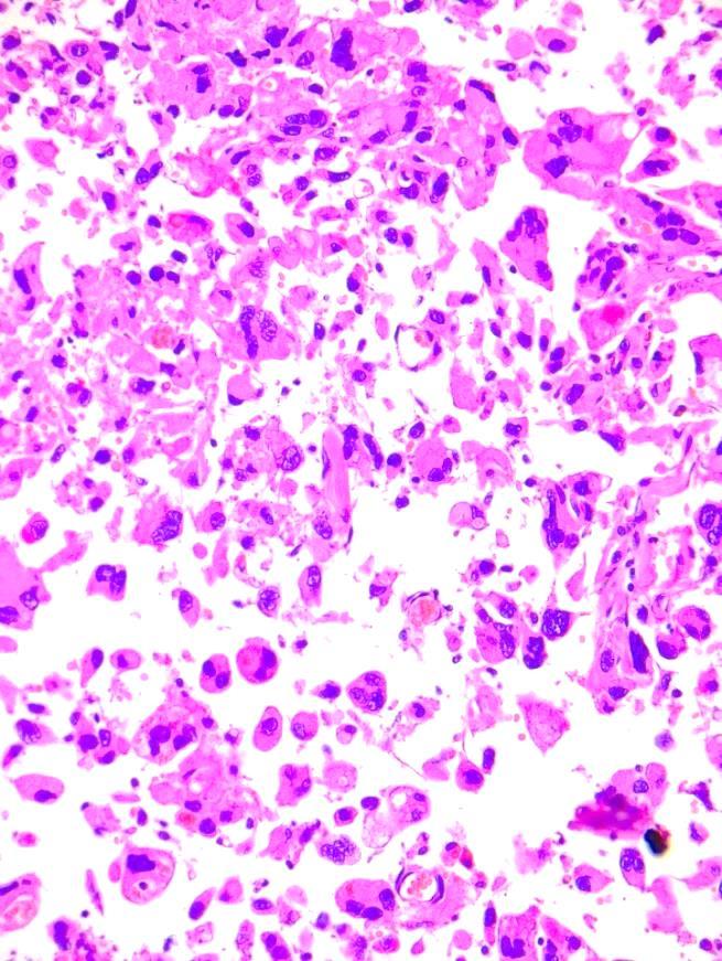
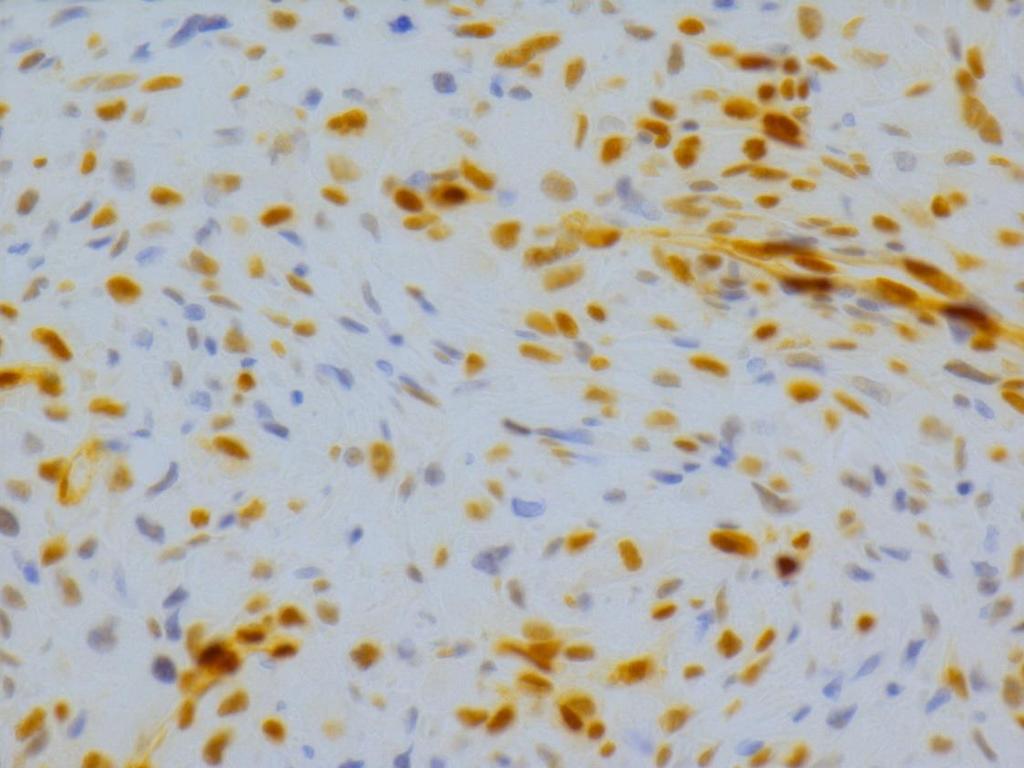
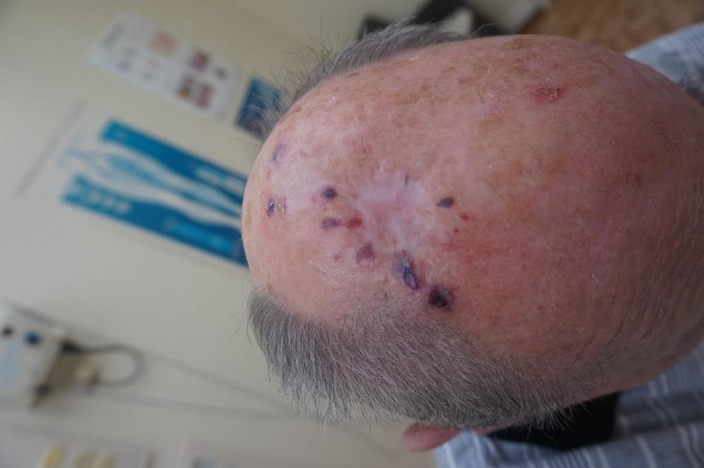
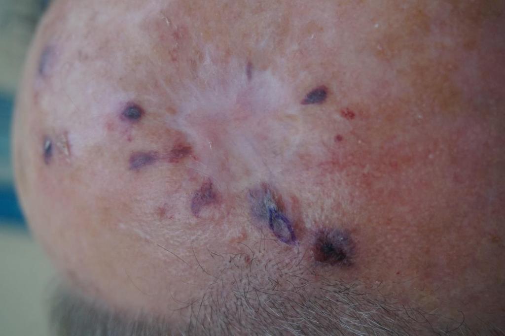
27 CASE Male of 78 years old with purpuric lesion on actinic damaged scalp. Takes anti-thrombotic medication
28

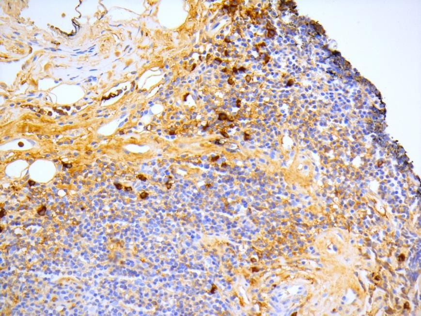
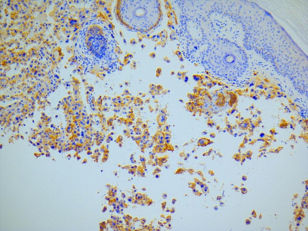
29 Keratin S100 CD34 CD31
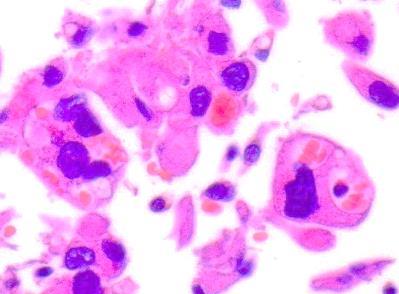
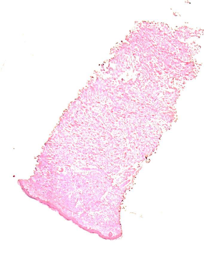
30 Histology Anaplastic sarcomatous proliferation Fibrotic spindled, cellular cuboidal and acantholytic split like or sinusoidal areas CK- not pseudosarcomatous squamous, S100- not melanoma, angio sarcoma? : CD34 also CD31- CD10 + Similar to previous biopsy and immuno findings : again compatible with fragment from AFX?
31 24/9/2017 Telefone call from referring dermatologist : recidivating and progressive circular purpuric lesions around scars of previous lesions Clinical story is not compatible with AFX!!!!! Revision and additional stains
32
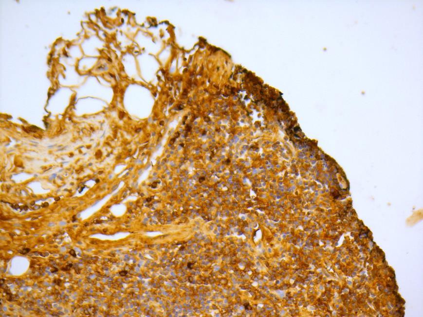
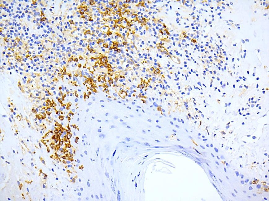
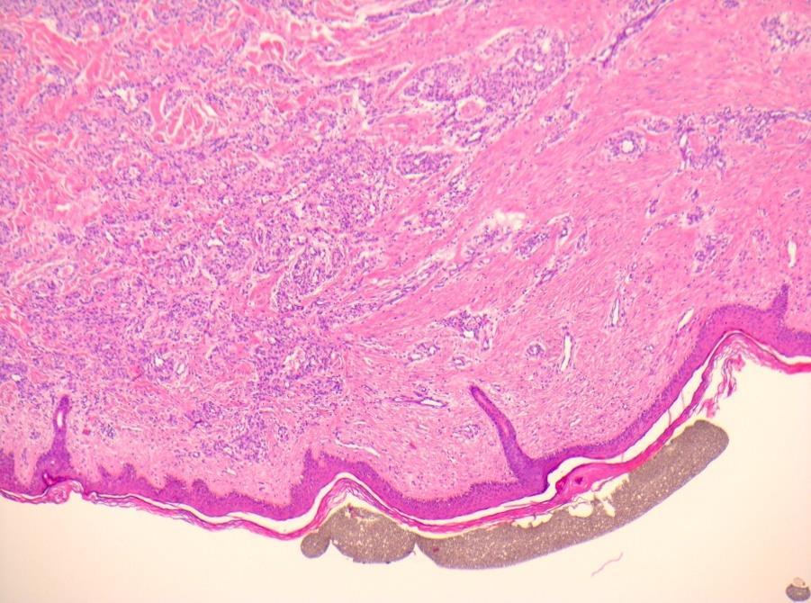
33 D2-40
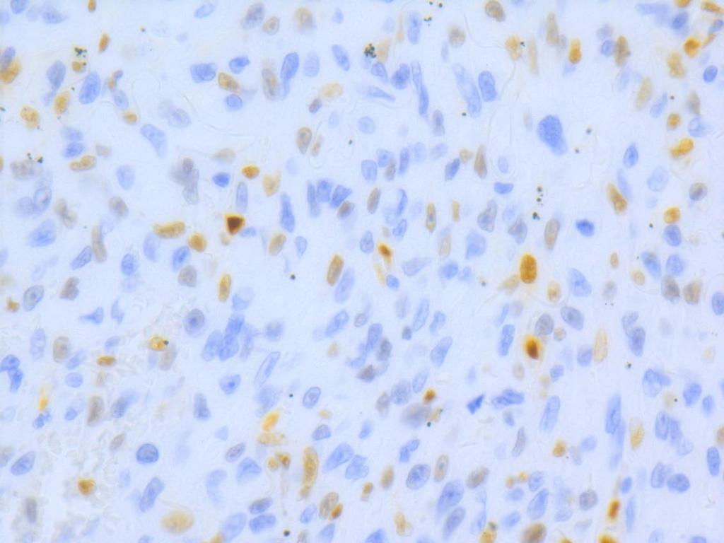
34 ERG C-MYC
35
36 Diagnosis AFX Sarcoma NOS Angiosarcoma Malignant Fibrous Histiocytoma Pseudosarcomatous Squamous Cell Carcinoma
37 Poorly differentiated angiosarcoma of the face and scalp in elderly patients DD with Melanoma and AFX can be difficult in solid areas! Vacuoles are clue Misleading immunos : CD34 - CD31-20 % of cases CD10 + Differential : D2-40 +, ERG +, C-myc + 40% of cases


38 lessons Clinical story! Erythematous maculae resembling hematoma in face or scalp elderly : Pannel of antibodies When a lesion recurs be cautious with previous interpretations

39 Self assesment Case 25

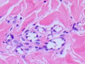
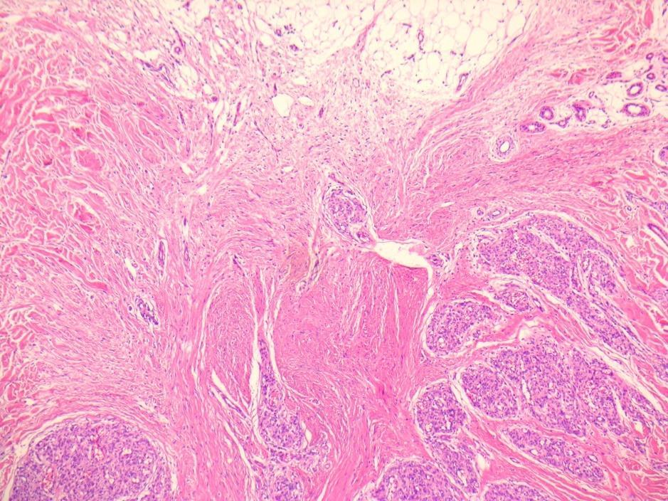
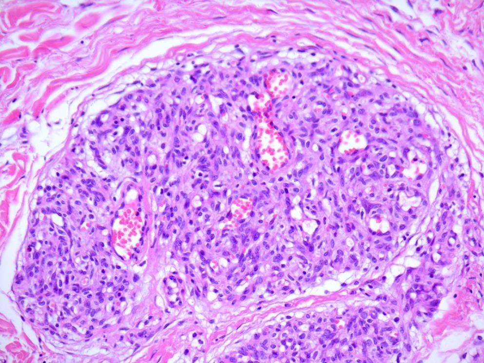
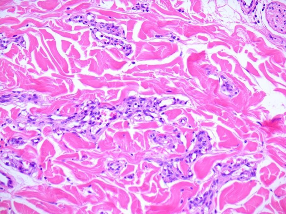
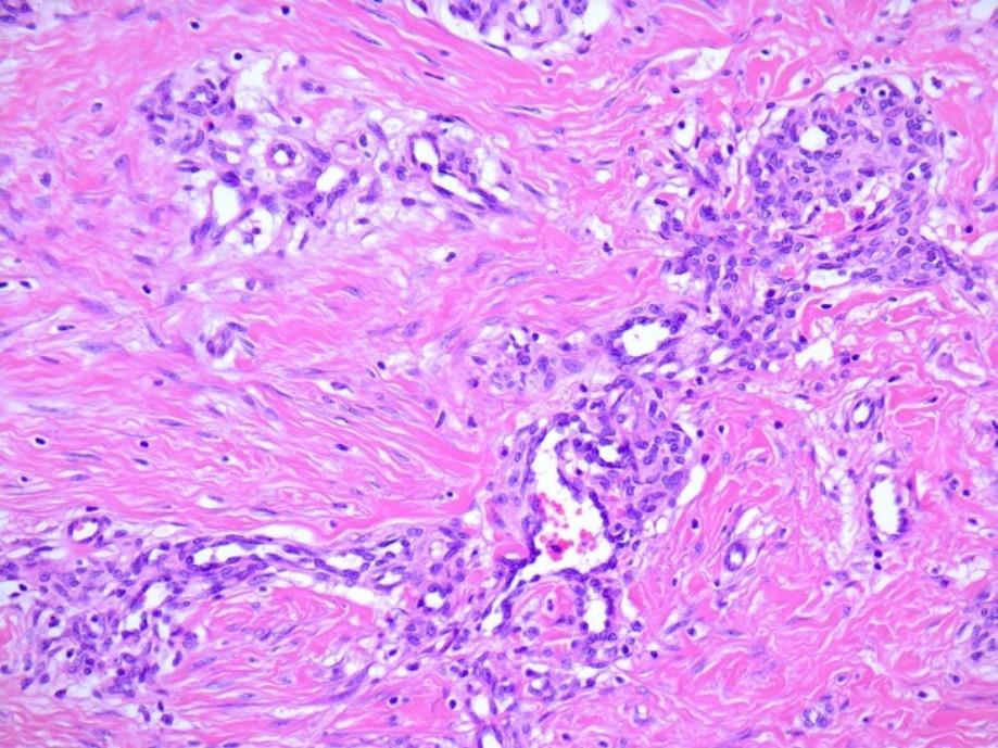
40 CASE Lady of 25 years old with erythematous infiltrated macula on central lumbar region since one month. Pseudolymphoma? Dermatofibroma? Malignant?
41
42

43
44

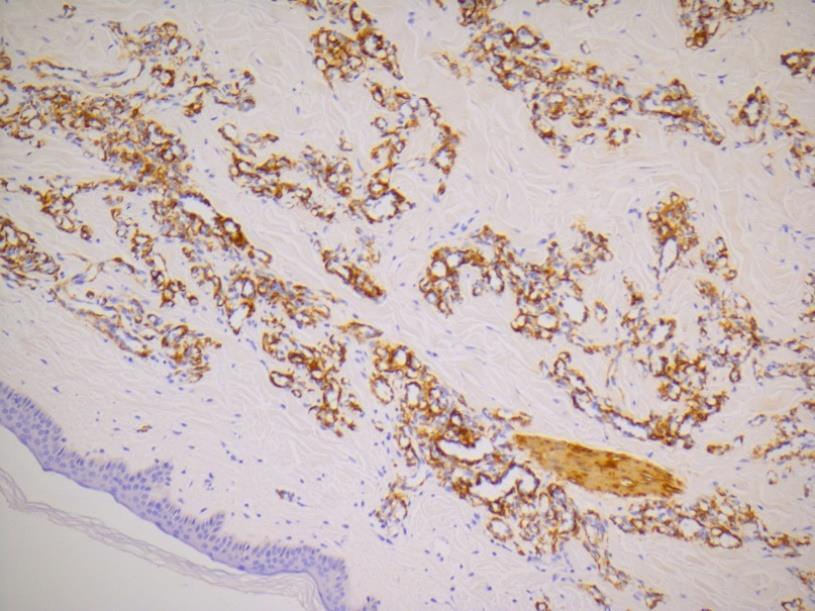
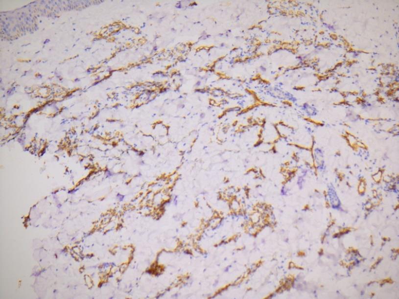
45 SMA D2-40 CD 31 CD 34
46 Diagnosis Angiomatosis Tufted angioma Hemangioma Microvenular angioma angiosarcoma
47 Microvenular Angioma Slow growing solitary asymptomatic plaque-like lesion Upper extremities, forearm, face, neck, trunk, lower extremities D2-40 neg CD31 +,CD34+ Pseudoinfiltrative growth DD Kaposi
48 Lesson learned Rare tumors do occur Not all what infiltrates is malignant!
49 THANK YOU
Actinic keratosis (AK): Dr Sarma s simple guide
: Dr Sarma s simple guide") Actinic keratosis (AK): Dr Sarma s simple guide Actinic keratosis is a very common lesion that you will see in your day-to-day practice. First, let me explain the name Actinic keratosis. It means keratosis
Actinic keratosis (AK): Dr Sarma s simple guide Actinic keratosis is a very common lesion that you will see in your day-to-day practice. First, let me explain the name Actinic keratosis. It means keratosis
Benign and malignant epithelial lesions: Seborrheic keratosis: A common benign pigmented epidermal tumor occur in middle-aged or older persons more
 Benign and malignant epithelial lesions: Seborrheic keratosis: A common benign pigmented epidermal tumor occur in middle-aged or older persons more common on the trunk; but extremities, head and neck are
Pathology of the skin. 2nd Department of Pathology, Semmelweis University
 Pathology of the skin 2nd Department of Pathology, Semmelweis University Histology of the skin Epidermis: Stratum corneum Stratum granulosum Stratum spinosum Stratum basale Dermis: papillary and reticular
Skin lesions The Good and the Bad. Dr Virginia Hubbard Ipswich Hospital NHS Trust Barts and the London School of Medicine and Dentistry
 Skin lesions The Good and the Bad Dr Virginia Hubbard Ipswich Hospital NHS Trust Barts and the London School of Medicine and Dentistry Case 1 32 year old woman Australian Lesion on back New hair growing
Chapter 6 Squamous Cell Carcinoma: Variants and Challenges
 Chapter 6 Squamous Cell Carcinoma: Variants and Challenges Michael B. Morgan EPIDEMIOLOGY: Second most common skin cancer, rare in the dark-skinned races. ETIOLOGY: Ultraviolet light, HPV infection. PATHOGENESIS:
Epidermolytic hyperkeratosis and acantholytic dyskeratosis. Porokeratoma ORIGINAL ARTICLE
 ORIGINAL ARTICLE Sarah N. Walsh, MD,* Mark A. Hurt, MD,w and Daniel J. Santa Cruz, MDw Abstract: Cornoid lamellation is a specific disorder of epidermal maturation manifested by a vertical column of parakeratosis
Disseminated epidermolytic acanthoma probably related to trauma
 Disseminated epidermolytic acanthoma probably related to trauma I. Sánchez-Carpintero, A. España and M.A. Idoate* Departments of Dermatology and *Pathology, University Clinic of Navarra, School of Medicine,
An Overview of Cutaneous Vascular Neoplasms
 An Overview of Cutaneous Vascular Neoplasms By Konstantinos Linos MD, FCAP, FASDP Bone, Soft Tissue and Dermatopathology Assistant Professor of Pathology Dartmouth-Hitchcock Medical Center Geisel School
Diploma examination. Dermatopathology: First paper. Tuesday 21 March Candidates must answer FOUR questions ONLY. Time allowed: Three hours
 Dermatopathology: First paper Tuesday 21 March 2017 1. Discuss the role of fluorescent in-situ hybridization (FISH) and emerging molecular techniques in the diagnosis of cutaneous melanocytic lesions,
Important Decisions in Dermatopathology: The Clinico- Pathologic Correlation. Dermatopathology Specialists Needed. Changing Trends
 Important Decisions in Dermatopathology: The Clinico- Pathologic Correlation Uma Sundram, MD, PhD Departments of Pathology and Dermatology Stanford University May 29, 2008 Dermatopathology Specialists
NEOPLASMS OF THE SURFACE EPITHELIUM (KERATINOCYTES)
") NEOPLASMS OF THE SURFACE EPITHELIUM (KERATINOCYTES) Papillary Lesions Precancerous Lesions Keratinocyte Proliferations Carcinomas Melanotic Lesions Melanomas Normal Mucosa Keratin layer Spinous layer Basal
NEOPLASMS OF THE SURFACE EPITHELIUM (KERATINOCYTES) Papillary Lesions Precancerous Lesions Keratinocyte Proliferations Carcinomas Melanotic Lesions Melanomas Normal Mucosa Keratin layer Spinous layer Basal
CUTANEOUS PSEUDONEOPLASTIC MESENCHYMAL LESIONS ( PSEUDOSARCOMAS )
") CUTANEOUS PSEUDONEOPLASTIC MESENCHYMAL LESIONS ( PSEUDOSARCOMAS ) Mark R. Wick, M.D. University of Virginia Health System Charlottesville, VA www.markwickmd.com; mrw9c@virginia.edu PSEUDONEOPLASMS & PSEUDOTUMORS
CUTANEOUS PSEUDONEOPLASTIC MESENCHYMAL LESIONS ( PSEUDOSARCOMAS ) Mark R. Wick, M.D. University of Virginia Health System Charlottesville, VA www.markwickmd.com; mrw9c@virginia.edu PSEUDONEOPLASMS & PSEUDOTUMORS
Basal cell carcinoma 5/28/2011
 Goal of this Presentation A practical approach to the diagnosis of cutaneous carcinomas and their mimics Thaddeus Mully, MD University of California San Francisco To review common non-melanoma skin cancers
Multiple epidermolytic acanthomas mimicking condyloma: a retrospective study of 8 cases
 Report Multiple epidermolytic acanthomas mimicking condyloma: a retrospective study of 8 cases Tsung-Ju Lee 1, MD, and Yu-Hung Wu 1,2, MD 1 Department of Dermatology, Mackay Memorial Hospital, Taipei,
Acantholytic Anaplastic Extramammary Paget s Disease: A Case Report and Review of the Literature
 Ann Dermatol Vol. 23, Suppl. 2, 2011 http://dx.doi.org/10.5021/ad.2011.23.s2.s226 CASE REPORT Acantholytic Anaplastic Extramammary Paget s Disease: A Case Report and Review of the Literature Yu-Jin Oh,
Benign versus Cancerous Lesions How to tell the difference FMF 2014 Christie Freeman MD, CCFP, DipPDerm, MSc
 1 Benign versus Cancerous Lesions How to tell the difference FMF 2014 Christie Freeman MD, CCFP, DipPDerm, MSc Benign lesions Seborrheic Keratoses: Warty, stuck-on Genetics and birthdays Can start in late
Common Benign Lesions and Skin Cancers. 22nd May 2015 Dr Mark Foley
 Common Benign Lesions and Skin Cancers 22nd May 2015 Dr Mark Foley Thank you for downloading this file. This intended to supplement the presentation given at the NZ Wound Care Conference, it is not intended
IT S FUNDAMENTAL MY DEAR WATSON! A SHERLOCKIAN APPROACH TO DERMATOLOGY
 IT S FUNDAMENTAL MY DEAR WATSON! A SHERLOCKIAN APPROACH TO DERMATOLOGY Skin, Bones, and other Private Parts Symposium Dermatology Lectures by Debra Shelby, PhD, DNP, FNP-BC, FADNP, FAANP Debra Shelby,
Lesions & Lifestyles
 Lesions & Lifestyles attended a 3 hour Continuing Education Seminar on Oral Pathology presented by Nancy Dewhirst, RDH,BS on (date) at (location):. Course material is directly related patient care. Notes:
Pathology of the skin. Dr Fónyad László, 1sz. Patológiai és Kísérleti Rákkutató Intézet, SE
 Pathology of the skin Dr Fónyad László, 1sz. Patológiai és Kísérleti Rákkutató Intézet, SE The skin Biggest organ Kb. 1.8 nm Kb. 10 kg Most frequent site for tumor development (BCC) Pathology of the skin
Clinically Microscopically Pathogenesis: autoimmune not lifetime
 Vulvar Diseases: Can be divided to non-neoplastic and neoplastic diseases. The neoplastic diseases are much less common. Of those, squamous cell carcinoma is the most common. most common in postmenopausal
المركب النموذج--- سبيتز وحمة = Type Spitz's Nevus, Compound SPITZ NEVUS 1 / 7
 SPITZ NEVUS 1 / 7 Epidemiology An annual incidence rate of 1.4 cases of Spitz nevus per 100,000 individuals has been estimated in Australia, compared with 25.4 per 100,000 individuals for cutaneous melanoma
Supplementary Online Content
 Supplementary Online Content Tschandl P, Rosendahl C, Akay BN, et al. Expert-level diagnosis of nonpigmented skin cancer by combined convolutional neural networks. JAMA Dermatol. Published online November
Desmoplastic Melanoma R/O BCC. Clinical Information. 74 y.o. man with lesion on left side of neck r/o BCC
 R/O BCC Sabine Kohler, M.D. Professor of Pathology and Dermatology Dermatopathology Service Stanford University School of Medicine Clinical Information 74 y.o. man with lesion on left side of neck r/o
Premalignant skin tumours
 Chapter 14: Premalignant skin tumours page: 434 Premalignant skin tumours page: 435 Solar keratoses (senile keratoses) Raised red and well-defined plaques with a rough surface covered in scales of varying
Post-test Self-assessment Cases
 Post-test Self-assessment Cases Ibrahim Khalifeh, M.D. Associate Professor Department of Pathology American University of Beirut Medical Center Beirut, Lebanon Case I History A 69 year old gentleman presenting
04/09/2018. Squamous Cell Neoplasia and Precursor Lesions. Agenda. Squamous Dysplasia. Squamo-proliferative lesions. Architectural features
 Squamous Cell Neoplasia and Precursor Lesions Jennifer L. Hunt, MD, MEd Aubrey J. Hough Jr, MD, Endowed Professor of Pathology Chair of Pathology and Laboratory Medicine University of Arkansas for Medical
UNIVERSITY OF MEDICINE AND PHARMACY OF CRAIOVA FACULTY OF MEDICINE DOCTORAL THESIS SUMMARY
 UNIVERSITY OF MEDICINE AND PHARMACY OF CRAIOVA FACULTY OF MEDICINE DOCTORAL THESIS SUMMARY CLINICAL, HISTOPATHOLOGICAL AND IMMUNOHISTOCHEMICAL STUDY OF THE EPITHELIAL PRECANCEROUS LESIONS PRECURSORS OF
Diploma Examination. Dermatopathology: First paper. Tuesday 20 March Candidates must answer FOUR questions. Time allowed: 3 hours
 Dermatopathology: First paper Tuesday 20 March 2018 Candidates must answer FOUR questions Time allowed: 3 hours 1. Give an account of the genetic aberrations encountered in Spitzoid neoplasms and how these
Cutanous Manifestation of Lupus Erythematosus. Presented By: Dr. Naif S. Al Shahrani Salman Bin Abdaziz university
 Cutanous Manifestation of Lupus Erythematosus Presented By: Dr. Naif S. Al Shahrani Salman Bin Abdaziz university A 50-year old lady, who is otherwise healthy, presented to the dermatology clinic with
Clinical Differential Diagnosis 4/16/2018 DERMATOPATHOLOGY OF THE GENITALIA AND BREAST NO CONFLICTS TO DISCLOSE
 DERMATOPATHOLOGY OF THE GENITALIA AND BREAST JOHN S. METCALF, MD Professor of Pathology and Dermatology MUSC NO CONFLICTS TO DISCLOSE Clinical Differential Diagnosis Plaques: Erythematous Inflammatory
Spindle Cell Lesions Of The Breast. Emad Rakha Professor of Breast Pathology and Consultant Pathologist
 Spindle Cell Lesions Of The Breast Emad Rakha Professor of Breast Pathology and Consultant Pathologist * SCLs comprise a wide spectrum of diseases, ranging from reactive processes to aggressive malignant
DERMCASE. Doc, my baby s all spotty! Case 1
 Test Your Knowledge With Multiple-Choice Cases This month 5 cases: Case 1 1. Doc, my baby s all spotty! 2. A Mediterranean Matter 3. Mommy, what s wrong with my head? 4. Armed with Lesions 5. It s spreading!
Pathology. Skin Tumor. Bayan N. Mohammad 15/10/2015. Mohammad al-orjani. Page 0 of 23
 #7 35 Pathology Skin Tumor Bayan N. Mohammad 15/10/2015 Mohammad al-orjani Page 0 of 23 بسم هللا الرحمن الرحيم GREETINGS This lecture is about skin tumors, all the slides are included and every slide will
Clinicopathologic Self- Assessment S003 AAD 2017
 Clinicopathologic Self- Assessment S003 AAD 2017 Clay J. Cockerell, M.D. Director, Cockerell Dermatopathology Director, Division of Dermatopathology UT Southwestern Medical Center July 2017 No relevant
Histopathology: skin pathology
 Histopathology: skin pathology These presentations are to help you identify, and to test yourself on identifying, basic histopathological features. They do not contain the additional factual information
Papillary Lesions of the breast
 Papillary Lesions of the breast Emad Rakha Professor of Breast Pathology The University of Nottingham Papillary lesions of the breast are a heterogeneous group of disease, which are characterised by neoplastic
Uropathology January Jon Oxley
 Uropathology January 2012 Jon Oxley Background to seminar These slides were available to view via the web from scanned slides The junior pathologists answered questions on them via the web The answers
A dinical and histopathologic entity associated with an increased risk of nonmelanoma skin cancer
 PUVA keratosis A dinical and histopathologic entity associated with an increased risk of nonmelanoma skin cancer M. C. G. van Praag, MD, a J. N. Bouwes Bavinck, MD, a W. Bergman, MD, PhD, a F. R. Rosendaal,
Dermatology for the PCP Deanna G. Brown, MD, FAAD Susong Dermatology Consulting Staff at CHI Memorial
 Dermatology for the PCP Deanna G. Brown, MD, FAAD Susong Dermatology Consulting Staff at CHI Memorial Cutaneous Oncology for the PCP Deanna G. Brown, MD, FAAD Susong Dermatology Consulting Staff at CHI
Case 18. M75. Excision of mass on scalp. Clinically SCC. The best diagnosis is:
 Case 18 M75. Excision of mass on scalp. Clinically SCC. The best diagnosis is: A. Pilomatrical carcinoma B. Adnexal carcinoma NOS C. Metastatic squamous cell carcinoma D.Primary squamous cell carcinoma
Diseases of the penis & testis
 Diseases of the penis & testis Done by : Saef B AL-Abbadi Diseases of penis, Condyloma Acuminatum A benign tumor *Tend to recur but only rarely progress into in situ or invasive cancers read this = genital
MULLERIAN PAPILLOMA ENTITY RECOGNITION FAILURE 04/04/2016 OUT OF SIGHT, OUT OF MIND: LESSER KNOWN LESIONS OF THE VULVOVAGINAL TRACT
 OUT OF SIGHT, OUT OF MIND: LESSER KNOWN LESIONS OF THE VULVOVAGINAL TRACT 23 rd ANNUAL SEMINAR IN PATHOLOGY 30 APRIL 2016 W. Dwayne Lawrence MD MSc (Path.) Chief of Pathology and Laboratory Medicine Women
Gross Appearance & Histology of Skin Cancer. Kyle Mannion M.D. January 21, 2005
 Gross Appearance & Histology of Skin Cancer Kyle Mannion M.D. January 21, 2005 Actinic Keratosis 5-20% will develop squamous/basal cell ca Almost solely from solar damage Usually develop during 4 th decade
Update on Cutaneous Mesenchymal Tumors. Thomas Brenn
 Update on Cutaneous Mesenchymal Tumors Thomas Brenn Cutaneous Mesenchymal Tumours Wide morphological and biological spectrum Myofibroblastic, smooth muscle, neural, vascular, apidocytic, undifferentiated;
Clinic Clinic Information Suitable for Referral Not Suitable for Referral
 PBC Services Clinic Clinic Information Suitable for Referral Not Suitable for Referral Diabetes One Off Clinic Donnington Medical The Health Centre, Wrekin Drive Donnington TF2 8EA Contact: Bryan Henshall
Cerebral Parenchymal Lesions: I. Metastatic Neoplasms
 Chapter 4 Cerebral Parenchymal Lesions: I. Metastatic Neoplasms After one has reasonably ruled out the possibility of a nonneoplastic diagnosis (see Chap. 3), one is left with considering a diagnosis of
Malignant non-melanocytic lesions
 Malignant non-melanocytic lesions Course C023: Fundamentals of Dermoscopy March 4, 2019, 11:20 AM - 11:50 PM Room: 146B Jason B. Lee, MD Professor & Vice Chair Director of Dermatopathology & Pigmented
Dermatopathology: The tumor is composed of keratinocytes which show atypia, increase mitoses and abnormal mitoses.
 Squamous cell carcinoma (SCC): A common malignant tumor of keratinocytes arising in the epidermis, usually from a precancerous condition: 1- UV induced actinic keratosis, usually of low grade malignancy.
ISPUB.COM. A Case of Actinic Lichen Planus. K Choi, H Kim, H Kim, Y Park INTRODUCTION CASE REPORT
 ISPUB.COM The Internet Journal of Dermatology Volume 8 Number K Choi, H Kim, H Kim, Y Park Citation K Choi, H Kim, H Kim, Y Park.. The Internet Journal of Dermatology. 2009 Volume 8 Number. Abstract The
Richard Turner Consultant Dermatologist
 Old Problems & New Treatments Photo Album by Administrator Richard Turner Consultant Dermatologist Plan for tonight? Refresher on SCC and solar keratosis How to distinguish the two Classic therapy than
A Retrospective Study of Treatment of Squamous Cell Carcinoma In situ. Övermark, Meri.
 https://helda.helsinki.fi A Retrospective Study of Treatment of Squamous Cell Carcinoma In situ Övermark, Meri 2016 Övermark, M, Koskenmies, S & Pitkanen, S 2016, ' A Retrospective Study of Treatment of
Mucinoses Diverse group of disorders which have in common deposition of basophilic, finely granular and stringy material in the connective tissues of
 Cutaneous Mucinoses Nathan C. Walk, M.D. Mucinoses Diverse group of disorders which have in common deposition of basophilic, finely granular and stringy material in the connective tissues of the dermis.
Diseases of the breast (1 of 2)
") Diseases of the breast (1 of 2) Introduction A histology introduction Normal ducts and lobules of the breast are lined by two layers of cells a layer of luminal cells overlying a second layer of myoepithelial
Diseases of the breast (1 of 2) Introduction A histology introduction Normal ducts and lobules of the breast are lined by two layers of cells a layer of luminal cells overlying a second layer of myoepithelial
Lung Tumor Cases: Common Problems and Helpful Hints
 Lung Tumor Cases: Common Problems and Helpful Hints Brandon T. Larsen, MD, PhD Senior Associate Consultant Department of Laboratory Medicine and Pathology Mayo Clinic Arizona Arizona Society of Pathologists
DERMCASE. A Shiny, Pink, Nose Lesion. Case 1
 Test your knowledge with multiple-choice cases This month 5 cases: 1. A Shiny, Pink, Nose Lesion p.43 2. A Red Patch on the Forehead p.44 3. An Ulcerated Nodule on the Thigh p.45 4. A Large Lump on the
Challenging Cases in Dermatopathology. Rosalie Elenitsas, M.D. Professor of Dermatology Director, Dermatopathology University of Pennsylvania
 Challenging Cases in Dermatopathology Rosalie Elenitsas, M.D. Professor of Dermatology Director, Dermatopathology University of Pennsylvania DISCLOSURE OF RELATIONSHIPS WITH INDUSTRY Rosalie Elenitsas
Cutaneous Adnexal Tumors
 Cutaneous Adnexal Tumors Lesions with Predominant Follicular Differentiation Special Emphasis on Basal Cell Carcinoma 2014-04-01 Prof. Dr. med. Katharina Glatz Pathologie Cutaneous Adnexal Tumors Hair
Skin Adnexal Tumors - A Histopathological Spectrum at a Tertiary Care Hospital
 Original Article GCSMC J Med Sci Vol (VI) No (I) January-June 2017 Skin Adnexal Tumors - A Histopathological Spectrum at a Tertiary Care Hospital Neeraja Barve*, Hansa Goswami**, Urvi Parikh *** Abstract
Common skin tumors. Benign Epidermal Tumors. Topic. Clinicopathologic Variants. Seborrheic keratoses
 Common skin tumors Topic Benign epidermal tumors Skin cyst and adnexal neoplasms Other common skin tumor Common skin malignancy สมศ กด ต นร ตนากร 26/02/2015 Benign Epidermal Tumors Seborrheic keratosis
Penile cancer teams in UK. Common variants. Penile cancer teams. Basaloid squamous carcinoma. The Pathology of Penile Tumours
 The Pathology of Penile Tumours Dr Jonathan H Shanks The Christie NHS Foundation Trust, Manchester, UK Penile cancer teams in UK 12 centres for penile cancer work (10 in England and Wales, 2 in Scotland)
ENCR RECOMMENDATIONS
 E N C R EUROPEAN NETWORK OF CANCER REGISTRIES (ENCR) ENCR RECOMMENDATIONS Non-Melanoma Skin Cancers Members of the Working Group: Dr T. Davies, East Anglian Cancer Registry, Cambridge, UK Mrs M. Page,
ISPUB.COM. Seborrheic Keratosis: A Pictorial Review of the Histopathologic Variations. D Sarma, S Repertinger
 ISPUB.COM The Internet Journal of Dermatology Volume 7 Number 2 Seborrheic Keratosis: A Pictorial Review of the Histopathologic Variations D Sarma, S Repertinger Citation D Sarma, S Repertinger.. The Internet
Classification (1) Classification (3) Classification (2) Spindle cell lesions. Spindle cell lesions of bladder (Mills et al.
 Classification (3) Classification (2) Spindle cell lesions. Spindle cell lesions of bladder (Mills et al.") Non-epithelial tumours and nonepithelial tumour-like lesions of the bladder Dr Jonathan H Shanks The Christie NHS Foundation Trust, Manchester, UK Classification (1) Myofibroblastic proliferations and
Non-epithelial tumours and nonepithelial tumour-like lesions of the bladder Dr Jonathan H Shanks The Christie NHS Foundation Trust, Manchester, UK Classification (1) Myofibroblastic proliferations and
Non-Melanocytic Pattern Dermoscopy
 Non-Melanocytic Pattern Dermoscopy I have no conflicts of interest to disclose Except that I LOVE dermoscopy Michelle Tarbox, MD Assistant Professor of Dermatology and Dermatopathology Texas Tech University
Premalignant lesions may expose to a promoting. factor & may be induced to undergo malignant. Carcinoma in situ displays the cytologic features of
 بسم رلاهللا Def. Premalignant lesions may expose to a promoting factor & may be induced to undergo malignant transformation. Carcinoma in situ displays the cytologic features of malignancy without invasion
Hemangiomas and Other Vascular Tumors
 facebook.com/cincykidsrad Hemangiomas and Other Vascular Tumors Disclosures No relevant financial disclosures Bernadette L. Koch, M.D. Departments of Radiology and Pediatrics Cincinnati Children s Hospital
David B. Troxel, MD. Common Medicolegal Situations: Misdiagnosis of Melanoma
 Common Medicolegal Situations: Misdiagnosis of Melanoma David B. Troxel, MD Medical Director, The Doctors Company, Napa, California Clinical Professor Emeritus, University of California at Berkeley Past
Pigmented Seborrheic Keratosis (Melanoacanthoma) of Nipple A case report with review of literature
 of Nipple A case report with review of literature") Case Report Pigmented Seborrheic Keratosis (Melanoacanthoma) of Nipple A case report with review of literature Aparna Narasimha, Harendra Kumar ML, Divyarani MN, Bhaskaran A* Department of Pathology and
Case Report Pigmented Seborrheic Keratosis (Melanoacanthoma) of Nipple A case report with review of literature Aparna Narasimha, Harendra Kumar ML, Divyarani MN, Bhaskaran A* Department of Pathology and
Corporate Medical Policy
 Corporate Medical Policy File Name: Origination: Last CAP Review: Next CAP Review: Last Review: mohs_micrographic_surgery 07/2004 11/2017 11/2018 11/2017 Description of Procedure or Service Mohs Micrographic
Skin Deep: Cutaneous Lupus. Dr Sarah Sasson Immunology Registrar, Liverpool Hospital 2016
 Skin Deep: Cutaneous Lupus Dr Sarah Sasson Immunology Registrar, Liverpool Hospital 2016 Introduction: Cutaneous lupus erythematosus LE is an autoimmune disease with a range of clinical manifestations
Case year female. Routine Pap smear
 Case 1 57 year female Routine Pap smear Diagnosis? 1. Atypical glandular cells of unknown significance (AGUS) 2. Endocervical AIS 3. Endocervical adenocarcinoma 4. Endometrial adenocarcinoma 5. Adenocarcinoma
Table of Contents. Preface xi. Acknowledgments xiii. Part I Overview of the Diagnostic Process 1. 1 Overview of Grading and Staging 3
 Table of Contents Preface xi Acknowledgments xiii Part I Overview of the Diagnostic Process 1 1 Overview of Grading and Staging 3 Identification of the process 3 Identification of tumor types 5 Grading
When Immunostains Can Get You in Trouble: Gynecologic Pathology p16: Panacea or Pandora s Box?
 When Immunostains Can Get You in Trouble: Gynecologic Pathology p16: Panacea or Pandora s Box? Teri A. Longacre, MD Stanford Medicine Stanford California pi6 in Gynecologic Pathology: Panacea or Pandora
Primer of Immunohistochemistry (Leukocytic)
") Primer of Immunohistochemistry (Leukocytic) Paul K. Shitabata, M.D. Dermatopathology Institute Torrance, CA BENIGN LYMPHOID SKIN LESIONS CAPABLE OF SIMULATING LYMPHOMA -Jessner s lymphoid infiltrate -Dermal-subcutaneous
Primer of Immunohistochemistry (Leukocytic) Paul K. Shitabata, M.D. Dermatopathology Institute Torrance, CA BENIGN LYMPHOID SKIN LESIONS CAPABLE OF SIMULATING LYMPHOMA -Jessner s lymphoid infiltrate -Dermal-subcutaneous
Progressive symmetrical Erythrokeratoderma: A case report and literature review.
 214 Case report Thai J Dermatol, October-December 2010 Progressive symmetrical Erythrokeratoderma: A case report and literature review. Pasu Piamphongsant MD, Kowit Kampirapap MD. ABSTRACT: PIAMPHONGSANT
Abid Irshad, MD Director Breast Imaging. Medical University of South Carolina Charleston
 Abid Irshad, MD Director Breast Imaging Medical University of South Carolina Charleston Cases Financial disclosure: I or my family have no financial interest related to the material discussed in this presentation
JPRAS Open 6 (2015) 25e30. Contents lists available at ScienceDirect. JPRAS Open. journal homepage:
 25e30. Contents lists available at ScienceDirect. JPRAS Open. journal homepage:") JPRAS Open 6 (2015) 25e30 Contents lists available at ScienceDirect JPRAS Open journal homepage: http://www.journals.elsevier.com/ jpras-open Case report Treatment of rare nodular amyloidosis on the nose:
JPRAS Open 6 (2015) 25e30 Contents lists available at ScienceDirect JPRAS Open journal homepage: http://www.journals.elsevier.com/ jpras-open Case report Treatment of rare nodular amyloidosis on the nose:
LUMPS AND BUMPS: AN ORGANIZED APPROACH TO DIAGNOSIS AND MANAGEMENT
 LUMPS AND BUMPS: AN ORGANIZED APPROACH TO DIAGNOSIS AND MANAGEMENT Tammy P. Than, M.S., O.D., F.A.A.O. The University of Alabama at Birmingham / School of Optometry 1716 University Blvd. Birmingham, AL
العصوي الوعاي ي الورام = angiomatosis Bacillary
 1 / 7 BACILLARY ANGIOMATOSIS Epidemiology BA is most commonly seen in patients with acquired immunodeficiency syndrome (AIDS) and a CD4 count less than 50 cells/mm 3, with an incidence of 1.2 cases per
Case Report Nevus Lipomatosus Superficialis with a Folliculosebaceous Component: Report of 2 Cases
 SAGE-Hindawi Access to Research Pathology Research International Volume 2011, Article ID 105973, 4 pages doi:10.4061/2011/105973 Case Report Nevus Lipomatosus Superficialis with a Folliculosebaceous Component:
Introduction. Results. Discussion. Histopathologic and immunohistochemical findings. Results. conclusions,
 1/5 2/5 Carcinoma distinctive carcinoma. form erysipeloides (CE), metastasis. which clinically Itfrom has resembles been termed erysipelas, is an uncommon, but may extend It164 toclassically back, presents
Erich M. Gaertner. 1. Introduction. 3. Results. 2. Material and Methods
 Skin Cancer Volume 2011, Article ID 645743, 5 pages doi:10.1155/2011/645743 Research Article Incidental Cutaneous Reaction Patterns: Epidermolytic Hyperkeratosis, Acantholytic Dyskeratosis, and Hailey-Hailey-Like
Verrucous porokeratosis: A case report
 Case Report Verrucous porokeratosis: case report Hojat Eftekhari, MD 1 Rana Rafiei, MD 1 Fatemeh Shamsa, MD 1 ehnam Rafiee, MD 2 1. Skin Research Center, Dermatology Department, Guilan University of Medical
Dermoscopy: Recognizing Top Five Common In- Office Diagnoses
 Dermoscopy: Recognizing Top Five Common In- Office Diagnoses Vu A. Ngo, DO Department of Family Medicine and Dermatology Choctaw Nation Health Services Authority Learning Objectives Introduction to dermoscopy
Citation The Journal of Dermatology, 37(8), available at
, available at") NAOSITE: Nagasaki University's Ac Title Two cases of blaschkitis with promi Author(s) Utani, Atsushi Citation The Journal of Dermatology, 37(8), Issue Date 2010-08 URL Right http://hdl.handle.net/10069/25634
NAOSITE: Nagasaki University's Ac Title Two cases of blaschkitis with promi Author(s) Utani, Atsushi Citation The Journal of Dermatology, 37(8), Issue Date 2010-08 URL Right http://hdl.handle.net/10069/25634
Vulvar Carcinoma. Definition: Cases should be classified as carsinoma of the vulva when the primary site growth is in the vulva Malignant melanoma sho
 Carcinoma Vulva & Vagina Subdivisi Onkologi Ginekologi Bagian Obgin FK USU Vulvar Carcinoma. Definition: Cases should be classified as carsinoma of the vulva when the primary site growth is in the vulva
Clinical characteristics
 Skin Cancer Fernando Vega, MD Seattle Healing Arts Clinical characteristics Precancerous lesions Common skin cancers ACTINIC KERATOSIS Precancerous skin lesions Actinic keratoses Dysplastic melanocytic
History A 89 year old gentleman presenting with a scalp/forehead nodule. Patient had squamous cell carcinoma 18 m at same site, excised. Outside diagn
 Case III History A 89 year old gentleman presenting with a scalp/forehead nodule. Patient had squamous cell carcinoma 18 m at same site, excised. Outside diagnoses: Squamous cell carcinoma. R/O: SCC, Melanoma,
CODING TUMOUR MORPHOLOGY. Otto Visser
 CODING TUMOUR MORPHOLOGY Otto Visser INTRODUCTION The morphology describes the tissue of the tumour closest to normal tissue Well differentiated tumours are closest to normal Undifferentiated tumours show
PATHOLOGY OF THE SKIN 2. Tumours of the skin
 PATHOLOGY OF THE SKIN 2. Tumours of the skin Máirín E. McMenamin MB MRCPI FRCPath Dip (Dermatopathol) RCPath St. James s Hospital and University of Dublin, Trinity College Tumour (Neoplasia) Benign or
Squamous Cell Neoplasia and Precursor Lesions
 Squamous Cell Neoplasia and Precursor Lesions Jennifer L. Hunt, MD, MEd Aubrey J. Hough Jr, MD, Endowed Professor of Pathology Chair of Pathology and Laboratory Medicine University of Arkansas for Medical
VULVAR CARCINOMA. Page 1 of 5
 VULVAR CARCINOMA EXAMPLE OF A VULVAR CARCINOMA USING PROPOSED TEMPLATE Case: Invasive squamous cell carcinoma arising in D-VIN Tumor in left labia major Left partial vaginectomy and sentinel lymph node
Diseases of the vulva
 Diseases of the vulva 1. Bartholin Cyst - Infection of the Bartholin gland produces an acute inflammation within the gland (adenitis) and may result in an abscess. Bartholin duct cysts - Are relatively
21/07/2017. Hobnail endothelial cells are not the same as epithelioid endothelial cells
 UPDATE IN CUTANEOUS VASCULAR S DERMATOPATHOLOGY SESSION BELFAST PATHOLOGY JUNE 21/2017 Dr E Calonje St John s Institute of Dermatology, London, United Kingdom THE FAMILY OF VASCULAR S WITH EPITHELIOID
Disorders of the vulva
 Vulval lesions Disorders of the vulva Terminology standardised by the International Society for the Study of Vulvovaginal Disease(ISSVD) Classification 1.Nonneoplastic epithelial disorders of vulva Lichen
Objectives. 1. Recognizing benign skin lesions. 2.Know which patients will likely need surgical intervention.
 The Joy of Pediatric Skin Dr. Claire Sanger University of Kentucky Plastic & Reconstructive Surgery Objectives 1. Recognizing benign skin lesions 2.Know which patients will likely need surgical intervention.
Case Report A Rare Cutaneous Adnexal Tumor: Malignant Proliferating Trichilemmal Tumor
 Case Reports in Medicine Volume 2015, Article ID 742920, 4 pages http://dx.doi.org/10.1155/2015/742920 Case Report A Rare Cutaneous Adnexal Tumor: Malignant Proliferating Trichilemmal Tumor Omer Alici,