BSD SELF-ASSESSMENT CASES 21-24
|
|
|
- Mervyn Wright
- 5 years ago
- Views:
Transcription
1 BSD SELF-ASSESSMENT CASES EDINBURGH, 2 JULY 2018 LASZLO IGALI
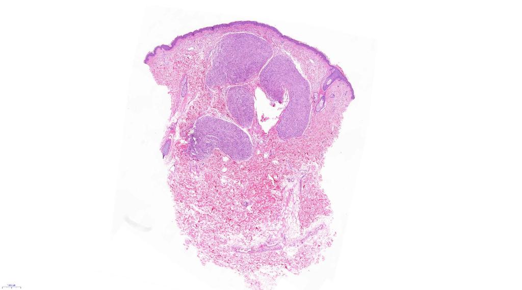
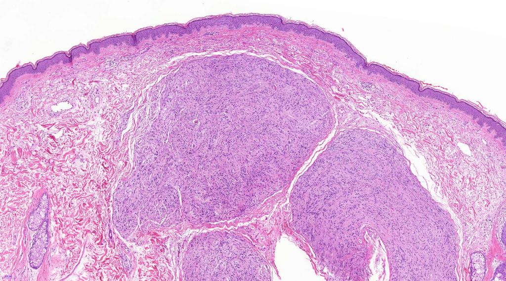
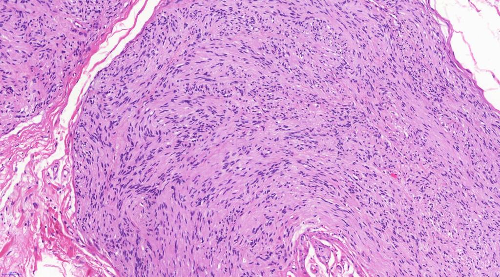
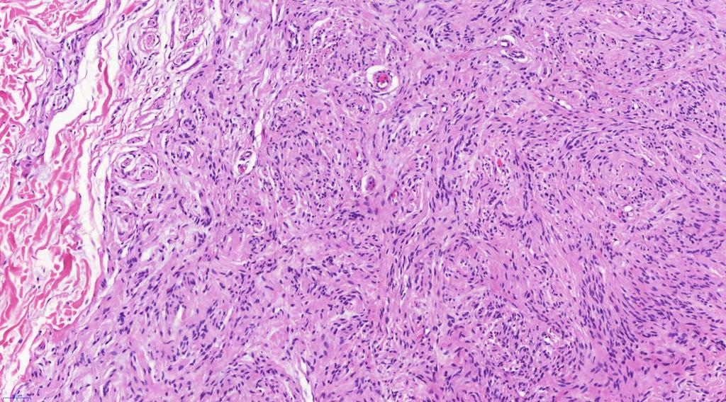
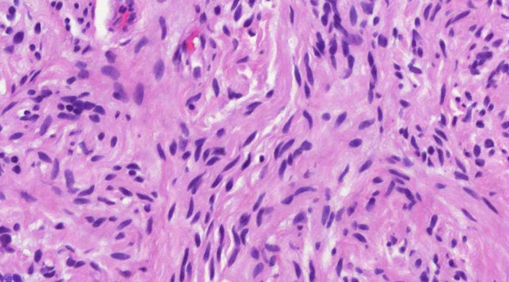
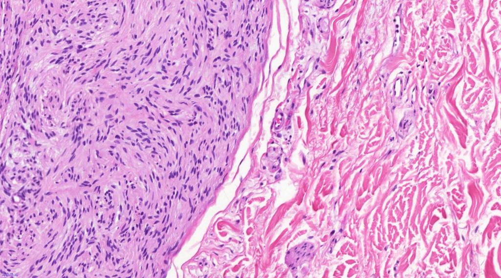
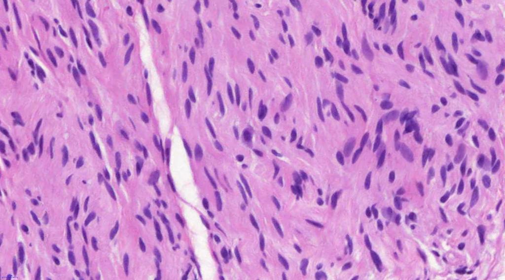
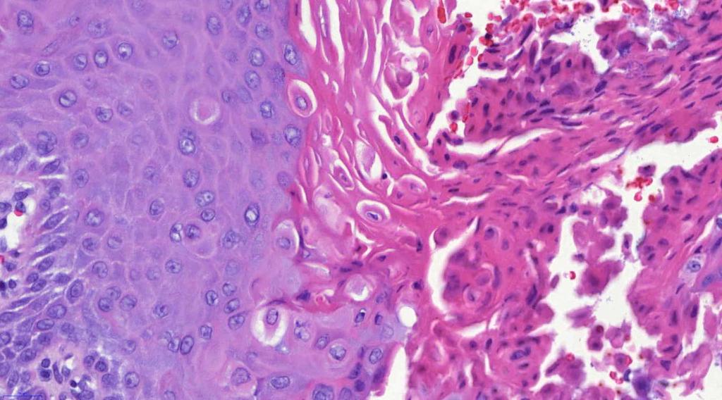
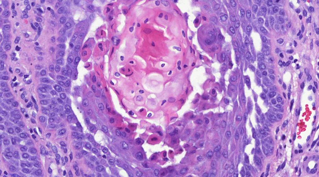
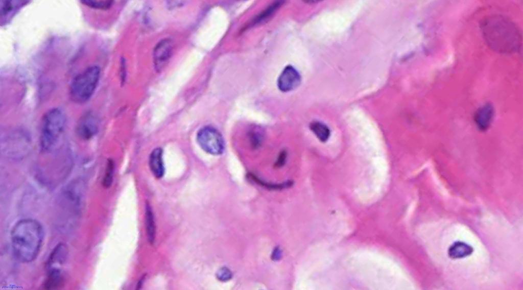
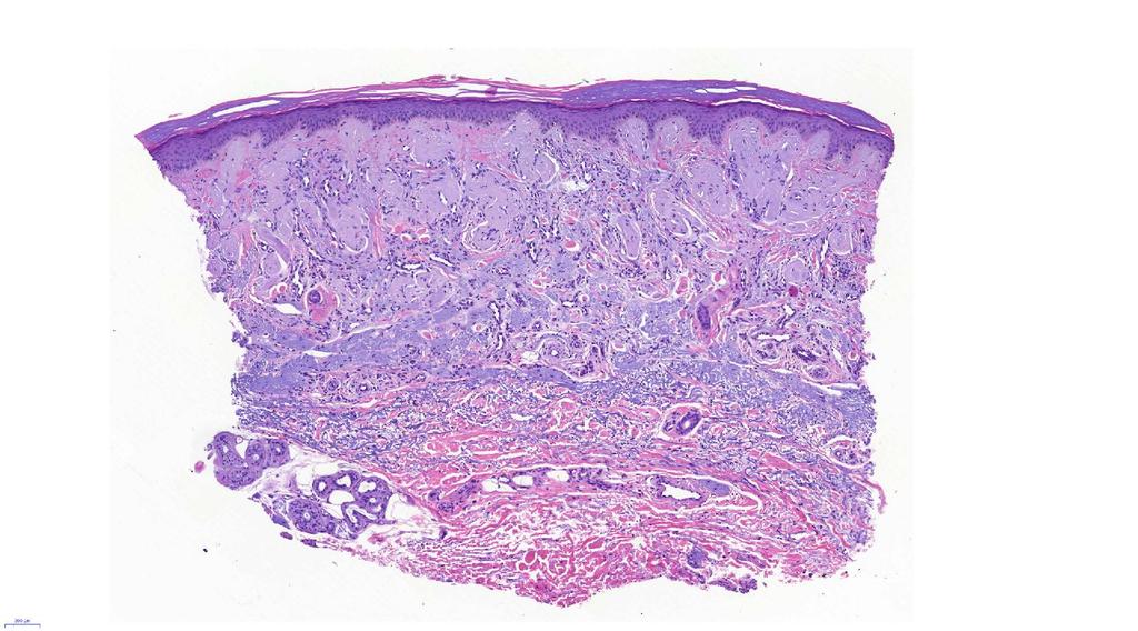
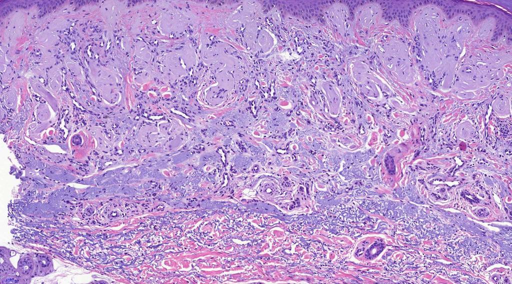
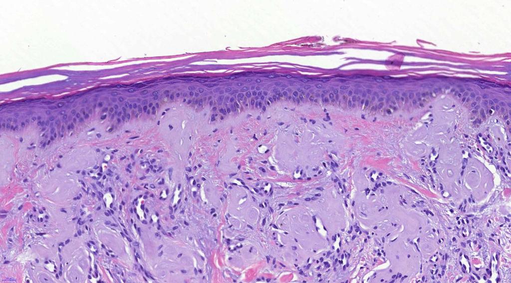
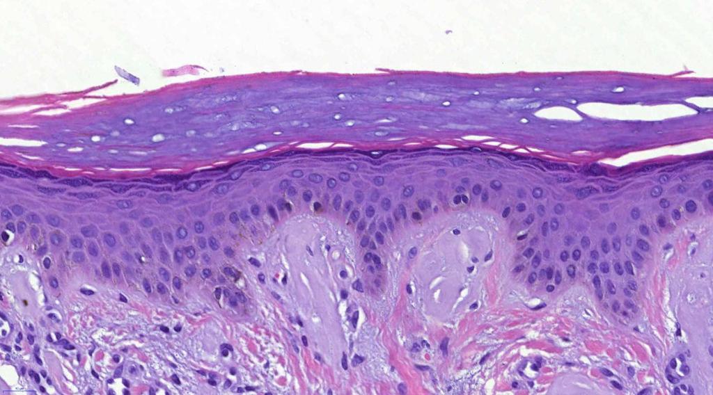
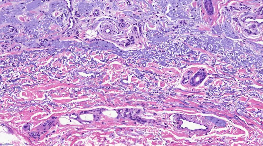
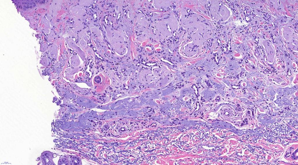
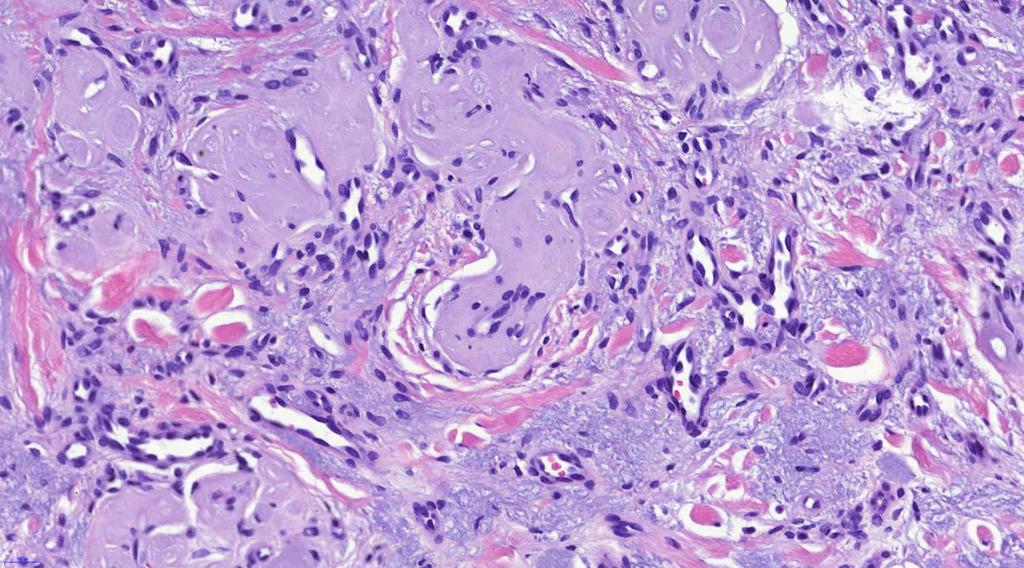
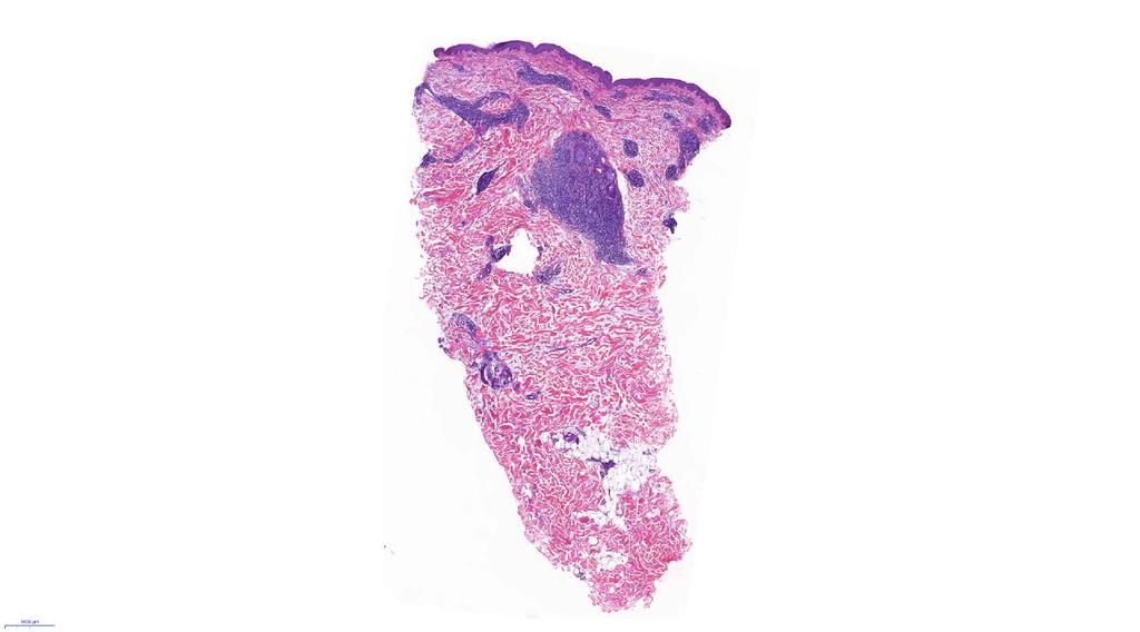
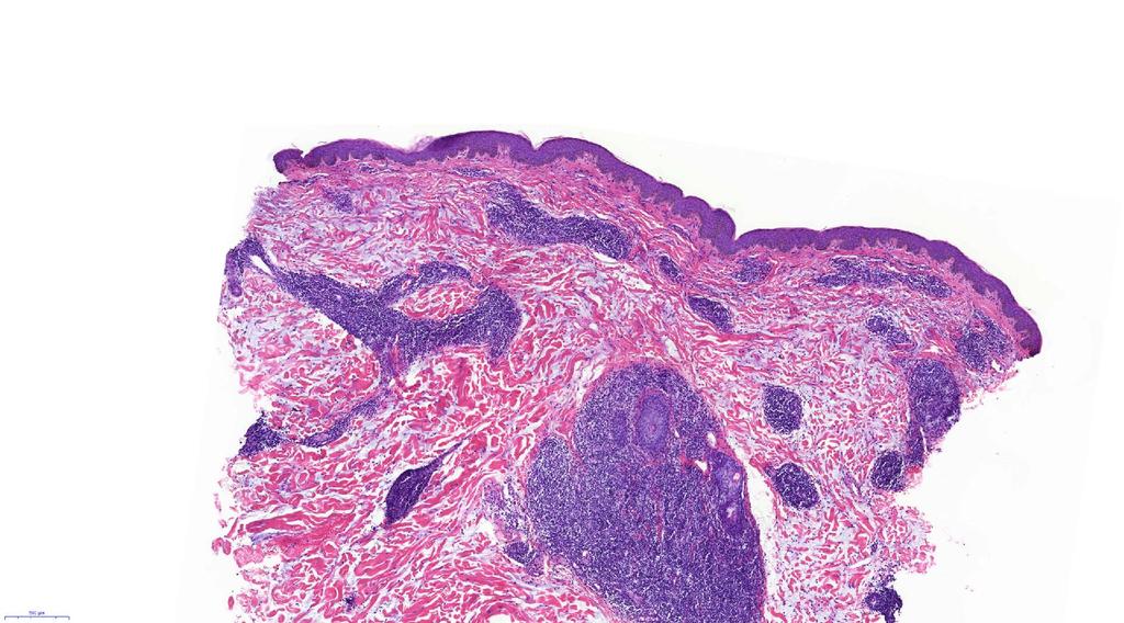
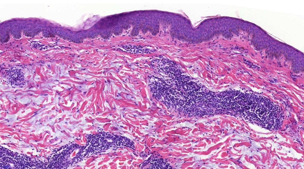
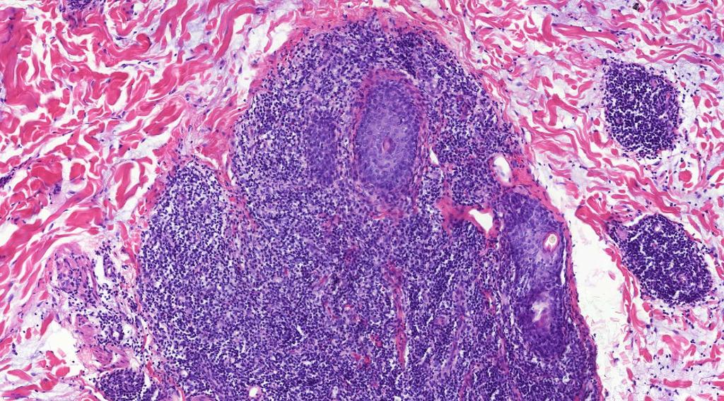
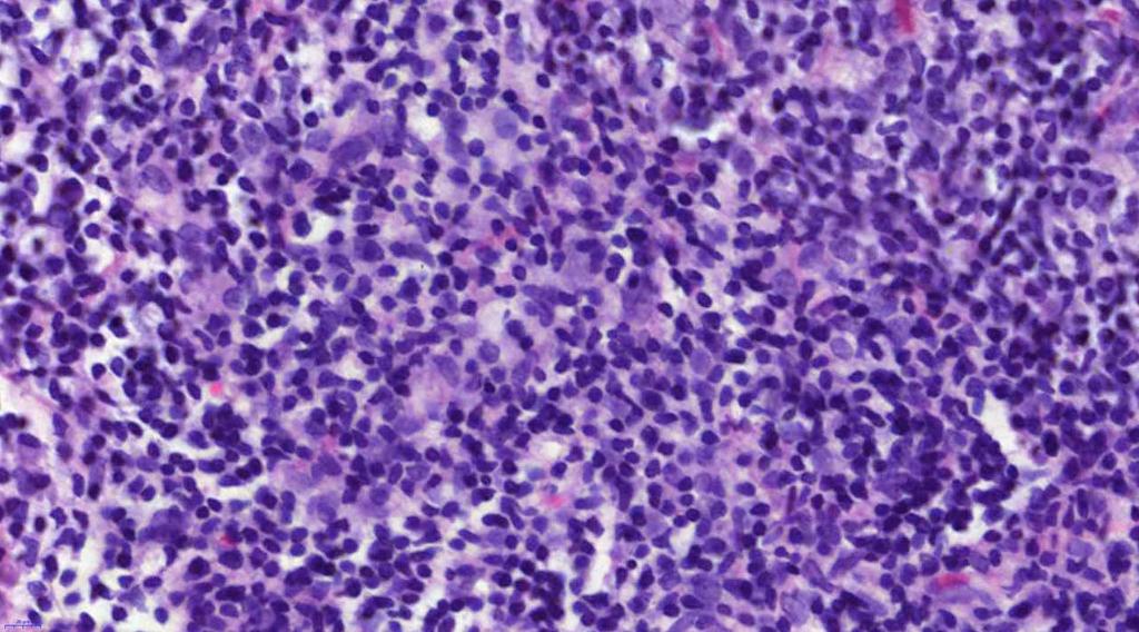
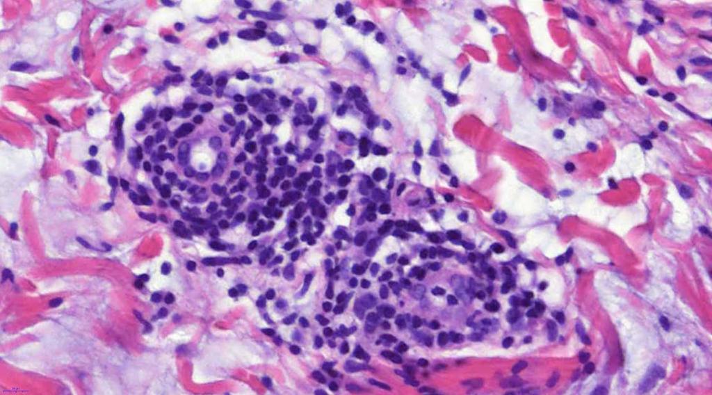
2 CASE 21 CLINICAL HISTORY Female, 43 years, nodule on left side of face/jaw angle area. Slow growth over years. MACRO (NOT GIVEN) Skin coloured nodule, up to 8 mm, contained within a punch biopsy. Single slice of the punch taken.
3 V-SLIDE
4
5
6
7
8
9
10
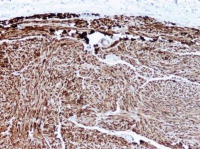
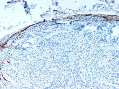
11 S100 EMA
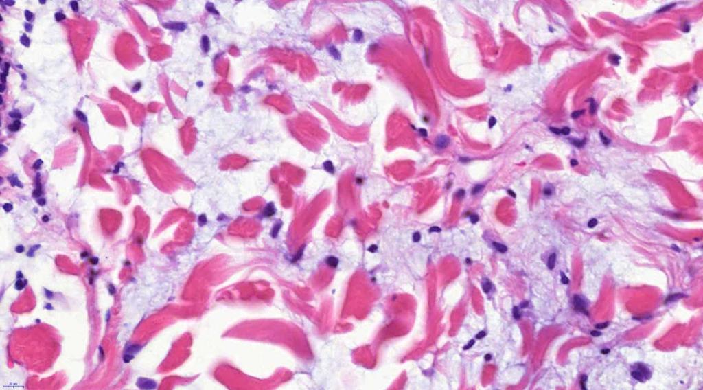
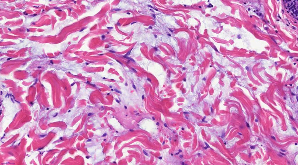
12 CASE 21 DIFFERENTIALS Schwannoma - unocmmon in dermis, complete perineual capsule, can be GFAP positive, could have Antoni A and B areas neurofibroma - no capsule, can be GFAP positive, frequent mast cells, neural fibroblasts and fibrillary collagen (CD34 and polarised light), can be associated with NF leiomyioma - S100 vs SMA/desmin positivity palisaded encapsulated neuroma - solitary, S100 pos schwann cells, weak peripheral EMA pos capsule, no Antoni A/B areas
13 CASE 21 HISTOLOGY FEATURES OF THIS CASE usually solitary, dermal or subcutaneous/submucosal, could be multifocal or plexiform (hyalinised vessels) poor/partial encapsulation retraction artefact predominantly spindled schwann cells - micro-bundles only vague palisading 90% - face, lips, oral cavity
14 CASE 21 PREFERRED DIAGNOSIS palisaded encapsulated neuroma
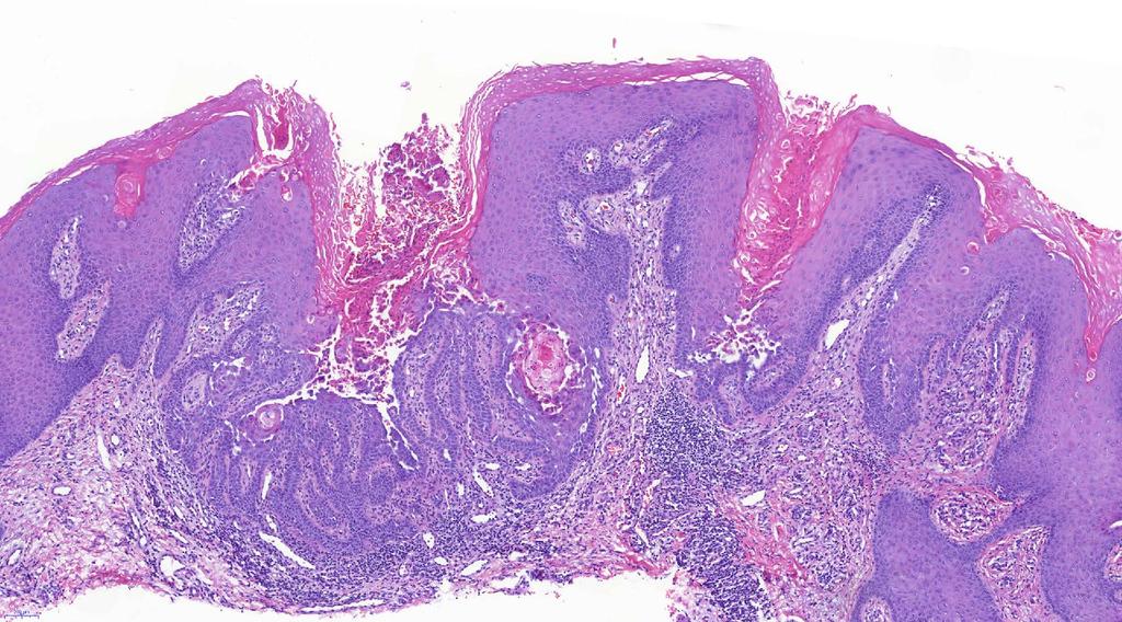
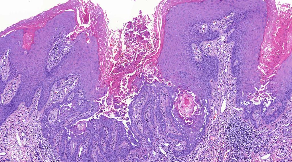
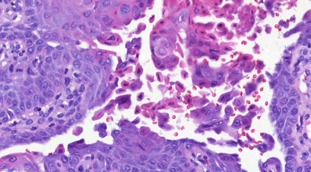
15 CASE 22 CLINICAL HISTORY Male, 53, nodule on scalp MACRO (NOT GIVEN) Skin ellipse - 2.4x1/8 cm, with a central depression up to 0.5 cm diameter with slightly raised edge and?keratotic debris in the centre.
16 V-SLIDE
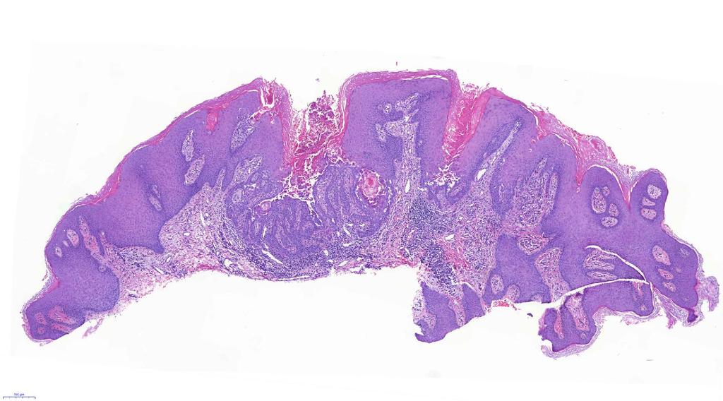
17
18
19
20
21
22
23
24 CASE 22 DIFFERENTIALS Hailey-Hailey disease - follicular syskeratosis, AD, 2-3rd decade, intertriginous sites, hspca1(golgi ATPase - Ca signalling); dilapidated brick wall Darier s disease - follicular dyskeratosis, AD, 2nd decde, seborrhoeic and acral distribution, SERCA2 (endoplasmic reticulum ATPase - Ca signalling), corps ronds, grains Grover s disease - can be similar to either (and other acanthilytic diseases), 6th decade or later, male predominance, transient Warty dyskeratoma - isolated follicular keratosis, head-neck, related to actinic keratosis or is it follicular neoplasia, Hellwig - isolated Darier s disease, no isolated reproducible molecular defect

25 CASE 22 DermatologyEdited by Jean L.BologniaJulie V.SchafferLorenzoCerroniFourth editionchina: Elsevier, 2018,

26 CASE 22 DermatologyEdited by Jean L.BologniaJulie V.SchafferLorenzoCerroniFourth editionchina: Elsevier, 2018,
27 CASE 22 PREFERRED DIAGNOSIS warty dykeratoma
28 CASE 23 CLINICAL HISTORY Male 33, blistering and crusting on back of the hands. Worse at summer. MACRO (NOT GIVEN) 6 mm punch biopsy with unremarkable epidermis, bisected.
29 V-SLIDE
30
31
32
33
34
35
36
37 CASE 23 DIFFERENTIALS hidroa vacciniforme - photo-aggravated /photodermatosis - childhood -first 2 decades, epidermal necrosis,?? EBV PCT - subepidermal blister, festooning, thinkened vessel walls (igg/m deposition in walls, woods light, urine red->brown if exposed to light, porphyrin screen ) EBA - mechanobullous disease, similar histology, blood tests, IMF Bullous lupus - can have identical picture, Ig deposition,

38 CASE 23 DermatologyEdited by Jean L.BologniaJulie V.SchafferLorenzoCerroniFourth editionchina: Elsevier, 2018,
39 CASE 23 DIAGNOSIS porphyria cutanea tarda
40 CASE 24 CLINICAL HISTORY Female, 70, extensive erythema on chest. MACRO (NOT GIVEN) 4 mm punch biopsy with unremarkable epidermis, bisected.
41 V-SLIDE
42
43
44
45
46
47
48
49
50 CASE 24 DIFFERENTIALS Scleredema - may be associated with diabtetes (late onset type1, male), thickened collagen +/- interstitial mucin Lupus erythematosus (?tumid) - could be plaque/pacth like, DIF positive, serology positive, tumid LE us photo-inducible, response to antimalarials REM - 3-4th decade of life, plaques/patches -in midline of chest/back, serology negative, could represenet ff of LE? Polymorphous light eruption - photodermatosis, mainly papules, rarely plaques, oedema, spongiosis, perivascular lymphoid infiltrate
51 CASE 24 PREFERRED DIAGNOSIS Reticulate erythematous mucinosis (REM)
52 THANKS FOR YOUR ATTENTION
53
Mucinoses Diverse group of disorders which have in common deposition of basophilic, finely granular and stringy material in the connective tissues of
 Cutaneous Mucinoses Nathan C. Walk, M.D. Mucinoses Diverse group of disorders which have in common deposition of basophilic, finely granular and stringy material in the connective tissues of the dermis.
Update in deposition diseases
 Genoa, Italy Update in deposition diseases Prof. Franco Rongioletti, Section of Dermatology, Chair of Dermatopathology, University of Genoa,Italy Cutaneous deposition disorders Endogenous Exogenous Cutaneous
BSD Self Assessment Workshop 7 th July 2013 CASE 27 RAC6123
 BSD Self Assessment Workshop 7 th July 2013 CASE 27 RAC6123 M55. 4/7 tender lesions on knee, legs and arms. Also iritis/ weight loss/headache, synovitis.?vasculitis. Sarcoidosis. Biopsy from left elbow
Dr Saleem Taibjee. Consultant Dermatologist & Dermatopathologist
 Dr Saleem Taibjee saleem.taibjee@dchft.nhs.uk Consultant Dermatologist & Dermatopathologist Case S14-10797 and S15-4023 F50. Previous blistering, now marked milia on dorsum of hands. 4mm punch biopsy The
السكري للداء مرافقة فقاعات diabeticorum= Bullosis
 1 / 6 Bullosis diabeticorum Bullous disease of diabetes (bullosis diabeticorum) is a distinct, spontaneous, noninflammatory, blistering condition of acral skin unique to patients with diabetes mellitus.
Cutanous Manifestation of Lupus Erythematosus. Presented By: Dr. Naif S. Al Shahrani Salman Bin Abdaziz university
 Cutanous Manifestation of Lupus Erythematosus Presented By: Dr. Naif S. Al Shahrani Salman Bin Abdaziz university A 50-year old lady, who is otherwise healthy, presented to the dermatology clinic with
Case Report A Rare Clinical Presentation of Darier s Disease
 Hindawi Publishing Corporation Case Reports in Dermatological Medicine Volume 2013, Article ID 419797, 5 pages http://dx.doi.org/10.1155/2013/419797 Case Report A Rare Clinical Presentation of Darier s
Pathology of the skin. 2nd Department of Pathology, Semmelweis University
 Pathology of the skin 2nd Department of Pathology, Semmelweis University Histology of the skin Epidermis: Stratum corneum Stratum granulosum Stratum spinosum Stratum basale Dermis: papillary and reticular
Case No. 5; Slide No. B13/8956/2
 Interface diseases Case No. 5; Slide No. B13/8956/2 Histological findings Severe hydropic vacuolation of epidermal and follicular basal cells/ interface dermatitis Multifocally apoptotic keratinocytes
Paul K. Shitabata, M.D. Dermatopathology Institute
 Paul K. Shitabata, M.D. Dermatopathology Institute Technical Considerations Storage of slides at room temperature
Histopathology: skin pathology
 Histopathology: skin pathology These presentations are to help you identify, and to test yourself on identifying, basic histopathological features. They do not contain the additional factual information
Degos Disease: A Case Report and Review of Literature
 Degos Disease: A Case Report and Review of Literature Monira waked Egyptian Dermatology Online Journal 4 (1): 5, June 2008 Al Houd Al Marsod Hospital Submitted for publication: May 25 th, 2008 Accepted
COPYRIGHTED MATERIAL. Introduction CHAPTER 1. Introduction
 CHAPTER 1 Introduction OVERVIEW The clinical features of skin lesions are related to the underlying pathological processes. Broadly skin conditions fall into three clinical groups: (a) those with a well-defined
Erythema gyratumrepens-like eruption in a patient with epidermolysisbullosaacquisita associated with ulcerative colitis
 Erythema gyratumrepens-like eruption in a patient with epidermolysisbullosaacquisita associated with ulcerative colitis A. España C. Sitaru* M. Pretel L. Aguado J. Jimenez# Department of Dermatology, University
Actinic keratosis (AK): Dr Sarma s simple guide
: Dr Sarma s simple guide") Actinic keratosis (AK): Dr Sarma s simple guide Actinic keratosis is a very common lesion that you will see in your day-to-day practice. First, let me explain the name Actinic keratosis. It means keratosis
Actinic keratosis (AK): Dr Sarma s simple guide Actinic keratosis is a very common lesion that you will see in your day-to-day practice. First, let me explain the name Actinic keratosis. It means keratosis
DARRIERS DISEASE WITH BASAL CELL CARCINOMA: A CASE REPORT Anila P. Sunandini 1, Shanti 2, G. Suryanarayana 3, Padmasri Somala Y 4
 DARRIERS DISEASE WITH BASAL CELL CARCINOMA: A Anila P. Sunandini 1, Shanti 2, G. Suryanarayana 3, Padmasri Somala Y 4 HOW TO CITE THIS ARTICLE: Anila P. Sunandini, Shanti, G. Suryanarayana, Padmasri Somala
The Relevance of Cytologic Atypia in Cutaneous Neural Tumors
 The Relevance of Cytologic Atypia in Cutaneous Neural Tumors Recent Findings - New Developments New Problems Zsolt B. Argenyi, M.D. Professor of Pathology & Dermatology Director of Dermatopathology Department
My Method for Approaching Skin Biopsies
 My Method for Approaching Skin Biopsies P A U L H A U N, MD, MS, F A A D A S S I S T A N T P R O F E S S O R D E R M A T O L O G Y A N D D E R M A T O P A T H O L O G Y D E P A R T M E N T O F D E R M
Skin Deep: Cutaneous Lupus. Dr Sarah Sasson Immunology Registrar, Liverpool Hospital 2016
 Skin Deep: Cutaneous Lupus Dr Sarah Sasson Immunology Registrar, Liverpool Hospital 2016 Introduction: Cutaneous lupus erythematosus LE is an autoimmune disease with a range of clinical manifestations
SMOOTH MUSCLE TUMOURS
 SMOOTH MUSCLE TUMOURS NORMAL SMOOTH MUSCLE Cytology Immunohistochemistry Ultrastructure Masson Trichrome Smooth Muscle Ultrastructure Many myofilaments running parallel to the long axis of the smooth
Essential Dermatopathology: Neoplastic American Academy of Dermatology Annual Meeting NEURAL AND SMOOTH MUSCLE NEOPLASMS
 Essential Dermatopathology: Neoplastic American Academy of Dermatology Annual Meeting NEURAL AND SMOOTH MUSCLE NEOPLASMS Kevin P. White M.D. Oregon Health and Science University Associate Professor of
Principi ed Aggiornamenti in Dermatologia Roma, 6-7 Aprile Grand rounds. Lorenzo Cerroni, Graz
 Principi ed Aggiornamenti in Dermatologia Roma, 6-7 Aprile 2018 Grand rounds Lorenzo Cerroni, Graz "Computer palms" Described in patient using computer keyboards for long periods; similar features described
Primer of Immunohistochemistry (Leukocytic)
") Primer of Immunohistochemistry (Leukocytic) Paul K. Shitabata, M.D. Dermatopathology Institute Torrance, CA BENIGN LYMPHOID SKIN LESIONS CAPABLE OF SIMULATING LYMPHOMA -Jessner s lymphoid infiltrate -Dermal-subcutaneous
Primer of Immunohistochemistry (Leukocytic) Paul K. Shitabata, M.D. Dermatopathology Institute Torrance, CA BENIGN LYMPHOID SKIN LESIONS CAPABLE OF SIMULATING LYMPHOMA -Jessner s lymphoid infiltrate -Dermal-subcutaneous
Diploma examination. Dermatopathology: First paper. Tuesday 21 March Candidates must answer FOUR questions ONLY. Time allowed: Three hours
 Dermatopathology: First paper Tuesday 21 March 2017 1. Discuss the role of fluorescent in-situ hybridization (FISH) and emerging molecular techniques in the diagnosis of cutaneous melanocytic lesions,
CLINCOPATHOLOGICAL CASE
 CLINCOPATHOLOGICAL CASE Generalized vesiculo-bullous and pustular eruption in an adult man Hassab El-Naby H, MD, El-Khalawany M, MD Department of Dermatology, Al-Azhar University, Cairo, Egypt CLINICAL
CPC. Chutika Srisuttiyakorn, M.D. Kobkul Aunhachoke, M.D. Phramongkutklao Hospital Bangkok, Thailand
 CPC Chutika Srisuttiyakorn, M.D. Kobkul Aunhachoke, M.D. Phramongkutklao Hospital Bangkok, Thailand A 53 year-old woman with fever, facial swelling and rashes on face, trunk and upper extremities for 3
Chapter 6 Squamous Cell Carcinoma: Variants and Challenges
 Chapter 6 Squamous Cell Carcinoma: Variants and Challenges Michael B. Morgan EPIDEMIOLOGY: Second most common skin cancer, rare in the dark-skinned races. ETIOLOGY: Ultraviolet light, HPV infection. PATHOGENESIS:
Questions. Answers. Share your photos and diagnoses with us!
 Illustrated quizzes on problems seen in everyday practice CASE 1 A 66-year-old male presents with ruddy-brown, pruritic papules on his chest and back that have been present for several years. The patient
Diploma Examination. Dermatopathology: First paper. Tuesday 20 March Candidates must answer FOUR questions. Time allowed: 3 hours
 Dermatopathology: First paper Tuesday 20 March 2018 Candidates must answer FOUR questions Time allowed: 3 hours 1. Give an account of the genetic aberrations encountered in Spitzoid neoplasms and how these
Patterns and mechanisms of inflammatory skin conditions: the pathologist s survival kit SALVADOR J. DIAZ-CANO BAHRAIN, APRIL 2017
 Patterns and mechanisms of inflammatory skin conditions: the pathologist s survival kit SALVADOR J. DIAZ-CANO 0000-0003-1245-2859 BAHRAIN, APRIL 2017 Basic Elements of Lesions Repair Injury Time & Intensity
Bullous Darier s Disease Mimicking Hailey-Hailey Disease
 Dermatol Ther (2011) 1(2):31-35. DOI 10.1007/s13555-011-0007-x CASE REPORT Bullous Darier s Disease Mimicking Hailey-Hailey Disease Aida Khaled Myriam Ben Hamida Samia Goucha Rachida Zermani Soumaya Rameh
Conflicts. Objectives. University of Texas Health Science Center at San Antonio. Pediatrics Grand Rounds 24 August Pediatric Dermatology 101
 Pediatric Dermatology 101 John C. Browning, MD, FAAD, FAAP Conflicts Investigator: ViroXis Advisor: ViroXis Advisory Board: TopMD Speaker: Galderma Objectives Understand the meaning and importance of cutaneous
Special slide seminar
 Special slide seminar Tomáš Rozkoš The Fingerland Department of Pathology Charles University Medical Faculty and Faculty Hospital in Hradec Králové Czech Republic Case history, 33 years old resistance
A case of bullous pemphigoid following pemphigus foliaceus
 #2228 A case of bullous pemphigoid following pemphigus foliaceus Priyanka Vedak MD 1, Danielle Levine MD 1,3, Lyn Duncan MD 2,3, Hensin Tsao 1,3, Daniela Kroshinsky MD MPH 1,3 1. Department of Dermatology,
Treatments used Topical including cleansers and moisturizer Oral medications:
 Discipline: Dermatology Extended Topic: Acne & Rosacea : Onset: Location: Face Chest Back Menses if female: Regular Irregular PCOS Treatments used Topical including cleansers and moisturizer Oral medications:
B. Autoimmune blistering diseases
 Go Back to the Top To Order, Visit the Purchasing Page for Details formation immediately above the basal layer. The dermal papillae, which are covered by basal cells in the single layer that is left in
أملس عضلي غرن = Leiomyosarcoma. Leiomyosarcoma 1 / 5
 Leiomyosarcoma 1 / 5 EPIDEMIOLOGY Exact incidence is unknown, but older studies suggest that leiomyosarcomas comprise approximately 3 percent of soft-tissue sarcomas. Superficial leiomyosarcoma occurs
=ﻰﻤاﻤﺤﻠا ﺔﻴﻘﻠﺤﻠا ﺔذﺒاﻨﻠا
 1 / 15 Erythema Annulare Centrifugum and Other Figurate Erythemas The figurate erythemas include a variety of eruptions characterized by annular and polycyclic lesions. Classification of this group has
SEBACEOUS NEOPLASMS. Dr. Prachi Saraogi Clinical Fellow in Dermatology
 SEBACEOUS NEOPLASMS Dr. Prachi Saraogi Clinical Fellow in Dermatology Sebaceous neoplasms Sebaceous adenoma (Benign) Sebaceous carcinoma (Malignant) SEBACEOUS ADENOMA Benign tumours composed of incompletely
B. Scleroderma. 6. Nodular cutaneous lupus mucinosis. 7. Bullous lupus erythematosus. 1. Systemic sclerosis (SSc)
") Go Back to the Top To Order, Visit the Purchasing Page for Details antiodies. Symptomatic therapies for the eruptions and the systemic symptoms are the main treatments. A pacemaker may e implanted in patients
Go Back to the Top To Order, Visit the Purchasing Page for Details antiodies. Symptomatic therapies for the eruptions and the systemic symptoms are the main treatments. A pacemaker may e implanted in patients
1/10/2018. Soft Tissue Tumors Showing Melanocytic Differentiation. Overview. Desmoplastic/ Spindle Cell Melanoma
 2016 MFMER slide-1 2016 MFMER slide-2 2016 MFMER slide-3 Soft Tissue Tumors Showing Melanocytic Differentiation Andrew L. Folpe, M.D. Professor of Laboratory Medicine and Pathology Mayo Clinic, Rochester,
Brief Report. Shivanand Gundalli 1, Smita Kadadavar 1, Somil Singhania 1, Rutuja Kolekar 2 INTRODUCTION. Melanocytic Nevus
 Our Dermatology Online Histopathological spectrum of benign melanocytic nevi our experience in a tertiary care centre Shivanand Gundalli 1, Smita Kadadavar 1, Somil Singhania 1, Rutuja Kolekar 2 1 Department
Retrospective 10 years review of 100 patients with psoriasis in the Kingdom of Saudi Arabia (KSA)
") Retrospective 10 years review of 100 patients with psoriasis in the Kingdom of Saudi Arabia (KSA) Ahmed Abdullah Alhumidi King saud university, Riyadh, kingdom of Saudi Arabia Abstract Background: This
Retrospective 10 years review of 100 patients with psoriasis in the Kingdom of Saudi Arabia (KSA) Ahmed Abdullah Alhumidi King saud university, Riyadh, kingdom of Saudi Arabia Abstract Background: This
Case Report Nevus Lipomatosus Superficialis with a Folliculosebaceous Component: Report of 2 Cases
 SAGE-Hindawi Access to Research Pathology Research International Volume 2011, Article ID 105973, 4 pages doi:10.4061/2011/105973 Case Report Nevus Lipomatosus Superficialis with a Folliculosebaceous Component:
Self assessment test handout Poznan, December 1 3, 2016
 1 Self assessment test handout Poznan, December 1 3, 2016 Wojciech Biernat MD PhD, Cases 1 6 Case 1 An 85 year old man presented with a tumor of the left buttock. The correct diagnosis is: C. Carcinoma
Grover s disease: A case report.
 320 Case report Thai J Dermatol, October-December 2011 ABSTRACT: Grover s disease: A case report. Supicha Chavanich MD, Praneet Sajjachareonpong MD. CHAVANICH C, SAJJACHAREONPONG P. GROVER S DISEASE: A
Early View Article: Online published version of an accepted article before publication in the final form.
 : Online published version of an accepted article before publication in the final form. Journal Name: International Journal of Case Reports and Images (IJCRI) Type of Article: Case Report Title: A Case
Epidermolysis Bullosa Acquisita
 Introduction Epidermolysis Bullosa Acquisita Pages with reference to book, From 192 To 194 Nasser Rashid Dar ( Departments of Dermatology, Combined Military Hospital, Peshawar. ) Ahsan Hameed, Ashfaq Ahmad
Dermatological Diseases of the Nose and Ears
 Dermatological Diseases of the Nose and Ears Bearbeitet von Can Baykal, K. Didem Yazganoglu 1. Auflage 2009. Buch. xi, 157 S. Hardcover ISBN 978 3 642 01558 8 Format (B x L): 21 x 27,9 cm Gewicht: 685
Malignant Peripheral Nerve Sheath Tumor
 C H A P T E R 120 Malignant Peripheral Nerve Sheath Tumor Currently, malignant peripheral nerve sheath tumor (MPNST) is the most commonly used generic name for the neoplasms known in the past as neurosarcoma,
DERMATOLOGY SKIN DISEASE: APPROACH TO DIAGNOSIS
 DERMATOLOGY SKIN DISEASE: APPROACH TO DIAGNOSIS History Clinical Examination List and Prioritise Differentials Diagnostic Testing/Trials (eg Treatment Trial) Correlate All Findings History Signalment age,
A cross-sectional study of clinical, histopathological and direct immmunofluorescence diagnosis in autoimmune bullous diseases
 Original Article A cross-sectional study of clinical, histopathological and direct immmunofluorescence diagnosis in autoimmune bullous diseases Anchal Jindal, MD 1 Rushikesh Shah, MBBS 2 Neela Patel, MD
Classification: 1. Infective: 2. Traumatic: 3. Idiopathic: Recurrent Aphthous Stomatitis (RAS) 4. Associated with systemic disease:
 4. Associated with systemic disease:") Classification: 1. Infective: 2. Traumatic: 3. Idiopathic: Recurrent Aphthous Stomatitis (RAS) 4. Associated with systemic disease: Hematological GIT Behcet s HIV 5. Associated with dermatological diseases:
Classification: 1. Infective: 2. Traumatic: 3. Idiopathic: Recurrent Aphthous Stomatitis (RAS) 4. Associated with systemic disease: Hematological GIT Behcet s HIV 5. Associated with dermatological diseases:
المركب النموذج--- سبيتز وحمة = Type Spitz's Nevus, Compound SPITZ NEVUS 1 / 7
 SPITZ NEVUS 1 / 7 Epidemiology An annual incidence rate of 1.4 cases of Spitz nevus per 100,000 individuals has been estimated in Australia, compared with 25.4 per 100,000 individuals for cutaneous melanoma
Self assesment Case 21
 17-18 MAY 2018 London Dermatopathology Symposium 2018 Self assesment Case 21 MARC HASPESLAGH CASE 21 1802-50585 48 year old lady with eczematous lesions at ear helix and red patch on nose bridge since
Diagnosis? What s Your. Why is my skin so fragile? What s your diagnosis? In this article: By Elizabeth Satter MD; and Catherine Maari, MD, FRCPC
 What s Your Diagnosis? Why is my skin so fragile? By Elizabeth Satter MD; and Catherine Maari, MD, FRCPC Mr. Young, a 67-year-old homeless male, was admitted to the hospital for a course of intravenous
Vesiculobullous Diseases
 Vesiculobullous Diseases Larkin Community Hospital/NSU-COM Presenters: Yuri Kim, DO, Sam Ecker, DO, Jennifer David, DO, MBA Program Director: Stanley Skopit, DO, MSE, FAOCD, FAAD We have no relevant disclosures
Background information of DIF
 Napa Dermatopathology Meeting 2018: Immunobullous Disease Whitney A. High, MD, JD, MEng whitney.high@ucdenver.edu Professor of Dermatology & Pathology Vice-Chairman, Dermatology Director of Dermatopathology
Skin Cut Up D R R O K I A H A L I C O N S U L T A N T D E R M A T O P A T H O L O G I S T S T H
 Skin Cut Up D R R O K I A H A L I C O N S U L T A N T D E R M A T O P A T H O L O G I S T S T H General Instructions 5.1.1 The type of biopsy is documented. 5.1.2 Orientated or not (suture indicating what
Benign and malignant epithelial lesions: Seborrheic keratosis: A common benign pigmented epidermal tumor occur in middle-aged or older persons more
 Benign and malignant epithelial lesions: Seborrheic keratosis: A common benign pigmented epidermal tumor occur in middle-aged or older persons more common on the trunk; but extremities, head and neck are
Benign versus Cancerous Lesions How to tell the difference FMF 2014 Christie Freeman MD, CCFP, DipPDerm, MSc
 1 Benign versus Cancerous Lesions How to tell the difference FMF 2014 Christie Freeman MD, CCFP, DipPDerm, MSc Benign lesions Seborrheic Keratoses: Warty, stuck-on Genetics and birthdays Can start in late
An Approach to Common and not so Common Rashes in the Office FMF 2014 Christie Freeman MD, CCFP, DipPDerm, MSc
 An Approach to Common and not so Common Rashes in the Office FMF 2014 Christie Freeman MD, CCFP, DipPDerm, MSc 1 Common Rashes Tinea Corporis: Annular- this is not the only criteria Advancing erythematous
Citation The Journal of Dermatology, 37(8), available at
, available at") NAOSITE: Nagasaki University's Ac Title Two cases of blaschkitis with promi Author(s) Utani, Atsushi Citation The Journal of Dermatology, 37(8), Issue Date 2010-08 URL Right http://hdl.handle.net/10069/25634
NAOSITE: Nagasaki University's Ac Title Two cases of blaschkitis with promi Author(s) Utani, Atsushi Citation The Journal of Dermatology, 37(8), Issue Date 2010-08 URL Right http://hdl.handle.net/10069/25634
HEMORRHAGIC BULLOUS HENOCH- SCHONLEIN PURPURA: A CASE REPORT
 HEMORRHAGIC BULLOUS HENOCH- SCHONLEIN PURPURA: A CASE REPORT Nirmala Ponnuthurai, Sabeera Begum, Lee Bang Rom Paediatric Dermatology Unit, Institute of Paediatric, Hospital Kuala Lumpur, Malaysia Abstract
Common Benign Lesions and Skin Cancers. 22nd May 2015 Dr Mark Foley
 Common Benign Lesions and Skin Cancers 22nd May 2015 Dr Mark Foley Thank you for downloading this file. This intended to supplement the presentation given at the NZ Wound Care Conference, it is not intended
Epidermolytic hyperkeratosis and acantholytic dyskeratosis. Porokeratoma ORIGINAL ARTICLE
 ORIGINAL ARTICLE Sarah N. Walsh, MD,* Mark A. Hurt, MD,w and Daniel J. Santa Cruz, MDw Abstract: Cornoid lamellation is a specific disorder of epidermal maturation manifested by a vertical column of parakeratosis
Some skin conditions
 Some skin conditions Some skin conditions Acute Inflammatory Dermatoses Chronic Inflammatory Dermatoses Blistering (Bullous) Diseases Panniculitis Disorders of Epidermal Appendages -Urticaria -Acute eczematous
1/14/2018. Objectives
 2018 Pathology CME Cutaneous Hematopathology Maui, HI Jan 18 th 26 th Pseudolymphomas Alejandro A. Gru, M.D. Assistant Professor of Pathology & Dermatology Dermatopathology Division and Fellowship Director
CASE REPORT GRANULOMA ANNULARE MIMICKING SARCOIDOSIS AND TREATED WITH ACITRETIN: A CASE REPORT
 GRANULOMA ANNULARE MIMICKING SARCOIDOSIS AND TREATED WITH ACITRETIN: A CASE REPORT M.G. Gopal 1, Divya Gupta 2, Sharath Kumar B.C 3, Ramesh M 4, Nandini 5 HOW TO CITE THIS ARTICLE: MG Gopal, Divya Gupta,
Darier's Disease: Report Of A New Case With A Rare Clinical Appearance
 ISPUB.COM The Internet Journal of Dermatology Volume 1 Number 2 Darier's Disease: Report Of A New Case With A Rare Clinical Appearance A Darjani, A Ramezanpour Citation A Darjani, A Ramezanpour.. The Internet
Skin lesions The Good and the Bad. Dr Virginia Hubbard Ipswich Hospital NHS Trust Barts and the London School of Medicine and Dentistry
 Skin lesions The Good and the Bad Dr Virginia Hubbard Ipswich Hospital NHS Trust Barts and the London School of Medicine and Dentistry Case 1 32 year old woman Australian Lesion on back New hair growing
Dermatopathology. Dr. Rafael Botella Estrada. Hospital La Fe de Valencia
 Dermatopathology Dr. Rafael Botella Estrada. Hospital La Fe de Valencia DERMATOPATHOLOGY CASE CHALLENGE: RECOGNIZING MIMIS AND MASQUERADERS Rosalie Elenitsas. University of Pennsylvania Spectrum Lupus
Common Cutaneous Signs of Medical Illnesses
 Common Cutaneous Signs of Medical Illnesses DR COLIN THENG MBBS, MMED (FAM. MED), MRCP(UK), FAMS SENIOR CONSULTANT DERMATOLOGIST THE SKIN SPECIALISTS & LASER CLINIC MOUNT ALVERNIA MEDICAL CENTRE D, #07-61
NEOPLASMS OF THE SURFACE EPITHELIUM (KERATINOCYTES)
") NEOPLASMS OF THE SURFACE EPITHELIUM (KERATINOCYTES) Papillary Lesions Precancerous Lesions Keratinocyte Proliferations Carcinomas Melanotic Lesions Melanomas Normal Mucosa Keratin layer Spinous layer Basal
NEOPLASMS OF THE SURFACE EPITHELIUM (KERATINOCYTES) Papillary Lesions Precancerous Lesions Keratinocyte Proliferations Carcinomas Melanotic Lesions Melanomas Normal Mucosa Keratin layer Spinous layer Basal
Name the condition: Canine sterile neutrophilic dermatosis (Sweet s syndrome)
") 5-year-old male miniature Schnauzer dog with acute onset of severe macular erythema and multiple tender violaceus plaques all over the body. Which of the following is the most likely diagnosis? 1. Canine
5-year-old male miniature Schnauzer dog with acute onset of severe macular erythema and multiple tender violaceus plaques all over the body. Which of the following is the most likely diagnosis? 1. Canine
EXPERIMENTAL THERMAL BURNS I. A study of the immediate and delayed histopathological changes of the skin.
 EXPERIMENTAL THERMAL BURNS I A study of the immediate and delayed histopathological changes of the skin. RJ Brennan, M.D. and B. Rovatti M.D. The purpose of this study was to determine the progressive
Eruptive Tumors of the Follicular Infundibulum: An Unexpected Diagnosis of Hypopigmented Macules
 Dermatol Ther (Heidelb) (2015) 5:207 211 DOI 10.1007/s13555-015-0079-0 CASE REPORT Eruptive Tumors of the Follicular Infundibulum: An Unexpected Diagnosis of Hypopigmented Macules Poonkiat Suchonwanit.
I have a skin lump doc! What s next? 12 th August 2017 Dr. Sue-Ann Ho Ju Ee
 I have a skin lump doc! What s next? 12 th August 2017 Dr. Sue-Ann Ho Ju Ee Some thoughts Is this skin cancer? How common is this? How likely is this in this patient? What happens next if it s something
1. Erythema. 2. Edema/papulation. 3. Excoriation 4. Lichenification
 How to Use EASI The EASI scoring system uses a defined process to grade the severity of the signs of eczema and the extent affected: 1. Select a body region Four body regions are considered separately:
A. Erythema multiforme and related diseases
 Go Back to the Top To Order, Visit the Purchasing Page for Details Chapter Erythema, Erythroderma (Exfoliative Dermatitis) Erythema is caused by telangiectasia or hyperemia in the papillary and reticular
Inflammatory Skins. Dr W. Merchant St. James Hospital Leeds
 Inflammatory Skins Dr W. Merchant St. James Hospital Leeds Case1 51 M long standing plaque on back Main Features Low power; Not obvious Rather square edged biopsy. Increased thickness to dermal collagen
The EASI scoring system uses a defined process to grade the severity of the signs of eczema and the extent affected:
 How to Use EASI The EASI scoring system uses a defined process to grade the severity of the signs of eczema and the extent affected: 1. Select a body region Four body regions are considered separately:
Pimples and Boils!! Dr Nathan Harvey Anatomical Pathology, PathWest
 Pimples and Boils!! Dr Nathan Harvey Anatomical Pathology, PathWest Overview & Learning Objectives Review the cardinal signs/symptoms of acute inflammation Review the histological features of acute inflammation
WHITE LESIONS OF THE UPPER AIRWAY
 WHITE LESIONS OF THE UPPER AIRWAY WHITE LESION CONFIGURATIONS Solitary vrs Multifocal Flat Plaque Verrucous/rippled Lacey White with red component Papular (curdled milk plaques) Pseudomembranous PLAQUES
Supplementary Online Content
 Supplementary Online Content Ross NA, Chung H-J, Li Q, Andrews JP, Keller MS, Uitto J. Pityriasis rubra pilaris: a case series of patients. Published online March 9, 26. JAMA Dermatol. doi:./jamadermatol.26.9.
Dermatopathology. Dr. Rafael Botella Estrada. Hospital La Fe de Valencia
 Dermatopathology Dr. Rafael Botella Estrada. Hospital La Fe de Valencia Melanoma and mimics Dr. Martin Mihm Malignant lesions result from the accumulation of mutations Class I lesions (benign) Class II
Myxo-inflammatory Fibroblastic sarcoma
 AKA Myxo-inflammatory Fibroblastic sarcoma Acral Myxoinflammatory fibroblastic sarcomaam.j.surg.path1998; 22; 911-924 Inflammatory myxoid tumour of soft parts with bizarre giant cells [Pathol.Res.Pract.
Learning Objectives. Tanning. The Skin. Classic Features. Sun Reactive Skin Type Classification. Skin Cancers: Preventing, Screening and Treating
 Learning Objectives Skin Cancers: Preventing, Screening and Treating Robert A. Baldor, MD, FAAFP Professor, Family Medicine & Community Health University of Massachusetts Medical School Distinguish the
Spindle Cell Lesions Of The Breast. Emad Rakha Professor of Breast Pathology and Consultant Pathologist
 Spindle Cell Lesions Of The Breast Emad Rakha Professor of Breast Pathology and Consultant Pathologist * SCLs comprise a wide spectrum of diseases, ranging from reactive processes to aggressive malignant
USCAP Pediatrics Evening Subspecialty Conference 2015
 USCAP Pediatrics Evening Subspecialty Conference 2015 Sunday 22 March 2015 Alexander Lazar MD/PhD Department of Pathology S Section of Bone Soft TIssue Pathology Sarcoma Research Center The Case Patient
MECHANISMS OF HUMAN DISEASE: LABORATORY SESSION PATHOLOGY OF THE SKIN LAB. Friday, February 12, :30 am 11:00 am
 MECHANISMS OF HUMAN DISEASE: LABORATORY SESSION PATHOLOGY OF THE SKIN LAB Friday, February 12, 2012 9:30 am 11:00 am FACULTY COPY GOALS: Describe the basic clinical and morphologic features of various
Proceedings of the Southern European Veterinary Conference - SEVC -
 Close this window to return to IVIS www.ivis.org Proceedings of the Southern European Veterinary Conference - SEVC - Sep. 30-Oct. 3, 2010, Barcelona, Spain Next SEVC Conference: Sep. 30-Oct. 2, 2011 -
Observations on the Pathology of Lesions Associated with Stephanofilaria dinniki Round, 1964 from the Black Rhinoceros (Diceros bicornis)
") Journal of Helminthology, ~ol. XXXVIII, Nos. 1/2, 1964, pp. 171-174. Observations on the Pathology of Lesions Associated with Stephanofilaria dinniki Round, 1964 from the Black Rhinoceros (Diceros bicornis)
Journal of Helminthology, ~ol. XXXVIII, Nos. 1/2, 1964, pp. 171-174. Observations on the Pathology of Lesions Associated with Stephanofilaria dinniki Round, 1964 from the Black Rhinoceros (Diceros bicornis)
Overview of Cutaneous Lymphomas: Diagnosis and Staging. Lauren C. Pinter-Brown MD, FACP Health Sciences Professor of Medicine and Dermatology
 Overview of Cutaneous Lymphomas: Diagnosis and Staging Lauren C. Pinter-Brown MD, FACP Health Sciences Professor of Medicine and Dermatology Definition of Lymphoma A cancer or malignancy that comes from
Important Decisions in Dermatopathology: The Clinico- Pathologic Correlation. Dermatopathology Specialists Needed. Changing Trends
 Important Decisions in Dermatopathology: The Clinico- Pathologic Correlation Uma Sundram, MD, PhD Departments of Pathology and Dermatology Stanford University May 29, 2008 Dermatopathology Specialists
Key words: Right ventricular heart failure, Uroporphyrin, Coproporphyrin, Protoporphyrin.
 Porphyria Cutanea Tarda with Constrictive Pericarditis in a Family Susumu ADACHI,1 MD, Jun AMANO,2 MD, Hiroshi ITO,1 MD, Takashi YAJIMA,3 MD, Toshizumi SHIRAI,2 MD, Yasuhiro MIYAHARA,3 MD, Fumiaki MARUMO,1
Acne necrotica (necrotizing lymphocytic folliculitis): An enigmatic and under-recognised dermatosis
: An enigmatic and under-recognised dermatosis") Australasian Journal of Dermatology (2018) 59, e53 e58 doi: 10.1111/ajd.12592 SMALL CASE SERIES Acne necrotica (necrotizing lymphocytic folliculitis): An enigmatic and under-recognised dermatosis Lucy
Australasian Journal of Dermatology (2018) 59, e53 e58 doi: 10.1111/ajd.12592 SMALL CASE SERIES Acne necrotica (necrotizing lymphocytic folliculitis): An enigmatic and under-recognised dermatosis Lucy
Cutaneous metastases. Thaddeus Mully. University of California, San Francisco Professor, Departments of Pathology and Dermatology
 Cutaneous metastases Thaddeus Mully University of California, San Francisco Professor, Departments of Pathology and Dermatology DISCLOSURE OF RELATIONSHIPS WITH INDUSTRY Thaddeus Mully Course C005 Essential
STUDIES ON THE PATHOGENESIS OF EPIDERMOLYSIS BULLOSA*
 STUDIES ON THE PATHOGENESIS OF EPIDERMOLYSIS BULLOSA* ROGER W. PEARSON, M.D. Clinical classification of the diseases in the epidermolysis bullosa group has led to little progress toward elucidation of