Malignant non-melanocytic lesions
|
|
|
- Curtis Kennedy
- 5 years ago
- Views:
Transcription


1 Malignant non-melanocytic lesions Course C023: Fundamentals of Dermoscopy March 4, 2019, 11:20 AM - 11:50 PM Room: 146B Jason B. Lee, MD Professor & Vice Chair Director of Dermatopathology & Pigmented Lesion Clinic Department of Dermatology and Cutaneous Biology Thomas Jefferson University
2 DISCLOSURE OF RELATIONSHIPS WITH INDUSTRY Jason B. Lee, MD Course C023: Fundamentals of Dermoscopy March 4, 2019, 11:20 AM - 11:50 PM Room: 146B DISCLOSURES I do not have any relevant relationships with industry.
3 Malignant Non-Melanocytic Lesions 1. Basal-cell carcinoma non-pigmented & pigmented 2. Solar (Actinic) Keratosis non-pigmented & pigmented 3. Squamous-cell carcinoma in-situ (Bowen s disease) 4. Keratoacanthoma/Squamous-cell carcinoma
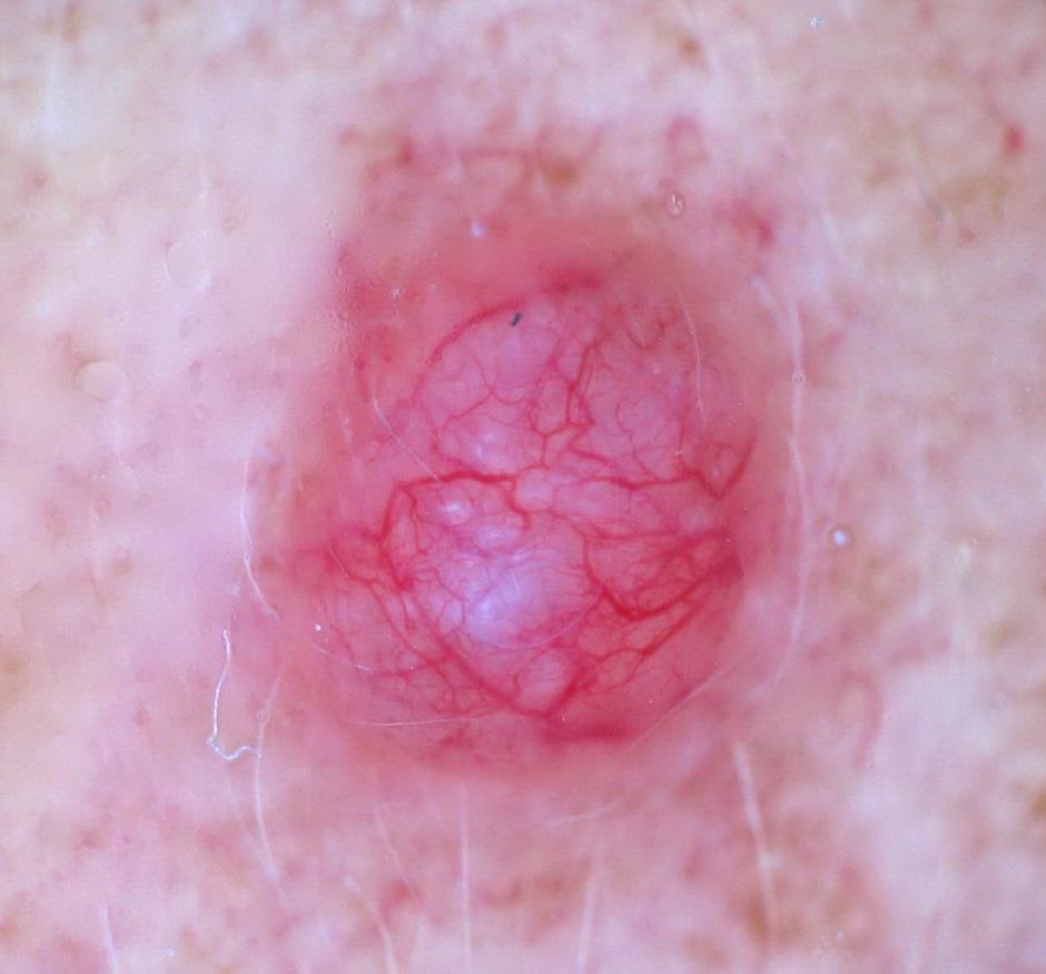
4 Basal-Cell Carcinoma (BCC)
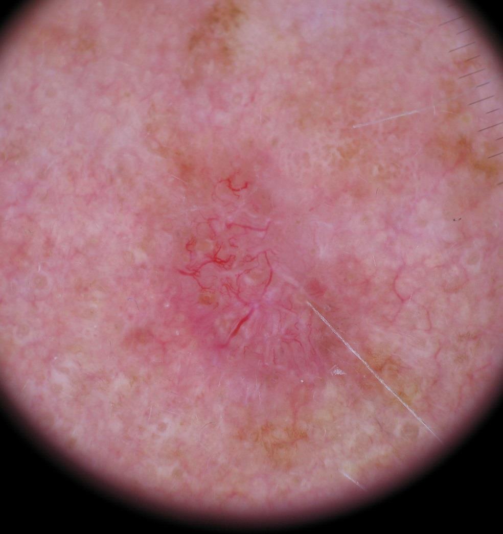
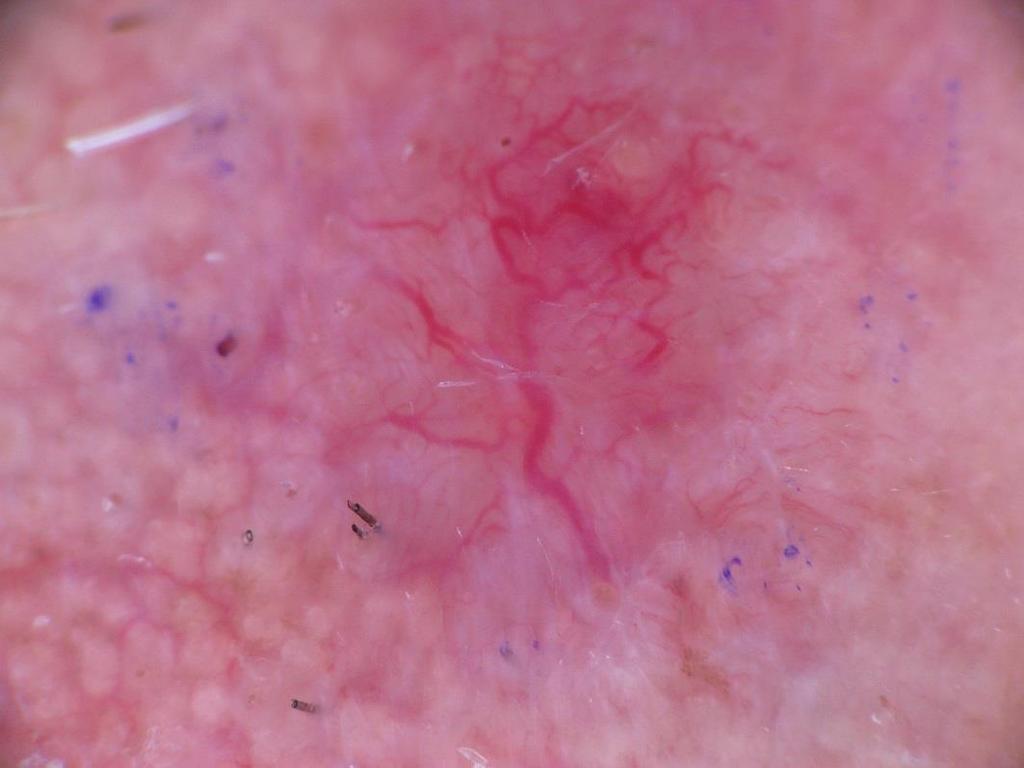
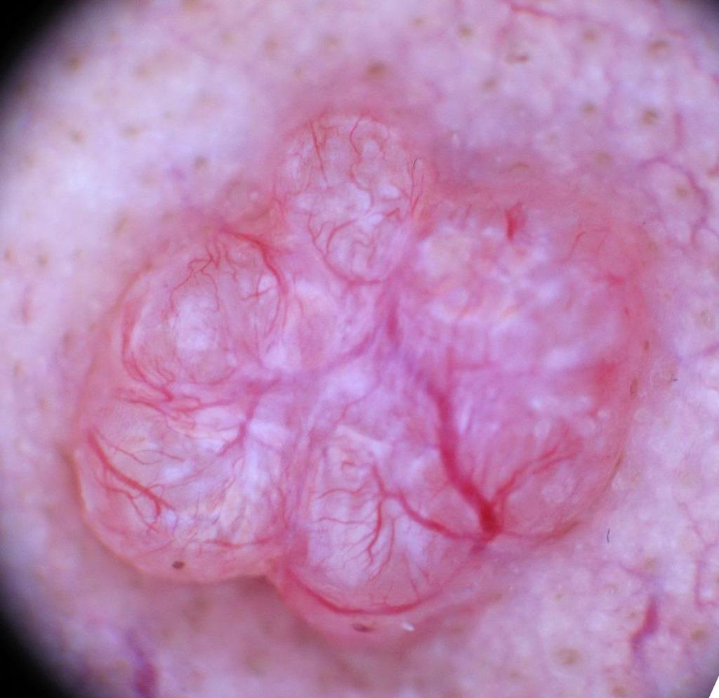
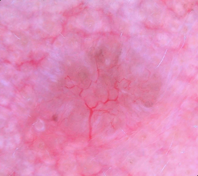
5 Non-Pigmented Basal-Cell Carcinoma Dermatoscopic features (semi) translucency white structures (crystalline, rosette, dot, strand, blotch) prominent vessels arborizing vessels short fine vessels shallow ulcers white to pink to reddish background = Non-Pigmented Basal-Cell Carcinoma
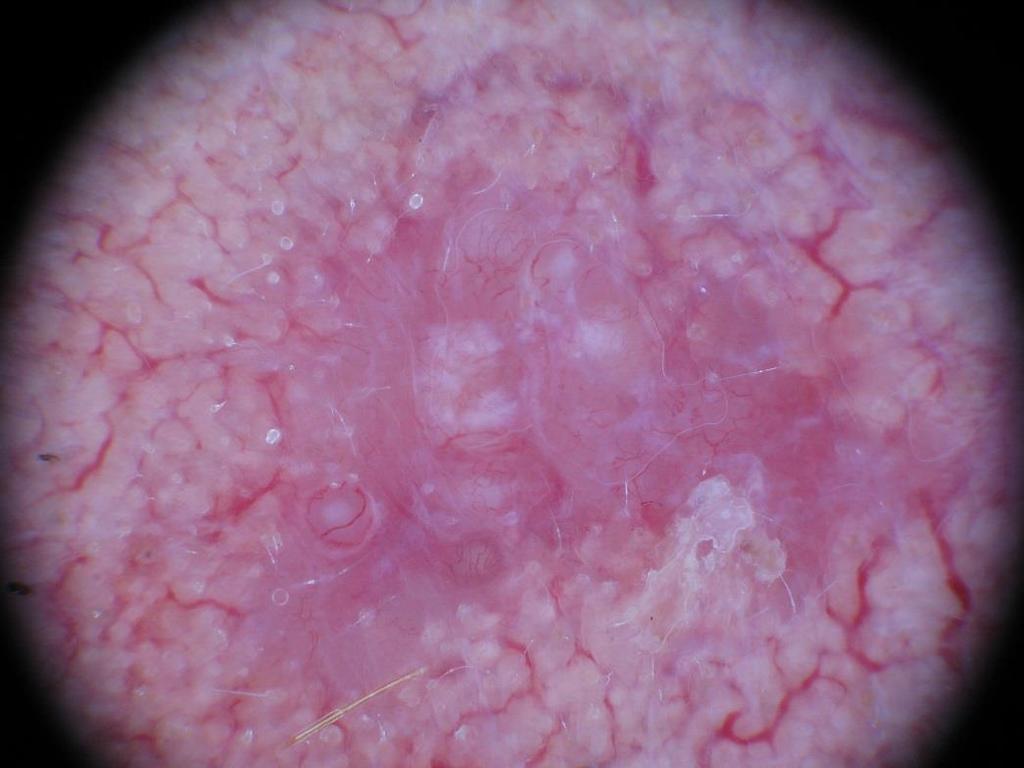
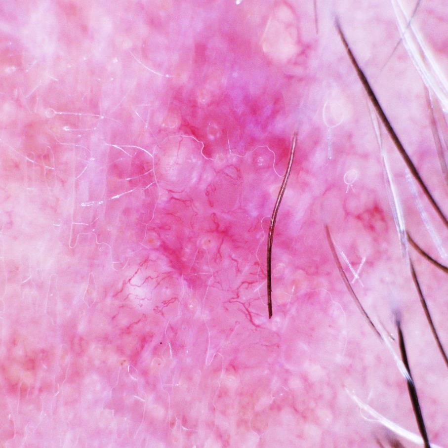
6 Nodular Basal-Cell Carcinoma (Semi)-translucency more prominent Larger caliber vessel

7 Nodular Basal-Cell Carcinoma

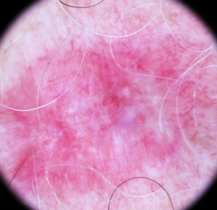
8 telangiectasia white streaks ulceration translucency pink to reddish background

9 Nodular BCC

10 Nodular BCC

11 Nodular BCC

12 white dot Though crystalline structures are the best known white structure associated with BCC, other white structures are also observed

13 Nodular BCC

14 Nodular BCC

15 Nodular BCC
16

17 Short fine vessels of BCC Background telangiectasia BCC change in vascular pattern from the background vasculature change is skin color, appearing semi-translucent
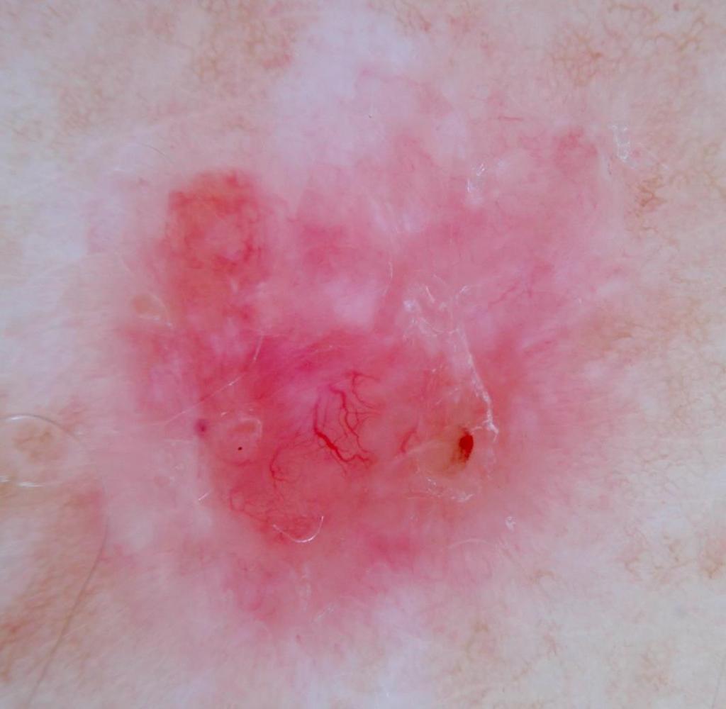
18 Superficial Basal-Cell Carcinoma (Semi)-translucency less prominent Smaller caliber vessel White structures more prominent White streaks crystalline structures White rosettes (clover) White dots White structureless areas/blotches

19 Superficial BCC Crystalline structures orthogonal bright white streaks visualized under polarized dermatoscopy Small vessels Small ulcerations Pink red background
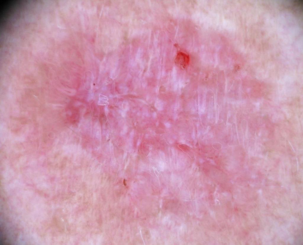
20 ulceration white streaks Superficial BCC
21 Superficial BCC
22 Superficial BCC
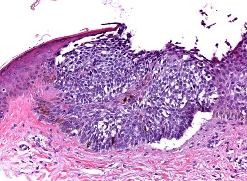
23 No dermatoscopic structures are pathognomonic Dermatoscopic structures need to be interpreted in the context of the patient Given the dermatoscopic findings associated with BCC, the pretest probability of BCC is much higher in patients with significant photodamage, history of skin cancer, Fitzpatrick skin phototype I & II, etc.
24 Dermatoscopic simulators of BCC Adnexal neoplasms Lichen planus-like keratosis Scar Dermatofibroma Melanocytic nevus Solar (actinic) keratosis Melanocytic nevus Melanoma Hemangioma Ruptured cyst/folliculitis

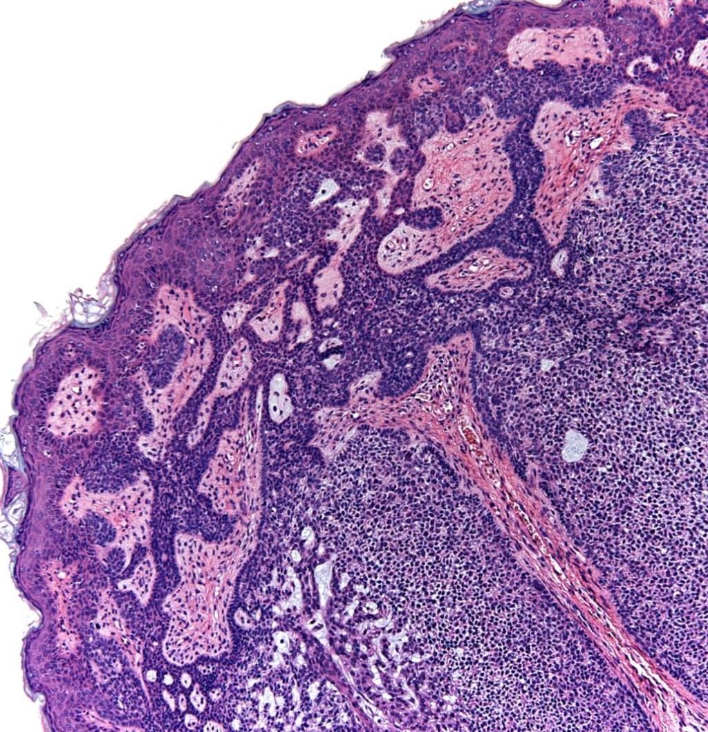
25 BCC Palisaded Encapsulated Neuroma
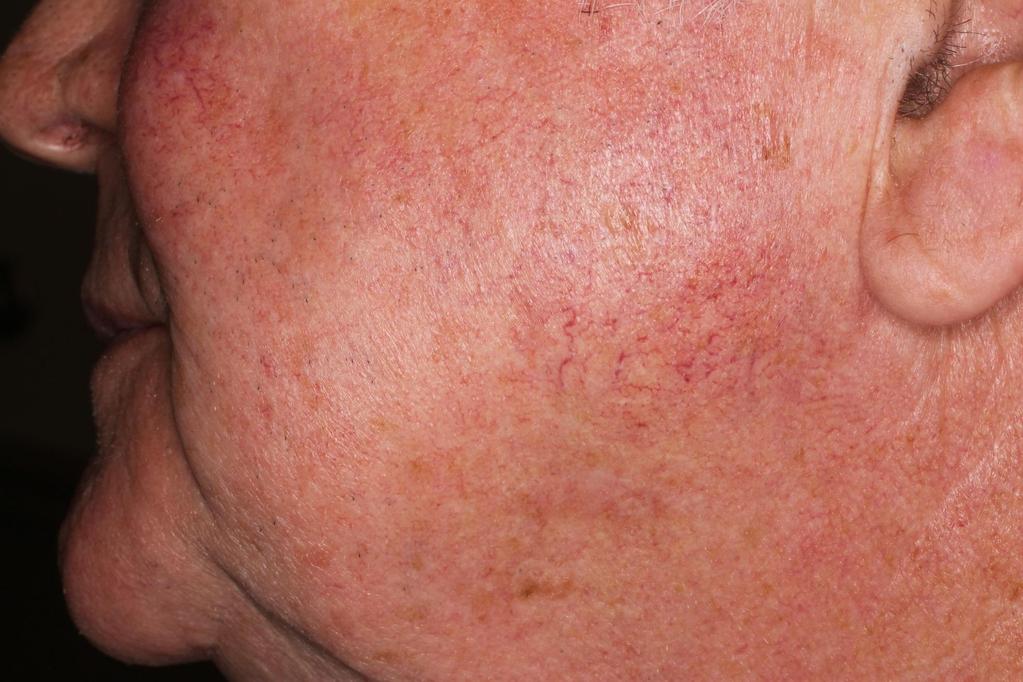
26 Scar BCC


27 Scar prominent vascularity pink white background
 Rosendahl C et al.")
28 In non-referral centers, it s impact on non-melanocytic lesions was more dramatic Sensitivity for non-melanocytic lesions 98.6% for basal cell carcinoma 86.5% for pigmented squamous cell ca 79.3% melanoma 10% LPLK (90.5% false+) Rosendahl C et al. J Acad Dermatol 2010;62:
29 Lichen Planus-Like Keratosis (LPLK) Dermatoscopic findings Pigmented granules Variable pink to light brown background color Look for remnant of a solar lentigo Lacks ulcer and characteristic BCC vascular pattern
30 Lichen Planus-Like Keratosis


31 LPLK pigmented granules pink to light brown background surface scale

32 sbcc LPLK Fine vessels
33 Dermatoscopic simulators of BCC Adnexal neoplasms Lichen planus-like keratosis Scar Dermatofibroma Melanocytic nevus Solar (actinic) keratosis Melanocytic nevus Melanoma Hemangioma Ruptured cyst/folliculitis
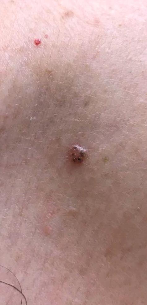
34 Pigmented Basal-Cell Carcinoma Dermatoscopic features (semi) translucency white structures(crystalline, rosette, dot, strand, blotch) prominent vessels Non-Pigmented arborizing vessels = short fine vessels Basal-Cell Carcinoma shallow ulcers white to pink to reddish background + Pigmented Islands blue-gray nests blue-gray globules = Pigmented Basal-Cell Carcinoma
35 Pigmented Basal-Cell Carcinoma Dermatoscopic features Frequency 1. large blue-gray ovoid nests 55% 2. arborizing treelike vessels 52% 3. multiple blue-gray globules 27% 4. ulceration 27% 5. maple leaf-like areas 17% 6. spoke wheel-like structures 10% Menzies SW. Dermoscopy of pigmented basal-cell carcinoma. Clinics in Dermatology 2002;20:268 9.
36 Pigmented Basal-Cell Carcinoma Dermatoscopic features Frequency 1. large blue-gray ovoid nests 55% 2 arborizing treelike vessels 52% 3 multiple blue-gray globules 27% 4 ulceration 27% 5. maple leaf-like areas 17% 6. spoke wheel-like structures 10% Menzies SW. Dermoscopy of pigmented basal-cell carcinoma. Clinics in Dermatology 2002;20:268 9.


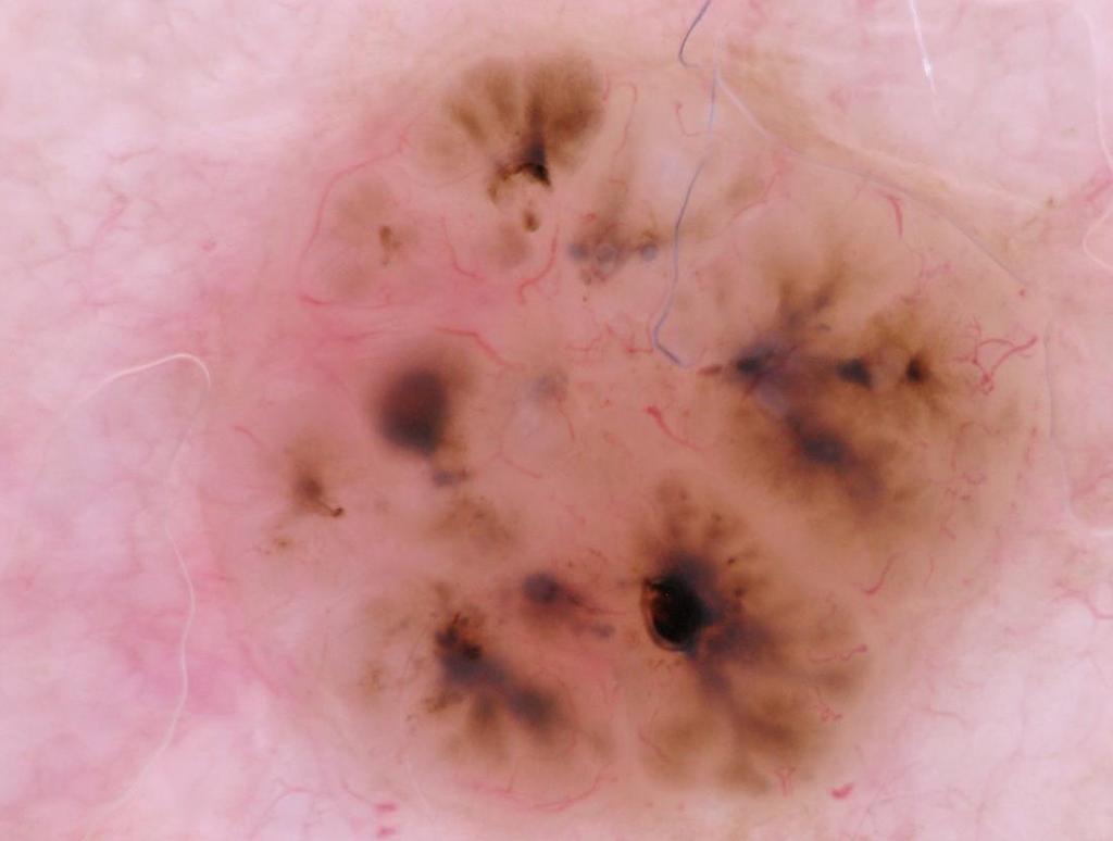
37 Large blue-gray ovoid nest

38 Blue-gray globules


39 Pigmented Nodular & Superficial BCC ulceration translucent area + telangiectasia pigmented islands


40 pigmented islands translucent area + telangiectasia Pigmented Nodular BCC

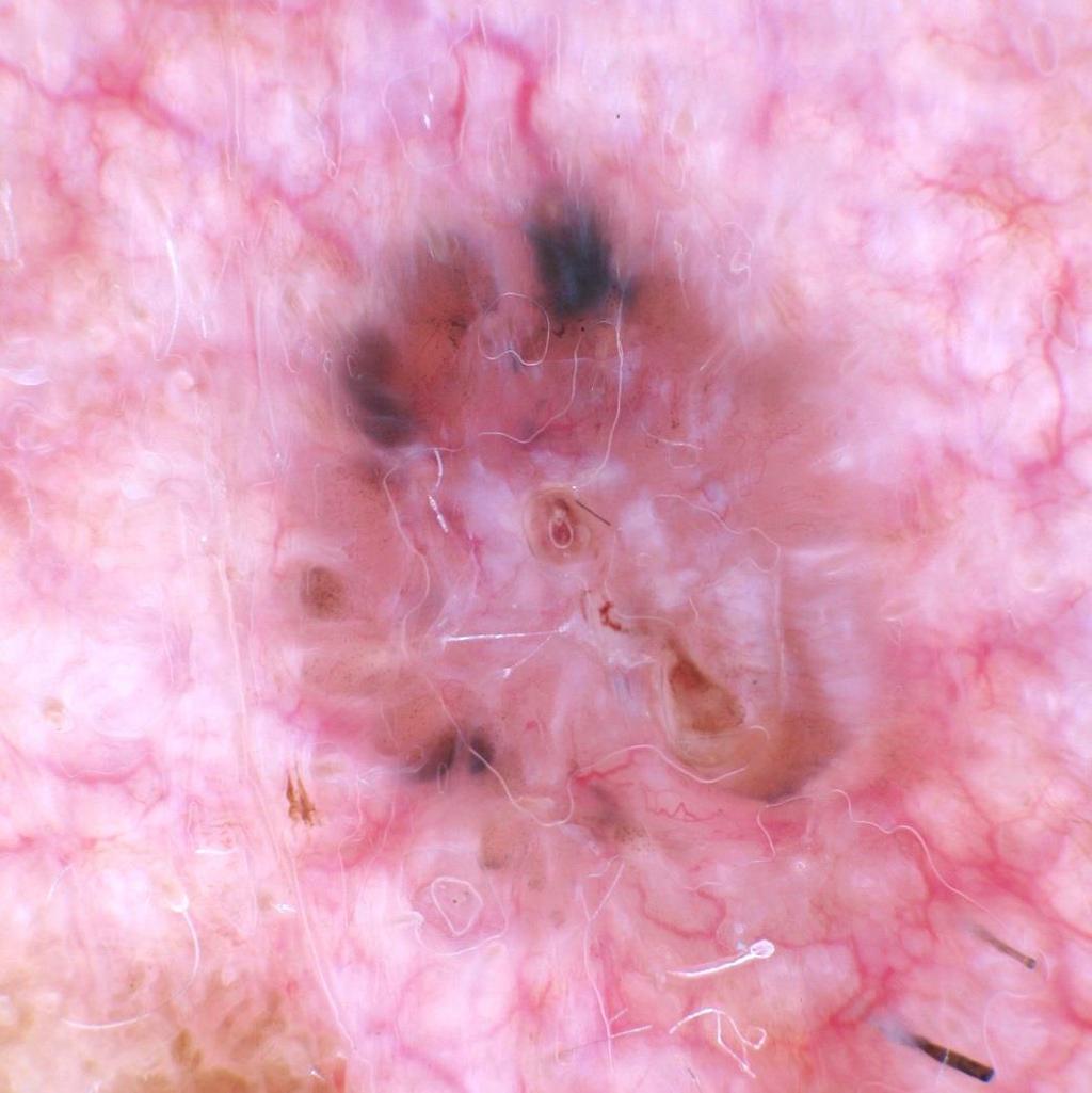
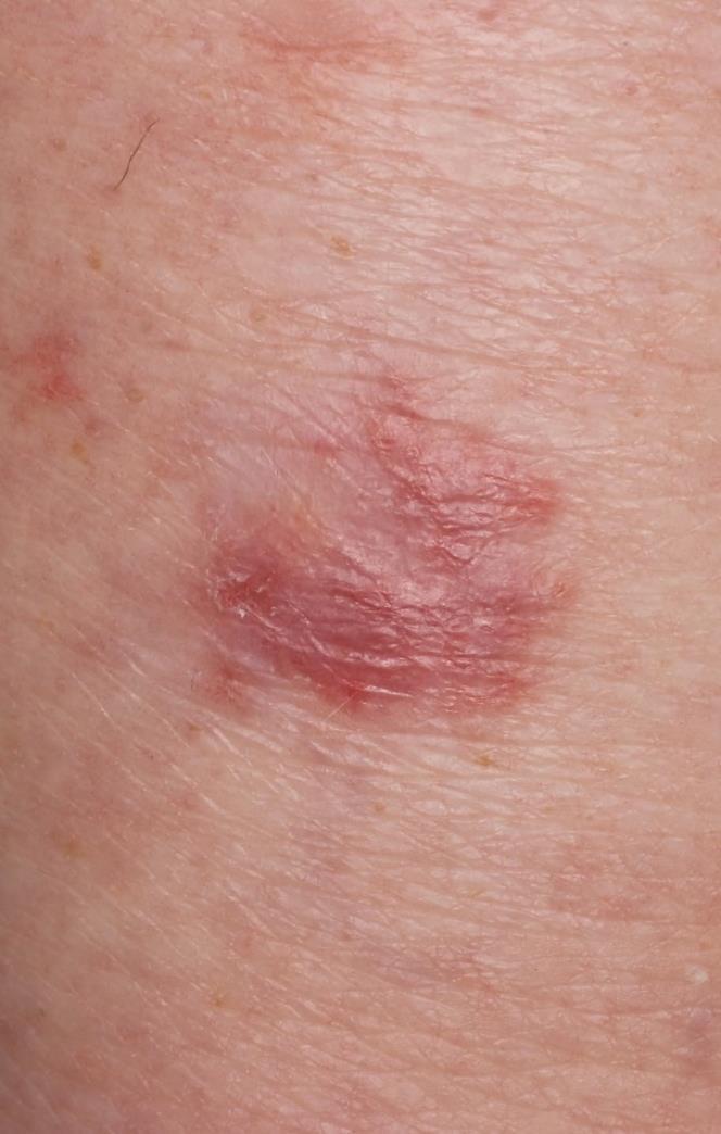
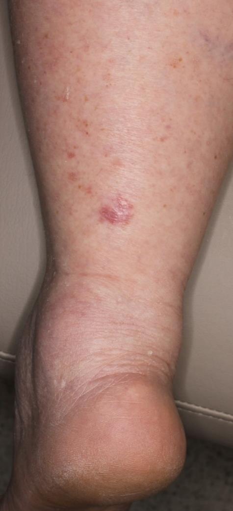
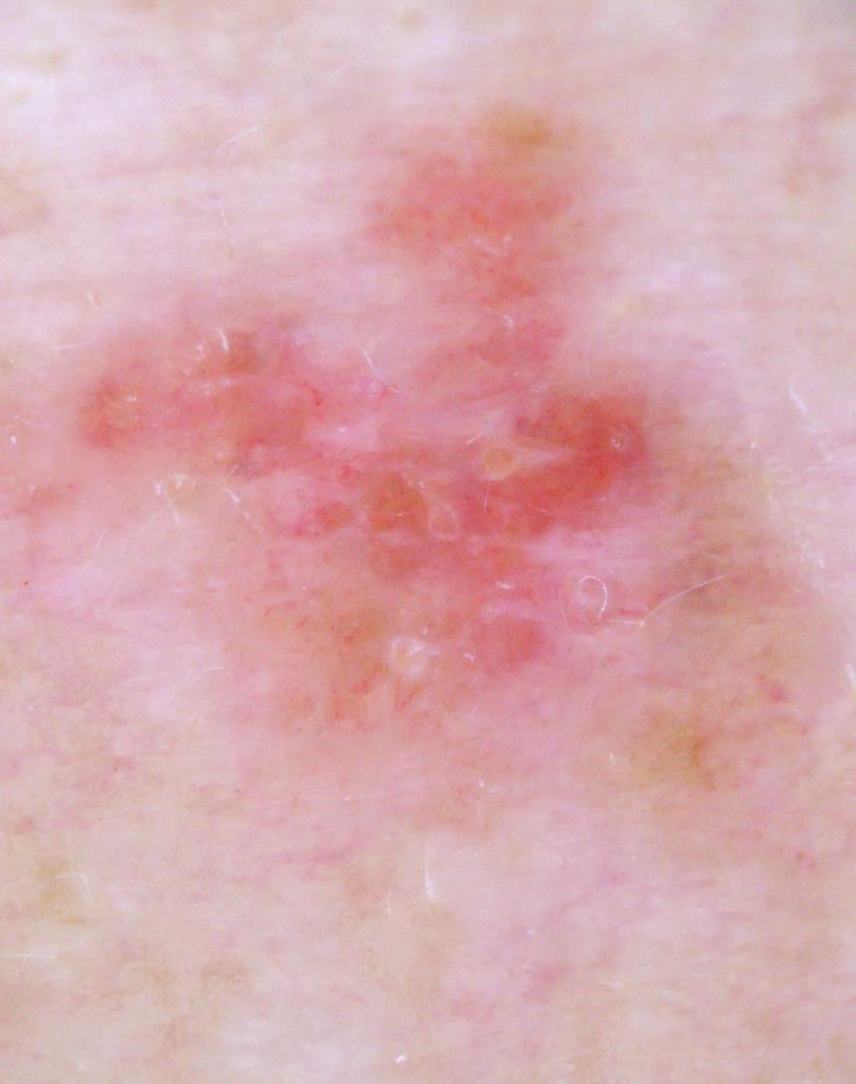
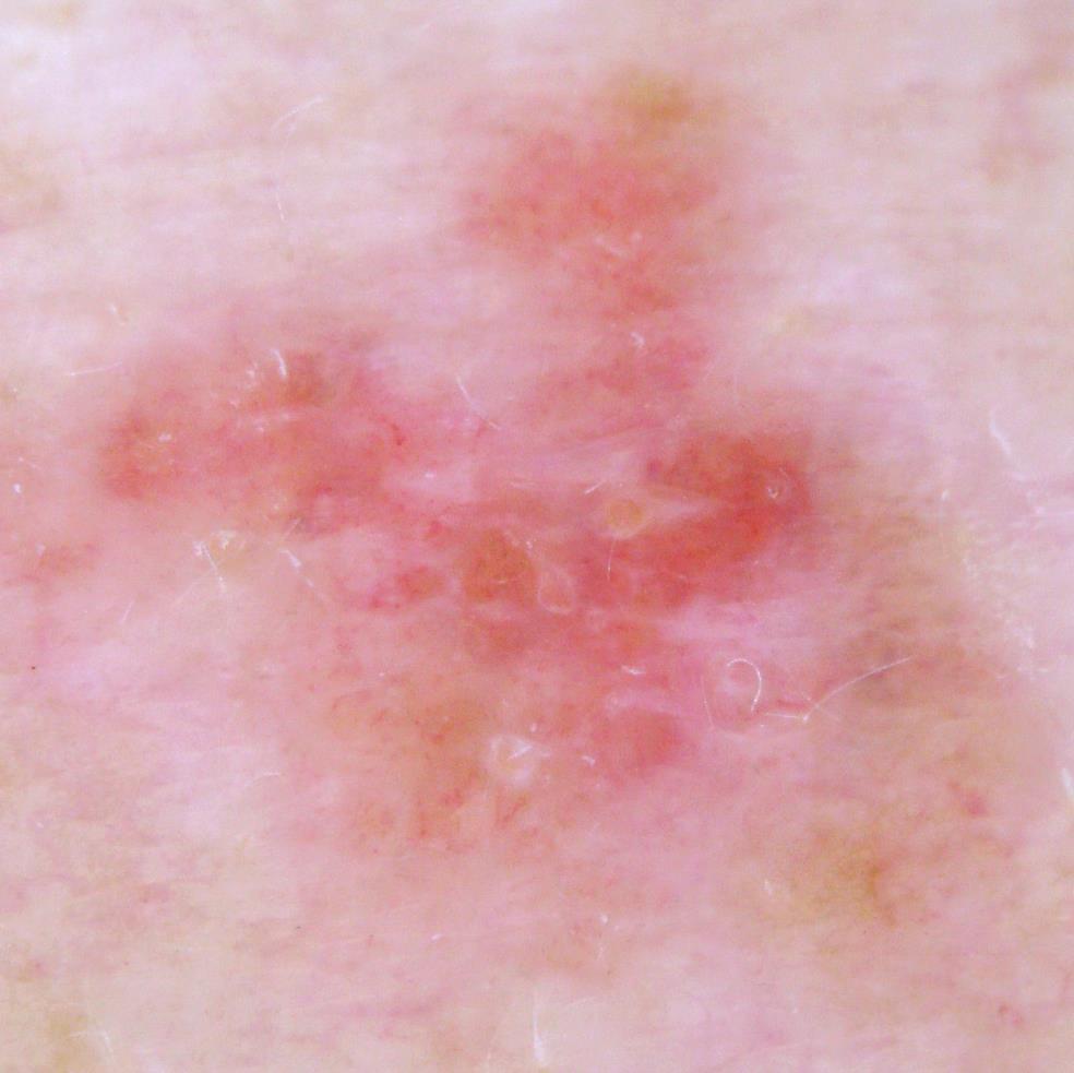
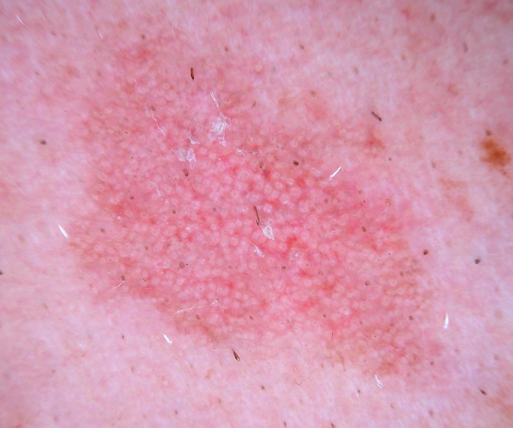
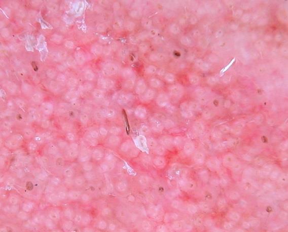
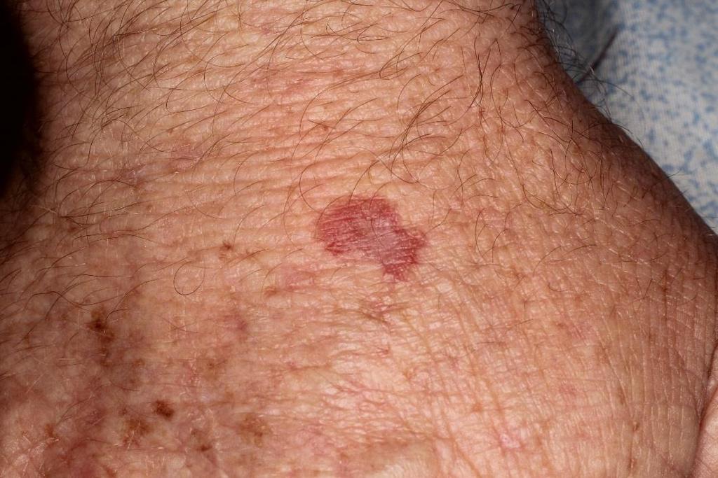
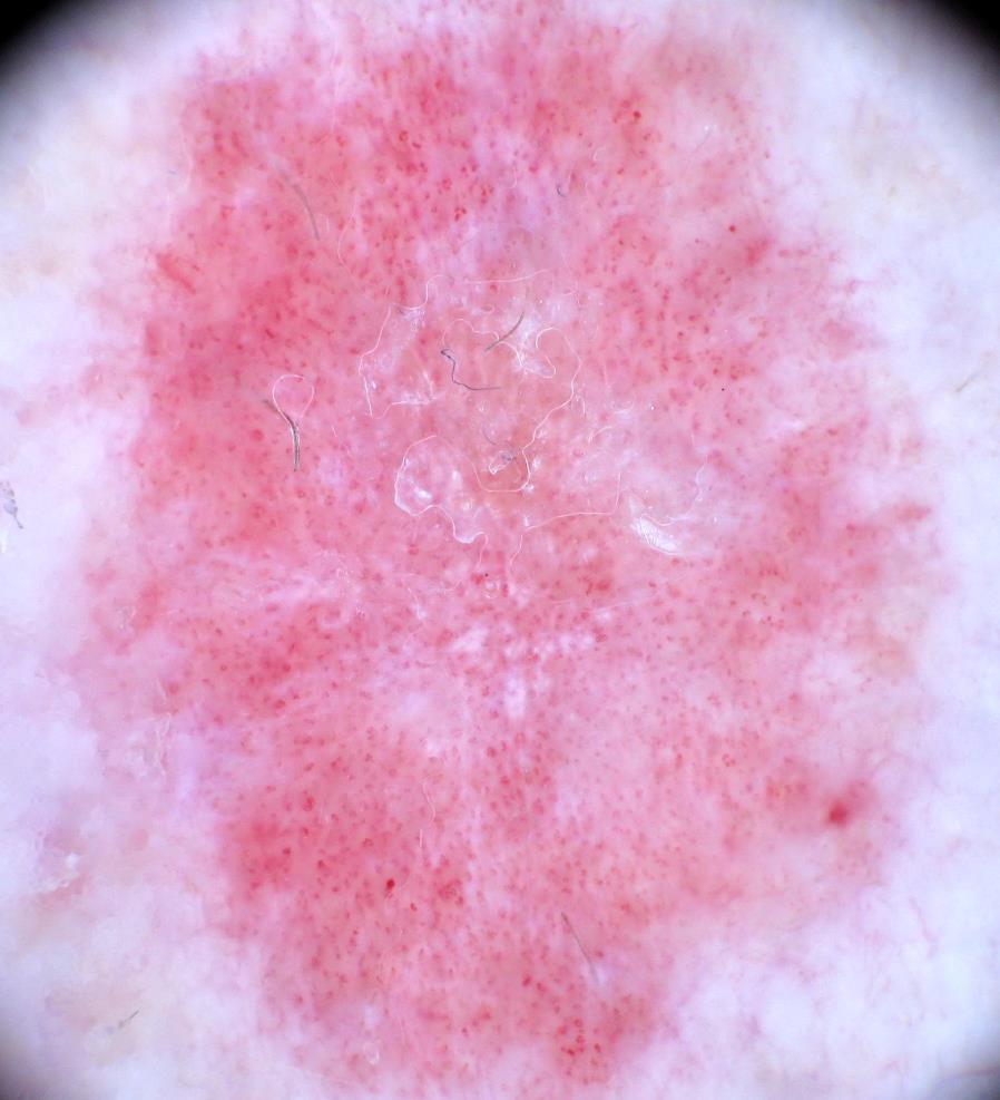
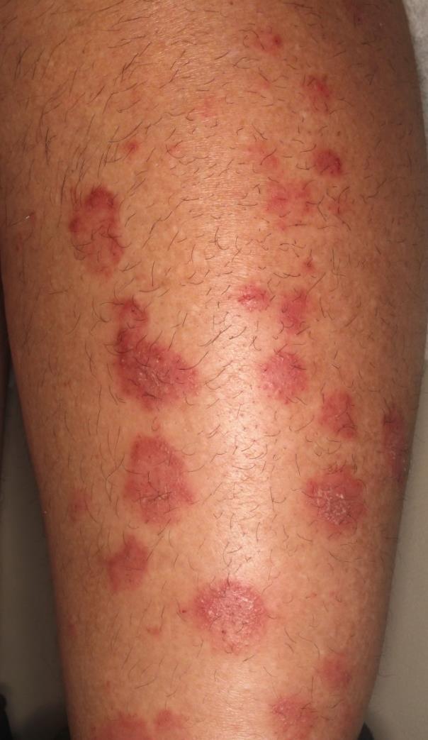
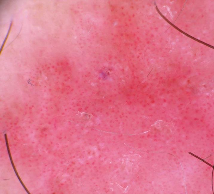
41 Pigmented Nodular BCC

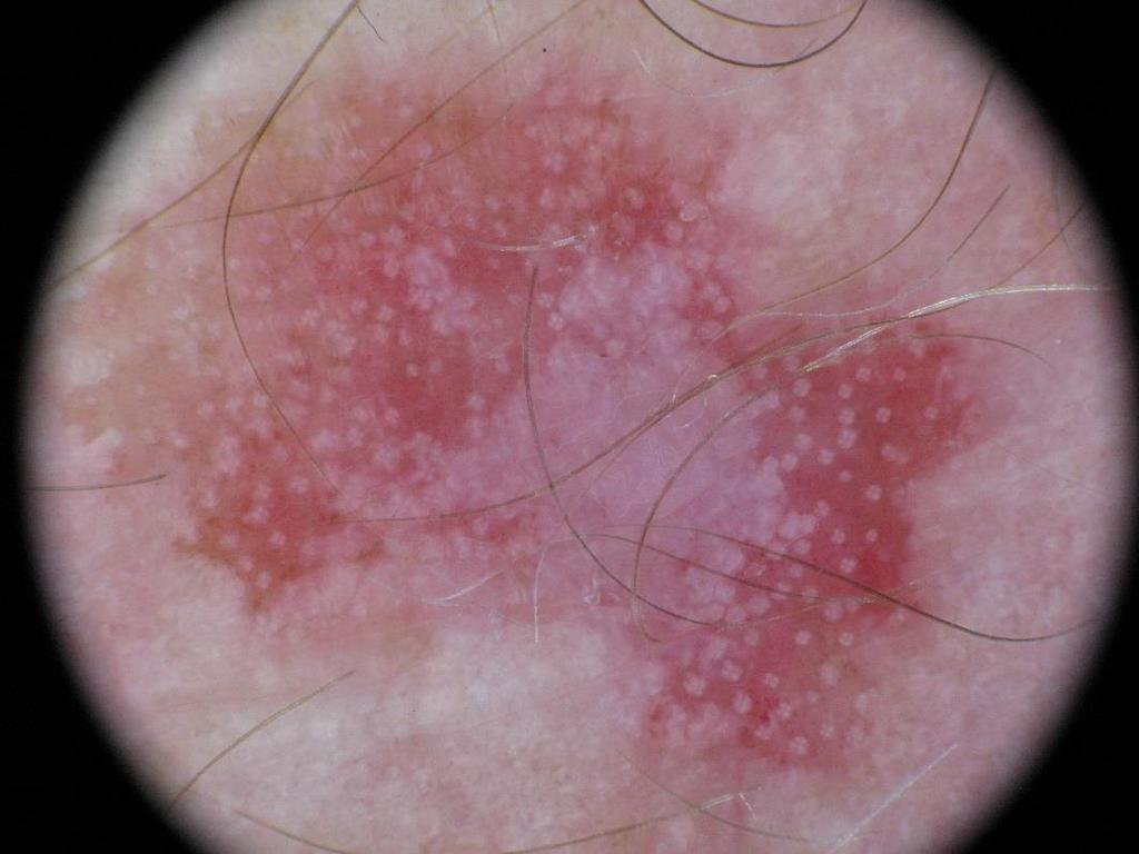
42 Spoke wheel-like areas Pigmented basal-cell carcinoma. Chapter 6a. Polsky D. page 58 in Atlas of dermoscopy. Ed Marghoob et al. Taylor & Francis. United Kingdom. 2006

43 Pigmented Nodular BCC

44 Submerged leaf-like areas


45 translucent area + telangiectasia ulceration pigmented islands Pigmented Nodular BCC


46 Pinkus BCC Dermatoscopic Findings white-network sharp circumscription polymorphous vascular pattern hairpin loops curvilinear comedo-like opening pink background with patchy pigmentation
47
48 Malignant Non-Melanocytic Lesions 1. Basal-cell carcinoma non-pigmented & pigmented 2. Solar (Actinic) Keratosis non-pigmented & pigmented 3. Squamous-cell carcinoma in-situ (Bowen s disease) 4. Keratoacanthoma/Squamous-cell carcinoma


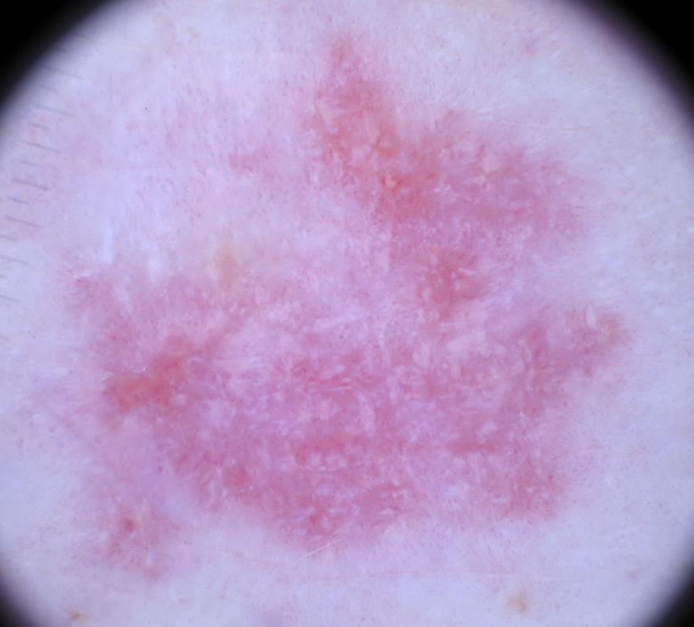
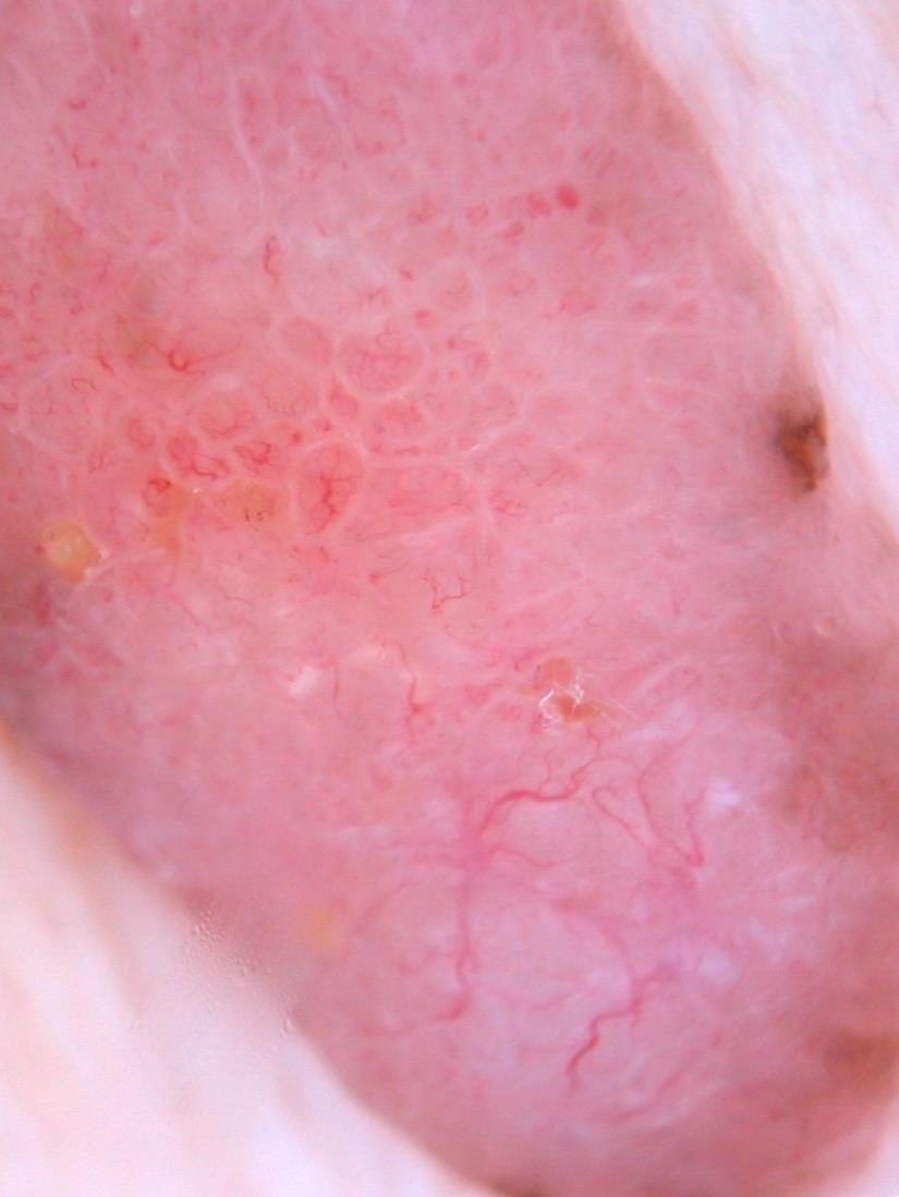
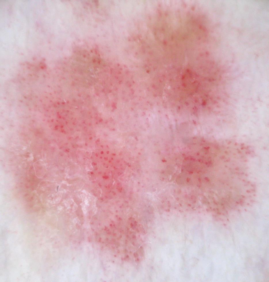
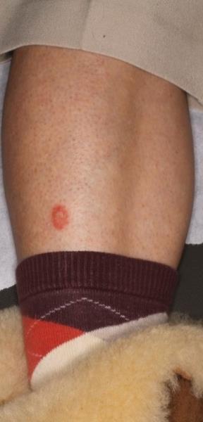
49 Solar (Actinic) Keratosis red (pseudo) network keratotic surface scale
:487-93.")
50 Solar (Actinic) Keratosis red network surface scale small white circles (halo, targetoid) white rosettes (4 leaf clover) Lee JH, Won CY, Kim GM, Kim SY. Dermoscopic features of actinic keratosis and follow up with dermoscopy: a pilot study. J Dermatol Jun;41(6):
51 2016 JEADV N=63 of 6108 (1%) showed rosettes Smaller rosettes were found to be caused by polarizing horny material in adnexal openings. Larger rosettes were found to be caused by perifollicular fibrosis. Rosettes observed in BCC SCC DF Nevus Melanoma Molluscum Dilated pore [Solar (actinic) keratosis]

52 Haspeslagh, M. et al. Rosettes and Other White Shiny Structures in Polarized Dermoscopy: Histological Correlate and Optical Explanation. JEADV 30.2 (2016):
53

54
55

56 white rosette
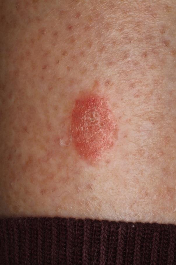
57 J Am Acad Dermatol 2013;68: Collision: solar keratosis and solar lentigo Dermatoscopic features solar (actinic) keratosis solar lentigo (most common)
")
58 Pigmented Solar (Actinic) Keratosis


59 On the right, note the red and brown network accompanied by keratotic scale. As a collision lesion, the proportion of the red and brown network will depend on the proportion of the 2 (or in some cases 3) lesions that make up the pigmented solar keratosis.


60 Pigmented Solar (Actinic) Keratosis
 Keratosis")
61 Pigmented Solar (Actinic) Keratosis white rosettes
")
62 Pigmented Solar (actinic) keratosis

63 Pigmented Solar (actinic) keratosis white circles white rosette

64 Pigmented AK vs Lentigo Maligna Akay et al. BJD 2010; 163: 1213 Conclusion: All the dermatoscopic structures observed in MIS may be also observed in pigmented actinic keratosis, except for black blotches There is significant overlap of dermatoscopic features between a pigmented solar (actinic) keratosis and MIS on the face. If in doubt, biopsy
65 Malignant Non-Melanocytic Lesions 1. Basal-cell carcinoma non-pigmented & pigmented 2. Solar (Actinic) Keratosis non-pigmented & pigmented 3. Squamous-cell carcinoma in-situ (Bowen s disease) 4. Keratoacanthoma/Squamous-cell carcinoma
66 Bowen s Disease Dermatoscopic findings coiled (glomerular) > dotted vessels surface scale pink to red background
67 Vascular Patterns Pan Y et al. JAAD 2008
68 Common Vascular Structures arborizing vessels: basal-cell carcinoma hairpin vessels: seborrheic keratosis dotted vessels: melanocytic neoplasms, psoriasis, porokeratosis, Bowen disease coiled vessels (glomerular): Bowen disease If one is relying on a dermatoscope with only a 10x magnification, the smaller caliber vessels are difficult to evaluate.


69 dotted vessels coiled (glomerular) vessels J Am Acad Dermatol 2010;63:361-74


70 Bowen Disease dotted vessels surface scale
71


72 Bowen s Disease dotted vessels surface scale ulceration 10x 10x

73 Bowen s Disease 20x 20x dotted vessels (20x mag)

74 Bowen s Disease
75 Coiled Vessels of Bowen s Disease
76 Psoriasiform Keratosis
77 Psoriasis
78 Bowen s Disease Dotted and coiled vessels may be seen in other diseases 1. Psoriasis: regularly dotted vessels 2. Psoriasiform keratosis: regularly dotted vessels 3. Pale-cell acanthoma: regularly dotted vessels sometimes in linearly dotted pattern ( pearls in a line ) 4. Porokeratosis: dotted vessels
79 Dermatoscopic findings surface scale coiled (glomerular) > dotted vessels pink to red background Bowen s Disease
80 Malignant Non-Melanocytic Lesions 1. Basal-cell carcinoma non-pigmented & pigmented 2. Solar (Actinic) Keratosis non-pigmented & pigmented 3. Squamous-cell carcinoma in-situ (Bowen s disease) 4. Keratoacanthoma/Squamous-cell carcinoma
:1386-1392.")
81 Keratoacanthoma white circles coiled vessels keratin blood spots Rosendahl C et al. Dermatol. 2012;148(12):


82 Keratoacanthoma white circles coiled vessels keratin Blood spots

83 Key to Success Correctly interpret the observed structures Look for presence and absence of dermatoscopic structures Do not rely on any one dermatoscopic finding Clinical context is important Look at all lesions
84 The End
Dermoscopy: Recognizing Top Five Common In- Office Diagnoses
 Dermoscopy: Recognizing Top Five Common In- Office Diagnoses Vu A. Ngo, DO Department of Family Medicine and Dermatology Choctaw Nation Health Services Authority Learning Objectives Introduction to dermoscopy
Non-melanocytic Patterns
 Non-melanocytic Lesions Non-melanocytic Patterns Michelle Tarbox, MD Assistant Professor of Dermatology and Dermatopathology Texas Tech University Health Sciences Center 2018 Seborrheic keratoses Acanthotic
Disclosure. Objectives. PAFP CME Conference Lou Mancano MD, FAAFP Reading Health System November 18, 2016
 PAFP CME Conference Lou Mancano MD, FAAFP Reading Health System November 18, 2016 1 Disclosure The speaker has no conflict of interest, financial agreement, or working affiliation with any group or organization.
Regression 2/3/18. Histologically regression is characterized: melanosis fibrosis combination of both. Distribution: partial or focal!
 Regression Margaret Oliviero MSN, ARNP Harold S. Rabinovitz MD Histologically regression is characterized: melanosis fibrosis combination of both Distribution: partial or focal! Dermatoscopic terminology
comedo-like openings (clods, brown or orange & circles) milia-like cysts (dots or clods, white) 1/29/18 Dotted vessels are also commonly seen in SCC
 milia-like cysts (dots or clods, white) 1/29/18 Dotted vessels are also commonly seen in SCC") Brown circles Dotted vessels are also commonly seen in SCC Step1 1. Nevus (unequivocal) 2. DF/IDN 3. BCC 4. SCC Network Patchy network Peripheral network & central hypopigmentation DF: network with central
Brown circles Dotted vessels are also commonly seen in SCC Step1 1. Nevus (unequivocal) 2. DF/IDN 3. BCC 4. SCC Network Patchy network Peripheral network & central hypopigmentation DF: network with central
Rosettes in actinic keratosis and squamous cell carcinoma: distribution, association to other dermoscopic signs and description of the rosette pattern
 DOI: 10.1111/jdv.14474 JEADV ORIGINAL ARTICLE Rosettes in actinic keratosis and squamous cell carcinoma: distribution, association to other dermoscopic signs and description of the rosette pattern B. Lozano-Masdemont,
Non-Melanocytic Pattern Dermoscopy
 Non-Melanocytic Pattern Dermoscopy I have no conflicts of interest to disclose Except that I LOVE dermoscopy Michelle Tarbox, MD Assistant Professor of Dermatology and Dermatopathology Texas Tech University
Features Causing Confusion between Basal Cell Carcinoma and Squamous Cell Carcinoma in Clinical Diagnosis
 TH Ryu, et al pissn 1013-9087ㆍeISSN 2005-3894 Ann Dermatol Vol. 30, No. 1, 2018 https://doi.org/10.5021/ad.2018.30.1.64 ORIGINAL ARTICLE Features Causing Confusion between Basal Cell Carcinoma and Squamous
6/17/2018. Breaking Bad (Part 1) Dermoscopy of Brown(ish) Things. Bad?
 Dermoscopy of Brown(ish) Things. Bad?") Breaking Bad (Part 1) Dermoscopy of Brown(ish) Things Jennie T. Clarke, MD ssociate Professor of Dermatology University of Utah School of Medicine Bad? 1 Brown(ish) Things Bad Melanoma Pigmented basal
Breaking Bad (Part 1) Dermoscopy of Brown(ish) Things Jennie T. Clarke, MD ssociate Professor of Dermatology University of Utah School of Medicine Bad? 1 Brown(ish) Things Bad Melanoma Pigmented basal
Prediction without Pigment: a decision algorithm for non-pigmented skin malignancy
 DERMATOLOGY PRACTICAL & CONCEPTUAL www.derm101.com Prediction without Pigment: a decision algorithm for non-pigmented skin malignancy Cliff Rosendahl 1, Alan Cameron 1, Philipp Tschandl 2, Agata Bulinska
Yes. Breaking Bad II: Dermoscopy of Pink-ish Things. Does it Fit? Yes 6/17/2018. Yes. Joslyn Kirby, MD, MS, MEd
 Breaking Bad II: Dermoscopy of Pink-ish Things Joslyn Kirby, MD, MS, MEd Yes Observe Yes Step 2. Fit a Benign Nevus Pattern? Does it Fit? Step 1: Melanocytic? pigment network, globules, homogeneous? No
Key factors in successfully integrating dermoscopy into your clinical practice
 Key factors in successfully integrating dermoscopy into your clinical practice S051 Dilemmas and challenges in skin cancer therapies and management Monday, March 4 th 2019 (9AM-12PM) Room 209A 10:56-11:09AM
Clinical and Dermoscopic Features of Thin Nodular Melanoma
 Clinical and Dermoscopic Features of Thin Nodular Melanoma A study of the International Dermoscopy Society Coordinator: Dr. Alexander J. Stratigos and colleagues, alstrat2@gmail.com ** Extended to May
Dermoscopy STFM Richard Usatine, MD 5/2/16. Disclosure Statement: Some Dermatoscopes. Dermoscopy Video. Thanks to Dr.
 Disclosure Statement: Dermoscopy STFM 2016 Richard P. Usatine, MD, FAAFP Professor, Family and Community Medicine Professor, Dermatology and Cutaneous Surgery Medical Director, Clinic University of Texas
22/04/2015. Dermoscopy of Melanoma. Ilsphi Browne. Overview
 Dermoscopy of Melanoma Ilsphi Browne Overview The device Dermoscopic criteria (terminology) Colour Patterns Global features Local features Approach to diagnosing pigmented lesions Other uses in general
Dermoscopy in everyday practice. What and Why? When in doubt cut it out? Trilokraj Tejasvi MD
 Dermoscopy in everyday practice Trilokraj Tejasvi MD Assistant Professor, Department of Dermatology, Director Teledermatology services, University of Michigan, Faculty Associate, GLOBAL REACH, Michigan
Appendix : Dermoscopy
 Go Back to the Top To Order, Visit the Purchasing Page for Details APP Appendix : Dermoscopy Dermoscopy, also known as dermatoscopy, epiluminoscopy and epiluminescent microscopy, is an effective non-invasive
It can be helpful in some cases of actinic keratosis, Bowen s disease and squamous cell carcinoma
 Dermoscopy Introduction, Terminology and Structures (to be read in conjunction with the Diagnostic Dermoscopic Algorithm) Copyright to Cunliffe TP (Jan. 2017) All rights reserved Introduction Dermoscopy
Introduction to Dermoscopy. Nicholas Compton, MD June 16, 2010
 Introduction to Dermoscopy Nicholas Compton, MD June 16, 2010 Overview What is dermoscopy Brief history Types of dermoscopy General approach to lesion of interest 2 step algorithm 3-point checklist Practice
Dermoscopy. Enhanced Diagnostic Ability: Pigmented Lesions. Ted Rosen, MD Baylor College of Medicine Houston, Texas
 Dermoscopy Enhanced Diagnostic Ability: Pigmented Lesions Ted Rosen, MD Baylor College of Medicine Houston, Texas Faculty Disclosure Statement No conflicts relevant to this workshop! Sir William Osler
Common Benign Lesions and Skin Cancers. 22nd May 2015 Dr Mark Foley
 Common Benign Lesions and Skin Cancers 22nd May 2015 Dr Mark Foley Thank you for downloading this file. This intended to supplement the presentation given at the NZ Wound Care Conference, it is not intended
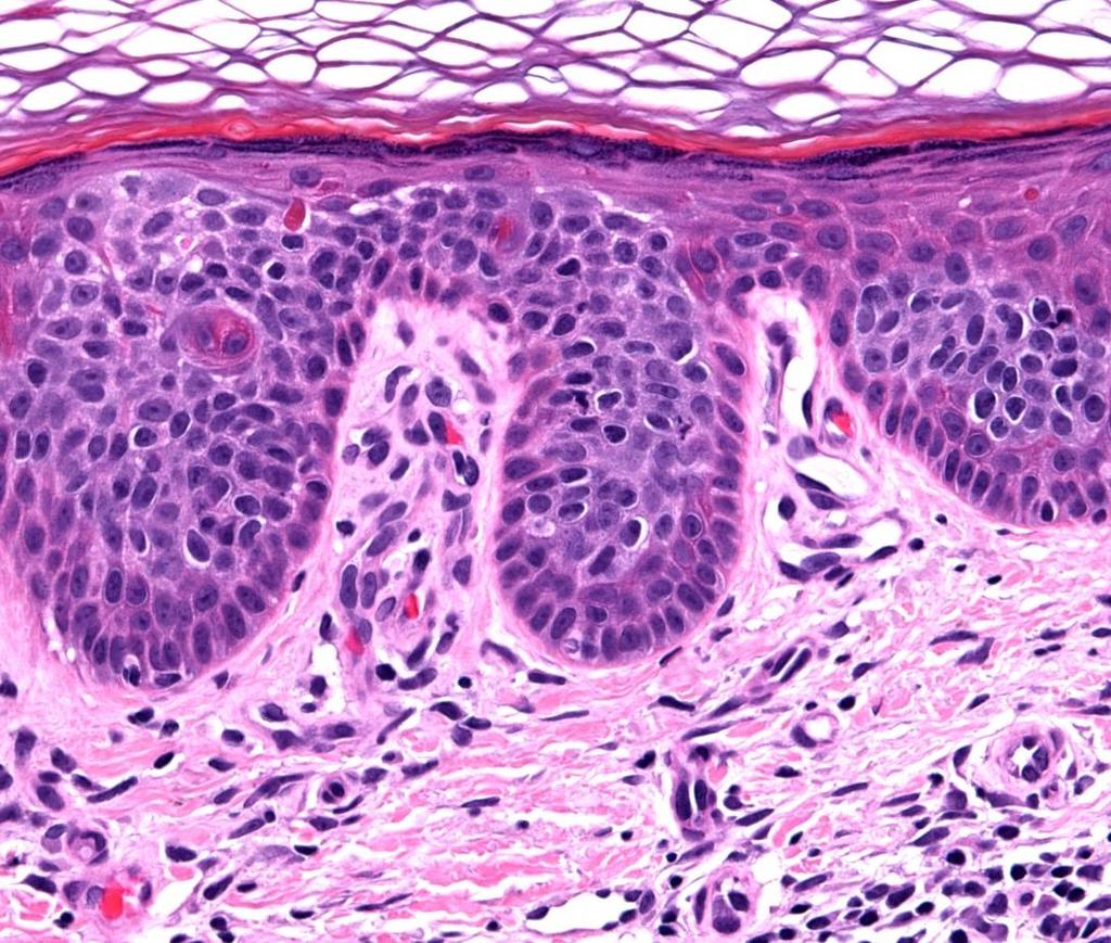
INVESTIGATION. The relation between dermoscopy and histopathology of basal cell carcinoma *
 INVESTIGATION The relation between dermoscopy and histopathology of basal cell carcinoma * 351 Nazan Emiroglu 1 Fatma Pelin Cengiz 1 Funda Kemeriz 2 DOI: http://dx.doi.org/10.1590/abd1806-4841.20153446
Review of vasculature visualized on dermoscopy
 doi: 10.1111/1346-8138.13686 Journal of Dermatology 2017; 44: 525 532 REVIEW ARTICLE Review of vasculature visualized on dermoscopy Yaei TOGAWA Department of Dermatology, Chiba University Graduate School
Fundamentals of dermoscopy
 Fundamentals of dermoscopy Learning objectives Upon completion of this session, participants should be able to: describe the basic principles of dermoscopy identify features associated with pigmented and
Dermoscopy. Sir William Osler. Dermoscopy. Dermoscopy. Melanoma USA Primary Care Update Faculty Disclosure Statement
 Diagnostic Ability: Pigmented Lesions Ted Rosen, MD Baylor College of Medicine Houston, Texas Enhanced 2010 Primary Care Update Faculty Disclosure Statement Ted Rosen, MD Speakers Bureau: Abbott, Amgen,
Benign versus Cancerous Lesions How to tell the difference FMF 2014 Christie Freeman MD, CCFP, DipPDerm, MSc
 1 Benign versus Cancerous Lesions How to tell the difference FMF 2014 Christie Freeman MD, CCFP, DipPDerm, MSc Benign lesions Seborrheic Keratoses: Warty, stuck-on Genetics and birthdays Can start in late
Chronology of lichen planus-like keratosis features by dermoscopy: a summary of 17 cases
 DERMATOLOGY PRACTICAL & CONCEPTUAL www.derm101.com Chronology of lichen planus-like keratosis features by dermoscopy: a summary of 17 cases Soko Watanabe 1, Mizuki Sawada 1, Itaru Dekio 1, Sumiko Ishizaki
Dermoscopy of non-pigmented skin lesions: a literature review
 Hong Kong J. Dermatol. Venereol. (2017) 25, 13-21 Review Article Dermoscopy of non-pigmented skin lesions: a literature review S Thomas, X Li, HP Soyer In this article, we will review benchmark dermoscopic
Dermoscopy, the use of a handheld
 ONLINE EXCLUSIVE Dermoscopy in family medicine: A primer Dermoscopy allows you to see deeper into the skin than with the naked eye. Here s how you can make use of it to spot malignant conditions sooner.
10/3/2018. Dermoscopy: Looking beneath the surface of the skin. Dermoscopy for Family Medicine 10/11/2018
 Dermoscopy for Family Medicine 10/11/2018 Jane M. Grant-Kels, MD, FAAD Founding Chair Emeritus, Dept of Dermatology Professor of Dermatology, Pathology & Pediatrics Director of the Cut Oncology Ctr & Melanoma
Basics in Dermoscopy
 Basics in Dermoscopy Manal Bosseila Professor of Dermatology, Cairo University Member of European Academy Dermatology & Venereology EADV Member of International Dermoscopy Society IDS Member of Aesthetic
Introduction to Dermoscopy. Disclosure. Introduction
 Introduction to Dermoscopy 1 Disclosure Dr. Deborah Bren has no conflict of interest, financial agreement, or working affiliation with any group or organization. 2 Introduction Deborah A. Bren, DO Family
STUDY. Dermoscopy of Squamous Cell Carcinoma and Keratoacanthoma
 ONLINE FIRST STUDY Dermoscopy of Squamous Cell Carcinoma and Keratoacanthoma Cliff Rosendahl, MBBS; Alan Cameron, MBBS; Giuseppe Argenziano, MD; Iris Zalaudek, MD; Philipp Tschandl, MD; Harald Kittler,
Dermoscopy-a BRIEF introduction
 Dermoscopy-a BRIEF introduction Aim of presentation -to tell you what dermoscopy is -to show some of what it can do -point the interested learner to further resources Overview of dermoscopy Dermoscopy
Revised Pattern Analysis: a method for the accurate diagnosis of pigmented skin lesions
 Dermatoscopy for Students A concise outline of: Revised Pattern Analysis: a method for the accurate diagnosis of pigmented skin lesions And Chaos and Clues: a decision algorithm for routine practice to
The dermatologist s stethoscope traditional and new applications of dermoscopy
 DERMATOLOGY PRACTICAL & CONCEPTUAL www.derm101.com The dermatologist s stethoscope traditional and new applications of dermoscopy Iris Zalaudek, M.D. 1,2, Aimilios Lallas, M.D. 1, Elvira Moscarella, M.D.
STUDY. Scott W. Menzies, MB,BS, PhD; Karin Westerhoff, MD; Harold Rabinovitz, MD; Alfred W. Kopf, MD; William H. McCarthy, MBBS, MEd; Brian Katz
 STUDY Surface Microscopy of Pigmented Basal Cell Carcinoma Scott W. Menzies, MB,BS, PhD; Karin Westerhoff, MD; Harold Rabinovitz, MD; Alfred W. Kopf, MD; William H. McCarthy, MBBS, MEd; Brian Katz Objectives:
Clinical characteristics
 Skin Cancer Fernando Vega, MD Seattle Healing Arts Clinical characteristics Precancerous lesions Common skin cancers ACTINIC KERATOSIS Precancerous skin lesions Actinic keratoses Dysplastic melanocytic
BLINCK A diagnostic algorithm for skin cancer diagnosis combining clinical features with dermatoscopy findings
 DERMATOLOGY PRACTICAL & CONCEPTUAL www.derm101.com BLINCK A diagnostic algorithm for skin cancer diagnosis combining clinical features with dermatoscopy findings Peter Bourne, MBBS 1, Cliff Rosendahl,
What is Dermoscopy? Early Dermoscopes. Deciphering Dermoscopy: Terminology, Features & Algorithms 6/17/2018
 Deciphering Dermoscopy: Terminology, Features & Algorithms Where did it come from and why do we use it? Jennie T. Clarke, MD Associate Professor of Dermatology University of Utah School of Medicine What
Diagnosis of Lentigo Maligna Melanoma. Steven Q. Wang, M.D. Memorial Sloan-Kettering Cancer Center Basking Ridge, NJ
 Diagnosis of Lentigo Maligna Melanoma Steven Q. Wang, M.D. Memorial Sloan-Kettering Cancer Center Basking Ridge, NJ Conflict of Interest: None Topics Epidemiology and Natural History Clinical and Histologic
Skin lesions The Good and the Bad. Dr Virginia Hubbard Ipswich Hospital NHS Trust Barts and the London School of Medicine and Dentistry
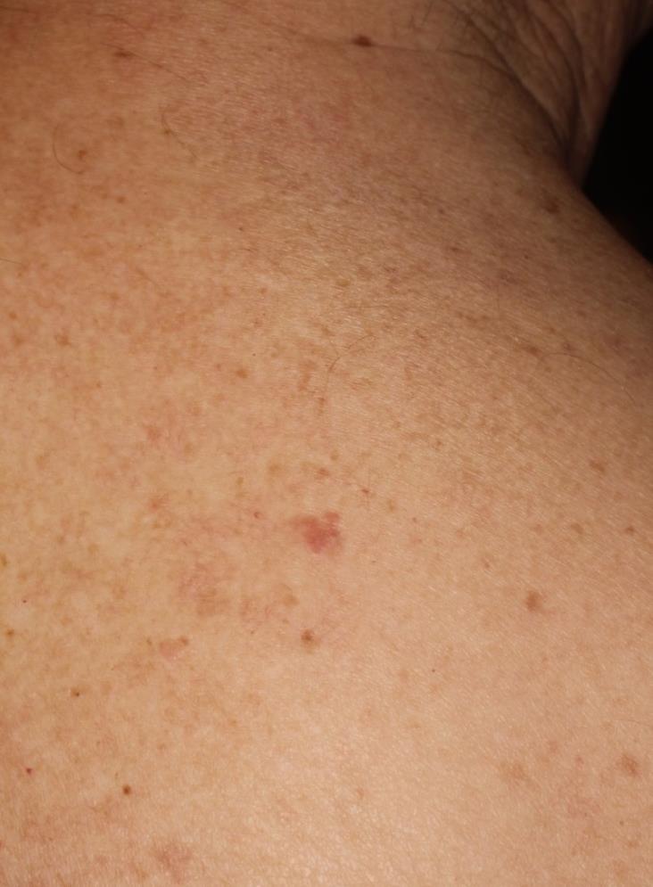
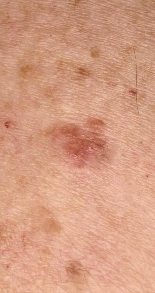
 Skin lesions The Good and the Bad Dr Virginia Hubbard Ipswich Hospital NHS Trust Barts and the London School of Medicine and Dentistry Case 1 32 year old woman Australian Lesion on back New hair growing
Skin Cancer A Personal Approach. Dr Matthew Strack Dunedin New Zealand
 Skin Cancer A Personal Approach Dr Matthew Strack Dunedin New Zealand Outline Dermoscopy Instruments and setup Photochemosurgery Clinical Aim: Leave with 2-3 ideas JLE Benign Junctional Nevus Management
50 interactive dermoscopic case discussions Dr Stephen Hayes
 50 interactive dermoscopic case discussions Dr Stephen Hayes Annotations will be found on your memory drive, as will 100 case discussions and other learning material Melanoma 2mm thick Ugly duckling-one
Mole mapping and monitoring. Dr Stephen Hayes. Associate Specialist in Dermatology, University Hospital Southampton
 Mole mapping and monitoring Dr Stephen Hayes Associate Specialist in Dermatology, University Hospital Southampton Outline of presentation The melanoma epidemic Benefits of early detection Risks of the
Supplementary Online Content
 Supplementary Online Content Tschandl P, Rosendahl C, Akay BN, et al. Expert-level diagnosis of nonpigmented skin cancer by combined convolutional neural networks. JAMA Dermatol. Published online November
Case Report A Case of Cystic Basal Cell Carcinoma Which Shows a Homogenous Blue/Black Area under Dermatoscopy
 Volume 20, Article ID 450472, 4 pages doi:0.55/20/450472 Case Report A Case of Cystic Basal Cell Carcinoma Which Shows a Homogenous Blue/Black Area under Dermatoscopy Akihiro Yoneta, Kohei Horimoto, Keiko
IT S FUNDAMENTAL MY DEAR WATSON! A SHERLOCKIAN APPROACH TO DERMATOLOGY
 IT S FUNDAMENTAL MY DEAR WATSON! A SHERLOCKIAN APPROACH TO DERMATOLOGY Skin, Bones, and other Private Parts Symposium Dermatology Lectures by Debra Shelby, PhD, DNP, FNP-BC, FADNP, FAANP Debra Shelby,
Squamous cell carcinoma: variation in dermatoscopic vascular features between well and non-well differentiated tumors
 DERMATOLOGY PRACTICAL & CONCEPTUAL www.derm101.com Squamous cell carcinoma: variation in dermatoscopic vascular features between well and non-well differentiated tumors John Pyne MBBS BOptom (Hons) MMed
Abrupt Intralesional Color Change on Dermoscopy as a New Indicator of Early Superficial Spreading Melanoma in a Japanese Woman
 Published online: June 24, 2015 1662 6567/15/0072 0123$39.50/0 This is an Open Access article licensed under the terms of the Creative Commons Attribution-NonCommercial 3.0 Unported license (CC BY-NC)
INTRODUCTION HOUSEKEEPING June 11 th Dr John Adams Dermatologist/Dermoscopist MOLEMAP NZ/Australia MOLESAFE USA
 INTRODUCTION HOUSEKEEPING June 11 th 2015 Dr John Adams Dermatologist/Dermoscopist MOLEMAP NZ/Australia MOLESAFE USA Program Skin cancer statistics. Dermoscopy description and usefulness. Patient /lesion
VACAVILLE DERMATOLOGY
 Connecting the Dots on those Spots NANDAN V. KAMATH, M.D. VACAVILLE DERMATOLOGY Sources All of the photos were taken with permission from the Dermnet NZ website - Dermnet New Zealand after communicating
Melanoma and Dermoscopy. Disclosure Statement: ABCDE's of melanoma. Co-President, Usatine Media
 Melanoma and Dermoscopy Richard P. Usatine, MD, FAAFP Professor, Family and Community Medicine Professor, Dermatology and Cutaneous Surgery Medical Director, University Skin Clinic University of Texas
Keratoacanthoma versus invasive squamous cell carcinoma: a comparison of dermatoscopic vascular features in 510 cases
 DERMATOLOGY PRACTICAL & CONCEPTUAL www.derm101.com Keratoacanthoma versus invasive squamous cell carcinoma: a comparison of dermatoscopic vascular features in 510 cases John H. Pyne 1, Graham Windrum 1,
Skin Cancer of the Nose: Common and Uncommon
 Skin Cancer of the Nose: Common and Uncommon Mark Russell, M.D. Associate Professor of Dermatology, Otolaryngology, and Pathology University of Virginia Objectives Review clinical presentations of select
Dermoscopy. Synonyms. Dermoscopy. Definition. Dermoscopy opens up a world of colour and structure that can t be seen with the naked eye
 Synonyms Dermoscopy Australasian College of Dermatologists G.P Training Module Dermoscopy Dermatoscopy Epiluminescence microscopy Skin surface microscopy Incident light microscopy Oil immersion microscopy
Antonella Tosti Fredric Brandt Endowed Professor of Dermatology & Cutaneous Surgery
 Dermoscopy in the evaluation and treatment of hair loss Antonella Tosti Fredric Brandt Endowed Professor of Dermatology & Cutaneous Surgery DISCLOSURE OF RELATIONSHIPS WITH INDUSTRY Antonella Tosti, MD
Dermoscopic Features of Non-Pigmented Eccrine Poromas in. Department of Dermatology, Shinshu University School of Medicine,
 Original article Dermoscopic Features of Non-Pigmented Eccrine Poromas in Association with their Histopathological Features Akane Minagawa, Hiroshi Koga,* Masaomi Takahashi, + Kenji Sano, + Ryuhei Okuyama,
Usefulness of Dermatoscopy for the Preoperative Assessment of the Histopathologic Aggressiveness of Basal Cell Carcinoma
 HS Kim, et al Ann Dermatol Vol. 27, No. 6, 2015 http://dx.doi.org/10.5021/ad.2015.27.6.682 ORIGINAL ARTICLE Usefulness of Dermatoscopy for the Preoperative Assessment of the Histopathologic Aggressiveness
Chapter 6 Squamous Cell Carcinoma: Variants and Challenges
 Chapter 6 Squamous Cell Carcinoma: Variants and Challenges Michael B. Morgan EPIDEMIOLOGY: Second most common skin cancer, rare in the dark-skinned races. ETIOLOGY: Ultraviolet light, HPV infection. PATHOGENESIS:
Basal cell carcinoma 5/28/2011
 Goal of this Presentation A practical approach to the diagnosis of cutaneous carcinomas and their mimics Thaddeus Mully, MD University of California San Francisco To review common non-melanoma skin cancers
Reports on Scientific Meetings
 Hong Kong J. Dermatol. Venereol. (2016) 24, 146-153 The Hong Kong Society of Dermatology and Venereology Annual Scientific Meeting 2016 Reported by BTH Chan, CT Chau, CW Chow, CC Koh, WYK Lam, BS Tong,
Management of patients with melanocytic and non-melanocytic neoplasms
 Management of patients with melanocytic and non-melanocytic neoplasms Ashfaq Marghoob MD Harold Rabinovitz MD Margaret Oliviero ARNP Harald Kittler MD Jupiter Cancer Centrer Characteristic Dermoscopic
Teaching point. Case 1 2/3/18. Challenging Cases. Examples of challenging cases?
 Challenging Cases Examples of challenging cases? 1. Challenge in diagnosis 2. Challenge in monitoring an off label treatment 3. Challenge where clinical diagnosis does not match the pathology diagnosis
DERMOSCOPY. October 23-24, 2015 INTERMEDIATE COURSE. 11th Annual. New York. is pleased to announce:
 is pleased to announce: October 23-24, 2015 11th Annual INTERMEDIATE COURSE COURSE LOCATION Memorial Sloan Kettering Cancer Center ZUCKERMAN RESEARCH CENTER 417 East 68th Street between First and York
World Journal of Medical and Surgical Case Reports
 Case Report World Journal of Medical and Surgical Case Reports Open Access Hair dye imitating multiple basal cell carcinoma of the scalp: A misdiagnosis prevented by dermoscopy Eldad Silberstein 1, Vasileios
The impact of GP sub-specialisation and dermatoscopy use on diagnostic accuracy for melanomas in Australia
 The impact of GP sub-specialisation and dermatoscopy use on diagnostic accuracy for melanomas in Australia Cliff Rosendahl, Gail Williams, Diann Eley, Tobias Wilson, Greg Canning, Jeffrey Keir, Ian McColl,
DIFFERENCES IN DERMOSCOPIC IMAGES FROM NON-POLARIZED DERMOSCOPE AND POLARIZED DERMOSCOPE INFLUENCE THE DIAGNOSTIC ACCURACY AND CONFIDENCE LEVEL.
 DIFFERENCES IN DERMOSCOPIC IMAGES FROM NON-POLARIZED DERMOSCOPE AND POLARIZED DERMOSCOPE INFLUENCE THE DIAGNOSTIC ACCURACY AND CONFIDENCE LEVEL. 1. Steven Q. Wang MD 1 (wangs@mskcc.org) 2. Stephen W. Dusza
Triage amalgamated dermoscopic algorithm (TADA) for skin cancer screening
 for skin cancer screening") DERMATOLOGY PRACTICAL & CONCEPTUAL www.derm101.com Triage amalgamated dermoscopic algorithm (TADA) for skin cancer screening Tova Rogers 1, Maria Marino 1, Stephen W. Dusza 1, Shirin Bajaj 1, Michael A.
DERMATOLOGY PRACTICAL & CONCEPTUAL www.derm101.com Triage amalgamated dermoscopic algorithm (TADA) for skin cancer screening Tova Rogers 1, Maria Marino 1, Stephen W. Dusza 1, Shirin Bajaj 1, Michael A.
Acral and Mucosal Dermoscopy
 Acral and Mucosal Dermoscopy Caroline C. Kim, MD Assistant Professor, Department of Dermatology Harvard Medical School Director, Pigmented Lesion Clinic Associate Director, Cutaneous Oncology Program Beth
Case Report Dermoscopy Clues in Pigmented Bowen s Disease
 Dermatology Research and Practice Volume 2010, Article ID 464821, 9 pages doi:10.1155/2010/464821 Case Report Dermoscopy Clues in Pigmented Bowen s Disease Daniela Gutiérrez-Mendoza, 1 Roberto Narro-Llorente,
Metaphoric and descriptive terminology in dermoscopy: Lessons from the cognitive sciences
 DERMATOLOGY PRACTICAL & CONCEPTUAL www.derm101.com Metaphoric and descriptive terminology in dermoscopy: Lessons from the cognitive sciences Jason Giacomel 1, Iris Zalaudek 2, Ashfaq A. Marghoob 3 1 Skin
EARLY ONLINE RELEASE
 EARLY ONLINE RELEASE Note: This article was posted on the Archives Web site as an Early Online Release. Early Online Release articles have been peer reviewed, copyedited, and reviewed by the authors. Additional
Work Place Carcinogens Solar Radiation and Skin Cancer. November 2013 Dr Mark Foley
 Work Place Carcinogens Solar Radiation and Skin Cancer November 2013 Dr Mark Foley Overview Work place carcinogens and skin cancer Who is a risk? Screening and Self skin exam Common skin cancers Many work
BJD British Journal of Dermatology. Summary. What s already known about this topic? CLINICAL AND LABORATORY INVESTIGATIONS
 CLINICAL AND LABORATORY INVESTIGATIONS BJD British Journal of Dermatology Pigmented nodular melanoma: the predictive value of dermoscopic features using multivariate analysis M.A. Pizzichetta, 1 H. Kittler,
MECHANISMS OF HUMAN DISEASE: LABORATORY SESSION PATHOLOGY OF THE SKIN LAB. Friday, February 13, :30 am 11:00 am
 MECHANISMS OF HUMAN DISEASE: LABORATORY SESSION PATHOLOGY OF THE SKIN LAB Friday, February 13, 2009 9:30 am 11:00 am FACULTY COPY GOALS: Describe the basic clinical and morphologic features of various
Supplementary Online Content
 Supplementary Online Content Chernoff KA, Marghoob AA, Lacouture ME, Deng L, Busam KJ, Myskowski PL. Dermoscopic findings in cutaneous metastases. JAMA Dermatol. Published online January 15, 2014. doi:10.1001/jamadermatol.2013.8502
Dermatoscopic features of cutaneous non-facial non-acral lentiginous growth pattern melanomas
 DERMATOLOGY PRACTICAL & CONCEPTUAL www.derm101.com Dermatoscopic features of cutaneous non-facial non-acral lentiginous growth pattern melanomas Jeff Keir 1 1 Department of Dermatology, School of Medicine,
MECHANISMS OF HUMAN DISEASE: LABORATORY SESSION PATHOLOGY OF THE SKIN LAB. Friday, February 12, :30 am 11:00 am
 MECHANISMS OF HUMAN DISEASE: LABORATORY SESSION PATHOLOGY OF THE SKIN LAB Friday, February 12, 2012 9:30 am 11:00 am FACULTY COPY GOALS: Describe the basic clinical and morphologic features of various
Discoid Lupus Erythematosus
 S023 Hair and Scalp Dermoscopy Discoid Lupus Erythematosus Bruna Duque Estrada, M.D. Instituto de Dermatologia Prof. Rubem David Azulay Rio de Janeiro, Brazil. Disclosure of Relationship with Industry
Apps and Telemedicine H. Peter Soyer Dermatology Research Centre
 Apps and Telemedicine H. Peter Soyer Dermatology Research Centre p.soyer@uq.edu.au https://twitter.com/hpsoyer William Gibson The future is already here it's just not very evenly distributed Vision 3D
SCREENING FOR SKIN CANCER IN PRIMARY CARE: IMPLEMENTATION OF DERMOSCOPY
 SCREENING FOR SKIN CANCER IN PRIMARY CARE: IMPLEMENTATION OF DERMOSCOPY A Dissertation Submitted to the Graduate Faculty of the North Dakota State University of Agriculture and Applied Science By Erin
Cutaneous Adnexal Tumors
 Cutaneous Adnexal Tumors Lesions with Predominant Follicular Differentiation Special Emphasis on Basal Cell Carcinoma 2014-04-01 Prof. Dr. med. Katharina Glatz Pathologie Cutaneous Adnexal Tumors Hair
Accepted Article. Dermoscopic diagnosis of amelanotic/hypomelanotic melanoma
 Received Date : 19-May-2016 Revised Date : 01-Sep-2016 Accepted Date : 20-Sep-2016 Article type : Research Letter Dermoscopic diagnosis of amelanotic/hypomelanotic melanoma M.A. Pizzichetta, 1 H. Kittler,
Fluorescence spectroscopy and microscopy of cutaneous tumours correlation between micro- and macro- spectral measurements
 Fluorescence spectroscopy and microscopy of cutaneous tumours correlation between micro- and macro- spectral measurements E. Borisova 1, L. Avramov 1, M. Lomova 2, O. Semyachkina-Glushkovskaya 2, D. Gorin
Conjunctival Melanoma: A New Clinical and Therapeutical Approach
 149 Conjunctival Melanoma: A New Clinical and Therapeutical Approach M. Rodríguez-Martín a J. Rodríguez-Martín c N. Merino de Paz a P. Contreras Ferrer a P. Rocha Cabrera c B. Rodríguez Martín d G. Gordillo
MODULE 1. LOCAL AND GENERAL CRITERIA IN PIGMENTED MELANOCYTIC LESIONS.
 DERMOSCOPY TEACHING PROGRAMME Dermoscopy Teaching Programme Module 1 MODULE 1. LOCAL AND GENERAL CRITERIA IN PIGMENTED MELANOCYTIC LESIONS. Dermoscopy is a non-invasive in vivo technique that provides
David B. Troxel, MD. Common Medicolegal Situations: Misdiagnosis of Melanoma
 Common Medicolegal Situations: Misdiagnosis of Melanoma David B. Troxel, MD Medical Director, The Doctors Company, Napa, California Clinical Professor Emeritus, University of California at Berkeley Past
Dermoscopy Quiz 3-Point Checklist Algorithm
 Dermoscopy Quiz 3-Point Checklist Algorithm GLOBAL PATTERN Globular LOCAL CRITERIA Aggregated globules Milia-like cysts 3 POINT CHECK LIST Symmetrical No abnormal net Slight Blue-white veil BENIGN MELANOCYTIC
LUMPS AND BUMPS: AN ORGANIZED APPROACH TO DIAGNOSIS AND MANAGEMENT
 LUMPS AND BUMPS: AN ORGANIZED APPROACH TO DIAGNOSIS AND MANAGEMENT Tammy P. Than, M.S., O.D., F.A.A.O. The University of Alabama at Birmingham / School of Optometry 1716 University Blvd. Birmingham, AL
Eccrine Poroma: A Clinical-Dermoscopic Study of Seven Cases
 Acta Derm Venereol 2009; 89: 160 164 CLINICAL REPORT Eccrine Poroma: A Clinical-Dermoscopic Study of Seven Cases Angela Ferrari 1, Pierluigi Buccini 1, Vitaliano Silipo 1, Paola De Simone 1, Giustino Mariani
Lumps and Bumps: An Organized Approach to Diagnosis and Management. Disclosure. Introduction. References. Structure of Skin.
 Lumps and Bumps: An Organized Approach to Diagnosis and Management Nothing to disclose Disclosure Tammy Pifer Than, MS, OD, FAAO Carl Vinson VAMC tammythan@bellsouth.net References Fitzpatrick's Color
Identifying Skin Cancer. Mary S. Stone MD Professor of Dermatology and Pathology University of Iowa Carver College of Medicine March, 2018
 Identifying Skin Cancer Mary S. Stone MD Professor of Dermatology and Pathology University of Iowa Carver College of Medicine March, 2018 American Cancer Society web site Skin Cancer Melanoma Non-Melanoma
Know who is at risk: LOOK! for ABCDs, rapidly changing lesions, do a biopsy when indicated
 Lindy P. Fox, MD Assistant Professor Director, Hospital Consultation Service Department of Dermatology University of California, San Francisco Applies to adults without history of malignancy or premalignant
Periocular Malignancies
 Periocular Malignancies Andrew Gurwood, O.D., F.A.A.O., Dipl. Marc Myers, O.D., F.A.A.O. Drs. Myers and Gurwood have no financial interests to disclose. Course Description Discussion of the most common
Multiple hyper-sonographic spots in basal cell carcinoma. Hisashi Uhara, MD, PhD; Koichi Hayashi, MD; Hiroshi Koga, MD;
 Multiple hyper-sonographic spots in basal cell carcinoma Hisashi Uhara, MD, PhD; Koichi Hayashi, MD; Hiroshi Koga, MD; Toshiaki Saida, MD, PhD Corresponding author: Hisashi Uhara MD, PhD Department of
Cover Page. The handle holds various files of this Leiden University dissertation.
 Cover Page The handle http://hdl.handle.net/1887/22172 holds various files of this Leiden University dissertation. Author: Rhee, Jasper Immanuel van der Title: Clinical characteristics and management of
Desmoplastic Melanoma R/O BCC. Clinical Information. 74 y.o. man with lesion on left side of neck r/o BCC
 R/O BCC Sabine Kohler, M.D. Professor of Pathology and Dermatology Dermatopathology Service Stanford University School of Medicine Clinical Information 74 y.o. man with lesion on left side of neck r/o
Know who is at risk: LOOK! for ABCDs, rapidly changing lesions, do a biopsy when indicated
 Lindy P. Fox, MD Associate Professor Director, Hospital Consultation Service Department of Dermatology University of California, San Francisco Applies to adults without history of malignancy or premalignant