Squamous papilloma Squamous acanthoma Keratoacanthoma Verruca vulgaris Condyloma acuminatum Focal epithelial hyperplasia Sino nasal papilloma
|
|
|
- Penelope Warren
- 5 years ago
- Views:
Transcription

1 Benign tumors
2 Epithelial origin Squamous papilloma Squamous acanthoma Keratoacanthoma Verruca vulgaris Condyloma acuminatum Focal epithelial hyperplasia Sino nasal papilloma
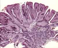
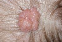
3 Squamous papilloma Exophytic growth made up of numerous small finger like projections which result in a lesion with a roughened verucous or cauliflower like surface. Painless pedunculated tumor or rarely sessile. Few millimeters to centimeters. Causative factors HPV 6 and 11

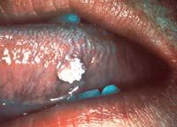
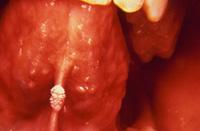
4 SQUAMOUS PAPILLOMA
5 Cowden s syndrome {multiple hamartomas neoplasia syndrome} Papilloma like or papillomatous lesions Fibromas in oral cavity Facial trichilemmomas Abnomalities in GIT, CNS, thyroid and musculoskeletal abnormalities
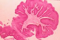
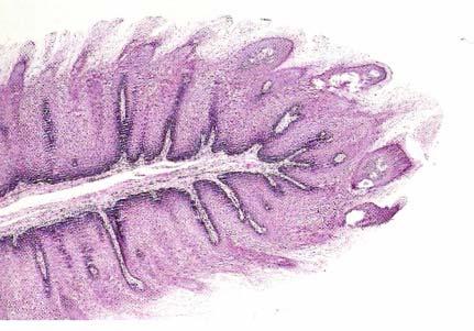
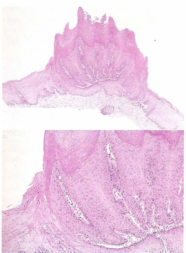
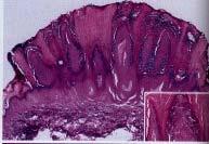
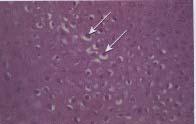
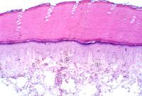
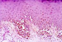
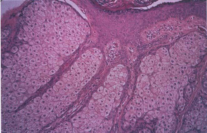
6 Histopathology Long, thin finger like projections made up of continuous layer of stratified squamous epithelium containing a thin central connective tissue core that support the nutrient blood vessels. Proliferation of spinous cells in a papillary pattern. Koilocytes {HPV altered epithelial cells with perinuclear spaces and nuclear pyknosis.}

7

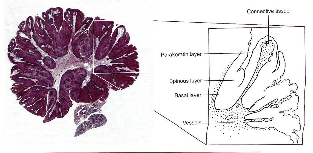
8 HISTOPATHOLOGY

9 papilloh1.jpg papilloh1.jpg papilloh1.jpg papilloh1.jpg


10 Treatment and Prognosis complete exicision including the base into which the pedicle or stalk inserts Its malignant potential is rare Alternative treatment includes conservative surgical exicision or curettage but liq nitrogen cryotherapy & topical application of keratolytic agent are also effective. Recurrence is rare.
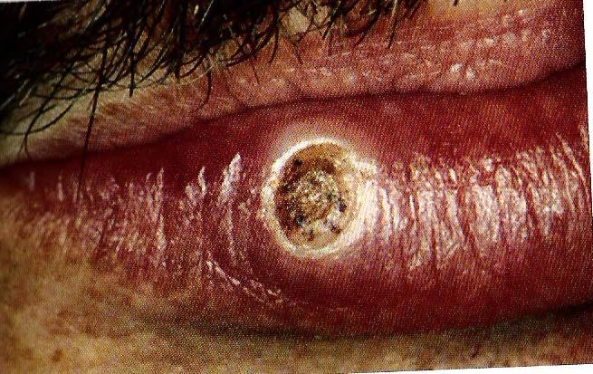
11 Keratoacanthoma Selfhealing carcinoma, molluscum pseudocarcinomatosam. Clinically and histologically resembles squamous cell carcinoma. Common low grade malignancy that originates in the pilosebaceous glands.
12 Etiology Chemical carcinogens tar and pitch Sunlight Trauma, HPV 9, 11, 13, 16, 18, 24, 25, 33,37, 57. Genetic factors chromosomal aberrations Gains on 8q, 1p, 9q with deletions on 3p, 9p, 19p and 19q. t(2;8)
13 Clinical features Older age groups Male preponderence Less common in dark skinned persons Common site sun exposed areas face, neck and dorsum of the upper extremities. Oral cavity lips and vermillion border.

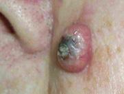
14 KERATOACANTHOMA
15 Solitary lesions, begin as firm, round, skin colored or reddish papules which progress in to dome shaped nodules with a smooth shiny surface with a central crateriform ulceration or keratin plug that may project like horn.

16 Keratoacanthoma Cup shaped symmetry and verruciform surface
17 Differential diagnosis: Keratoacanthoma Squamous cell carcinoma Tissue of origin : Hair follicle epithelium Main Pathologic Process: Benign proliferation Treatment: regresses spontaneously if left untreated


18 Verruca vulgaris Intra Oral

19 Verruca vulgaris
20 Condyloma acuminatum{venereal wart} One of the most common sexually transmitted diseases in the world. Etiology HPV 6, 11,30, 42, 43, 45, 46, 51,54,55 and 70.
21 Pathogenesis Inoculation in epithelium Viral Replication and transcription in basal cells Release of virions associated with epithelial proliferation except basal cells Virions assemble in cytoplasm of epithelial cells Acanthosis, hyperkeratosis Koilocytes in granular cells Release of virions along with desquamated cells


22 Condyloma acuminatum Soft pink nodules proliferate and coalesce to form diffuse pappilomatous clusters. Site : anogenital region skin. Oral site: dorsal tongue, buccal mucosa, palate, gingiva or alveolar ridge.

23 Condyloma Acuminatum Koilocytes

24 Focal epithelial hyperplasia Virus induced localized proliferation of oral sq epi ( HPV 13 & 32 ) C/F: Childhood condition Labial, buccal and lingual mucosa Multiple, soft non tender flattened or rounded papules clustered together Cobble stone appearance


25 Focal epithelial hyperplasia Acanthosis of epithelium Proliferation upwards Rete ridges are at the same depth and are widened and confluent Mitusoid cell Koilocytic changes Tests : DNA in-situ hybridisation IHC
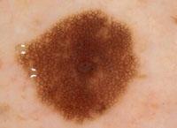
26 ORAL NEVI {oral melanocytic nevus, nevocellular nevus, mole} catogerised as hamartomas, nevi are they are benign proliferation of nevus cells in either connective or epithelial tissue. More common in whites. They are classified as congenital or acquired. On their histologic location basis, they are classified as junctional nevi: when nevi cells are limited to the basal cell layer. Compound nevi : when the cells are in epidermis dermis. Intradermal nevi or common mole : nests of nevus cells are entirely in the dermis.
27 Oral Nevi Junctional nevi that are first observed in infants and young adults typically mature into compound nevi, and during later adulthood into intramucosal nevi. As nevi cells penetrate into the mucosa, thee pigmentation diminishes. Most common of all nevi are the intramucosal nevi which account for 50% of all oral nevi followed by blue nevi {25 36%}.
28 ORAL NEVI
29 Intramucosal Nevus The pigmented nevus is characterized by a proliferation of nevus cells microscopically within the underlying connective tissue. Clinical Features: Most common lesion of the skin & mucosa. Exhibit as several or dozen scattered over the body. Smooth, flat lesion, may be elevated above the surface. May or may not show brown pigmentation. Often shows strands of hair growing from the surface. Common oral sites are hard palate & buccal mucosa.
30 Junctional Nevus The pigmented nevus is characterized by a proliferation of nevus cells microscopically within the basal cell layer of the surface epithelium. Clinical Features: usually on the skin and occasionally on the oral mucosa:hard palate or gingiva brown to black macular lesion
31 JUNCTIONAL NEVI
32 Histopathologic features nest of nevus cells in the basilar region of the epithelium epithelial rete pegs In the junctional nevus, the zone of demarcation between connective tissue and the overlying epithelium is absent and the cells contact and seem to blend with the surface epithelium. Overlying epithelium usually thin and irregular and shows the so called abtropfung or dropping off effect.

33 Histopathologic features
34 Main Pathologic Process: benign prolifertion Treatment: diagnostic excisional biopsy Prognosis: may transform to melanoma does not recur after excision
35 Compound Nevi The pigmented nevus is characterized by a proliferation of nevus cells microscopically within the basal cell layer of the surface epithelium and the underlying connective tissue. Clinical Features: more common on skin than oral mucosa pigmented papule or a macule hard palate or gingiva

36 COMPOUND NEVI
37 Differential Diagnosis: compound nevus melanotic macule amalgam tattoo melanotic macule Tissue of Origin: epithelium and connective tissue


38 Histologic Features: nevus cells in the basal region and adjacent conective tissue
39 Tissue of Origin: epithelium and connective tissue Main Pathologic Process: benign proliferation Treatment: diagnostic excisional biopsy Prognosis: does not recur
40 Blue Nevus A benign pigmented lesion that presents as a dark blue dome-shaped papule or as a flat macule on the skin or mucosa. Clinical Features: children & young adults female predilection most often on the hard palate 1-3 cm in diameter usually solitary elevated, smooth-surfaced papules or plaques that are gray-blue to bluish black in color

41 BLUE CELL NEVI


42 Histologic Features: melanin producing spindled and fusiform dendritic cells in the connectve tissue parallel to the normal overlying epithelium melanin containing macrophages
43 Differential Diagnosis: melanotic macule amalgam tattoo melanotic macule Tissue of Origin: connective tissue Main Pathologic Process: benign prolifertion Treatment: diagnostic excisional biopsy Prognosis: no tendency for malignant transformation
44 Verruciform xanthoma Hyperplastic condition of the epithelium of the mouth, skin and genitalia Accumulation of lipid laden histiocytes beneath the epithelium Not associated with virus Ass with immune response


45 Verruciform xanthoma Clinical features : Common in whites Age : 4 7 th decade Female predilection Oral : gingiva and alveolar mucosa Manifestation: well demarcated, soft, painless sessile slightly elevated mass with the papillary surface Lesions < 2 cm


46 Verruciform xanthoma Papillary acanthotic surface epithelium covered by thick para keratin Clefts or crypts present Numerous large macrophages with foamy cytoplasm confined to the conn tissue papillae Xanthoma cells Diastase resistant, PAS +ve
47 Seborrheic keratosis Common skin lesion of older people Benign proliferation of epidermal basal cells Hereditary Chronic sun exposure


48 Clinical features : Skin of face, trunk and extremities Above 4 th decade Multiple lesions Small tan to brown macules indistinguishable from actinic lentigenous Fissured, pitted sharply demarcated plaques Adhered to the skin Dermatosis papillosa nigra


49 H/P: Basilar epithelial hyperplasia with varying degrees of keratinisation Acanthosis Papillomatosis Lesion exhibits deep keratin filled invaginations Horn cysts, pseudo horn cysts
50 Sebaceous hyperplasia Localized proliferation of sebaceous glands of skin Similar to Basal cell carcinoma

51 C/F: Adults 40 years above Skin of face, forehead, and cheek One or more spft non tender papules, white or yellow Umblicated with small central depression from which a thick yellow white sebum can be expressed on compression Oral : BM papule or nodular mass with a cauliflower like appearance
52 Sebaceous hyperplasia
53 Ephelis ( Freckle ) Common small hyperpigmented macule of the skin Represents increased melanin production Face, arms, fair skinned blue eye persons Autosomal dominant Skin discoloration pronounced after sun exposure Age: 1 st decade and fades as age advances
 Brown")
54 Ephelis ( Freckle ) Brown macules
55 Actinic lentigo Lentigo solaris ; solar lentigo ; senile lentigo Continuation of ephelis Benign brown macule that results from chronic UV light exposure to the skin Age: 6 7 th decade ; Whites affected Frequent on facial skin, dorsum of hands Multiple but individual lesions pigmented brown to tan macules, well demarcated but irregular borders >1 cm in diameter Adjacent lesions coalesce and new ones arise with age No change in color intensity after exposure of UV light

56 H/P: Rete ridges elongated and club shaped Thinning of the epithelium above the connective tissue papillae Melanocytes
57 Lentigo simplex Benign cutaneous melanocytic hyperplasia of unknown cause Skin not exposed to sun light Color intensity does not change with exposure of sunlight Earlier stage of melanocytic nevus


58 Clinical features : Children Sharply demarcated macules with tan to dark brown color Reaches maximum size in a matter of months
59 H/P: Increased benign melanocytes

60 Melasma ( Mask of pregnancy) Symmetric hyper pigmentation of the sun exposed skin of the face and neck Associated with pregnancy Use of oral contraceptives
61 Oral melanotic macule Focal melanosis Brown mucosal discoloration due to focal increase in the no of melanocytes Unknown cause


62 Oral melanotic macule C/F: No age predilection Females Vermillion zone of lower lip- common site Solitary, well demarcated tan to brown asymptomatic round or oval macule


63 Oral melanotic macule Increase in melanin Melanin incontinence
NEOPLASMS OF THE SURFACE EPITHELIUM (KERATINOCYTES)
") NEOPLASMS OF THE SURFACE EPITHELIUM (KERATINOCYTES) Papillary Lesions Precancerous Lesions Keratinocyte Proliferations Carcinomas Melanotic Lesions Melanomas Normal Mucosa Keratin layer Spinous layer Basal
NEOPLASMS OF THE SURFACE EPITHELIUM (KERATINOCYTES) Papillary Lesions Precancerous Lesions Keratinocyte Proliferations Carcinomas Melanotic Lesions Melanomas Normal Mucosa Keratin layer Spinous layer Basal
Pigmented lesions of the Oral cavity
 Oral medicine أ.م.د احسان عبد هللا كميل Pigmented lesions of the Oral cavity Pigmented oral lesions are a large group of disorders in which the dark or brown color is the essential clinical characteristic.
Benign and malignant epithelial lesions: Seborrheic keratosis: A common benign pigmented epidermal tumor occur in middle-aged or older persons more
 Benign and malignant epithelial lesions: Seborrheic keratosis: A common benign pigmented epidermal tumor occur in middle-aged or older persons more common on the trunk; but extremities, head and neck are
المركب النموذج--- سبيتز وحمة = Type Spitz's Nevus, Compound SPITZ NEVUS 1 / 7
 SPITZ NEVUS 1 / 7 Epidemiology An annual incidence rate of 1.4 cases of Spitz nevus per 100,000 individuals has been estimated in Australia, compared with 25.4 per 100,000 individuals for cutaneous melanoma
LUMPS AND BUMPS: AN ORGANIZED APPROACH TO DIAGNOSIS AND MANAGEMENT
 LUMPS AND BUMPS: AN ORGANIZED APPROACH TO DIAGNOSIS AND MANAGEMENT Tammy P. Than, M.S., O.D., F.A.A.O. The University of Alabama at Birmingham / School of Optometry 1716 University Blvd. Birmingham, AL
Benign versus Cancerous Lesions How to tell the difference FMF 2014 Christie Freeman MD, CCFP, DipPDerm, MSc
 1 Benign versus Cancerous Lesions How to tell the difference FMF 2014 Christie Freeman MD, CCFP, DipPDerm, MSc Benign lesions Seborrheic Keratoses: Warty, stuck-on Genetics and birthdays Can start in late
Pathology of the skin. 2nd Department of Pathology, Semmelweis University
 Pathology of the skin 2nd Department of Pathology, Semmelweis University Histology of the skin Epidermis: Stratum corneum Stratum granulosum Stratum spinosum Stratum basale Dermis: papillary and reticular
MECHANISMS OF HUMAN DISEASE: LABORATORY SESSION PATHOLOGY OF THE SKIN LAB. Friday, February 13, :30 am 11:00 am
 MECHANISMS OF HUMAN DISEASE: LABORATORY SESSION PATHOLOGY OF THE SKIN LAB Friday, February 13, 2009 9:30 am 11:00 am FACULTY COPY GOALS: Describe the basic clinical and morphologic features of various
MECHANISMS OF HUMAN DISEASE: LABORATORY SESSION PATHOLOGY OF THE SKIN LAB. Friday, February 12, :30 am 11:00 am
 MECHANISMS OF HUMAN DISEASE: LABORATORY SESSION PATHOLOGY OF THE SKIN LAB Friday, February 12, 2012 9:30 am 11:00 am FACULTY COPY GOALS: Describe the basic clinical and morphologic features of various
Periocular Malignancies
 Periocular Malignancies Andrew Gurwood, O.D., F.A.A.O., Dipl. Marc Myers, O.D., F.A.A.O. Drs. Myers and Gurwood have no financial interests to disclose. Course Description Discussion of the most common
LENTIGO SIMPLEX. Epidemiology
 LENTIGO SIMPLEX Epidemiology The frequency of lentigo simplex in children and adults has not been determined. There does not appear to be a racial or gender predilection. Lentigo simplex is the most common
Lid Lesions: Relax or Refer
 Lid Lesions: Relax or Refer Blair Lonsberry, MS, OD, MEd., FAAO Professor of Optometry Pacific University College of Optometry blonsberry@pacificu.edu Agenda Benign vs. Malignant lesions Benign Eyelid
The Integumentary System. Mosby items and derived items 2010, 2006, 2002, 1997, 1992 by Mosby, Inc., an affiliate of Elsevier Inc.
 The Integumentary System The Skin Structure two primary layers called epidermis and dermis Epidermis Outermost and thinnest primary layer of skin Composed of several layers of stratified squamous epithelium
4Ps LUMPS AND BUMPS B.L.&T. BUMPS, LUMPS, AND TATTOOS. Most Common BUMP in the oral cavity Fibroma INTERDENTAL PAPILLAE LESIONS
 B.L.&T. BUMPS, LUMPS, AND TATTOOS LUMPS AND BUMPS DIFFERENTIAL DIAGNOSIS FOR LUMPS AND BUMPS Traumatic Fibroma Papilloma Epulis Fissuratum Inflammatory Papillary Hyperplasia Lesions of Attached Gingiva
Dermatopathology: The tumor is composed of keratinocytes which show atypia, increase mitoses and abnormal mitoses.
 Squamous cell carcinoma (SCC): A common malignant tumor of keratinocytes arising in the epidermis, usually from a precancerous condition: 1- UV induced actinic keratosis, usually of low grade malignancy.
Lumps and Bumps: The Dermatology of Lid Lesions
 Lumps and Bumps: The Dermatology of Lid Lesions Thomas J. Joly, MD, PhD Assistant Professor of Ophthalmology Eastern Virginia Medical School Ophthalmic Plastic Surgery Service Virginia Eye Consultants
IT S FUNDAMENTAL MY DEAR WATSON! A SHERLOCKIAN APPROACH TO DERMATOLOGY
 IT S FUNDAMENTAL MY DEAR WATSON! A SHERLOCKIAN APPROACH TO DERMATOLOGY Skin, Bones, and other Private Parts Symposium Dermatology Lectures by Debra Shelby, PhD, DNP, FNP-BC, FADNP, FAANP Debra Shelby,
Skin lesions The Good and the Bad. Dr Virginia Hubbard Ipswich Hospital NHS Trust Barts and the London School of Medicine and Dentistry
 Skin lesions The Good and the Bad Dr Virginia Hubbard Ipswich Hospital NHS Trust Barts and the London School of Medicine and Dentistry Case 1 32 year old woman Australian Lesion on back New hair growing
Clinical characteristics
 Skin Cancer Fernando Vega, MD Seattle Healing Arts Clinical characteristics Precancerous lesions Common skin cancers ACTINIC KERATOSIS Precancerous skin lesions Actinic keratoses Dysplastic melanocytic
Actinic keratosis (AK): Dr Sarma s simple guide
: Dr Sarma s simple guide") Actinic keratosis (AK): Dr Sarma s simple guide Actinic keratosis is a very common lesion that you will see in your day-to-day practice. First, let me explain the name Actinic keratosis. It means keratosis
Actinic keratosis (AK): Dr Sarma s simple guide Actinic keratosis is a very common lesion that you will see in your day-to-day practice. First, let me explain the name Actinic keratosis. It means keratosis
Doctors of Optometry Course Notes
 Doctors of Optometry Course Notes OD19 1CE COPE: 43871-AS Eyelid Lumps and Bumps Sunday, February 26, 2017 2:40 pm 3:30 pm Regency C 3 rd Floor Presenter: Blair Lonsberry, OD, FAAO Dr. Lonsberry is a Full
Premalignant skin tumours
 Chapter 14: Premalignant skin tumours page: 434 Premalignant skin tumours page: 435 Solar keratoses (senile keratoses) Raised red and well-defined plaques with a rough surface covered in scales of varying
Histopathology: skin pathology
 Histopathology: skin pathology These presentations are to help you identify, and to test yourself on identifying, basic histopathological features. They do not contain the additional factual information
Appendix : Dermoscopy
 Go Back to the Top To Order, Visit the Purchasing Page for Details APP Appendix : Dermoscopy Dermoscopy, also known as dermatoscopy, epiluminoscopy and epiluminescent microscopy, is an effective non-invasive
Oral Epithelial Tumors, Melanocytic Nevi, and Melanoma (I)
") Introduction: Oral Epithelial Tumors, Melanocytic Nevi, and Melanoma (I) Oral Epithelial Tumors may be: Benign tumors Sequamous cell Papilloma Malignant tumors Sequamous cell carcinoma, Basal cell carcinoma
Introduction: Oral Epithelial Tumors, Melanocytic Nevi, and Melanoma (I) Oral Epithelial Tumors may be: Benign tumors Sequamous cell Papilloma Malignant tumors Sequamous cell carcinoma, Basal cell carcinoma
Dermoscopy: Recognizing Top Five Common In- Office Diagnoses
 Dermoscopy: Recognizing Top Five Common In- Office Diagnoses Vu A. Ngo, DO Department of Family Medicine and Dermatology Choctaw Nation Health Services Authority Learning Objectives Introduction to dermoscopy
Learning Objectives. Tanning. The Skin. Classic Features. Sun Reactive Skin Type Classification. Skin Cancers: Preventing, Screening and Treating
 Learning Objectives Skin Cancers: Preventing, Screening and Treating Robert A. Baldor, MD, FAAFP Professor, Family Medicine & Community Health University of Massachusetts Medical School Distinguish the
Diagnostic difficulties with lesions of the oral mucosa
 BDIAP London, November 2010 School of Clinical Dentistry University of Sheffield Diagnostic difficulties with lesions of the oral mucosa Paul M Speight Dept Oral & Maxillofacial Pathology University of
Basics in Dermoscopy
 Basics in Dermoscopy Manal Bosseila Professor of Dermatology, Cairo University Member of European Academy Dermatology & Venereology EADV Member of International Dermoscopy Society IDS Member of Aesthetic
Disclosure. Objectives. PAFP CME Conference Lou Mancano MD, FAAFP Reading Health System November 18, 2016
 PAFP CME Conference Lou Mancano MD, FAAFP Reading Health System November 18, 2016 1 Disclosure The speaker has no conflict of interest, financial agreement, or working affiliation with any group or organization.
Common Benign Lesions and Skin Cancers. 22nd May 2015 Dr Mark Foley
 Common Benign Lesions and Skin Cancers 22nd May 2015 Dr Mark Foley Thank you for downloading this file. This intended to supplement the presentation given at the NZ Wound Care Conference, it is not intended
comedo-like openings (clods, brown or orange & circles) milia-like cysts (dots or clods, white) 1/29/18 Dotted vessels are also commonly seen in SCC
 milia-like cysts (dots or clods, white) 1/29/18 Dotted vessels are also commonly seen in SCC") Brown circles Dotted vessels are also commonly seen in SCC Step1 1. Nevus (unequivocal) 2. DF/IDN 3. BCC 4. SCC Network Patchy network Peripheral network & central hypopigmentation DF: network with central
Brown circles Dotted vessels are also commonly seen in SCC Step1 1. Nevus (unequivocal) 2. DF/IDN 3. BCC 4. SCC Network Patchy network Peripheral network & central hypopigmentation DF: network with central
Identifying Skin Cancer. Mary S. Stone MD Professor of Dermatology and Pathology University of Iowa Carver College of Medicine March, 2018
 Identifying Skin Cancer Mary S. Stone MD Professor of Dermatology and Pathology University of Iowa Carver College of Medicine March, 2018 American Cancer Society web site Skin Cancer Melanoma Non-Melanoma
Diseases of the vulva
 Diseases of the vulva 1. Bartholin Cyst - Infection of the Bartholin gland produces an acute inflammation within the gland (adenitis) and may result in an abscess. Bartholin duct cysts - Are relatively
Basal cell carcinoma 5/28/2011
 Goal of this Presentation A practical approach to the diagnosis of cutaneous carcinomas and their mimics Thaddeus Mully, MD University of California San Francisco To review common non-melanoma skin cancers
Lesions & Lifestyles
 Lesions & Lifestyles attended a 3 hour Continuing Education Seminar on Oral Pathology presented by Nancy Dewhirst, RDH,BS on (date) at (location):. Course material is directly related patient care. Notes:
Non-melanocytic Patterns
 Non-melanocytic Lesions Non-melanocytic Patterns Michelle Tarbox, MD Assistant Professor of Dermatology and Dermatopathology Texas Tech University Health Sciences Center 2018 Seborrheic keratoses Acanthotic
Premalignant lesions may expose to a promoting. factor & may be induced to undergo malignant. Carcinoma in situ displays the cytologic features of
 بسم رلاهللا Def. Premalignant lesions may expose to a promoting factor & may be induced to undergo malignant transformation. Carcinoma in situ displays the cytologic features of malignancy without invasion
Describe the functions of the vertebrate integumentary system. Discuss the structure of the skin and how it relates to function.
 Chapter 5 Describe the functions of the vertebrate integumentary system. Discuss the structure of the skin and how it relates to function. Explain the basis for different skin colors. Describe the structure
Integumentary System
 Integumentary System The integumentary system is commonly known as the Skin Largest organ of human body 10% total body weight and would cover over 20 square feet Functions of Skin 1. Protection Barrier
Cowden Syndrome PTEN Hamartoma Tumor Syndrome. ACCME/Disclosure. 1. Background. Outline
 MASSACHUSETTS GENERAL HOSPITAL HARVARD MEDICAL SCHOOL PATHOLOGY Cowden Syndrome PTEN Hamartoma Tumor Syndrome ACCME/Disclosure Vania Nosé, MD, PhD Professor of Pathology Director of Anatomic Pathology
Lumps and Bumps: An Organized Approach to Diagnosis and Management. Disclosure. Introduction. References. Structure of Skin.
 Lumps and Bumps: An Organized Approach to Diagnosis and Management Nothing to disclose Disclosure Tammy Pifer Than, MS, OD, FAAO Carl Vinson VAMC tammythan@bellsouth.net References Fitzpatrick's Color
PDF of Trial CTRI Website URL -
 Clinical Trial Details (PDF Generation Date :- Wed, 25 Jul 2018 13:50:16 GMT) CTRI Number Last Modified On 10/05/2013 Post Graduate Thesis Type of Trial Type of Study Study Design Public Title of Study
Desmoplastic Melanoma R/O BCC. Clinical Information. 74 y.o. man with lesion on left side of neck r/o BCC
 R/O BCC Sabine Kohler, M.D. Professor of Pathology and Dermatology Dermatopathology Service Stanford University School of Medicine Clinical Information 74 y.o. man with lesion on left side of neck r/o
ISPUB.COM. Seborrheic Keratosis: A Pictorial Review of the Histopathologic Variations. D Sarma, S Repertinger
 ISPUB.COM The Internet Journal of Dermatology Volume 7 Number 2 Seborrheic Keratosis: A Pictorial Review of the Histopathologic Variations D Sarma, S Repertinger Citation D Sarma, S Repertinger.. The Internet
CASE REPORT PLAQUE TYPE ORAL VERRUCOUS HYPERPLASIA AND IRRITATIONAL FIBROMA: A REPORT OF CONJOINT OCCURRENCE
 CASE REPORT PLAQUE TYPE ORAL VERRUCOUS HYPERPLASIA AND IRRITATIONAL FIBROMA: A REPORT OF CONJOINT OCCURRENCE Alphy Alphonsa Sebastian, Hasan Subhi 1. Phd student, Department of Oral Medicine and Oral Pathology,
Pigmented Seborrheic Keratosis (Melanoacanthoma) of Nipple A case report with review of literature
 of Nipple A case report with review of literature") Case Report Pigmented Seborrheic Keratosis (Melanoacanthoma) of Nipple A case report with review of literature Aparna Narasimha, Harendra Kumar ML, Divyarani MN, Bhaskaran A* Department of Pathology and
Case Report Pigmented Seborrheic Keratosis (Melanoacanthoma) of Nipple A case report with review of literature Aparna Narasimha, Harendra Kumar ML, Divyarani MN, Bhaskaran A* Department of Pathology and
Gross Appearance & Histology of Skin Cancer. Kyle Mannion M.D. January 21, 2005
 Gross Appearance & Histology of Skin Cancer Kyle Mannion M.D. January 21, 2005 Actinic Keratosis 5-20% will develop squamous/basal cell ca Almost solely from solar damage Usually develop during 4 th decade
Diseases of the breast (1 of 2)
") Diseases of the breast (1 of 2) Introduction A histology introduction Normal ducts and lobules of the breast are lined by two layers of cells a layer of luminal cells overlying a second layer of myoepithelial
Diseases of the breast (1 of 2) Introduction A histology introduction Normal ducts and lobules of the breast are lined by two layers of cells a layer of luminal cells overlying a second layer of myoepithelial
It can be helpful in some cases of actinic keratosis, Bowen s disease and squamous cell carcinoma
 Dermoscopy Introduction, Terminology and Structures (to be read in conjunction with the Diagnostic Dermoscopic Algorithm) Copyright to Cunliffe TP (Jan. 2017) All rights reserved Introduction Dermoscopy
Regression 2/3/18. Histologically regression is characterized: melanosis fibrosis combination of both. Distribution: partial or focal!
 Regression Margaret Oliviero MSN, ARNP Harold S. Rabinovitz MD Histologically regression is characterized: melanosis fibrosis combination of both Distribution: partial or focal! Dermatoscopic terminology
IN THE NAME OF GOD Dr. Kheirandish Oral and maxillofacial pathology
 IN THE NAME OF GOD Dr. Kheirandish Oral and maxillofacial pathology ORAL FOCAL MUCINOSIS Uncommon Tumorlike Cutaneous myxoid cyst Overproduction of hyaluronic acid by firoblasts Young adults Female Gingiva
04/09/2018. Squamous Cell Neoplasia and Precursor Lesions. Agenda. Squamous Dysplasia. Squamo-proliferative lesions. Architectural features
 Squamous Cell Neoplasia and Precursor Lesions Jennifer L. Hunt, MD, MEd Aubrey J. Hough Jr, MD, Endowed Professor of Pathology Chair of Pathology and Laboratory Medicine University of Arkansas for Medical
ORAL MELANOTIC NEVI: A CASE REPORT AND REVIEW OF LITERATURE
 ORAL MELANOTIC NEVI: A CASE REPORT AND REVIEW OF LITERATURE Abstract: *V.T Beena **Isha Chauhan *** R. Heera ***R. Rajeev Oral melanotic nevi are uncommon oral lesions causing focal. Melanotic nevi are
 Page 1 of 15 Title Authored By Course No Contact Hours 2 Skin Cancer the Real Picture for Early Detection and Treatment Cheryl Sommer RN, MSN, ARNP SC120604 Purpose The purpose of this course is to provide
Ex. 7: Integumentary
 Collin County Community College BIOL. 2401 Ex. 7: Integumentary. Skin or Integument Consists of three major regions Epidermis outermost superficial region Dermis middle region Hypodermis (superficial fascia)
ORAL MELANOMA Definition Epidemiology Clinical Presentation
 ORAL MELANOMA Definition Melanoma is a highly malignant neoplasia, arising from melanocytes, the cells that produce the brownish pigment melanin. Melanin is the determinant in skin colour and protects
Pathology. Skin Tumor. Bayan N. Mohammad 15/10/2015. Mohammad al-orjani. Page 0 of 23
 #7 35 Pathology Skin Tumor Bayan N. Mohammad 15/10/2015 Mohammad al-orjani Page 0 of 23 بسم هللا الرحمن الرحيم GREETINGS This lecture is about skin tumors, all the slides are included and every slide will
Dermatological Manifestations in the Elderly. Sanjay Siddha Staff Dermatologist UHN & MSH
 Dermatological Manifestations in the Elderly Sanjay Siddha Staff Dermatologist UHN & MSH Disclosure No actual or potential conflicts of interest or commercial relationships to declare Objectives Recognize
Keratinocyte tumors. Actinic Keratosis. Squamous cell carcinoma in situ. Squamous Cell Carcinoma. (aka Bowen s disease)
") Actinic Keratosis Keratinocyte tumors Prepared by Kurt Schaberg Precancerous, risk of malignancy ~8-20% per year (progresses to SCC); Due to chronic sun exposure Rough scaly plaque; typically due to sun
Actinic Keratosis Keratinocyte tumors Prepared by Kurt Schaberg Precancerous, risk of malignancy ~8-20% per year (progresses to SCC); Due to chronic sun exposure Rough scaly plaque; typically due to sun
Lagophthalmos. Lagophthalmos: signs. Lagophthalmos: clinical tips. Lagophthalmos: treatment plan. Madarosis
 Lagophthalmos Def: incomplete closure of the eyelid SX: FBS, irritation, red, burn, dry, chronic morning corneal irritation Lagophthalmos: signs 2-5 mm lid separation with slit lamp during blink can force
Clinically Microscopically Pathogenesis: autoimmune not lifetime
 Vulvar Diseases: Can be divided to non-neoplastic and neoplastic diseases. The neoplastic diseases are much less common. Of those, squamous cell carcinoma is the most common. most common in postmenopausal
General information about skin cancer
 Skin Cancer General information about skin cancer Key points Skin cancer is a disease in which malignant (cancer) cells form in the tissues of the skin. There are different types of cancer that start in
ARTICLE INFO ABSTRACT
 Melanocytic Pigmentation: A Single Manifestation of Myriad of Pathologies [PP: 05-09] Dr. Swapna Honwad Department of Oral Pathology dr.swapnahonwad@gmail.com Dr. Elsy P. Simon Department of Endodontics
Large majority caused by sun exposure Often sun exposure before age 20 Persons who burn easily and tan poorly are at greatest risk.
 Basics of Skin Cancer Detection and Treatment of Non- Melanoma Skin Cancers Large majority caused by sun exposure Often sun exposure before age 20 Persons who burn easily and tan poorly are at greatest
Brief Report. Shivanand Gundalli 1, Smita Kadadavar 1, Somil Singhania 1, Rutuja Kolekar 2 INTRODUCTION. Melanocytic Nevus
 Our Dermatology Online Histopathological spectrum of benign melanocytic nevi our experience in a tertiary care centre Shivanand Gundalli 1, Smita Kadadavar 1, Somil Singhania 1, Rutuja Kolekar 2 1 Department
6/17/2018. Breaking Bad (Part 1) Dermoscopy of Brown(ish) Things. Bad?
 Dermoscopy of Brown(ish) Things. Bad?") Breaking Bad (Part 1) Dermoscopy of Brown(ish) Things Jennie T. Clarke, MD ssociate Professor of Dermatology University of Utah School of Medicine Bad? 1 Brown(ish) Things Bad Melanoma Pigmented basal
Breaking Bad (Part 1) Dermoscopy of Brown(ish) Things Jennie T. Clarke, MD ssociate Professor of Dermatology University of Utah School of Medicine Bad? 1 Brown(ish) Things Bad Melanoma Pigmented basal
Polypoid Melanoma, A Virulent Variant of the Nodular Growth Pattern
 Polypoid Melanoma, A Virulent Variant of the Nodular Growth Pattern ELIZABETH A. MANCI, M.D., CHARLES M. BALCH, M.D..TARIQ M. MURAD, M.D., PH.D., AND SENG/JAW SOONG, PH.D. Manci, Elizabeth A., Balch, Charles
Skin and Body Membranes Body Membranes Function of body membranes Cover body surfaces Line body cavities Form protective sheets around organs
 Skin and Body Membranes Body Membranes Function of body membranes Cover body surfaces Line body cavities Form protective sheets around organs Classification of Body Membranes Epithelial membranes Cutaneous
Skin Cancer - Non-Melanoma
 Skin Cancer - Non-Melanoma Introduction Each year, millions of people find out that they have skin cancer. Skin cancer is almost 100% curable if found early and treated right away. It is possible to prevent
Dermoscopy in everyday practice. What and Why? When in doubt cut it out? Trilokraj Tejasvi MD
 Dermoscopy in everyday practice Trilokraj Tejasvi MD Assistant Professor, Department of Dermatology, Director Teledermatology services, University of Michigan, Faculty Associate, GLOBAL REACH, Michigan
Chapter 8 Skin Disorders and Diseases
 Chapter 8 Skin Disorders and Diseases Attitude is more important than the past, than education, than money, than circumstances, than what people do or say. It is more important than appearance, giftedness,
Histopathology of Melanoma
 THE YALE JOURNAL OF BIOLOGY AND MEDICINE 48, 409-416 (1975) Histopathology of Melanoma G. J. WALKER SMITH Department ofpathology, Yale University School ofmedicine, 333 Cedar Street, New Haven, Connecticut
Human Anatomy & Physiology
 PowerPoint Lecture Slides prepared by Barbara Heard, Atlantic Cape Community College Ninth Edition Human Anatomy & Physiology C H A P T E R 5 Annie Leibovitz/Contact Press Images 2013 Pearson Education,
الفتوي الاصفر الحبيبوم = Xanthogranuloma_Juvenile JUVENILE XANTHOGRANULOMA 1 / 9
 JUVENILE XANTHOGRANULOMA 1 / 9 Clinical Findings CUTANEOUS LESIONS JXG is a benign, self-healing disorder that is characterized by asymptomatic yellowish papulonodular lesions of the skin and other organs
Malignant Melanoma Early Stage. A guide for patients
 This melanoma patient brochure is designed to help educate melanoma patients and their caregivers. It was developed under the guidance of Dr. Michael Smylie, Professor, Department of Oncology, University
Skin (Integumentary System) Wheater, Chap. 9
 Wheater, Chap. 9") Skin (Integumentary System) Wheater, Chap. 9 Skin (Integument) Consists of skin and associated derivatives Largest organ of body (21 ft 2 ; 9 lbs.; has 11 miles of blood vessels) Functions: Protection
Skin (Integumentary System) Wheater, Chap. 9 Skin (Integument) Consists of skin and associated derivatives Largest organ of body (21 ft 2 ; 9 lbs.; has 11 miles of blood vessels) Functions: Protection
Atlas of Eyelid and Conjunctival Tumors
 Atlas of Eyelid and Conjunctival Tumors Jerry A. Shields, M.D. Director, Ocular Oncology Service Wills Eye Hospital Professor of Ophthalmology Thomas Jefferson University Philadelphia, Pennsylvania Carol
Objectives. 1. Recognizing benign skin lesions. 2.Know which patients will likely need surgical intervention.
 The Joy of Pediatric Skin Dr. Claire Sanger University of Kentucky Plastic & Reconstructive Surgery Objectives 1. Recognizing benign skin lesions 2.Know which patients will likely need surgical intervention.
CH 05 THE INTEGUMENTARY SYSTEM
 CH 05 THE INTEGUMENTARY SYSTEM This system consists of skin and its derivatives. The skin is one of the largest organs of the body in terms of surface area. The functions of the integumentary system include:
SURGERY OF THE HAND. Giant Wart as a Cutaneous Horn on the Dorsum of the Hand INTRODUCTION CASE REPORT
 CASE REPORT pissn 1598-3889 eissn 2234-0998 J Korean Soc Surg Hand 2016;21(2):100-104. http://dx.doi.org/10.12790/jkssh.2016.21.2.100 JOURNAL OF THE KOREAN SOCIETY FOR SURGERY OF THE HAND Giant Wart as
PowerPoint Lecture Slide Presentation by Patty Bostwick-Taylor, Florence-Darlington Technical College Skin and Body Membranes
 PowerPoint Lecture Slide Presentation by Patty Bostwick-Taylor, Florence-Darlington Technical College Skin and Body Membranes 4 Body Membranes Function of body membranes Cover body surfaces Line body cavities
Identifying Benign and Malignant Skin Lesions. No Disclosures. Common Benign Lesions. Benign Lesions 2/25/2018. Stucco Keratoses.
 Dermatology in Primary Care Identifying Benign and Malignant Skin Lesions Christy Quire Baker, APRN, FNP-BC, DCNP Dermatology Certified Nurse Practitioner No Disclosures Common Benign Lesions Seborrheic
Skin and Body Membranes
 4 Skin and Body Membranes PowerPoint Lecture Slide Presentation by Jerry L. Cook, Sam Houston University ESSENTIALS OF HUMAN ANATOMY & PHYSIOLOGY EIGHTH EDITION ELAINE N. MARIEB Skin and Body Membranes
Know who is at risk: LOOK! for ABCDs, rapidly changing lesions, do a biopsy when indicated
 Lindy P. Fox, MD Assistant Professor Director, Hospital Consultation Service Department of Dermatology University of California, San Francisco Applies to adults without history of malignancy or premalignant
External Neoplasms in Goats: A Clinicopathological Study on Five Types. Abu-Seida, A.M and Kawkab, A. Ahmed
 External Neoplasms in Goats: A Clinicopathological Study on Five Types By Abu-Seida, A.M and Kawkab, A. Ahmed Introduction Introduction Neoplasia is occasionally diagnosed in goats. A survey of 800000
Simulators of melanoma
 Simulators of melanoma Philip E. LeBoit, M.D. Depts. of Pathology and Dermatology University of California, San Francisco Simulators of melanoma Simulators of melanoma in situ Melanocytic Non-melanocytic
Chapter 6 Squamous Cell Carcinoma: Variants and Challenges
 Chapter 6 Squamous Cell Carcinoma: Variants and Challenges Michael B. Morgan EPIDEMIOLOGY: Second most common skin cancer, rare in the dark-skinned races. ETIOLOGY: Ultraviolet light, HPV infection. PATHOGENESIS:
Unit 4 The Integumentary System
 Unit 4 The Integumentary System I. Classification of Body Membranes A. Epithelial Membranes (3) 1. Cutaneous Membrane > Stratified Squamous > Sits on Dense Connective Tissue > Skin: Epidermis & Dermis
Integumentary System (Script) Slide 1: Integumentary System. Slide 2: An overview of the integumentary system
 Slide 1: Integumentary System. Slide 2: An overview of the integumentary system") Integumentary System (Script) Slide 1: Integumentary System Slide 2: An overview of the integumentary system Skin is the body s largest and heaviest organ making up 15% of body weight. Most skin is 1 to
Integumentary System (Script) Slide 1: Integumentary System Slide 2: An overview of the integumentary system Skin is the body s largest and heaviest organ making up 15% of body weight. Most skin is 1 to
Integumentary System (Skin) Unit 6.3 (6 th Edition) Chapter 7.3 (7 th Edition)
 Unit 6.3 (6 th Edition) Chapter 7.3 (7 th Edition)") Integumentary System (Skin) Unit 6.3 (6 th Edition) Chapter 7.3 (7 th Edition) 1 Learning Objectives Identify the major components (anatomy) of skin Differentiate between the two types of skin glands Explain
Integumentary System (Skin) Unit 6.3 (6 th Edition) Chapter 7.3 (7 th Edition) 1 Learning Objectives Identify the major components (anatomy) of skin Differentiate between the two types of skin glands Explain
The Integumentary System
 The Integumentary System The Integumentary System Integument is skin Skin and its appendages make up the integumentary system (See if you can name some appendages) A fatty layer (hypodermis) lies deep
Integumentary System. Remember: Types of Membranes: Bio 250
 Integumentary System Bio 250 Remember: Tissue: Group of cells that are similar in appearance and perform similar function Organ: Two or more tissues grouped together and performing a specialized function
Figure 4.1. Using Figure 4.1, identify the following: 1) The region that contains adipose tissue is indicated by letter. Diff: 2 Page Ref: 115
 The region that contains adipose tissue is indicated by letter. Diff: 2 Page Ref: 115") Essentials of Anatomy and Physiology, 9e (Marieb) Chapter 4 Skin and Body Membranes Short Answer Figure 4.1 Using Figure 4.1, identify the following: 1) The region that contains adipose tissue is indicated
Essentials of Anatomy and Physiology, 9e (Marieb) Chapter 4 Skin and Body Membranes Short Answer Figure 4.1 Using Figure 4.1, identify the following: 1) The region that contains adipose tissue is indicated
Eruptive Tumors of the Follicular Infundibulum: An Unexpected Diagnosis of Hypopigmented Macules
 Dermatol Ther (Heidelb) (2015) 5:207 211 DOI 10.1007/s13555-015-0079-0 CASE REPORT Eruptive Tumors of the Follicular Infundibulum: An Unexpected Diagnosis of Hypopigmented Macules Poonkiat Suchonwanit.
The Integumentary System
 The Integumentary System The Integumentary System Integument is skin Skin and its appendages make up the integumentary system A fatty layer (hypodermis) lies deep to it Two distinct regions Epidermis Dermis
Describing and interpreting gross lesions. Prepared for VPM 4600, May 2018; Shannon Martinson
 Describing and interpreting gross lesions Prepared for VPM 4600, May 2018; Shannon Martinson How to Describe (and Interpret) Lesions Step 1 Step 2 Step 3 Step 4 Look at the specimen: Is it normal or abnormal
Medical History. Oral Medicine and General Medicine
 Medical History Oral Medicine and General Medicine Gingivitis herpetica acuta NECROTIZÁLÓ SIALOMETAPLASIA SOOR Medical History The life expectancy has recently increased and increasing By dental prevention
Know who is at risk: LOOK! for ABCDs, rapidly changing lesions, do a biopsy when indicated
 Lindy P. Fox, MD Associate Professor Director, Hospital Consultation Service Department of Dermatology University of California, San Francisco Applies to adults without history of malignancy or premalignant