WHITE LESIONS OF THE UPPER AIRWAY
|
|
|
- Eustacia Horton
- 5 years ago
- Views:
Transcription
1 WHITE LESIONS OF THE UPPER AIRWAY
2 WHITE LESION CONFIGURATIONS Solitary vrs Multifocal Flat Plaque Verrucous/rippled Lacey White with red component Papular (curdled milk plaques) Pseudomembranous
3 PLAQUES FRICTIONAL KERATOSES DENTURE, CHRONIC BITING LEUKOPLAKIA IDIOPATHIC SMOKELESS TOBACCO SANGUINARIA ACTINIC CHEILITIS WARTY DYSKERATOMA & GROVER S DISEASE LICHEN PLANUS (HYPERPLASTIC) DYSKERATOSIS CONGENTIA
4 Frictional Keratosis Cause/Effect Edentulous ridge crest, especially retromolar pad Ill Fitting prostheses Broken cusps and restorations Habitual tongue and Cheek biting Risk for cancer is low, perhaps nonexistant If smoking is a factor, biopsy should be undertaken
")
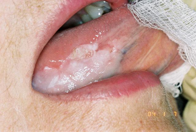
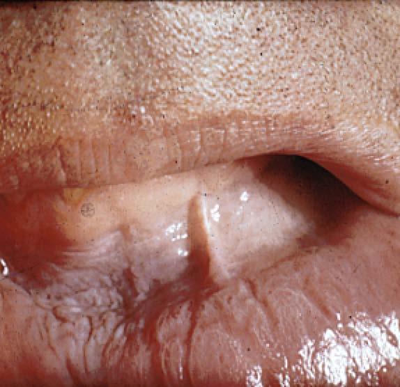
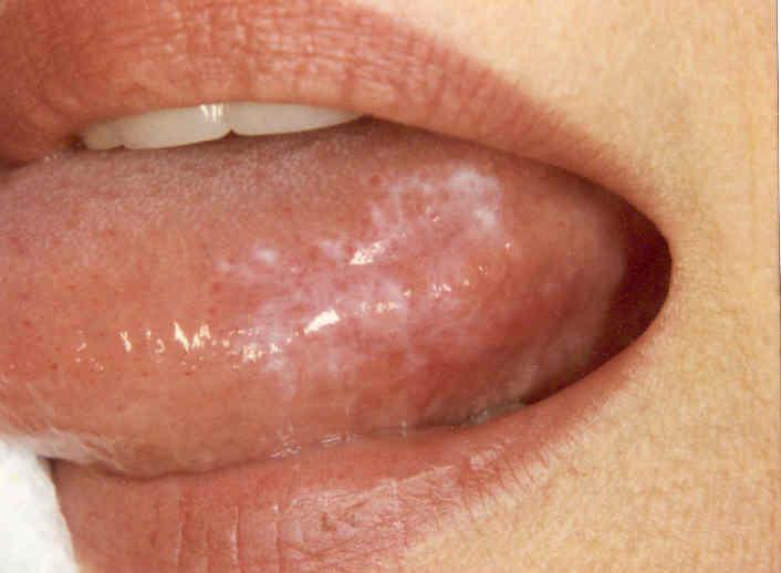
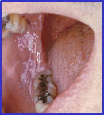
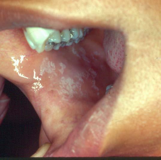
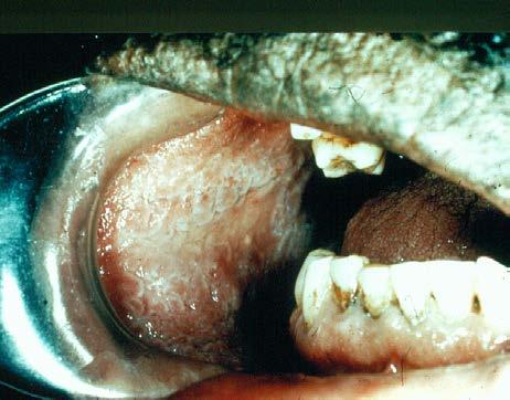
5 Cheek Bite Keratosis (Morsicasio Bucarum)

6 Frictional Keratosis
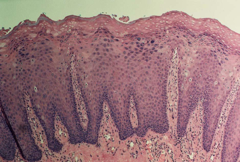
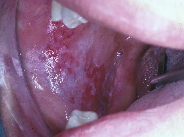
7 Leukoplakia An upper aerodigestive tract white lesion that: cannot be rubbed away does not have a cause (tobacco excluded) does not represent another clincially and microscopically defined disease
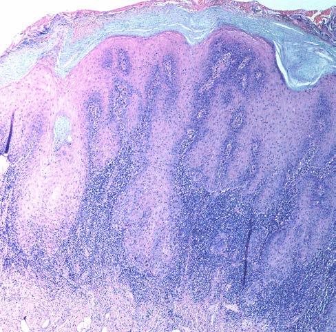
8 Leukoplakia 20% precancerous change histologically Floor of the Mouth 40% dysplastic 6% of all leukoplakias will progress to carcinoma within 5-7 years Biopsy: no dysplasia > periodic follow up or excision (laser ablation) Postive for dysplasia > excision with assessment of margins Ploidy assessment: aneuploid leukoplakias carry highest risk for progression to carcinoma
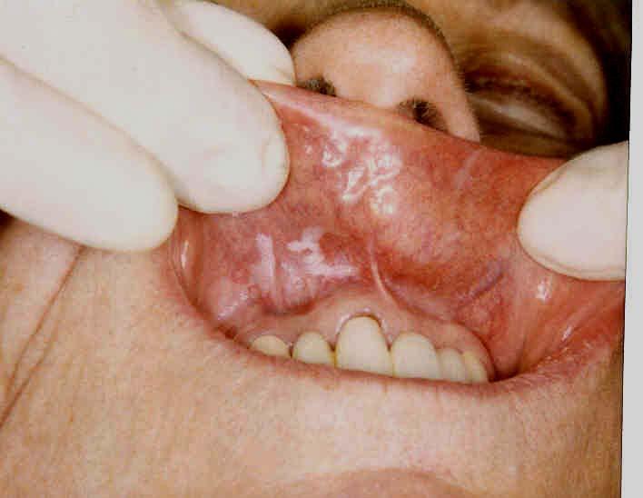
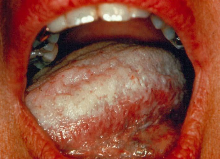
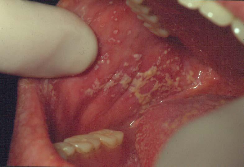
9 LEUKOPLAKIA
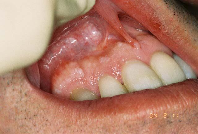
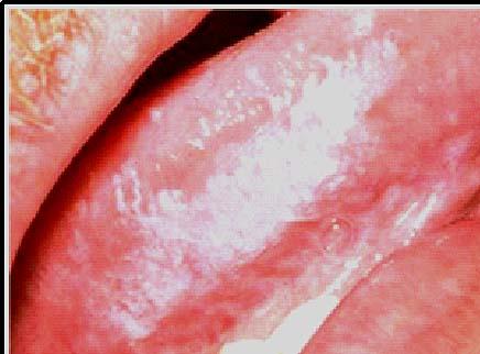
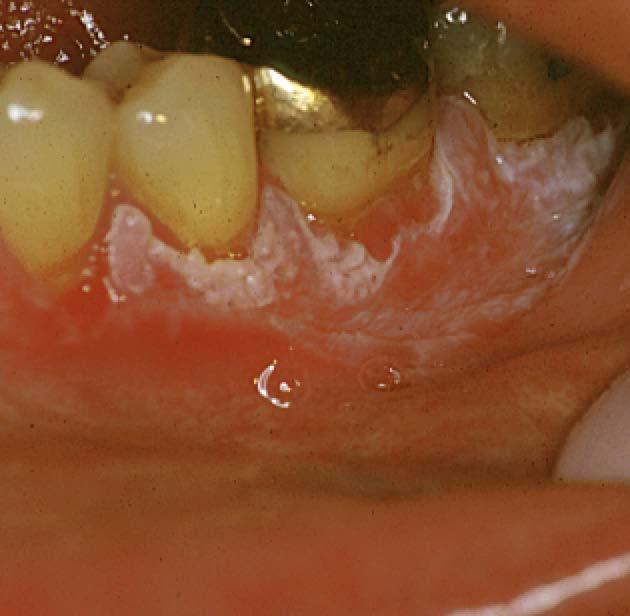
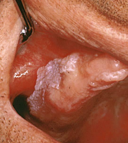
10 LEUKOPLAKIA
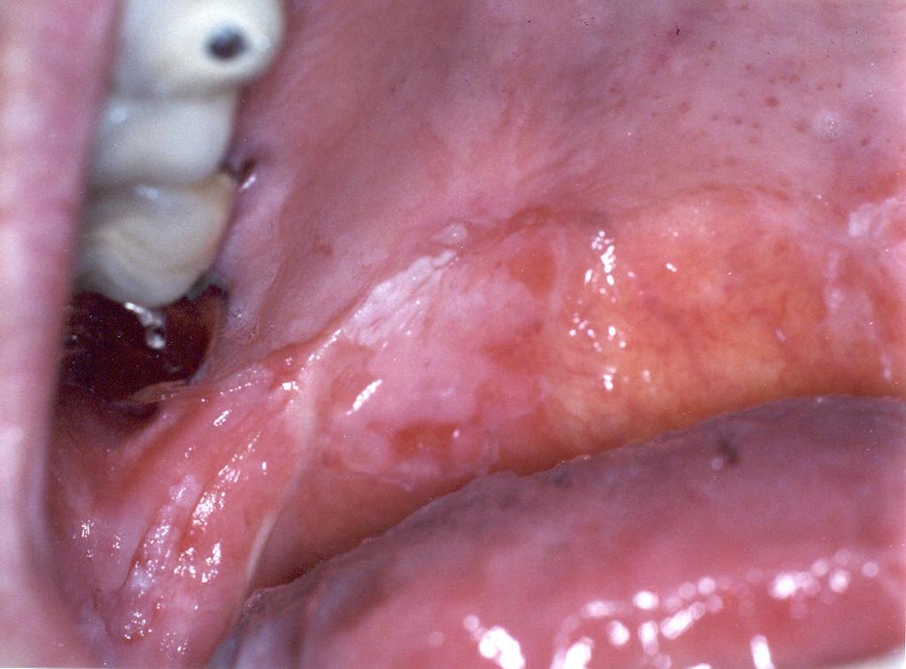
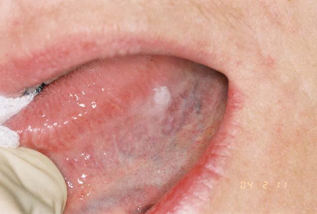
11 LEUKOPLAKIA
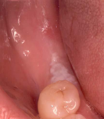
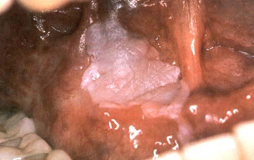
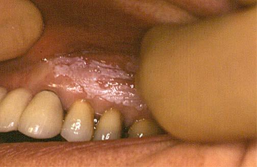
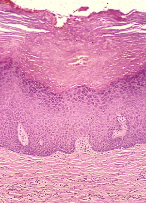
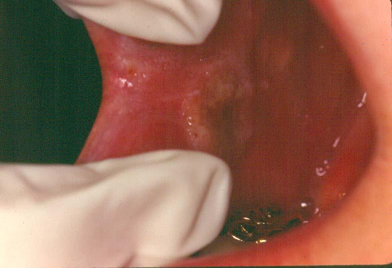
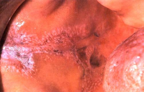
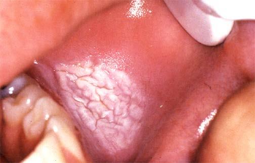
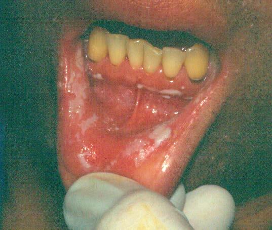
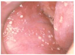
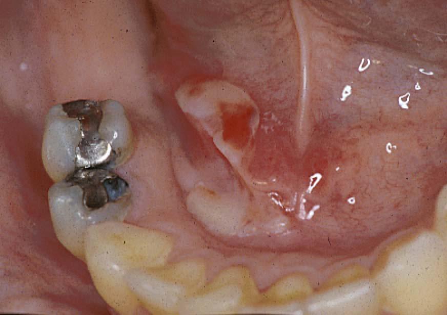
12 FOCAL LEUKOPLAKIA

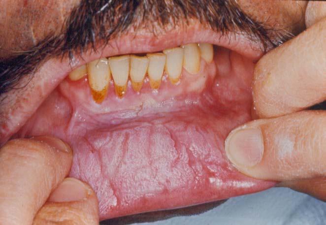
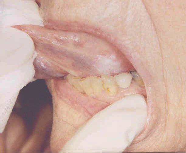
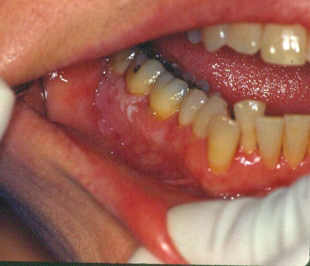
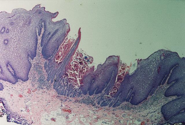
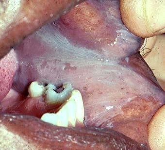
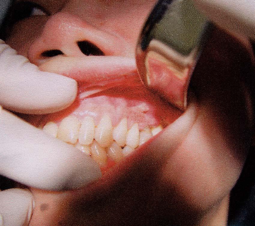
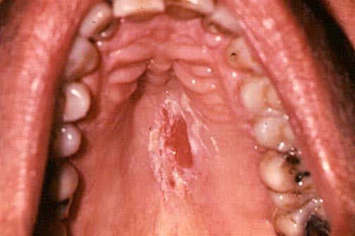
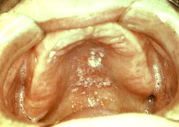
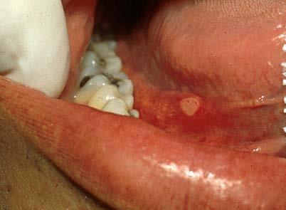
13 Smokeless Tobacco Keratosis
14 SANGUINARIA LEUKOPLAKIA
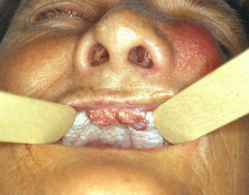
15 Actinic Cheilitis Solar irradiation to lower lip Diffuse leukoplakia across vermilion Light complected most prone Prolonged outdoor jobs or recreation Microscopically ranges from benign keratosis to dysplasia to carcinoma. Connective tissues show elastic degeneration. Tx: lip stripping or 5-fluorouracil cream


16 Actinic Keratosis of the Lip Proposed Excision areas inked

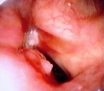
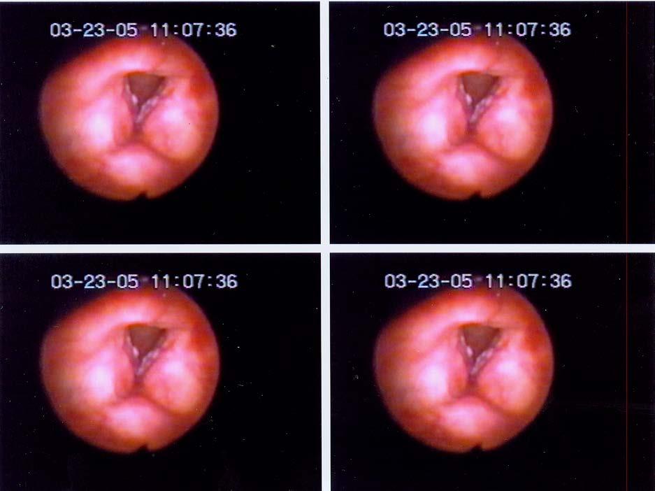
17 VOCAL CORD LEUKOPLAKIA DYSPLASIA SQUAMOUS CELL CA
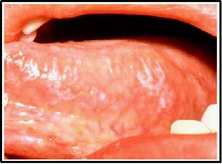
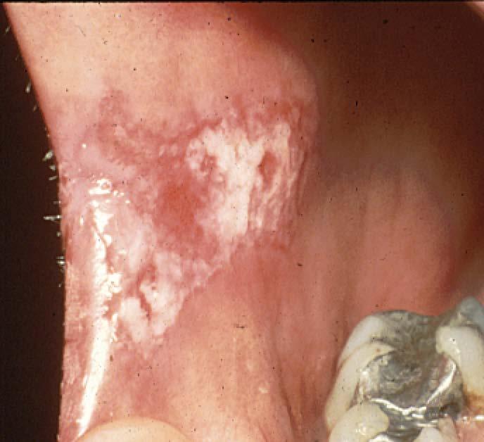
18 Proliferative Verrucous Leukoplakia A specific subtype of leukoplakia characterized by verrucous white lesions that tend to spread laterally and recur following excision Elderly females Gingiva and vestibule Smoking habit seen in less than 50% Microscopically, the lesions range from verrucous keratosis to atypical verrucous hyperplasia that may progress to either verrucous carcinoma or invasive squamous cancer Tx: aggressive excision by scalpel or laser with close periodic followup and re-excision when recurrences develope

19 PVL
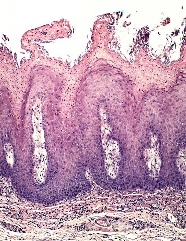
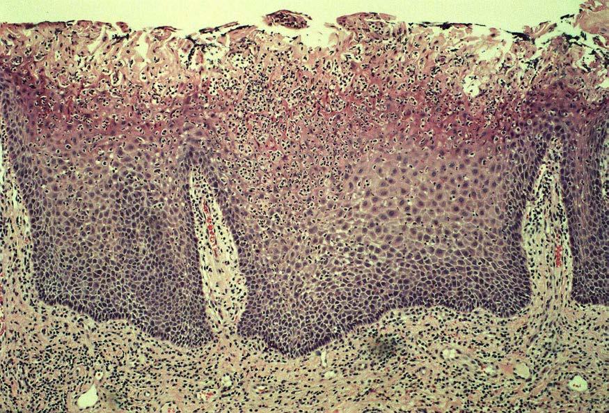
20 Leukoplakia Histology Benign Keratosis

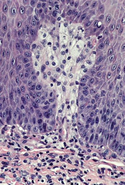
21 Dysplasia Leukoplakia Histology


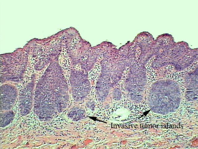
22 Leukoplakia Histology Squamous Cell Carcinoma, invasive islands of tumor
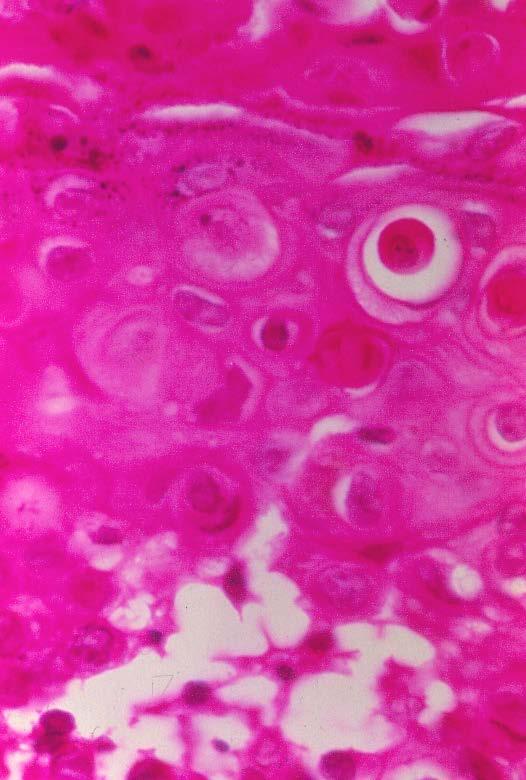
23 WARTY DYSKERATOMA Rare white lesion, often with an irregular, pebbly surface A benign keratosis with specific histologic features: Keratosis Individual cell keratinization Villous rete pegs Suprabasilar acantholysis (desmosome defect) Multiple lesions are termed Grover s disease (focal acantholytic dyskeratosis)
24 WARTY DYSKERATOMA
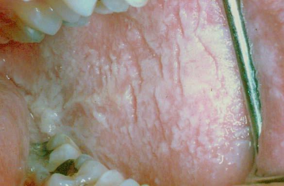
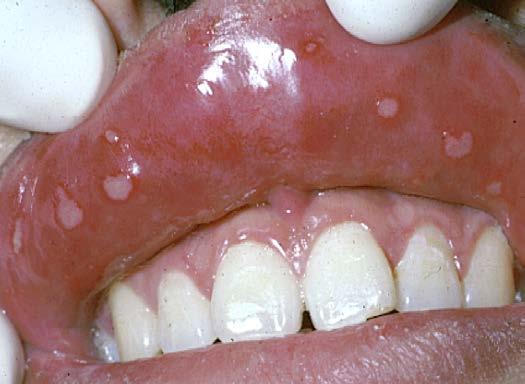
25 Lichen Planus Reticular, Erosive, Hyperplastic The Hyperplastic or Hypertrophic form resembles leukopakia clinically Tongue and Buccal Mucosa There may be marginal or adjacent stria Histology: marked hyperkeratosis, lichenoid mucositis

26 LICHEN PLANUS, Reticular

27 Lichen Planus Histology Keratosis, basal cell damage, thickening of the basement membrane, T lymphocyte infiltrate (chronic interface mucositis)

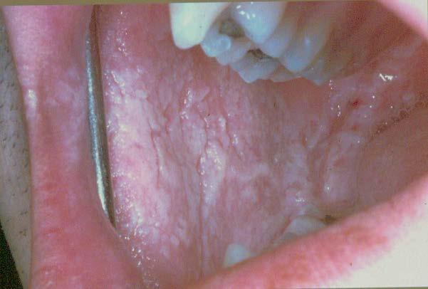
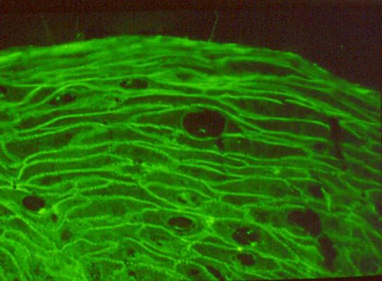
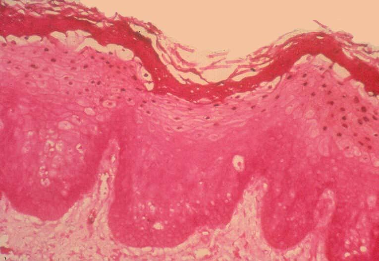
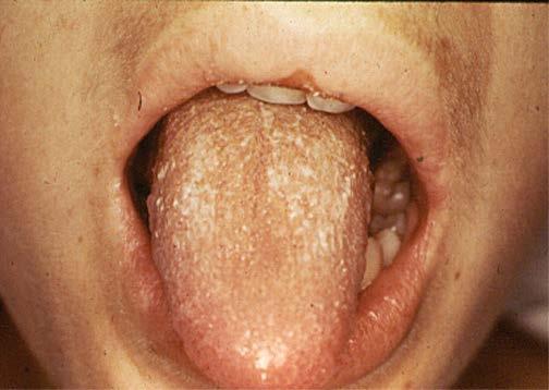
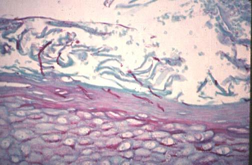
28 LICHEN PLANUS, HYPERPLASTIC


29 EROSIVE LICHEN PLANUS desquamation Anti-fibrinogen
30 Lichen Planus
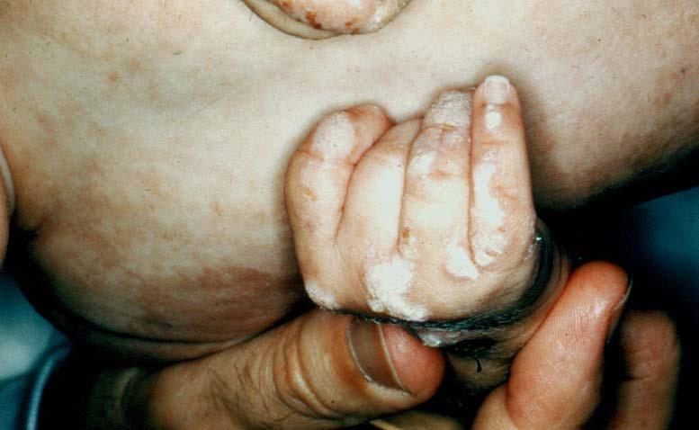
31 IS LICHEN PLANUS PRECANCEROUS? Lichen planus occurs in.5% of the population (1/200) and is therefore common. So CA could be coincidental 1-2% of patients with LP end up with an oral cancer (1/2000) Conclusion: LP is a risk factor for oral cancer in a subpopulation of patients Tumors reported to arise in an LP lesion as well as on nonlesional mucosa among LP patients

32 LICHEN PLANUS, CARCINOMA
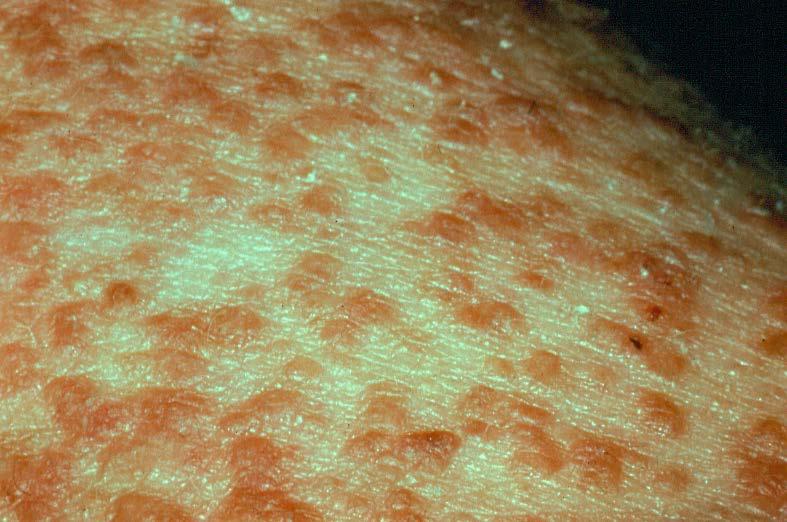
33 DIFFUSE BILATERAL BUCCAL MUCOSAL WHITE LESIONS LEUKOEDEMA WHITE SPONGE NEVUS RARE GENOKERATOSES CHEEK BITE KERATOSIS
34 Leukoedema Normal variation Diffuse sheetlike silky white sheen of buccal mucosa Dark skin populations Whiteness disappears or minimizes when the tissue is stretched It is not precancerous
35 Leukoedema
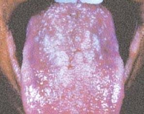
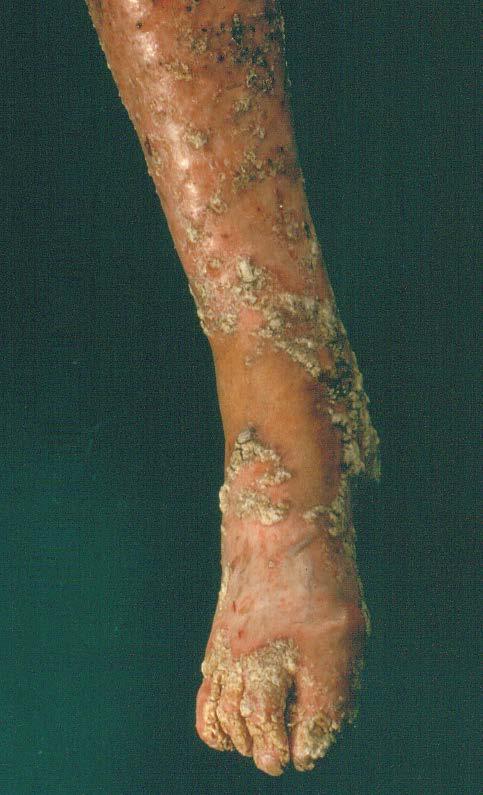
36 WHITE SPONGE NEVUS Thick curtain like folded white lesions, entire buccal mucosa bilaterally Similar lesions involve the genitourinary tract Hereditary: Autosomal dominant Parakeratosis with parakeratin chevrons and individual cell keratinization Tx: none

37 White Sponge Nevus

38 WSN Histology Parakeratosis, individual cell keratinization
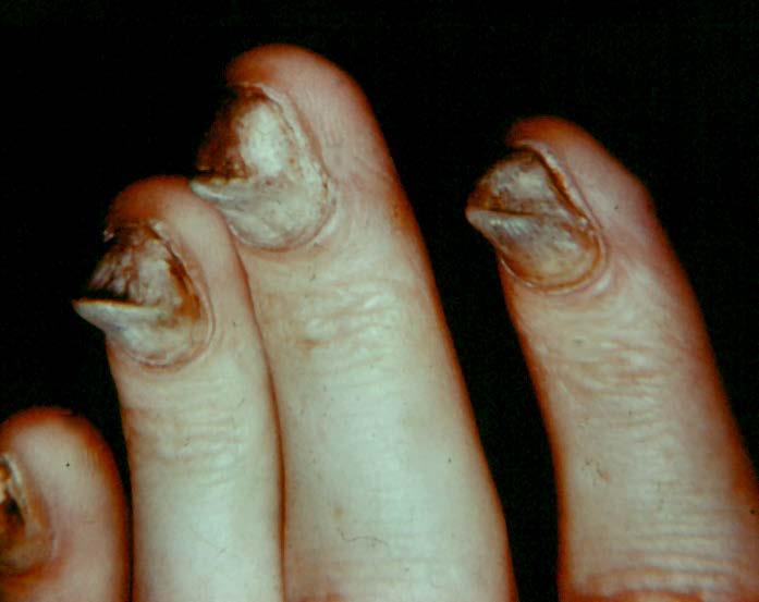
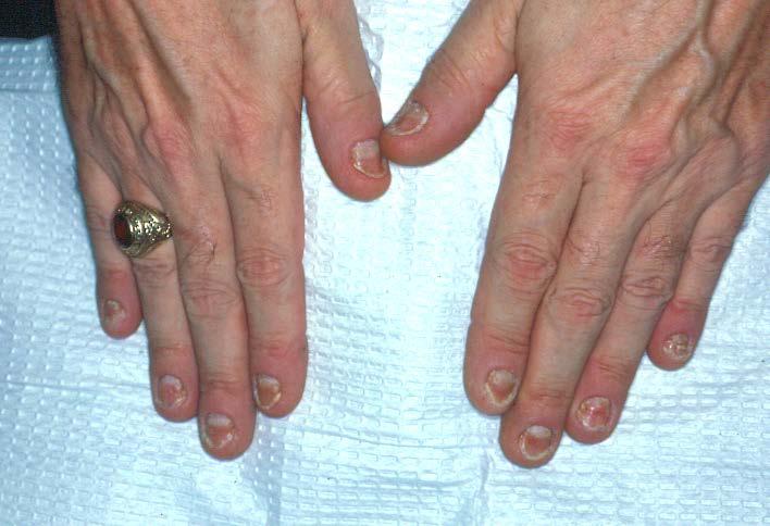
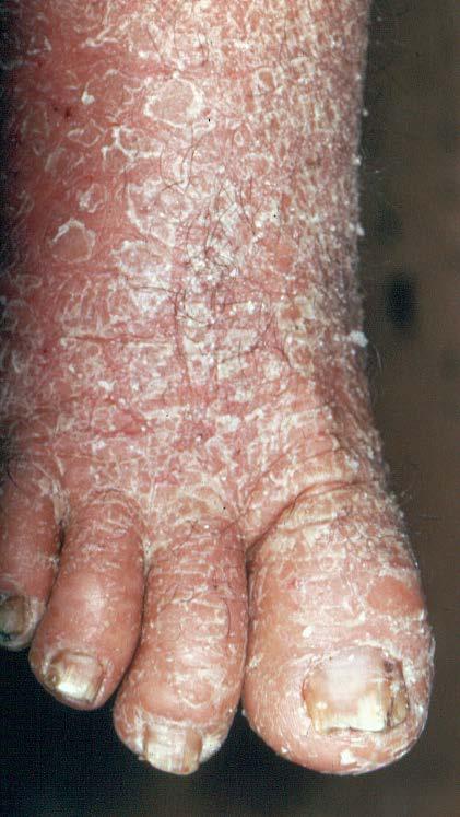
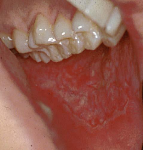
39 Pachonychia Congenita Patchy diffuse white lesions of buccal mucosa bilaterally Lesions may occur on other mucosal areas Massive thickening of toenails and fingernails Hereditary: Autosomal Dominant defect in keratinization Hyperparakeratosis, acanthosis Tx: none
40 Pachonychia Congenita
41 Hereditary Benign Intraepithelial Dyskeratosis (HBID, Red Eye) Hereditary defect in keratinization Racial isolate group in North Carolina Bilateral patchy lesions of buccal mucosa Scleral erythema Seasonal fluctuation in lesional severity Parakeratosis, acanthosis, individual cell keratinization Tx: none
42 Incontinentia Pigmenti Heritable disease Manifestations evolve during infancy Slate grey or brown diffuse cutaneous macular pigmentations Verrucoid crusty cutaneous lesions Oral white lesions
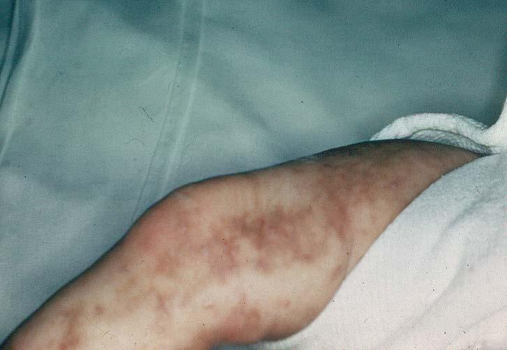

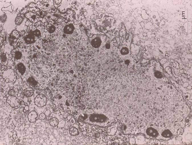
43 Incontinentia Pigmenti
44 Dyskeratosis Congenita Heritable disease Clinical manifestations evolve during infancy Oral white lesions, dysplastic, progress to carcinoma Multiple other anomalies Progression to carcinoma may occur during childhood
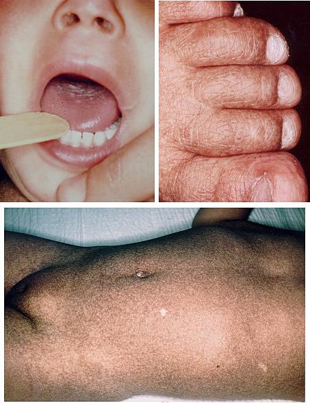
45 Dyskeratosis Congenita
46 LACEY/FRINGE BORDERS LICHEN PLANUS LICHENOID MUCOSITIS LUPUS ERYTHEMATOSUS ORAL HAIRY LEUKOPLAKIA
47 Reticular Lichen Planus Stria of Wickham, lacey, spiderweb, fringe borders Asymptomatic May progress to erosive form over time Adult onset Buccal mucosa, vestibule, gingiva Chronic lymphocytic interface mucositis

48 Reticular Lichen Planus
49 Lupus Erythematosus Females>Males Systemic and cutaneous (discoid) forms Oral lesions usually do not exist in the absence of skin lesions (classic discoid lesions and butterfly rash) White, lacey, finge borders Hyperkeratosis, acanthosis, chronic lymphocytic interface mucositis DIF: basement membrane IgM Serologic: ANA, anti-dna antibodies Tx: topical and systemic steroids
50 LUPUS ERYTHEMATOSUS
51 Oral Hairy Leukoplakia Epstein Barr virus etiology EBV receptors present on oropharyngeal keratinocytes HIV infected patients, appears when CD4 count is less than 500 Males Lateral tongue lesions with fringe (hairy) borders Often with superimposed Candida Tx: Acyclovir related drugs

52 ORAL HAIRY LEUKOPLAKIA

53 Candida ORAL HAIRY LEUKOPLAKIA EBV receptors EBV virions EBV DNA
54 VERRUCOUS/CORRUGATED/RIPPLED VERRUCIFORM BENIGN KERATOSIS RETROMOLAR FRICTIONAL KERATOSIS IDIOPATHIC VERRUCIFORM XANTHOMA SMOKELESS TOBACCO KERATOSIS FLAT WARTS (SESSILE PAPILLOMA) PROLIFERATIVE VERRUCOUS LEUKOPLAKIA ATYPICAL VERRUCOUS HYPERPLASIA VERRUCOUS CARCINOMA EXOPHYTIC, PAPILLARY SQUAMOUS CELL CARCINOMA
55 PROLIFERATIVE VERRUCOUS LEUKOPLAKIA A clinical term that defines a group of histologically distinct diagnoses ranging from atypical verrucous hyperplasia to verrucous carcinoma to invasive squamous cell carcinoma Thick white lesions with a rough, cauliflower, verrucous surface Females > Males Typically over age 50 < 50% with a history of tobacco use Gingival/Sulcus predilection Progressive lateral proliferation, increasing the area of involvement High recurrence after excision HPV association Tx: wide local excision with margin assessment, three month followup
56 PROLIFERATIVE VERRUCOUS LEUKOPLAKIA (PVL)

57 VERRUCOUS CARCINOMA

58 PVL HISTOLOGY ATYPICAL VERRUCOUS HYPERPLASIA VERRUCOUS CARCINOMA

59 VERRUCOUS CARCINOMA
60 Smokeless Tobacco Keratosis White lesions in mandibular vestibule Contact lesion: occurs at site of tobacco or snuff placement Subtle to overt white appearance with a wrinkled, corrugated appearance Reversible if habit is curtailed Gingival recession Cancer progression is extremely low

61 SMOKELESS TOBACCO KERATOSIS


62 Flat Warts Inked to show excisional margins
63 Verruciform Xanthoma White to yellow plaque with a rough surface Usually on the gingiva, yet can occur anywhere Skin counterpart is xanthalasma of the eyelids Benign keratosis with elongated rete ridges between which are found submucosal papilla with foam xanthoma cell infiltration Not known to be associated with hyperlipoproteinemia Tx: local excision
64 VERRUCIFORM XANTHOMA
65 MIXED RED AND WHITE LESIONS BENIGN MIGRATORY GLOSSITIS IRRITATIONAL KERATOSIS CANDIDIASIS LEUKOERYTHROPLAKIA (SPECKLED LEUKOPLAKIA) EROSIVE LICHEN PLANUS
66 Benign Migratory Glossitis Geographic tongue Circinate white lesions with a red denuded surface (depapillation) Lesions spontaneous resolve then reappear at other tongue sites Primarily on dorsum, yet can have lesions on ventral aspect or even lips and buccal mucosa (Erythema migrans) Parakeratosis with subacute mucositis Tx: none, brush tongue

67 Benign Migratory Glossitis
68 Candidiasis Infection with various Candida species Usually asymptomatic yet can experience mild burning Red appeance with white speckling White lesions can be rubbed away Denture sore mouth of the palate, angular cheilitis, other mucosal sites Smear positive for PAS mycelia Tx: topical or systemic specific antifungals
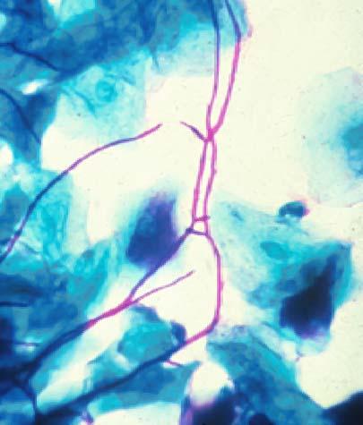
69 Candidiasis HIV PAS Post antibiotic Diabetes
70 Leukoerythroplakia Speckled leukoplakia Mixed red and white foci in which the white component cannot be rubbed away Soft palate, ventral tongue, floor of mouth Toluidine blue positivity in red areas: suspect dysplasia, biopsy should include a red focus Over 60% show dysplasia or carcinoma Wide excision evaluation of margins
71 LEUKOERYTHROPLAKIA
72 EROSIVE LICHEN PLANUS White stria often seen White lesions overlayed on an erythematous backgroung Desquation without bulla formation Painful, burning aggrevated by acidic foods Emotional upset, stress Tx: Topical and systemic steroids

73 EROSIVE LICHEN PLANUS

74 Erosive Lichen Planus Sub-basilar desquamation, Lymphocytic infiltration Direct Immunofluorescence positive For basement membrane fibrinogen
75 PAPULAR CANDIDIASIS DARIER WHITE DISEASE LICHEN PLANUS
76 CANDIDIASIS
77 CANDIDIASIS PAS STAINING MYCELIA
78 Keratosis Follicularis (Darier-White Disease) Autosomal dominant defect in keratinization Multiple orange papules and verrucoid keratoses of the skin Multiple white lesions, bilateral buccal mucosa and other mucosal sites Same histology as warty dyskeratoma with villous rete pegs, individual cell keratinization and suprabasilar clefting Tx: Vitamin A and retinoids
79 Keratosis Follicularis
80 PSEUDOMEMBRANOUS CANDIDIASIS ASPIRIN BURN BULLOUS DISEASES ULCERATIVE DISEASES VESICULAR DISEASES

81 ASPIRIN BURN
82 PSEUDOMEMBRANOUS VESICULO-BULLOUS PEMPHIGUS APHTHOUS STOMATITIS PYOSTOMATITIS VEGETANS PRIMARY HERPES
NEOPLASMS OF THE SURFACE EPITHELIUM (KERATINOCYTES)
") NEOPLASMS OF THE SURFACE EPITHELIUM (KERATINOCYTES) Papillary Lesions Precancerous Lesions Keratinocyte Proliferations Carcinomas Melanotic Lesions Melanomas Normal Mucosa Keratin layer Spinous layer Basal
NEOPLASMS OF THE SURFACE EPITHELIUM (KERATINOCYTES) Papillary Lesions Precancerous Lesions Keratinocyte Proliferations Carcinomas Melanotic Lesions Melanomas Normal Mucosa Keratin layer Spinous layer Basal
الطلاوة = Leukoplakia LEUKOPLAKIA
 LEUKOPLAKIA Leukoplakia is a clinical term that refers to a predominantly white lesion of the oral mucosa that cannot be rubbed off or characterized by any other definable lesion or known disease. 130
04/09/2018. Squamous Cell Neoplasia and Precursor Lesions. Agenda. Squamous Dysplasia. Squamo-proliferative lesions. Architectural features
 Squamous Cell Neoplasia and Precursor Lesions Jennifer L. Hunt, MD, MEd Aubrey J. Hough Jr, MD, Endowed Professor of Pathology Chair of Pathology and Laboratory Medicine University of Arkansas for Medical
Lesions & Lifestyles
 Lesions & Lifestyles attended a 3 hour Continuing Education Seminar on Oral Pathology presented by Nancy Dewhirst, RDH,BS on (date) at (location):. Course material is directly related patient care. Notes:
LEUKOPLAKIA Definition Epidemiology Clinical presentation
 LEUKOPLAKIA Definition Leukoplakia is the most common premalignant or "potentially malignant" lesion of the oral mucosa. Leukoplakia is a predominantly white lesion of the oral mucosa than cannot be clinicopathologically
INFLAMMATORY DISEASES PART I. Immunopathology Part I
 INFLAMMATORY DISEASES PART I Immunopathology Part I Nonspecific & T Cell Mediated Mucosal Inflammatory Lesions Nonspecific and Idiopathic Mucositis Hypersensitivity and Autoimmune T cell mediated Immunoglobulin
Squamous Cell Neoplasia and Precursor Lesions
 Squamous Cell Neoplasia and Precursor Lesions Jennifer L. Hunt, MD, MEd Aubrey J. Hough Jr, MD, Endowed Professor of Pathology Chair of Pathology and Laboratory Medicine University of Arkansas for Medical
Premalignant lesions may expose to a promoting. factor & may be induced to undergo malignant. Carcinoma in situ displays the cytologic features of
 بسم رلاهللا Def. Premalignant lesions may expose to a promoting factor & may be induced to undergo malignant transformation. Carcinoma in situ displays the cytologic features of malignancy without invasion
LESIONS OF THE ORAL CAVITY ORAL CAVITY. Oral Cavity Subsites 4/10/2013 LIPS TEETH GINGIVA ORAL MUCOUS MEMBRANES PALATE TONGUE ORAL LYMPHOID TISSUES
 LESIONS OF THE ORAL CAVITY David I. Kutler, MD, FACS Associate Professor Division of Head and Neck Surgery Department of Otolaryngology HNS Weill Cornell Medical Center ORAL CAVITY LIPS TEETH GINGIVA ORAL
Autoimmune Diseases with Oral Manifestations
 Autoimmune Diseases with Oral Manifestations Martin S. Greenberg DDS, FDS RCSEd Professor Emeritus Department of Oral Medicine University of Pennsylvania Disclosure Statement I have no actual or potential
Oral cavity cancer accounts for approximately 3% of all malignancies and is a significant worldwide health problem.
 Oral cavity cancer accounts for approximately 3% of all malignancies and is a significant worldwide health problem. Majority are SCC ( 5-year survival rate only about 50-60% ) Many SCC arrive from premalignant
That. Name QUIZ. 60 SEPTEMBER 2017 // dentaltown.com
 QUIZ Name That General dentists are first in the line of practitioners that patients see for an oral lesion evaluation; therefore, a sound understanding of oral mucosal diseases and their clinical presentation
Oral Health & HIV. Professor Sudeshni Naidoo Department of Community Dentistry University of the Western Cape
 Oral Health & HIV Professor Sudeshni Naidoo Department of Community Dentistry University of the Western Cape Importance & relevance of Oral HIV Lesions >70% of HIV+ve patients present with oral manifestations
Diagnostic difficulties with lesions of the oral mucosa
 BDIAP London, November 2010 School of Clinical Dentistry University of Sheffield Diagnostic difficulties with lesions of the oral mucosa Paul M Speight Dept Oral & Maxillofacial Pathology University of
IN THE NAME OF GOD. Dr.kheirandish DDS,MSC Oral and maxillofacial pathology
 IN THE NAME OF GOD Dr.kheirandish DDS,MSC Oral and maxillofacial pathology Dermatologic Diseases Chapter 16 ECTODERMAL DYSPLASIA o Two or more ectodermally derived anatomic structures fail to develop o
TANYA A. WRIGHT, DDS OBJECTIVES
 TANYA A. WRIGHT, DDS OBJECTIVES One will be able to recognize pathological entities One will be able to establish a reasonable differential diagnosis One will be able to identify various types of lesions
Contents. 3 Diagnostic Tests and Studies Introduction Examination... 27
 Contents 1 Normal Anatomy... 1 1.1 Introduction... 1 1.2 Surface Landmarks... 1 1.3 Oral Mucosa... 3 1.4 Tongue... 5 1.5 Floor of Mouth... 6 1.6 Palate... 6 1.7 Dentition... 7 1.8 Temporomandibular Joint...
White Oral Lesions: How to Distinguish the Benign From the Deadly
 ABSTRACT: Chronic irritation from smoking is the most common cause of white mucosal lesions. Because benign leukoplakic growths are virtually impossible to distinguish from carcinoma, biopsy is essential.
Oral Manifestations of Dermatologic Disease: A Focus on Lichenoid Lesions. Proceedings of the NASHNP Companion Meeting, March, 2011, San Antonio, TX
 1 Oral Manifestations of Dermatologic Disease: A Focus on Lichenoid Lesions Proceedings of the NASHNP Companion Meeting, March, 2011, San Antonio, TX Susan Müller, DMD, MS Professor Department of Pathology
Actinic keratosis (AK): Dr Sarma s simple guide
: Dr Sarma s simple guide") Actinic keratosis (AK): Dr Sarma s simple guide Actinic keratosis is a very common lesion that you will see in your day-to-day practice. First, let me explain the name Actinic keratosis. It means keratosis
Actinic keratosis (AK): Dr Sarma s simple guide Actinic keratosis is a very common lesion that you will see in your day-to-day practice. First, let me explain the name Actinic keratosis. It means keratosis
Oral Cancer and Common Oral Lesions seen in HIV Seropositive Patients. Gwen Cohen Brown DDS, FAAOMP Professor New York City College of Technology
 Oral Cancer and Common Oral Lesions seen in HIV Seropositive Patients Gwen Cohen Brown DDS, FAAOMP Professor New York City College of Technology Program Objectives Recognize the oral health needs of the
Role of the Dental Hygienist in Oral Pathology. Role of the Dental Hygienist in Oral Pathology. Cancers of the Oral Cavity.
 Gum Gardeners Study Club April 25, 2016 Early Detection of Oral Cancer Cindy Kleinegger, DDS, MS NW Oral Pathology Tigard, OR nworalpathology.com Role of the Dental Hygienist in Oral Pathology Work closely
Contents. 1 Normal Anatomy Introduction... 17
 Contents 1 Normal Anatomy... 1 Introduction... 1 Surface Landmarks... 1 Oral Mucosa... 1 Tongue... 4 Floor of Mouth... 6 Palate... 7 Dentition... 7 Temporomandibular Joint... 9 Innervation... 10 Jaws and
Oral Medicine. Dr. Qianming Ian CHEN
 Oral Medicine Dr. Qianming Ian CHEN ORAL MEDICINE Oral medicine is the specialty of dentistry that is concerned with the oral health care of medically compromised patients and with the diagnosis and nonsurgical
Oral Medicine Update for the dental practitioner Oral white patches
 IN BRIEF Most white lesions in the mouth are inconsequential and caused by friction or trauma. However, cancer and some systemic diseases such as lichen planus and candidosis may present in this way. Biopsy
WHITE LESIONS OF THE ORAL CAVITY - diagnostic appraisal & management strategies
 WHITE LESIONS OF THE ORAL CAVITY - diagnostic appraisal & management strategies * Joshy V.R ** Hari.S * Reader, Dept of Oral Pathology, Yenepoya Dental College, Yenepoya University, Mangalore 575 018.
The Oral Cavity. Image source:
 The Oral Cavity Anatomy Image source: http://anatomyforlayla.blogspot.co.za/2007/04/blog-post.html The major structures of the oral cavity are the lips, the teeth, the alveolar ridges (bony areas that
Dysplasia, Mimics and Other Controversies
 Dysplasia, Mimics and Other Controversies Mary S. Richardson, MD Dept. of Pathology Medical University of South Carolina Charleston, SC Notice of Faculty Disclosure In accordance with ACGME guidelines,
DENIS P. LYNCH, DDS, PHD
 140 TH ANNUAL MEETING MAY 6 MAY 7, 2010 JEWEL OF THE GREAT LAKES DENIS P. LYNCH, DDS, PHD FRIDAY, MAY 7, 2010 9:00 A.M. TO 12:00 NOON ORAL CANCER AND RELATED PREMALIGNANCY Oral Cancer and Premalignancy
Oral Epithelial Tumors, Melanocytic Nevi, and Melanoma (I)
") Introduction: Oral Epithelial Tumors, Melanocytic Nevi, and Melanoma (I) Oral Epithelial Tumors may be: Benign tumors Sequamous cell Papilloma Malignant tumors Sequamous cell carcinoma, Basal cell carcinoma
Introduction: Oral Epithelial Tumors, Melanocytic Nevi, and Melanoma (I) Oral Epithelial Tumors may be: Benign tumors Sequamous cell Papilloma Malignant tumors Sequamous cell carcinoma, Basal cell carcinoma
Red and White Tissue Reactions: A white appearance of the oral mucosa may be caused by: An increased production of keratin (hyperkeratosis).
.") Burket, chapter 4 Red and White Tissue Reactions: A white appearance of the oral mucosa may be caused by: An increased production of keratin (hyperkeratosis). An abnormal but benign thickening o stratum
Burket, chapter 4 Red and White Tissue Reactions: A white appearance of the oral mucosa may be caused by: An increased production of keratin (hyperkeratosis). An abnormal but benign thickening o stratum
Benign and malignant epithelial lesions: Seborrheic keratosis: A common benign pigmented epidermal tumor occur in middle-aged or older persons more
 Benign and malignant epithelial lesions: Seborrheic keratosis: A common benign pigmented epidermal tumor occur in middle-aged or older persons more common on the trunk; but extremities, head and neck are
Index. Dent Clin N Am 49 (2005) Note: Page numbers of article titles are in boldface type.
 Note: Page numbers of article titles are in boldface type.") Dent Clin N Am 49 (2005) 273 278 Index Note: Page numbers of article titles are in boldface type. A Acanthosis nigricans, familial, 251 Amalgam tattoo, 197 198 Amphotericin B, 62 Ankyloglossia, 11 Anti-inflammatory
Dent Clin N Am 49 (2005) 273 278 Index Note: Page numbers of article titles are in boldface type. A Acanthosis nigricans, familial, 251 Amalgam tattoo, 197 198 Amphotericin B, 62 Ankyloglossia, 11 Anti-inflammatory
A clinical diagnosis of oral leukoplakia; A guide for dentists
 Journal section: Oral Medicine and Pathology Publication Types: Review doi:10.4317/medoral.22292 http://dx.doi.org/doi:10.4317/medoral.22292 ; A guide for dentists Vinicius C. Carrard 1, Isaäc van der
Diseases of oral cavity
 Diseases of oral cavity Diseases of Teeth and Supporting Structures Inflammatory/Reactive Lesions Infections Oral Manifestations of Systemic Disease Precancerous and Cancerous Lesions Odontogenic Cysts
Pathology of the skin. 2nd Department of Pathology, Semmelweis University
 Pathology of the skin 2nd Department of Pathology, Semmelweis University Histology of the skin Epidermis: Stratum corneum Stratum granulosum Stratum spinosum Stratum basale Dermis: papillary and reticular
Review Article- Leukoplakia: A mysterious white patch.
 International Journal Of Scientific Research And Education Volume 2 Issue 9 Pages 1824-1830 September-2014 ISSN (e): 2321-7545 Website: http://ijsae.in Review Article- Leukoplakia: A mysterious white patch.
Oral Leukoplakia: An Insight
 Oral Leukoplakia: An Insight Gigi Roy 1, Anu Vijayan 2, Shamji Shajahan 3, Anuja S 4, Rashmi Elizabeth Mathen 5 1,3,4,5-Post Graduate, Department of Oral Medicine and Radiology, Mar Baselios Dental College,
Morsicatio Mucosae Oris A Chronic Oral Frictional Keratosis, Not a Leukoplakia
 J Oral Maxillofac Surg 67:140-146, 2009 Morsicatio Mucosae Oris A Chronic Oral Frictional Keratosis, Not a Leukoplakia Sook-Bin Woo, DMD,* and Dorothy Lin Purpose: Morsicatio mucosae oris (MMO) presents
Differential Diagnosis of Oral Lesions. An Interactive Lecture Using Audience Response Polling. John L. Alonge, MS, DDS
 Differential Diagnosis of Oral Lesions An Interactive Lecture Using Audience Response Polling John L. Alonge, MS, DDS Goals 1. Review the diagnostic process needed to formulate a differential diagnosis
REF: Chap 1 (Pemphigus vulgaris/etiology and
 Chapter 1: Vesiculobullous Diseases Test Bank MULTIPLE CHOICE 1. Intercellular deposits of IgG are consistently found in oral epithelium in which of the following? a. Cicatricial pemphigoid b. Lichen planus
ANS: C REF: Chap 1 (Pemphigus vulgaris/etiology and pathogenesis), p 11
, p 11") Chapter 1: Vesiculobullous Diseases Test Bank MULTIPLE CHOICE 1. Intercellular deposits of IgG are consistently found in oral epithelium in which of the following? a. Cicatricial pemphigoid b. Lichen planus
Chapter 1: Vesiculobullous Diseases Test Bank MULTIPLE CHOICE 1. Intercellular deposits of IgG are consistently found in oral epithelium in which of the following? a. Cicatricial pemphigoid b. Lichen planus
Dermatopathology: The tumor is composed of keratinocytes which show atypia, increase mitoses and abnormal mitoses.
 Squamous cell carcinoma (SCC): A common malignant tumor of keratinocytes arising in the epidermis, usually from a precancerous condition: 1- UV induced actinic keratosis, usually of low grade malignancy.
Premalignant lesion is a morphologically altered tissue in which cancer is more likely to occur, than its apparently normal counter parts.
 Oral Premalignancy Premalignant lesion is a morphologically altered tissue in which cancer is more likely to occur, than its apparently normal counter parts. Premalignant condition is a generalized state
Squamous papilloma Squamous acanthoma Keratoacanthoma Verruca vulgaris Condyloma acuminatum Focal epithelial hyperplasia Sino nasal papilloma
 Benign tumors Epithelial origin Squamous papilloma Squamous acanthoma Keratoacanthoma Verruca vulgaris Condyloma acuminatum Focal epithelial hyperplasia Sino nasal papilloma Squamous papilloma Exophytic
Diagnostic sieve. Looking Beyond the Vermillion Border. Time bombs for medical GPs! Normal oral mucosa
 Sat 12 June 2010 Millennium WS 28 + 38 2.00-2.55; 3.05-4.00 PM Looking Beyond the Vermillion Border Laurence J. Walsh BDSc, PhD, DDSc, FFOP(RCPA), GCEd, FICD, FPFA, FADI, FIADFE The University of Queensland
Benign versus Cancerous Lesions How to tell the difference FMF 2014 Christie Freeman MD, CCFP, DipPDerm, MSc
 1 Benign versus Cancerous Lesions How to tell the difference FMF 2014 Christie Freeman MD, CCFP, DipPDerm, MSc Benign lesions Seborrheic Keratoses: Warty, stuck-on Genetics and birthdays Can start in late
Pigmented lesions of the Oral cavity
 Oral medicine أ.م.د احسان عبد هللا كميل Pigmented lesions of the Oral cavity Pigmented oral lesions are a large group of disorders in which the dark or brown color is the essential clinical characteristic.
Chapter 2 Variants of Normal and Common Benign Conditions
 Chapter 2 Variants of Normal and Common Benign Conditions Summary Fundamental to diagnosing oral pathologic conditions is the ability to recognize the spectrum of clinical findings that represents variation
Leukoplakia is a white patch on the oral mucous membrane, which is undeliable and can not diagnose neither clinically nor pathologically as an other
 Leukoplakia Leukoplakia is a white patch on the oral mucous membrane, which is undeliable and can not diagnose neither clinically nor pathologically as an other disease. (Pindborg. 1978) Precancerous lesion
A five year study on differential diagnosis of verruciform penile lesions
 Original Research Article A five year study on differential diagnosis of verruciform penile lesions S. Sujatha 1, V. Srinivas Kumar 2*, K. Durga 3 1 Associate Professor, 2 Assistant Professor, 3 Professor
Classification: 1. Infective: 2. Traumatic: 3. Idiopathic: Recurrent Aphthous Stomatitis (RAS) 4. Associated with systemic disease:
 4. Associated with systemic disease:") Classification: 1. Infective: 2. Traumatic: 3. Idiopathic: Recurrent Aphthous Stomatitis (RAS) 4. Associated with systemic disease: Hematological GIT Behcet s HIV 5. Associated with dermatological diseases:
Classification: 1. Infective: 2. Traumatic: 3. Idiopathic: Recurrent Aphthous Stomatitis (RAS) 4. Associated with systemic disease: Hematological GIT Behcet s HIV 5. Associated with dermatological diseases:
WOMEN'S INTERAGENCY HIV STUDY ORAL PROTOCOL FORM OP 4: ORAL MUCOSAL TISSUE EXAM
 WOMEN'S INTERAGENCY HIV STUDY ORAL PROTOCOL FORM OP 4: ORAL MUCOSAL TISSUE EXAM COMPLETING THE FORM GENERAL INFORMATION Affix the Participant ID label in the space indicated. Record the visit number. Be
Differential Diagnosis of Oral Ulcerations
 Differential Diagnosis of Oral Ulcerations Dr. Nagamani Narayana Department of Oral Biology University of Nebraska Medical Center College of Dentistry Objectives Differential diagnosis of oral ulcerations
Linear Leukoplakia on the Right Lateral Border of the Tongue
 Continuing Education Brought to you by Linear Leukoplakia on the Right Lateral Border of the Tongue Course Author(s): Anne Cale Jones, DDS; H. Stan McGuff, DDS; Michaell A. Huber, DDS Online Case: www.dentalcare.com/en-us/professional-education/case-challenges/case-challenge-058
Dr Rodney Itaki Lecturer Division of Pathology Anatomical Pathology Discipline
 Oral Lesions & Oral Cancer Dr Rodney Itaki Lecturer Division of Pathology Anatomical Pathology Discipline University of Papua New Guinea School of Medicine & Health Sciences Division of Pathology Overview
Epidemiological and clinicopathological study of oral leukoplakia
 Study Epidemiological and clinicopathological study of oral Minati Mishra, Janardan Mohanty*, Sujata Sengupta, Satyabrata Tripathy Department of Dermatology & Venereology, S.C.B. Medical College, *Department
Premalignant skin tumours
 Chapter 14: Premalignant skin tumours page: 434 Premalignant skin tumours page: 435 Solar keratoses (senile keratoses) Raised red and well-defined plaques with a rough surface covered in scales of varying
Basal cell carcinoma 5/28/2011
 Goal of this Presentation A practical approach to the diagnosis of cutaneous carcinomas and their mimics Thaddeus Mully, MD University of California San Francisco To review common non-melanoma skin cancers
Orofacial Disease: Update For The Dental Clinical Team: 3. White Lesions
 ORAL MEDICINE Orofacial Disease: Update For The Dental Clinical Team: 3. White Lesions Crispian Scully and Stephen Porter Abstract: White lesions usually contain an increased amount of keratin. Some are
Stomatitis.
 Stomatitis http://www.entusa.com/oral_photographs/20080102-stomatitis-palate_small.jpg Oral inflammation and ulcers, known as stomatitis, may be mild and localized or severe and widespread. They are invariably
a. viral INFECTIONS OF ORAL MUCOSA HERPETIC STOMATITIS HERPETIC STOMATITIS
 a. viral INFECTIONS OF ORAL MUCOSA laboratory confirmation rather long diagnosis based mainly on clinical features basic diagnostic methods: (1) culture of viral particles; (2) morphologic changes and
Oral Cancer and Precancerous Lesions Brad W. Neville and Terry A. Day. DOI: /canjclin This information is current as of June 15, 2011
 Oral Cancer and Precancerous Lesions Brad W. Neville and Terry A. Day CA Cancer J Clin 2002;52;195-215 DOI: 10.3322/canjclin.52.4.195 This information is current as of June 15, 2011 The online version
Chapter 6 Squamous Cell Carcinoma: Variants and Challenges
 Chapter 6 Squamous Cell Carcinoma: Variants and Challenges Michael B. Morgan EPIDEMIOLOGY: Second most common skin cancer, rare in the dark-skinned races. ETIOLOGY: Ultraviolet light, HPV infection. PATHOGENESIS:
ORAL LEUKOPLAKIA IN A SOUTH AFRICAN SAMPLE: A CLINICOPATHOLOGICAL STUDY
 ORAL LEUKOPLAKIA IN A SOUTH AFRICAN SAMPLE: A CLINICOPATHOLOGICAL STUDY Rakesh Chandran A research report submitted to the Faculty of Health Sciences, University of Witwatersrand, Johannesburg, in partial
White and Red Lesions of the Oral Mucosa
 White and Red Lesions of the Oral Mucosa Maryam Jessri, Hani Mawardi, Camile S. Farah, and Sook-Bin Woo Abstract There are several conditions that can present as white or red macular, papular, and/or plaquelike
Original Article- A CYTOLOGICAL STUDY OF LEUKOPLASTIC LESIONS IN ORAL CAVITY
 Original Article- A CYTOLOGICAL STUDY OF LEUKOPLASTIC LESIONS IN ORAL CAVITY I. GUJRAL*, P. SINGH**, S. SHARMA***, N. GANGANE*** ABSTRACT Oral white lesions that cannot be clinically or pathologically
Proliferative Verrucous Leukoplakia of the Gingiva, Report of two Cases with Malignant Transformation
 Journal of Clinical and Anatomic Pathology Case Report Open Access Proliferative Verrucous Leukoplakia of the Gingiva, Report of two Cases with Malignant Transformation Nadereh Ghanee DMD, Selene Saraf
PACIFIC JOURNAL OF MEDICAL SCIENCES {Formerly: Medical Sciences Bulletin} ISSN:
 PACIFIC JOURNAL OF MEDICAL SCIENCES {Formerly: Medical Sciences Bulletin} ISSN: 2072 1625 Pac. J. Med. Sci. (PJMS) www.pacjmedsci.com. Email: pacjmedsci@gmail.com. EROSIVE LICHEN PLANUS A CASE REPORT *Prathima
Tissue Conditions and the
 Management of Oral Soft Tissue Conditions and the Use of Medications 29 August 2017 Mike Brennan DDS, MHS Department of Oral Medicine Carolinas Medical Center Charlotte, NC Objectives Differential diagnosis
Benign Oral cavity lesions. Mohammed ALESSA MBBS,FRCSC Assistant Professor Consultant Otolaryngology, Head & Neck Surgery
 Benign Oral cavity lesions Mohammed ALESSA MBBS,FRCSC Assistant Professor Consultant Otolaryngology, Head & Neck Surgery Anatomy Histology Physiology Pathology Clinical cases Introduction The oral cavity
Vascular. Extravasated blood. Melanocytic. Tattoo. Epidermolysis bullosa. Lichen planus. Pemphigoid Pemphigus Lupus. Candidosis. Surface Epithelial
 Oral Soft Tissue Pathology Epithelial Thickening (white) Combination Erythema migrans Epithelial atrophy (red) Surface Lesions Clinical Impression Enlargements Surface Debris Pigmented Vesicular Ulcerated
Papillary and verrucous lesions of the oral mucosa
 Papillary and verrucous lesions of the oral mucosa Gareth J Thomas A William Barrett Abstract A variety of verrucous and papillary lesions affect the oral mucosa. Those which are benign and reactive, for
MUCOCUTANEOUS LESIONS Normal structures in epithelium cell adhesion to each other and to underlying connective tissue:
 ORAL DERMATOSES AND MUCOSAL/GINGIVAL LESIONS MUCOCUTANEOUS LESIONS Normal structures in epithelium cell adhesion to each other and to underlying connective tissue: Diagram taken from: Oral and Maxillofacial
Clinically Microscopically Pathogenesis: autoimmune not lifetime
 Vulvar Diseases: Can be divided to non-neoplastic and neoplastic diseases. The neoplastic diseases are much less common. Of those, squamous cell carcinoma is the most common. most common in postmenopausal
UNC Cancer Network Lecture
 UNC Cancer Network Lecture Oral Cancer: A Comprehensive Overview Samip N. Patel, MD, FACS Trevor Hackman, MD, FACS Ricardo Padilla, DDS Definition of Oral Cancer Odontogenic Other Malignancies Sarcomas
From the Cradle to the Grave: Oral pathology through the life span
 From the Cradle to the Grave: Oral pathology through the life span Conditions Exhibited in Infants and Children: Dental Lamina Cysts Etiology: Developmental Clinical appearance: Cystic nodules on alveolar
OROPHYRENGEAL CANCERS
 OROPHYRENGEAL CANCERS INTRODUCTION 2 % 4 % of all malignant Tumors in west Asia India 40% Men ^ Age :Over 60 yrs 90% of all oral cancers results from Tobacco and Alcohol Pan (Betel Leaf,Nut, Lime), Reverse
Papillary verrucous lesion of the oral mucosa: A need for detailed histopathological examination
 Case report DOI: http://dx.doi.org/10.18320/jimd/201502.03163 JOURNAL OF INTERNATIONAL MEDICINE AND DENTISTRY To search..to know...to share p-issn: 2454-8847 e-issn: 2350-045X Papillary verrucous lesion
Finding Dangerous Mucosa
 Finding Dangerous Mucosa 2 Oral Cancer Squamous Cell Carcinoma Salivary Gland Adenocarcinoma Malignant Lymphoma Metastatic Carcinoma Sarcoma 4 Incidence of Cancer in the United States For Oral and Oropharyngeal
Case Report II Sri Lanka Dental Journal 2018; 48(01) 41-45
 41-45") Case Report II Sri Lanka Dental Journal 2018; 48(01) 41-45 White sponge nevus in the oral cavity: case report and P.V.K.S. Hettiarachchi, J.C.M. Jayasinghe, B.S.M.S. Siriwardena, R.D. Jayasinghe Abstract
Case Report II Sri Lanka Dental Journal 2018; 48(01) 41-45 White sponge nevus in the oral cavity: case report and P.V.K.S. Hettiarachchi, J.C.M. Jayasinghe, B.S.M.S. Siriwardena, R.D. Jayasinghe Abstract
A Speckled Lesion. Angela C. Chi, DMD; Michele Carter Ravenel, DMD
 A Speckled Lesion Angela C. Chi, DMD; Michele Carter Ravenel, DMD The following Case Challenge is provided in conjunction with the American Academy of Oral and Maxillofacial Pathology. Case Summary This
2018 Oregon Dental Conference Course Handout Denis Lynch, DDS, PhD
 2018 Oregon Dental Conference Course Handout Denis Lynch, DDS, PhD Course 9121: Infectious Hazards in Dentistry or What You Never Thought You'd Have to Worry about after You Passed National Boards Thursday,
=ﻰﻤاﻤﺤﻠا ﺔﻴﻘﻠﺤﻠا ﺔذﺒاﻨﻠا
 1 / 15 Erythema Annulare Centrifugum and Other Figurate Erythemas The figurate erythemas include a variety of eruptions characterized by annular and polycyclic lesions. Classification of this group has
Smoking Habits Among Patients Diagnosed with Oral Lichen Planus
 TOBACCO INDUCED DISEASES Vol. 2, No. 2: 103-108 (2004) PTID Society Smoking Habits Among Patients Diagnosed with Oral Lichen Planus Meir Gorsky, 1 Joel B. Epstein, 2 Haya Hasson-Kanfi, 1 Eliezer Kaufman
Oral leukoplakia, the ongoing discussion on definition and terminology
 Journal section: Oral Medicine and Pathology Publication Types: Review doi:10.4317/medoral.21007 http://dx.doi.org/doi:10.4317/medoral.21007, the ongoing discussion on definition and terminology Isaäc
Evaluation and Management of Head and Neck Cancer in Patients with Fanconi anemia David I. Kutler, M.D., F.A.C.S.
 Evaluation and Management of Head and Neck Cancer in Patients with Fanconi anemia David I. Kutler, M.D., F.A.C.S. Residency Site Director Weill Cornell Medical Center Associate Professor Division of Head
The relevance of uniform reporting in oral leukoplakia: Definition, certainty factor and staging based on experience with 275 patients
 Journal section: Oral Medicine and Pathology Publication Types: Research doi:10.4317/medoral.18756 http://dx.doi.org/doi:10.4317/medoral.18756 The relevance of uniform reporting in oral leukoplakia: Definition,
Oral Cancer- Improving Early Detection
 Oral Cancer- Improving Early Detection GDC Recommended Subject Aims: To give an overview of the dental team's role in detecting the early signs of oral cancer; to give an overview of the risk factors associated
Benign Lichenoid Keratosis
 Benign Lichenoid Keratosis ALAN F. FRIGY, M.D. AND PHILIP H. COOPER, M.D. The microscopic spectrum of benign lichenoid keratosis (BLK) was studied by examination of 30 examples. BLK consists of a segment
Pattern of oral lesions Cytohistopathological study in tertiary care centre.
 International Journal of Current Research in Medical Sciences ISSN: 2454-5716 P-ISJN: A4372-3064, E -ISJN: A4372-3061 www.ijcrims.com Original Research Article Volume 3, Issue 10-2017 Pattern of oral lesions
HEAD AND NECK PATHOLOGY
 Bosnian-British School of Pathology November 2012 HEAD AND NECK PATHOLOGY Slide seminar: Oral Pathology Preferred Diagnoses Dr A Sandison, Slide seminar: Pathology of the Oral Cavity Page 1 of 5 1. Female
Chapter 5. Oxygenated Hemoglobin Diffuse Reflectance Ratio for In Vivo Detection of oral Pre-cancer
 Chapter 5 Oxygenated Hemoglobin Diffuse Reflectance Ratio for In Vivo Detection of oral Pre-cancer This work is published in: JB0 (SPIE) 13(4):041306 (1-10), 2008 Oxygenated Hemoglobin Diffuse Reflectance
HPV induced Proliferative verrucous Leukoplakia : Case Report
 Case Report: HPV induced Proliferative verrucous Leukoplakia : Case Report Dr Gopal Sharma, Dr Deepa Das, Dr Prachi Naik, Dr Jaya Mukherjee Oral Medicine and Radiology Department, YMT Dental College and
Index. Note: Page numbers of article titles are in boldface type.
 Index Note: Page numbers of article titles are in boldface type. A Actinic cheilitis, 123 124 Addison disease, 175 Adenopathy, due to Myobacterium tuberculosis, 6 from histoplasmosis abcess, 6 Alveolar
VIRUS. Viral infection causing, or associated with diseases of the oral mucosa : Herpes Simpleks 1 & 2
 VIRUS Viral infection causing, or associated with diseases of the oral mucosa : VIRUS Herpes Simpleks 1 & 2 Varicella - Zoster Coxsakie A PENYAKIT Primary Gingivostomatitis Herpetica Herpes Labialis Recurrent
LENTIGO SIMPLEX. Epidemiology
 LENTIGO SIMPLEX Epidemiology The frequency of lentigo simplex in children and adults has not been determined. There does not appear to be a racial or gender predilection. Lentigo simplex is the most common
Allergic contact stomatitis is a rare disorder,
 Allergic Contact Stomatitis: A Case Report and Review of Literature P Lokesh, T Rooban, Joshua Elizabeth, K Umadevi, K Ranganathan Abstract Allergic contact stomatitis is a well-recognized entity, which
CASE REPORT PLAQUE TYPE ORAL VERRUCOUS HYPERPLASIA AND IRRITATIONAL FIBROMA: A REPORT OF CONJOINT OCCURRENCE
 CASE REPORT PLAQUE TYPE ORAL VERRUCOUS HYPERPLASIA AND IRRITATIONAL FIBROMA: A REPORT OF CONJOINT OCCURRENCE Alphy Alphonsa Sebastian, Hasan Subhi 1. Phd student, Department of Oral Medicine and Oral Pathology,
4/16/2018. Bumps & Lumps. What s HPV Got To Do With It? 2 hour Oral & Pharyngeal Pathology Review Nancy Dewhirst RDH,BS
 4/6/08 Bumps & Lumps. What s HPV Got To Do With It? hour Oral & Pharyngeal Pathology Review Nancy Dewhirst RDH,BS www.nancydewhirst.com Bumps & Lumps What s HPV Got To Do With It? Patient Assessment Clinical