The impact of GP sub-specialisation and dermatoscopy use on diagnostic accuracy for melanomas in Australia
|
|
|
- Myles Todd
- 5 years ago
- Views:
Transcription
1 The impact of GP sub-specialisation and dermatoscopy use on diagnostic accuracy for melanomas in Australia Cliff Rosendahl, Gail Williams, Diann Eley, Tobias Wilson, Greg Canning, Jeffrey Keir, Ian McColl, David Wilkinson. J Am Acad Dermatol /j.jaad

2 How good are we at finding melanomas?
3
4
5
6
7
8 Does it matter?
9
10
11

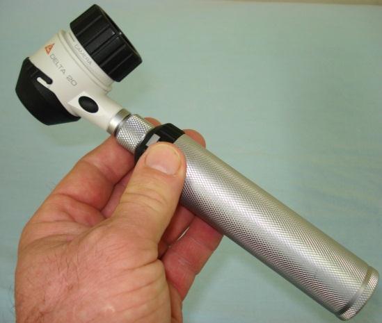
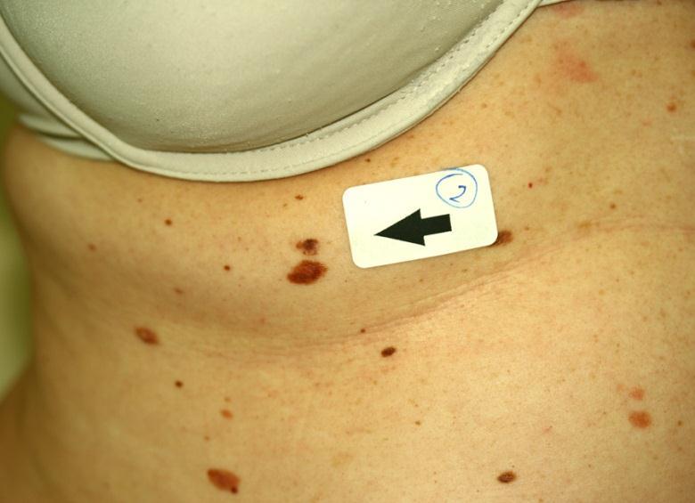
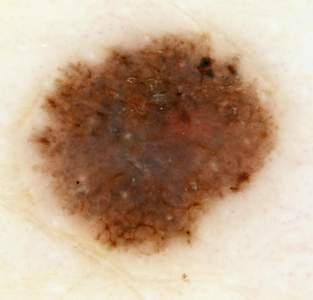
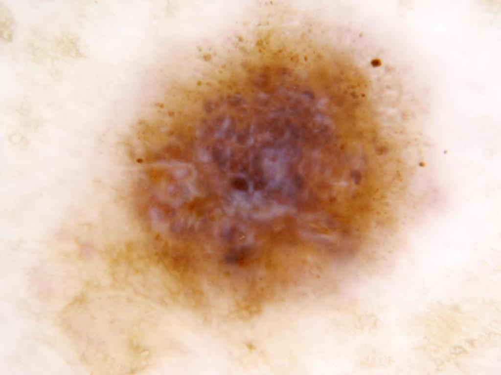
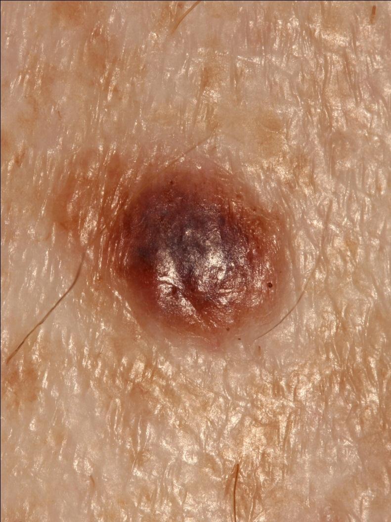
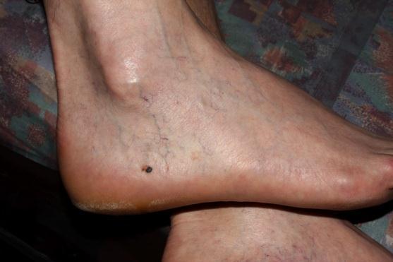
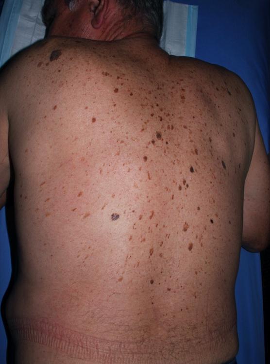
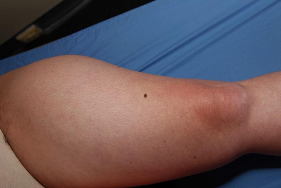
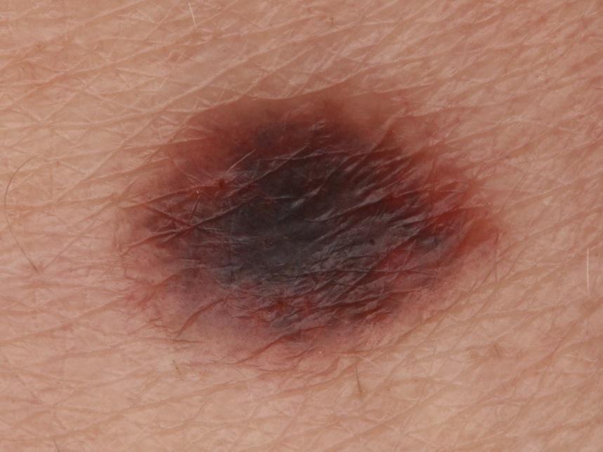
12 Photograph by Alan Cameron
13 NNT Number Needed to Treat for each melanoma diagnosed Number of lesions treated for each melanoma diagnosed Measure of specificity (not sensitivity) Diverse methods of calculation
14 NNT previous studies Naevi + Melanomas/Melanomas ( ) Naevi + Melanomas + Seb K/Melanomas (19-30) All lesions EXCISED/Melanomas (39) All predicted Melanomas/Melanomas (4)
15 Previous NNT studies numbers of melanomas
16 Previous NNT studies NNT values


17 Skin Cancer Audit Research Database
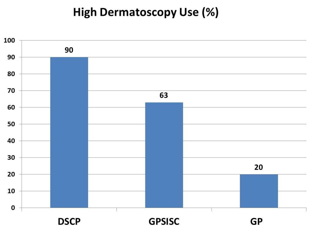
18 Doctor type Dedicated skin cancer practitioner (DSCP) GP with a special interest in skin cancer (GPSISC) General practitioner (GP)
19 Dermatoscopy use High all pigmented skin lesions Medium most days Low/none less than weekly/not at all
20 NNT Lesions tested to exclude melanoma / melanomas Prior declaration of intent!
21 RESULTS
22
23 Lesions tested to exclude melanoma (51) (86) (56)
24
 (8.1-9.0) (8.9-10.")
25 (95% CI P <0.0001) ( ) ( ) ( ) Doctor-type
 (8.6-9.")
26 (95% CI P <0.0001) ( ) ( ) ( ) Dermatoscopy-use
27 The association between NNT and doctor type remained (P< ) when adjusted for dermatoscopy use and other variables. The association between NNT and dermatoscopy use disappeared (P = 0.41) when adjusted for practice type and other variables (doctor-type and dermatoscopy use were too inter-dependant)
28 Fact General practitioners sub-specialized in skin cancer treatment had a higher use of dermatoscopy and excised half the number of benign lesions for each melanoma detected compared to their generalist colleagues Opinion The role of such sub-specialized general practitioners should be defined and the factors that make their performance more effective including trained dermatoscopy use should be promoted
29 For the first time it is shown that there is a measureable benefit of sub-specialisation in General Practice

30 Skin Thank you! Cancer Audit Research Database


31 Dermatoscopy in Routine Practice Chaos & Clues Presented by Dr Cliff Rosendahl & Dr Alan Cameron

32 (1920) Dermatology Dermatologists Dermatoscope Dermatoscopy
33 Conflict of interest a method which cannot be taught is barely a method at all.

34
35 ALL pigmented lesions Detects malignancy ANY type No need to decide whether melanocytic Can be applied at examination speed Efficacy similar to the other algorithms

36 What is dermatoscopy? a non-invasive diagnostic technique for skin lesions a low powered microscope designed for visualisation of pigmentation and vessels by reducing the amount of light reflected off the skin surface by either Contact fluid immersion polarising filters


37 Clinical examination looks at lesions in the horizontal plane In contrast, conventional microscopy looks at lesions in the vertical plane


38 Because melanin appears as different colours at different depths in the skin dermatoscopy provides information in both the horizontal and vertical planes. It provides a 3-dimensional view Atlas of Dermoscopy Marghoob Braun Kopf. Page 11
39 What do you put your dermatoscope on? Every lesion? Selected lesions which break the pattern?

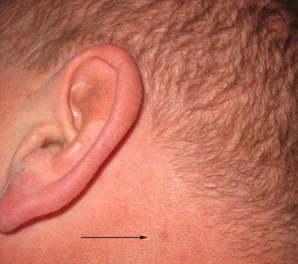
40 Photograph Cliff Rosendahl
41 Photograph Cliff Rosendahl

42 Photograph Cliff Rosendahl
43 Photograph Cliff Rosendahl

44 Do you need dermatoscopy? Photograph Cliff Rosendahl
45 Photograph Cliff Rosendahl

46 Photograph Cliff Rosendahl

47
48 Instruments and flight charts
49 Algorithms 1987 Pehamberger Classic Pattern Analysis 1989 First hand-held dermatoscope consensus meeting metaphoric terminology 1994 Stolz ABCD rule 1996 Menzies method 1998 Argenziano 7 point checklist 2000 Soyer/Argenziano 3 point checklist 2007 CASH (color, architecture, symmetry, and homogeneity) version of pattern Analysis
50 Algorithms 1987 Pehamberger Classic Pattern Analysis 1989 First hand-held dermatoscope consensus meeting metaphoric terminology 1994 Stolz ABCD rule 1996 Menzies method 1998 Argenziano 7 point checklist 2000 Soyer/Argenziano 3 point checklist 2007 CASH (color, architecture, symmetry, and homogeneity) version of pattern Analysis
51 Do you use Pattern Recognition?
52 In the next slide I will present 2 images One will be innocent and one suspicious I want you to see how long it takes to decide
53 Pattern recognition is innate human behaviour Photographs Cliff Rosendahl

54 In contrast to pattern recognition Pattern analysis is the process of describing and analysing the objects in a logical fashion so they can be defined in a repeatable and teachable manner
55 Algorithms 1987 Pehamberger Classic Pattern Analysis 1989 First hand-held dermatoscope consensus meeting metaphoric terminology 1994 Stolz ABCD rule 1996 Menzies method 1998 Argenziano 7 point checklist 2000 Soyer/Argenziano 3 point checklist 2007 CASH (color, architecture, symmetry, and homogeneity) version of pattern Analysis
56 Melanocytic Criteria Network Pseudo network Aggregated brown globules Pseudopods/radial streaming Parallel ridge pattern (acral) Homogenous blue pigmentation


57 Ink spot lentigo Pigmented solar keratosis Solar lentigo/piec Solar lentigo Network occurs in many non-melanocytic lesions


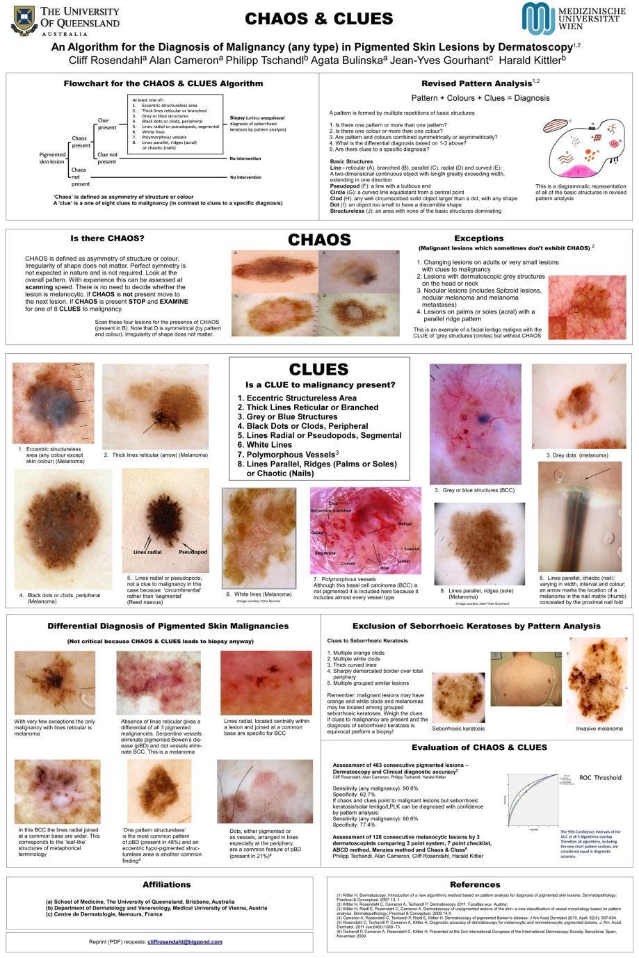
58 Photographs Cliff Rosendahl Pseudonetwork occurs in facial solar lentigines and facial solar keratoses


59 Photograph Cliff Rosendahl Aggregated brown globules occur in BCCs Parallel ridge pattern (acral) occurs in corneal haemorrhage Homogenous blue pigmentation occurs in tattoos

60

61 Melanocytic Criteria You need a microscope to see melanocytes!

62 Why a new method??
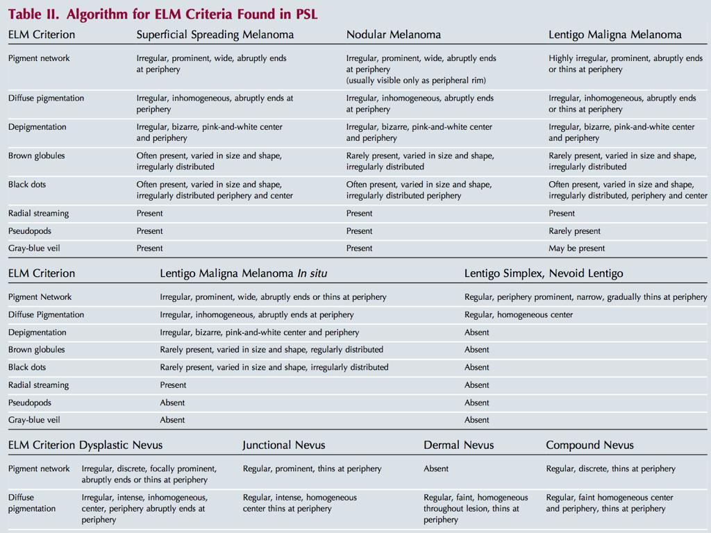
63
64 There is no doubt that the method will have to be improved. There is also no doubt that methods will have to be found to make pattern analysis more objective Pehamberger H, Binder M, Steiner A, Wolff K. In vivo epiluminescence microscopy: improvement of early diagnosis of melanoma. J. Invest. Dermatol. 1993;100(3):356S- 362S.
65 What makes Pattern Analysis difficult? Lack of clear definitions Descriptions that depend on diagnosis Inconsistent metaphorical terminology No clear diagnostic pathway

66 Photograph Alan Cameron

67 2007 Revised pattern Analysis Harald Kittler
68 Revised Pattern Analysis Rebuilds from the firm foundation of Classic pattern Analysis (no subsequent system gave better results) Iconoclastic - Poorly defined confusing metaphorical language replaced by clearly defined geometric terminology No need to decide melanocytic status as a (hazardous) first step
69 Towards a more objective Pattern Analysis Descriptions precede diagnosis clear and objectively defined descriptions are the same regardless of diagnosis no metaphoric descriptions A clear structure to reach a diagnosis
70 Pattern + Colours + Clues = Diagnosis DESCRIPTION PRECEDES DIAGNOSIS! Describing a lesion assists the cognitive process. Metaphoric terms with preconceived diagnostic implications are not used.
71 Pattern Analysis There are only 5 basic elements.
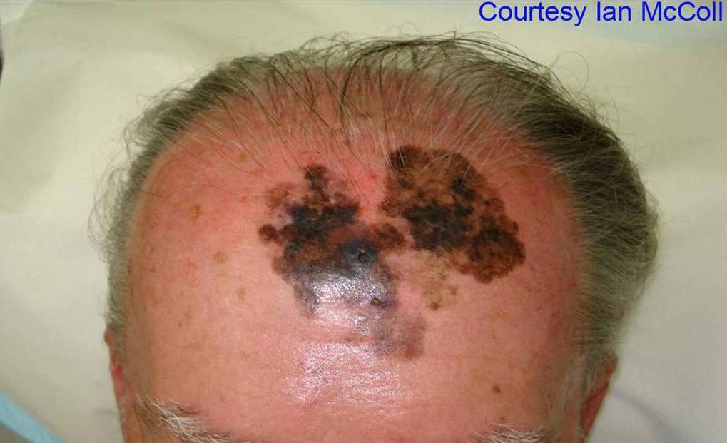
72 Lines Reticular Pseudopods Branched Circles parallel Clods Radial Dots Curved
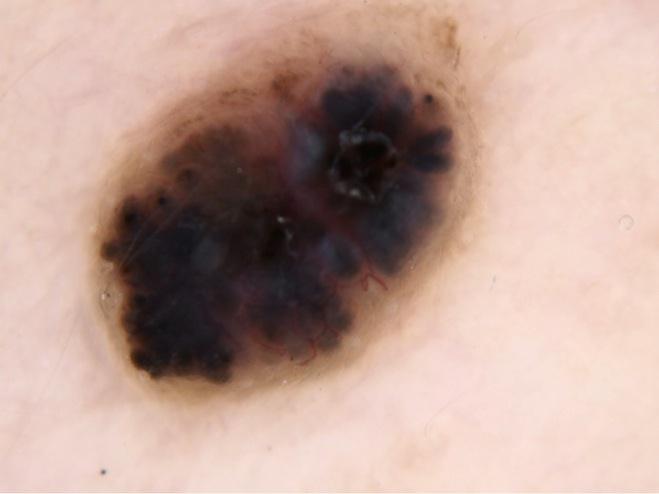
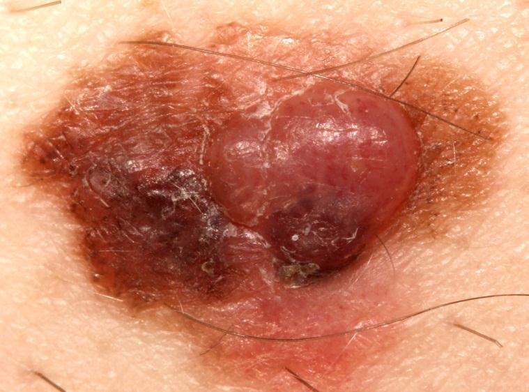
73 Definitions Line : a two-dimensional continuous object with length greatly exceeding width Pseudopod : a line with a bulbous end Circle : a curved line equidistant from a central point Clod : any well circumscribed, solid object larger than a dot. Clods may take any shape Dot : an object too small to have a discernable shape Structureless : An area with no basic structure predominating
74 Pattern There are only 5 basic elements. A pattern consists of multiple repetitions of basic elements.

75 Line a two-dimensional continuous object with length greatly exceeding width. Acral naevus Photograph Alan Cameron

76 Photograph Alan Cameron
77 Courtesy Stelios Minas

78 Photograph Alan Cameron

79 Pseudopod a line with a bulbous end. Reed naevus Photograph Alan Cameron


80 Photograph Alan Cameron Circle a curved line equidistant from a central point. Melanoma in situ face

81 Photograph Alan Cameron Dot an object too small to have a discernable shape. Melanoma courtesy Ian McColl
82 Clod any well circumscribed, solid object larger than a dot. Clods may take any shape. Invasive melanoma Photograph Alan Cameron

83 Photographs Alan Cameron
84 Structureless an area without basic elements, or where no basic element predominates. Photograph Alan Cameron
85 Pattern + Colours + Clues = Diagnosis

86 Black Melanin in Stratum corneum, congealed blood Melanin Dark brown Light brown Melanin in the epidermis, dense Melanin in the epidermis, delicate Grey Melanin in the papillary dermis Blue Melanin in the reticular dermis Keratin Orange Yellow Combination of melanin and keratin, serum crust Keratin White Absence of melanin, sclerosis of the dermis, keratin Haemoglobin Red Purple Blood Blood (poorly oxygenized)
87 Pattern + Colours + Clues = Diagnosis
88 Pattern + Colours + Clues = Diagnosis Clues to a specific diagnosis Clues to malignancy ( Chaos and Clues )
89 CLUES TO MALIGNANCY (Chaos and Clues)

90 each person should develop their own method modified by Jeff Keir
91 Polygons Photographs Cliff Rosendahl

92 Evaluating Chaos & Clues Photograph Cliff Rosendahl


93 Tested on 463 consecutive pigmented lesions in an Australian general practice


94
95 CONCLUSIONS A simplified algorithm based on pattern analysis (CHAOS & CLUES) is a useful tool for all types of pigmented lesions, including those which are non-melanocytic Sensitivity % Specificity %
96

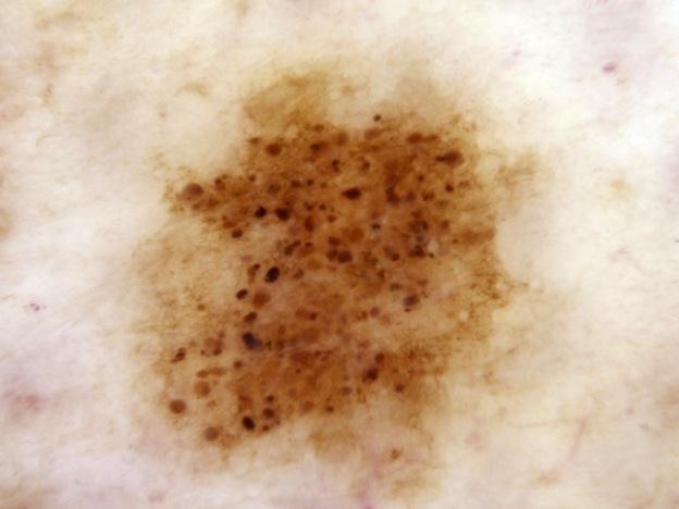
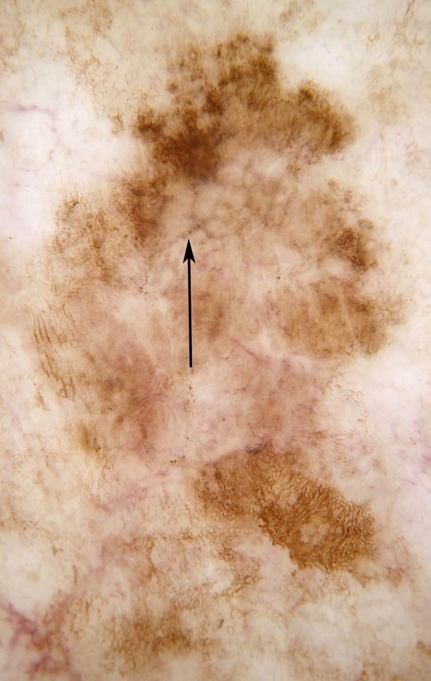
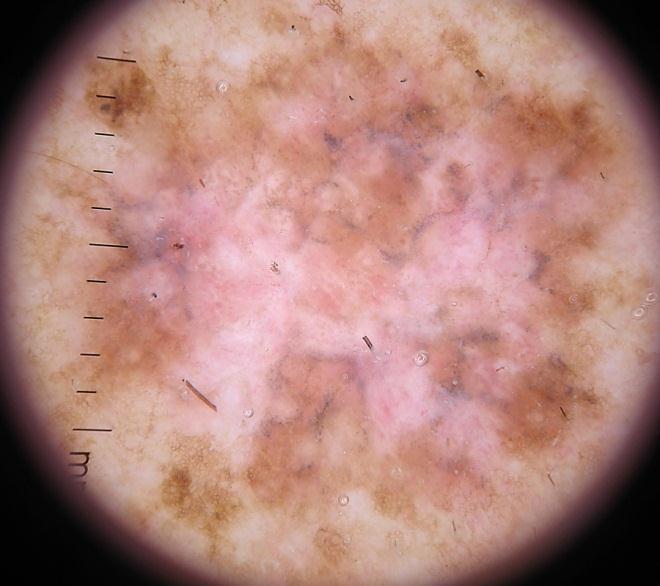
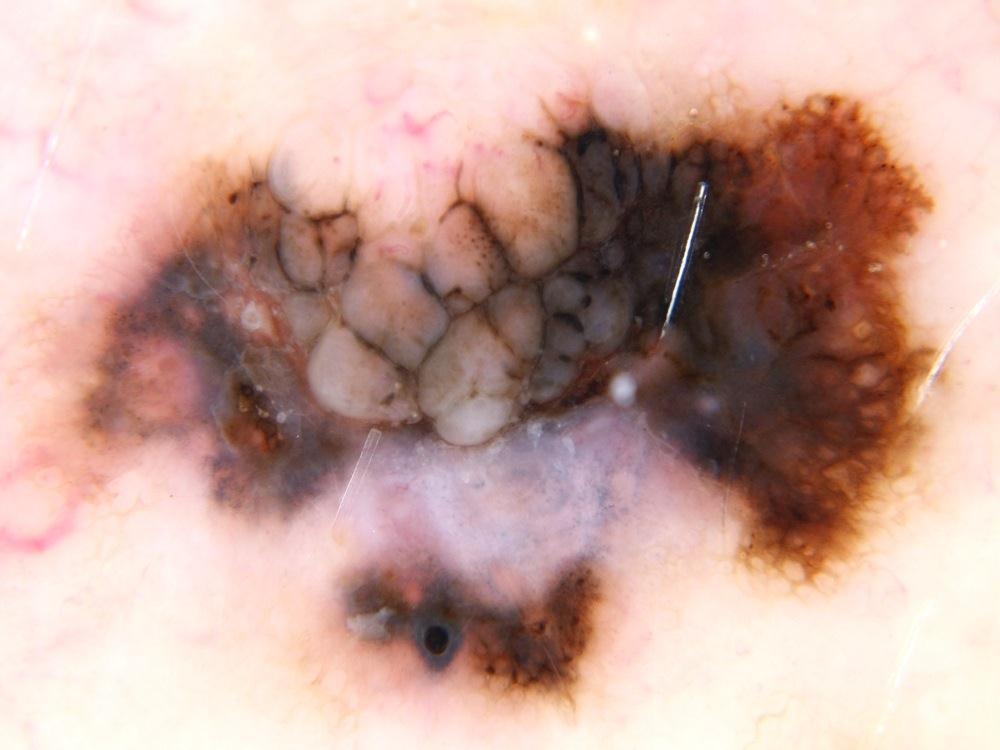
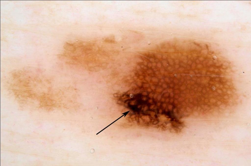
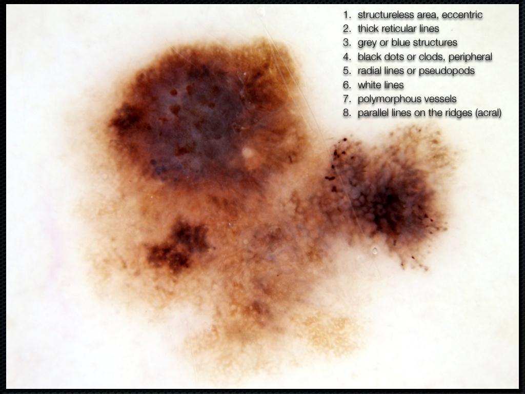
97 Asymmetry of structure or colour


98 Chaos No chaos Judge on pattern and colour, not on outline

99 Photograph Alan Cameron

100 Photograph Alan Cameron concentric = no chaos Be suspicious of other combinations of more than one pattern or colour

101 Photograph Alan Cameron

102 Photograph Alan Cameron
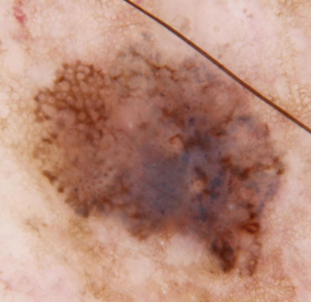
103 CHAOS In a Blink! SCAN for CHAOS The presence or absence of CHAOS can usually be assessed at the speed of a blink! With practice it is a scanning assessment If in doubt assume chaos and assess for clues

104
105 In contrast to scanning for CHAOS the search for CLUES involves thoughtful examination
106 1. Eccentric structureless area
107 Eccentric Structureless Area Any colour except skin colour If white usually represents regression Often scattered grey/blue dots Red- increased blood flow Coloured- dense chaotic melanin deposition

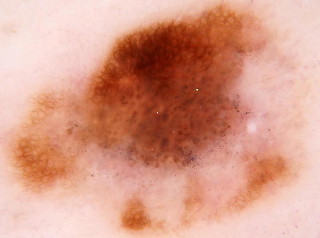
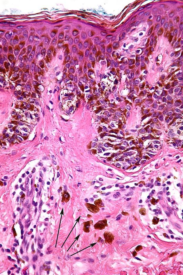
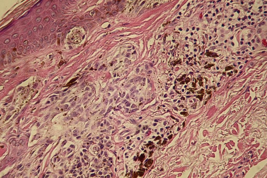
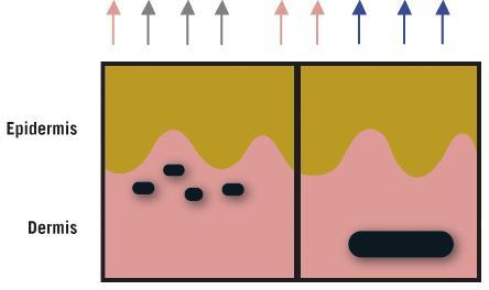
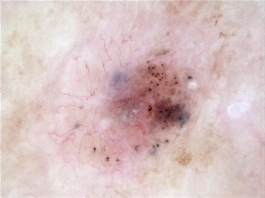
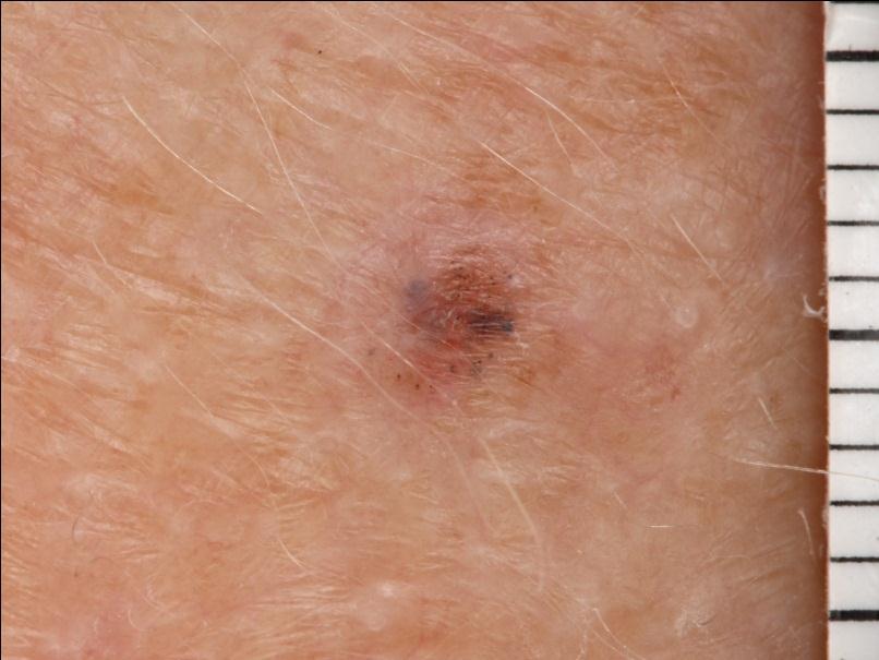
108 Photograph Cliff Rosendahl

109 Photographs Cliff Rosendahl

110 Photograph Alan Cameron

111 Photograph Cliff Rosendahl
112 2. Thick lines reticular or branched
113 Thick Lines Reticular or Branched in benign lesions, the lines are thinner than the holes they enclose Lines thicker than the holes they enclose are seen in melanoma melanomas usually display variability of line thickness
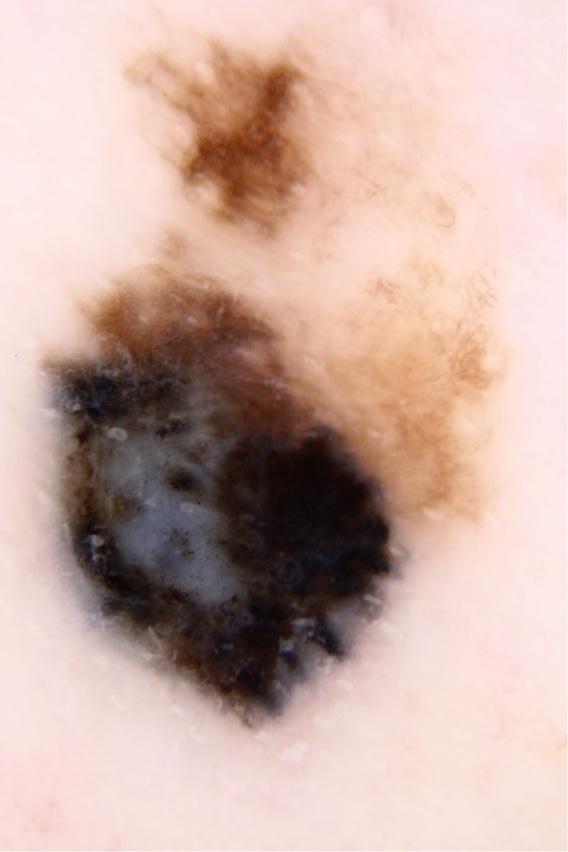
114 Photographs Cliff Rosendahl

115 Photograph Alan Cameron

116 Photographs Alan Cameron
117 3. Grey or blue structures

118 Grey or Blue Structures Lines, circles, clods or dots Represent melanin in the dermis Melanoma cells or melanophages or free melanin
119 Photographs Cliff Rosendahl

120 Photographs Cliff Rosendahl


121 Photographs Cliff Rosendahl Dermatomicrographs Richard Williamson Rosendahl C, Cameron A, Bulinska A, Williamson R, Kittler H. Dermatoscopy of a minute melanoma. Australas. J. Dermatol Feb;52(1):76 8.

122 Photograph Cliff Rosendahl
123 4. Black dots or clods, peripheral
124 Black Dots or Clods, Peripheral Black dots and clods can occur centrally in naevi. They can occur anywhere in a melanoma, so peripheral black dots and/or clods are a clue to malignancy.

125 Photograph Alan Cameron Photograph Alan Cameron

126 Photograph Alan Cameron
127 Photographs Alan Cameron
128 Photographs Cliff Rosendahl
129 5. Lines radial or pseudopods, segmental
130 Lines Radial or Pseudopods, Segmental Melanocytic lesions: Represent radial proliferation of nests of melanocytes Only requires one pseudopod or 1 radial line Basal cell carcinomas: Radial lines converging

131 Photographs Cliff Rosendahl

132 Photographs Cliff Rosendahl


133 Lines radial segmental also a clue to piec Photographs Cliff Rosendahl
134 6. White lines
135 White Lines the colour white is the exception to the rule that structures are defined by pigment. lines are white when they are lighter than surrounding skin Skin coloured lines are occasionally a weak clue- not to be ignored

136 Photograph Alan Cameron

137 Photograph Peter Bourne

138 Photograph Alan Cameron

139 Photograph Alan Cameron
140 Photographs Alan Cameron

141 Nodular melanomas may be symmetrical Photographs Cliff Rosendahl
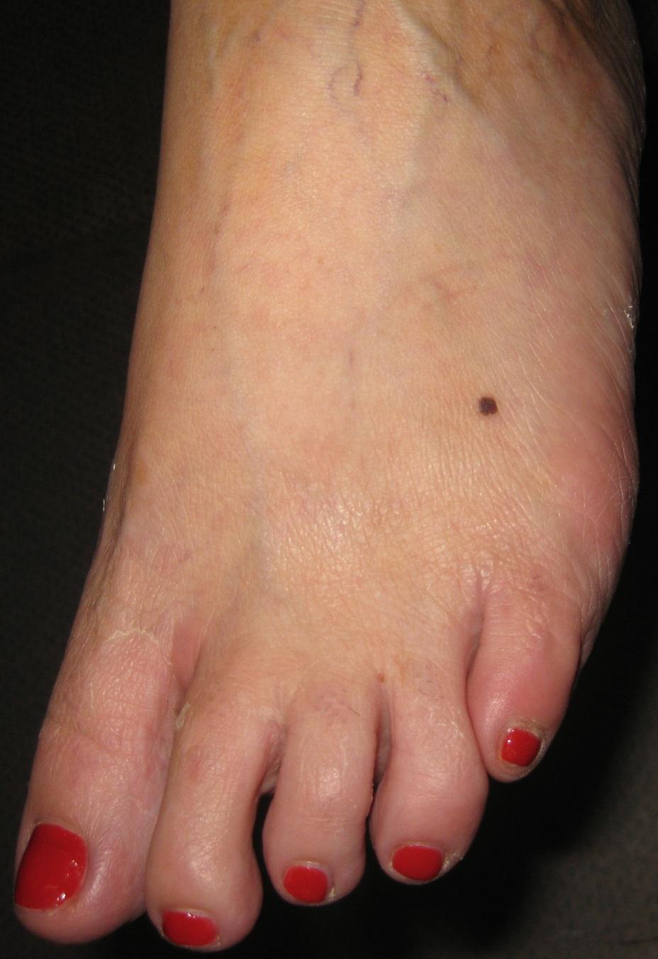
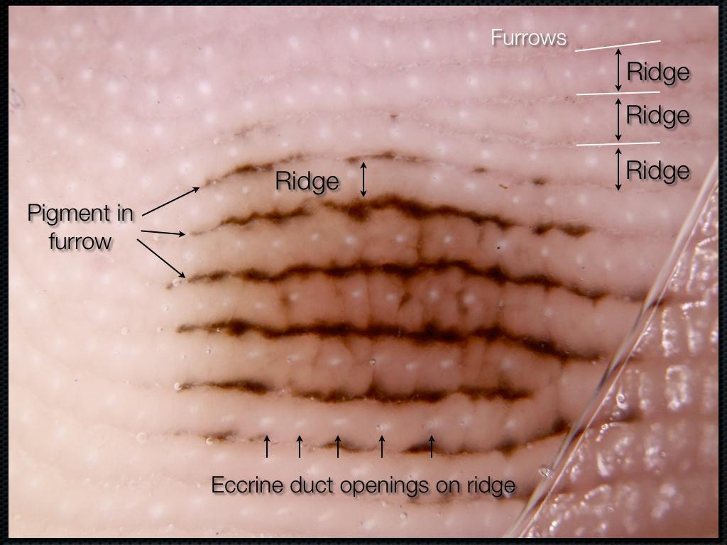
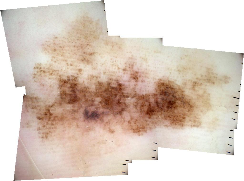
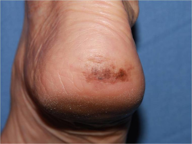
142 7. Polymorphous vessels
143 Polymorphous Vessels A relatively weak clue (pigment structures should take priority when present) More than one vessel pattern Dot vessels often seen in melanomas


144 Vessel patterns Vessel structure left: A- dots; B- clods; C- lines; D- looped; E- curved; F- serpentine; G- helical; H- coiled Vessel arrangement right: A- random; B- clustered; C- serpiginous, D- linear; E- centred; F- radial; G -reticular; H- branched

145 Photograph Cliff Rosendahl

146 Looped Serpentine Helical Photograph Cliff Rosendahl

147 Photographs Cliff Rosendahl

148 Photograph Cliff Rosendahl
149 Photographs Cliff Rosendahl

150 Photograph Cliff Rosendahl
151 8. Lines parallel, ridges (acral) or chaotic (nails)
152 Lines parallel, ridges (acral) or chaotic (nails) Acral skin has a pattern of pattern of ridges and furrows Pigmentation tends to follow either the ridges or the furrows, creating parallel lines In naevi, pigment follows the furrows
153 Photograph Alan Cameron
154 Photograph Alan Cameron Photograph Alan Cameron
155 Lines parallel, ridges (acral) or chaotic (nails) Acral skin has a pattern of pattern of ridges and furrows Pigmentation tends to follow these ridges and furrows Pigmentation on the ridges is a clue to melanoma even in a symmetrical lesion

156 courtesy Jean-Yves Gourhant
157 Photographs Alan Cameron
158 Photographs Alan Cameron
159 Lines parallel, ridges (acral) or chaotic (nails) Lines parallel on nails, varying in width interval and colour
160 Photograph Cliff Rosendahl
161 Photograph Cliff Rosendahl

162 Photograph Cliff Rosendahl


163 1 Poster 1 smart kid + = 1 rare in-situ melanoma
164
165 Excluding seborrhoeic keratoses

166 Photograph Alan Cameron

167 Photograph Alan Cameron
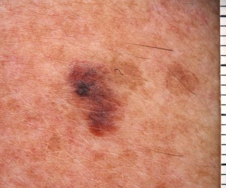
168 Excluding seborrhoeic keratoses Multiple orange or yellow clods Multiple white clods Thick curved or reticular lines Well demarcated border Vessels as loops Multiple grouped similar lesions Malignant conditions can have individual criteria Weigh the clues to arrive at a diagnosis If in doubt at all - BIOPSY

169 Photograph Alan Cameron

170 Photograph Alan Cameron

171 Photograph Alan Cameron

172 Photograph Alan Cameron

173 Photograph Alan Cameron

174 Photographs Alan Cameron
175 Photographs Alan Cameron

176 Photograph Alan Cameron

177 The Aoraki Principle If with respect to any skin lesion you remain unable to make a CONFIDENT SPECIFIC BENIGN Diagnosis BIOPSY IS INDICATED Photograph Cliff Rosendahl

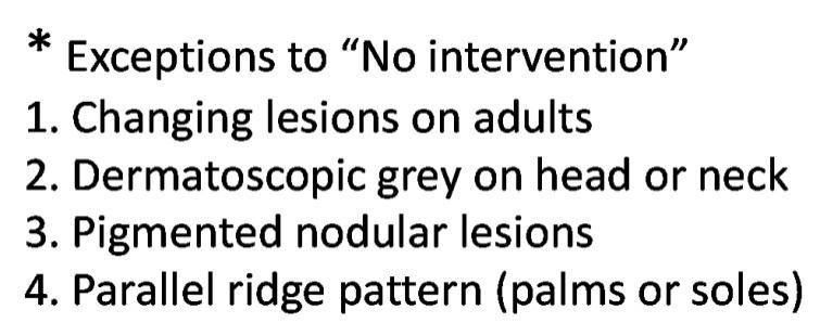
178 Photographs Cliff Rosendahl

179 Photograph Cliff Rosendahl Level 1 melanoma No clues to malignancy but he was not aware how long it had been there
180 Chaos and Clues in everyday practice
181
182

183
184
185
186
187 Exceptions
188 Photographs Alan Cameron

189 Photograph Alan Cameron
190 The most valuable clinical sign of all

191 The break in the pattern The almost inaudible snap of a twig

192 Photographs Cliff Rosendahl Photograph Cliff Rosendahl

193 Photograph Cliff Rosendahl

194 Photograph Cliff Rosendahl

195 Photograph Cliff Rosendahl
196 The signature of an assassin Melanoma writes its message on the skin with its own ink and it is there for all to see Dr Neville Davis Modern concepts of melanoma and its management Annals of Plastic Surgery 1978:1:
197 Look for the break in the pattern Scan for chaos Examine for clues
198 Take out the assassin
199
200 If there is more than one way to skin a cat why not choose the easiest method? Harald Kittler


201 If there is more than one way to skin a cat why not choose the easiest method? Harald Kittler Thank you!
Revised Pattern Analysis: a method for the accurate diagnosis of pigmented skin lesions
 Dermatoscopy for Students A concise outline of: Revised Pattern Analysis: a method for the accurate diagnosis of pigmented skin lesions And Chaos and Clues: a decision algorithm for routine practice to
22/04/2015. Dermoscopy of Melanoma. Ilsphi Browne. Overview
 Dermoscopy of Melanoma Ilsphi Browne Overview The device Dermoscopic criteria (terminology) Colour Patterns Global features Local features Approach to diagnosing pigmented lesions Other uses in general
Dermoscopy in everyday practice. What and Why? When in doubt cut it out? Trilokraj Tejasvi MD
 Dermoscopy in everyday practice Trilokraj Tejasvi MD Assistant Professor, Department of Dermatology, Director Teledermatology services, University of Michigan, Faculty Associate, GLOBAL REACH, Michigan
It can be helpful in some cases of actinic keratosis, Bowen s disease and squamous cell carcinoma
 Dermoscopy Introduction, Terminology and Structures (to be read in conjunction with the Diagnostic Dermoscopic Algorithm) Copyright to Cunliffe TP (Jan. 2017) All rights reserved Introduction Dermoscopy
MODULE 1. LOCAL AND GENERAL CRITERIA IN PIGMENTED MELANOCYTIC LESIONS.
 DERMOSCOPY TEACHING PROGRAMME Dermoscopy Teaching Programme Module 1 MODULE 1. LOCAL AND GENERAL CRITERIA IN PIGMENTED MELANOCYTIC LESIONS. Dermoscopy is a non-invasive in vivo technique that provides
Dermoscopy: Recognizing Top Five Common In- Office Diagnoses
 Dermoscopy: Recognizing Top Five Common In- Office Diagnoses Vu A. Ngo, DO Department of Family Medicine and Dermatology Choctaw Nation Health Services Authority Learning Objectives Introduction to dermoscopy
6/17/2018. Breaking Bad (Part 1) Dermoscopy of Brown(ish) Things. Bad?
 Dermoscopy of Brown(ish) Things. Bad?") Breaking Bad (Part 1) Dermoscopy of Brown(ish) Things Jennie T. Clarke, MD ssociate Professor of Dermatology University of Utah School of Medicine Bad? 1 Brown(ish) Things Bad Melanoma Pigmented basal
Breaking Bad (Part 1) Dermoscopy of Brown(ish) Things Jennie T. Clarke, MD ssociate Professor of Dermatology University of Utah School of Medicine Bad? 1 Brown(ish) Things Bad Melanoma Pigmented basal
BLINCK A diagnostic algorithm for skin cancer diagnosis combining clinical features with dermatoscopy findings
 DERMATOLOGY PRACTICAL & CONCEPTUAL www.derm101.com BLINCK A diagnostic algorithm for skin cancer diagnosis combining clinical features with dermatoscopy findings Peter Bourne, MBBS 1, Cliff Rosendahl,
INTRODUCTION HOUSEKEEPING June 11 th Dr John Adams Dermatologist/Dermoscopist MOLEMAP NZ/Australia MOLESAFE USA
 INTRODUCTION HOUSEKEEPING June 11 th 2015 Dr John Adams Dermatologist/Dermoscopist MOLEMAP NZ/Australia MOLESAFE USA Program Skin cancer statistics. Dermoscopy description and usefulness. Patient /lesion
Dermatoscopic features of cutaneous non-facial non-acral lentiginous growth pattern melanomas
 DERMATOLOGY PRACTICAL & CONCEPTUAL www.derm101.com Dermatoscopic features of cutaneous non-facial non-acral lentiginous growth pattern melanomas Jeff Keir 1 1 Department of Dermatology, School of Medicine,
Disclosure. Objectives. PAFP CME Conference Lou Mancano MD, FAAFP Reading Health System November 18, 2016
 PAFP CME Conference Lou Mancano MD, FAAFP Reading Health System November 18, 2016 1 Disclosure The speaker has no conflict of interest, financial agreement, or working affiliation with any group or organization.
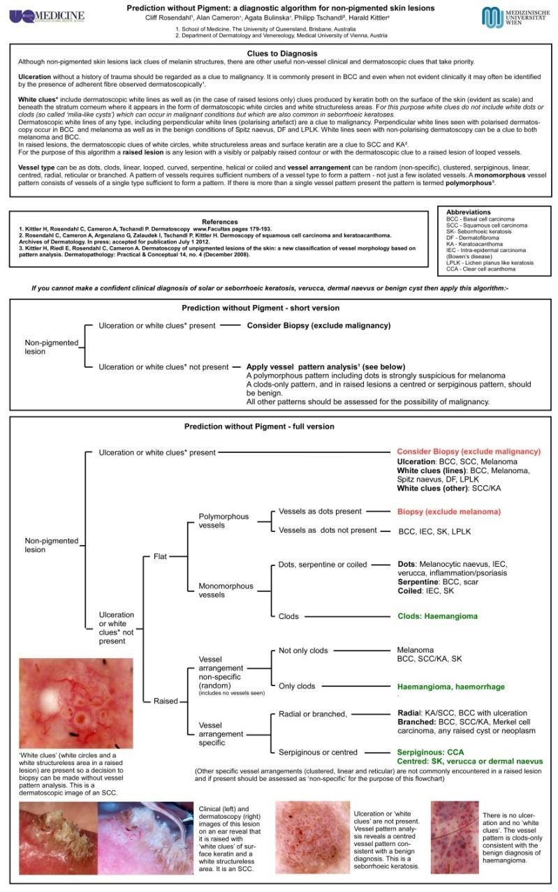
Prediction without Pigment: a decision algorithm for non-pigmented skin malignancy
 DERMATOLOGY PRACTICAL & CONCEPTUAL www.derm101.com Prediction without Pigment: a decision algorithm for non-pigmented skin malignancy Cliff Rosendahl 1, Alan Cameron 1, Philipp Tschandl 2, Agata Bulinska
Basics in Dermoscopy
 Basics in Dermoscopy Manal Bosseila Professor of Dermatology, Cairo University Member of European Academy Dermatology & Venereology EADV Member of International Dermoscopy Society IDS Member of Aesthetic
Case Report Micromelanomas: A Review of Melanomas 2mmand a Case Report
 Case Reports in Oncological Medicine, Article ID 206260, 4 pages http://dx.doi.org/10.1155/2014/206260 Case Report Micromelanomas: A Review of Melanomas 2mmand a Case Report Sharad P. Paul 1,2,3 1 Skin
10/3/2018. Dermoscopy: Looking beneath the surface of the skin. Dermoscopy for Family Medicine 10/11/2018
 Dermoscopy for Family Medicine 10/11/2018 Jane M. Grant-Kels, MD, FAAD Founding Chair Emeritus, Dept of Dermatology Professor of Dermatology, Pathology & Pediatrics Director of the Cut Oncology Ctr & Melanoma
Fundamentals of dermoscopy
 Fundamentals of dermoscopy Learning objectives Upon completion of this session, participants should be able to: describe the basic principles of dermoscopy identify features associated with pigmented and
Skin Cancer A Personal Approach. Dr Matthew Strack Dunedin New Zealand
 Skin Cancer A Personal Approach Dr Matthew Strack Dunedin New Zealand Outline Dermoscopy Instruments and setup Photochemosurgery Clinical Aim: Leave with 2-3 ideas JLE Benign Junctional Nevus Management
Dermoscopy. Enhanced Diagnostic Ability: Pigmented Lesions. Ted Rosen, MD Baylor College of Medicine Houston, Texas
 Dermoscopy Enhanced Diagnostic Ability: Pigmented Lesions Ted Rosen, MD Baylor College of Medicine Houston, Texas Faculty Disclosure Statement No conflicts relevant to this workshop! Sir William Osler
Introduction to Dermoscopy. Nicholas Compton, MD June 16, 2010
 Introduction to Dermoscopy Nicholas Compton, MD June 16, 2010 Overview What is dermoscopy Brief history Types of dermoscopy General approach to lesion of interest 2 step algorithm 3-point checklist Practice
What is Dermoscopy? Early Dermoscopes. Deciphering Dermoscopy: Terminology, Features & Algorithms 6/17/2018
 Deciphering Dermoscopy: Terminology, Features & Algorithms Where did it come from and why do we use it? Jennie T. Clarke, MD Associate Professor of Dermatology University of Utah School of Medicine What
50 interactive dermoscopic case discussions Dr Stephen Hayes
 50 interactive dermoscopic case discussions Dr Stephen Hayes Annotations will be found on your memory drive, as will 100 case discussions and other learning material Melanoma 2mm thick Ugly duckling-one
Dermoscopy. Sir William Osler. Dermoscopy. Dermoscopy. Melanoma USA Primary Care Update Faculty Disclosure Statement
 Diagnostic Ability: Pigmented Lesions Ted Rosen, MD Baylor College of Medicine Houston, Texas Enhanced 2010 Primary Care Update Faculty Disclosure Statement Ted Rosen, MD Speakers Bureau: Abbott, Amgen,
Appendix : Dermoscopy
 Go Back to the Top To Order, Visit the Purchasing Page for Details APP Appendix : Dermoscopy Dermoscopy, also known as dermatoscopy, epiluminoscopy and epiluminescent microscopy, is an effective non-invasive
Dermoscopy-a BRIEF introduction
 Dermoscopy-a BRIEF introduction Aim of presentation -to tell you what dermoscopy is -to show some of what it can do -point the interested learner to further resources Overview of dermoscopy Dermoscopy
Mole mapping and monitoring. Dr Stephen Hayes. Associate Specialist in Dermatology, University Hospital Southampton
 Mole mapping and monitoring Dr Stephen Hayes Associate Specialist in Dermatology, University Hospital Southampton Outline of presentation The melanoma epidemic Benefits of early detection Risks of the
Dermoscopy. Synonyms. Dermoscopy. Definition. Dermoscopy opens up a world of colour and structure that can t be seen with the naked eye
 Synonyms Dermoscopy Australasian College of Dermatologists G.P Training Module Dermoscopy Dermatoscopy Epiluminescence microscopy Skin surface microscopy Incident light microscopy Oil immersion microscopy
comedo-like openings (clods, brown or orange & circles) milia-like cysts (dots or clods, white) 1/29/18 Dotted vessels are also commonly seen in SCC
 milia-like cysts (dots or clods, white) 1/29/18 Dotted vessels are also commonly seen in SCC") Brown circles Dotted vessels are also commonly seen in SCC Step1 1. Nevus (unequivocal) 2. DF/IDN 3. BCC 4. SCC Network Patchy network Peripheral network & central hypopigmentation DF: network with central
Brown circles Dotted vessels are also commonly seen in SCC Step1 1. Nevus (unequivocal) 2. DF/IDN 3. BCC 4. SCC Network Patchy network Peripheral network & central hypopigmentation DF: network with central
Clinical and Dermoscopic Features of Thin Nodular Melanoma
 Clinical and Dermoscopic Features of Thin Nodular Melanoma A study of the International Dermoscopy Society Coordinator: Dr. Alexander J. Stratigos and colleagues, alstrat2@gmail.com ** Extended to May
Regression 2/3/18. Histologically regression is characterized: melanosis fibrosis combination of both. Distribution: partial or focal!
 Regression Margaret Oliviero MSN, ARNP Harold S. Rabinovitz MD Histologically regression is characterized: melanosis fibrosis combination of both Distribution: partial or focal! Dermatoscopic terminology
Growth rate of melanoma in vivo and correlation with dermatoscopic and dermatopathologic findings
 Dermatology Practical & Conceptual www.derm101.com Growth rate of melanoma in vivo and correlation with dermatoscopic and dermatopathologic findings Jürgen Beer, M.D. 1, Lina Xu, M.D. 1, Philipp Tschandl,
Pathology of the skin. 2nd Department of Pathology, Semmelweis University
 Pathology of the skin 2nd Department of Pathology, Semmelweis University Histology of the skin Epidermis: Stratum corneum Stratum granulosum Stratum spinosum Stratum basale Dermis: papillary and reticular
STUDY. Scott W. Menzies, MB,BS, PhD; Karin Westerhoff, MD; Harold Rabinovitz, MD; Alfred W. Kopf, MD; William H. McCarthy, MBBS, MEd; Brian Katz
 STUDY Surface Microscopy of Pigmented Basal Cell Carcinoma Scott W. Menzies, MB,BS, PhD; Karin Westerhoff, MD; Harold Rabinovitz, MD; Alfred W. Kopf, MD; William H. McCarthy, MBBS, MEd; Brian Katz Objectives:
Abrupt Intralesional Color Change on Dermoscopy as a New Indicator of Early Superficial Spreading Melanoma in a Japanese Woman
 Published online: June 24, 2015 1662 6567/15/0072 0123$39.50/0 This is an Open Access article licensed under the terms of the Creative Commons Attribution-NonCommercial 3.0 Unported license (CC BY-NC)
STUDY. Dermoscopy of Squamous Cell Carcinoma and Keratoacanthoma
 ONLINE FIRST STUDY Dermoscopy of Squamous Cell Carcinoma and Keratoacanthoma Cliff Rosendahl, MBBS; Alan Cameron, MBBS; Giuseppe Argenziano, MD; Iris Zalaudek, MD; Philipp Tschandl, MD; Harald Kittler,
Histopathological and SIAscopic Correlation of Pigmented Skin Lesions
 Histopathological and SIAscopic Correlation of Pigmented Skin Lesions Professor Sujatha Fernando MBBS(Hon), MSc(London, Distinction), FRSTM&H, FRCPA, FIAC, FACTM Senior Consultant in Anatomical Pathology,
Keratoacanthoma versus invasive squamous cell carcinoma: a comparison of dermatoscopic vascular features in 510 cases
 DERMATOLOGY PRACTICAL & CONCEPTUAL www.derm101.com Keratoacanthoma versus invasive squamous cell carcinoma: a comparison of dermatoscopic vascular features in 510 cases John H. Pyne 1, Graham Windrum 1,
Key factors in successfully integrating dermoscopy into your clinical practice
 Key factors in successfully integrating dermoscopy into your clinical practice S051 Dilemmas and challenges in skin cancer therapies and management Monday, March 4 th 2019 (9AM-12PM) Room 209A 10:56-11:09AM
Dermoscopy STFM Richard Usatine, MD 5/2/16. Disclosure Statement: Some Dermatoscopes. Dermoscopy Video. Thanks to Dr.
 Disclosure Statement: Dermoscopy STFM 2016 Richard P. Usatine, MD, FAAFP Professor, Family and Community Medicine Professor, Dermatology and Cutaneous Surgery Medical Director, Clinic University of Texas
Diagnosis of Lentigo Maligna Melanoma. Steven Q. Wang, M.D. Memorial Sloan-Kettering Cancer Center Basking Ridge, NJ
 Diagnosis of Lentigo Maligna Melanoma Steven Q. Wang, M.D. Memorial Sloan-Kettering Cancer Center Basking Ridge, NJ Conflict of Interest: None Topics Epidemiology and Natural History Clinical and Histologic
Malignant non-melanocytic lesions
 Malignant non-melanocytic lesions Course C023: Fundamentals of Dermoscopy March 4, 2019, 11:20 AM - 11:50 PM Room: 146B Jason B. Lee, MD Professor & Vice Chair Director of Dermatopathology & Pigmented
STUDY. Characteristic Epiluminescent Microscopic Features of Early Malignant Melanoma on Glabrous Skin
 Characteristic Epiluminescent Microscopic Features of Early Malignant Melanoma on Glabrous Skin A Videomicroscopic Analysis STUDY Shinji Oguchi, MD; Toshiaki Saida, MD, PhD; Yoko Koganehira, MD; Sachiko
Common Benign Lesions and Skin Cancers. 22nd May 2015 Dr Mark Foley
 Common Benign Lesions and Skin Cancers 22nd May 2015 Dr Mark Foley Thank you for downloading this file. This intended to supplement the presentation given at the NZ Wound Care Conference, it is not intended
Histopathology: skin pathology
 Histopathology: skin pathology These presentations are to help you identify, and to test yourself on identifying, basic histopathological features. They do not contain the additional factual information
Cover Page. The handle holds various files of this Leiden University dissertation.
 Cover Page The handle http://hdl.handle.net/1887/22172 holds various files of this Leiden University dissertation. Author: Rhee, Jasper Immanuel van der Title: Clinical characteristics and management of
STUDY. Epiluminescence Microscopy for the Diagnosis of Doubtful Melanocytic Skin Lesions
 STUDY Epiluminescence Microscopy for the Diagnosis of Doubtful Melanocytic Skin Lesions Comparison of the ABCD Rule of Dermatoscopy and a New 7-Point Checklist Based on Pattern Analysis Giuseppe Argenziano,
Assisting diagnosis of melanoma through the noninvasive biopsy of skin lesions
 Assisting diagnosis of melanoma through the noninvasive biopsy of skin lesions Symon D Oyly Cotton Ela Claridge School of Computer Science, The University of Birmingham Birmingham B15 2TT, UK Per Hall
Non-Melanocytic Pattern Dermoscopy
 Non-Melanocytic Pattern Dermoscopy I have no conflicts of interest to disclose Except that I LOVE dermoscopy Michelle Tarbox, MD Assistant Professor of Dermatology and Dermatopathology Texas Tech University
Dr Amanda Oakley. Dermatologist Dept of Dermatology, Health Waikato Adjunct Associate Professor, Waikato Clinical Campus
 Dr Amanda Oakley Dermatologist Dept of Dermatology, Health Waikato Adjunct Associate Professor, Waikato Clinical Campus 14:00-16:00 WS #14: Dermoscopy Part 1 Skin Lesions and Dermatoscopy 16 August 2018
Age-related prevalence of dermatoscopic patterns of acral melanocytic nevi
 DERMATOLOGY PRACTICAL & CONCEPTUAL www.derm101.com Age-related prevalence of dermatoscopic patterns of acral melanocytic nevi Reiko Suzaki 1, Sumiko Ishizaki 1, Hitoshi Iyatomi 2, Masaru Tanaka 1 1 Department
Dermoscopy Quiz 3-Point Checklist Algorithm
 Dermoscopy Quiz 3-Point Checklist Algorithm GLOBAL PATTERN Globular LOCAL CRITERIA Aggregated globules Milia-like cysts 3 POINT CHECK LIST Symmetrical No abnormal net Slight Blue-white veil BENIGN MELANOCYTIC
Review of vasculature visualized on dermoscopy
 doi: 10.1111/1346-8138.13686 Journal of Dermatology 2017; 44: 525 532 REVIEW ARTICLE Review of vasculature visualized on dermoscopy Yaei TOGAWA Department of Dermatology, Chiba University Graduate School
STUDY. Identification of Clinically Featureless Incipient Melanoma Using Sequential Dermoscopy Imaging
 STUDY Identification of Clinically Featureless Incipient Melanoma Using Sequential Dermoscopy Imaging Harald Kittler, MD; Pascale Guitera, MD; Elisabeth Riedl, MD; Michelle Avramidis, MD; Ligia Teban,
Dermoscopy, the use of a handheld
 ONLINE EXCLUSIVE Dermoscopy in family medicine: A primer Dermoscopy allows you to see deeper into the skin than with the naked eye. Here s how you can make use of it to spot malignant conditions sooner.
Skin lesions The Good and the Bad. Dr Virginia Hubbard Ipswich Hospital NHS Trust Barts and the London School of Medicine and Dentistry
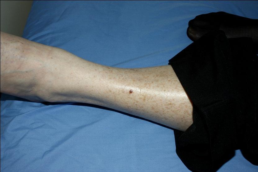
 Skin lesions The Good and the Bad Dr Virginia Hubbard Ipswich Hospital NHS Trust Barts and the London School of Medicine and Dentistry Case 1 32 year old woman Australian Lesion on back New hair growing
Sensitivity and Specificity of Confocal Laser-Scanning Microscopy for In Vivo Diagnosis of Malignant Skin Tumors
 193 Sensitivity and Specificity of Confocal Laser-Scanning Microscopy for In Vivo Diagnosis of Malignant Skin Tumors Armin Gerger, MD 1 Silvia Koller, MD 2 Wolfgang Weger, MD 2 Erika Richtig, MD 2 Helmut
Melanoma and Dermoscopy. Disclosure Statement: ABCDE's of melanoma. Co-President, Usatine Media
 Melanoma and Dermoscopy Richard P. Usatine, MD, FAAFP Professor, Family and Community Medicine Professor, Dermatology and Cutaneous Surgery Medical Director, University Skin Clinic University of Texas
Non-melanocytic Patterns
 Non-melanocytic Lesions Non-melanocytic Patterns Michelle Tarbox, MD Assistant Professor of Dermatology and Dermatopathology Texas Tech University Health Sciences Center 2018 Seborrheic keratoses Acanthotic
Features Causing Confusion between Basal Cell Carcinoma and Squamous Cell Carcinoma in Clinical Diagnosis
 TH Ryu, et al pissn 1013-9087ㆍeISSN 2005-3894 Ann Dermatol Vol. 30, No. 1, 2018 https://doi.org/10.5021/ad.2018.30.1.64 ORIGINAL ARTICLE Features Causing Confusion between Basal Cell Carcinoma and Squamous
Morphologic Features of Melanocytes, Pigmented Keratinocytes, and Melanophages by In Vivo Confocal Scanning Laser Microscopy
 Morphologic Features of Melanocytes, Pigmented Keratinocytes, and Melanophages by In Vivo Confocal Scanning Laser Microscopy Klaus J. Busam, M.D., Carlos Charles, M.D., Grace Lee, M.D., Allan C Halpern,
Development and validation of a scoring system for SIAscopic diagnosis of pigmented skin lesions in primary care
 Development and validation of a scoring system for SIAscopic diagnosis of pigmented skin lesions in primary care J Hunter 1,2, FM Walter 3,5, M Moncrieff 1, S Cotton 4 PN Hall 1, J Emery 3,5 1 Dept of
INCREASE IN incidence and mortality rates for
 Skin Research and Technology 2005; 11: 236 241 Copyright & Blackwell Munksgaard 2005 Printed in Denmark. All rights reserved Skin Research and Technology Pigment distribution in melanocytic lesion images:
Case Report A Case of Cystic Basal Cell Carcinoma Which Shows a Homogenous Blue/Black Area under Dermatoscopy
 Volume 20, Article ID 450472, 4 pages doi:0.55/20/450472 Case Report A Case of Cystic Basal Cell Carcinoma Which Shows a Homogenous Blue/Black Area under Dermatoscopy Akihiro Yoneta, Kohei Horimoto, Keiko
Triage amalgamated dermoscopic algorithm (TADA) for skin cancer screening
 for skin cancer screening") DERMATOLOGY PRACTICAL & CONCEPTUAL www.derm101.com Triage amalgamated dermoscopic algorithm (TADA) for skin cancer screening Tova Rogers 1, Maria Marino 1, Stephen W. Dusza 1, Shirin Bajaj 1, Michael A.
DERMATOLOGY PRACTICAL & CONCEPTUAL www.derm101.com Triage amalgamated dermoscopic algorithm (TADA) for skin cancer screening Tova Rogers 1, Maria Marino 1, Stephen W. Dusza 1, Shirin Bajaj 1, Michael A.
R J M E Romanian Journal of Morphology & Embryology
 Rom J Morphol Embryol 2013, 54(2):315 320 ORIGINAL PAPER R J M E Romanian Journal of Morphology & Embryology http://www.rjme.ro/ Correlation of dermatoscopy with the histopathological changes in the diagnosis
From colour to tissue histology: Physics based interpretation of images of pigmented skin lesions
 From colour to tissue histology: Physics based interpretation of images of pigmented skin lesions Ela Claridge 1, Symon Cotton 2,PerHall 3, Marc Moncrieff 4 1 School of Computer Science, The University
Histopathology of Melanoma
 THE YALE JOURNAL OF BIOLOGY AND MEDICINE 48, 409-416 (1975) Histopathology of Melanoma G. J. WALKER SMITH Department ofpathology, Yale University School ofmedicine, 333 Cedar Street, New Haven, Connecticut
Benign versus Cancerous Lesions How to tell the difference FMF 2014 Christie Freeman MD, CCFP, DipPDerm, MSc
 1 Benign versus Cancerous Lesions How to tell the difference FMF 2014 Christie Freeman MD, CCFP, DipPDerm, MSc Benign lesions Seborrheic Keratoses: Warty, stuck-on Genetics and birthdays Can start in late
Diagnostic Applicability of In Vivo Confocal Laser Scanning Microscopy in Melanocytic Skin Tumors
 See related Commentaries on pages v, vi and viii Diagnostic Applicability of In Vivo Confocal Laser Scanning Microscopy in Melanocytic Skin Tumors Armin Gerger, Silvia Koller, Thomas Kern, Cesare Massone,
DERMATOLOGY PRACTICAL & CONCEPTUAL. Gabriel Salerni 1,2, Teresita Terán 3, Carlos Alonso 1,2, Ramón Fernández-Bussy 1 ABSTRACT
 DERMATOLOGY PRACTICAL & CONCEPTUAL www.derm101.com The role of dermoscopy and digital dermoscopy follow-up in the clinical diagnosis of melanoma: clinical and dermoscopic features of 99 consecutive primary
Principles of Dermatoscopy of Pigmented Skin Lesions
 Principles of Dermatoscopy of Pigmented Skin Lesions Wilhelm Stolz, U. Semmelmayer, K. Johow, and Walter H. C. Burgdorf There has been a dramatic increase in the incidence of malignant melanoma in most
DIFFERENCES IN DERMOSCOPIC IMAGES FROM NON-POLARIZED DERMOSCOPE AND POLARIZED DERMOSCOPE INFLUENCE THE DIAGNOSTIC ACCURACY AND CONFIDENCE LEVEL.
 DIFFERENCES IN DERMOSCOPIC IMAGES FROM NON-POLARIZED DERMOSCOPE AND POLARIZED DERMOSCOPE INFLUENCE THE DIAGNOSTIC ACCURACY AND CONFIDENCE LEVEL. 1. Steven Q. Wang MD 1 (wangs@mskcc.org) 2. Stephen W. Dusza
Algorithmic reproduction of asymmetry and border cut-off parameters according to the ABCD rule for dermoscopy
 JEADV ISSN 1468-3083 Blackwell Publishing Ltd ORIGINAL ARTICLE Algorithmic reproduction of asymmetry and border cut-off parameters according to the ABCD rule for dermoscopy G Pellacani,* C Grana, S Seidenari
Case Report Dermoscopy Clues in Pigmented Bowen s Disease
 Dermatology Research and Practice Volume 2010, Article ID 464821, 9 pages doi:10.1155/2010/464821 Case Report Dermoscopy Clues in Pigmented Bowen s Disease Daniela Gutiérrez-Mendoza, 1 Roberto Narro-Llorente,
Clinical characteristics
 Skin Cancer Fernando Vega, MD Seattle Healing Arts Clinical characteristics Precancerous lesions Common skin cancers ACTINIC KERATOSIS Precancerous skin lesions Actinic keratoses Dysplastic melanocytic
Acral and Mucosal Dermoscopy
 Acral and Mucosal Dermoscopy Caroline C. Kim, MD Assistant Professor, Department of Dermatology Harvard Medical School Director, Pigmented Lesion Clinic Associate Director, Cutaneous Oncology Program Beth
Page: 1 of 16. Optical Diagnostic Devices for Evaluating Skin Lesions Suspected of Malignancy
 Last Review Status/Date: December 2014 Page: 1 of 16 Skin Lesions Suspected of Malignancy Description There is interest in non-invasive devices that will improve the diagnosis of malignant skin lesions.
DERMATOLOGY PRACTICAL & CONCEPTUAL. Introduction. Dermoscopy. Hiroshi Sakai 1, Kyoko Tonomura 1, Hirotsugu Shirabe 1, Masaru Tanaka 2
 DERMATOLOGY PRACTICAL & CONCEPTUAL www.derm101.com Assessment of the colors of melanin pigment in acral compound nevus by using a novel dermoscopy technique with surgical light illumination and saturation
Malignant tumors of melanocytes : Part 3. Deba P Sarma, MD., Omaha
 Malignant tumors of melanocytes : Part 3 Deba P Sarma, MD., Omaha Let s go over one case of melanoma using the following worksheet. Of the various essential information that needs to be included in the
Decision Support System for Skin Cancer Diagnosis
 The Ninth International Symposium on Operations Research and Its Applications (ISORA 10) Chengdu-Jiuzhaigou, China, August 19 23, 2010 Copyright 2010 ORSC & APORC, pp. 406 413 Decision Support System for
Skin Cancer. Dr Elizabeth Ogden Associate Specialist in Dermatology East and North Herts Dr Elizabeth Ogden
 Skin Cancer Dr Elizabeth Ogden Associate Specialist in Dermatology East and North Herts 13.10.16 Skin Cancer Melanoma mole cancer - is a true cancer which can metastasize and kill Non Melanoma skin cancer
Dermoscopy of non-pigmented skin lesions: a literature review
 Hong Kong J. Dermatol. Venereol. (2017) 25, 13-21 Review Article Dermoscopy of non-pigmented skin lesions: a literature review S Thomas, X Li, HP Soyer In this article, we will review benchmark dermoscopic
Aspects on in vivo imaging techniques for diagnostics of pigmented skin lesions
 Thesis for the Degree of Doctor of Philosophy Aspects on in vivo imaging techniques for diagnostics of pigmented skin lesions Karin Terstappen (Westerhoff) Department of Dermatology and Venereology Institure
Yes. Breaking Bad II: Dermoscopy of Pink-ish Things. Does it Fit? Yes 6/17/2018. Yes. Joslyn Kirby, MD, MS, MEd
 Breaking Bad II: Dermoscopy of Pink-ish Things Joslyn Kirby, MD, MS, MEd Yes Observe Yes Step 2. Fit a Benign Nevus Pattern? Does it Fit? Step 1: Melanocytic? pigment network, globules, homogeneous? No
This is a repository copy of Easily missed? Amelanotic melanoma. White Rose Research Online URL for this paper:
 This is a repository copy of Easily missed? Amelanotic melanoma. White Rose Research Online URL for this paper: http://eprints.whiterose.ac.uk/127789/ Version: Accepted Version Article: Muinonen-Martin,
INVESTIGATION. The relation between dermoscopy and histopathology of basal cell carcinoma *
 INVESTIGATION The relation between dermoscopy and histopathology of basal cell carcinoma * 351 Nazan Emiroglu 1 Fatma Pelin Cengiz 1 Funda Kemeriz 2 DOI: http://dx.doi.org/10.1590/abd1806-4841.20153446
Benign and malignant epithelial lesions: Seborrheic keratosis: A common benign pigmented epidermal tumor occur in middle-aged or older persons more
 Benign and malignant epithelial lesions: Seborrheic keratosis: A common benign pigmented epidermal tumor occur in middle-aged or older persons more common on the trunk; but extremities, head and neck are
Management of patients with melanocytic and non-melanocytic neoplasms
 Management of patients with melanocytic and non-melanocytic neoplasms Ashfaq Marghoob MD Harold Rabinovitz MD Margaret Oliviero ARNP Harald Kittler MD Jupiter Cancer Centrer Characteristic Dermoscopic
Rosettes in actinic keratosis and squamous cell carcinoma: distribution, association to other dermoscopic signs and description of the rosette pattern
 DOI: 10.1111/jdv.14474 JEADV ORIGINAL ARTICLE Rosettes in actinic keratosis and squamous cell carcinoma: distribution, association to other dermoscopic signs and description of the rosette pattern B. Lozano-Masdemont,
The most common mistakes on dermatoscopy of melanocytic lesions
 Review paper The most common mistakes on dermatoscopy of melanocytic lesions Grażyna Kamińska-Winciorek 1, Waldemar Placek 2 1 The Center for Diagnostics and Treatment of Skin Diseases, Katowice, Poland
MELANOCYTIC LESIONS: EFFECTIVE MANAGEMENT IN PRIMARY CARE: Part 2
 MELANOCYTIC LESIONS: EFFECTIVE MANAGEMENT IN PRIMARY CARE: Part 2 In the second part of our feature on pigmented skin lesions, dermatology specialist Dr Sweta Rai describes the steps to rational decision-making
Finding Melanoma. Is not easy!
 Finding Melanoma Is not easy! Finding Melanoma Victoria mean depth at diagnosis is 1.5 mm. Melanoma 1.5mm Has Stage 1B Mortality 10% Melanoma Spotting a killer! Spotting a killer Visual Clues What are
IV.4. Early Evolution of Melanoma (Small-Diameter Melanoma)
") Chapter Early Evolution of Melanoma (Small-Diameter Melanoma) Robert J. Friedman, Melanie Warycha, Michele Farber, Dina Gutkowicz-Krusin, Harold Rabinovitz, David Polsky, Margaret Oliviero, Darrell S.
Chapter Early Evolution of Melanoma (Small-Diameter Melanoma) Robert J. Friedman, Melanie Warycha, Michele Farber, Dina Gutkowicz-Krusin, Harold Rabinovitz, David Polsky, Margaret Oliviero, Darrell S.
Dr Stephen Hayes Associate Specialist in Dermatology University Hospital Southampton
 South East Dermatology Transformation and Sustainability Network Guildford, 19 th April 2018 Dermoscopy as an effective skin lesion triage tool in GP surgeries Dr Stephen Hayes Associate Specialist in
=C 0= C8E4 <4;0=><0 3806=>B8B
 4558284=C 0=3 45542C8E4 B8B Wednesday 20 th November 2002 Centenary Institute of Cancer Medicine & Cell Biology Royal Prince Alfred Hospital Missenden Road Camperdown NSW WORKSHOP SUMMARY
In vivo confocal scanning laser microscopy of pigmented Spitz nevi: Comparison of in vivo confocal images with dermoscopy and routine histopathology
 In vivo confocal scanning laser microscopy of pigmented Spitz nevi: Comparison of in vivo confocal images with dermoscopy and routine histopathology Giovanni Pellacani, MD, a Anna Maria Cesinaro, MD, b
VACAVILLE DERMATOLOGY
 Connecting the Dots on those Spots NANDAN V. KAMATH, M.D. VACAVILLE DERMATOLOGY Sources All of the photos were taken with permission from the Dermnet NZ website - Dermnet New Zealand after communicating
Automated Detection and Analysis of Dermoscopic Structures on Dermoscopy Images
 Automated Detection and Analysis of Dermoscopic Structures on Dermoscopy Images Maryam Sadeghi Computing Science Simon Fraser University Burnaby, BC, Canada msa68@sfu.ca Tim K. Lee Cancer Control Research
Multispectral Digital Skin Lesion Analysis. Summary
 Subject: Multispectral Digital Skin Lesion Analysis Page: 1 of 8 Last Review Status/Date: March 2016 Multispectral Digital Skin Lesion Analysis Summary There is interest in noninvasive devices that will