Common skin tumors. Benign Epidermal Tumors. Topic. Clinicopathologic Variants. Seborrheic keratoses
|
|
|
- Charlene Wilson
- 5 years ago
- Views:
Transcription


1 Common skin tumors Topic Benign epidermal tumors Skin cyst and adnexal neoplasms Other common skin tumor Common skin malignancy สมศ กด ต นร ตนากร 26/02/2015 Benign Epidermal Tumors Seborrheic keratosis Dermatosis papulosa nigra Stucco keratosis Inverted follicular keratosis Acrokeratosis verruciformis Clear cell acanthoma Large cell acanthoma Porokeratosis Epidermal nevus Inflammatory linear verrucous epidermal nevus Nevus comedonicus Epidermolytic acanthoma Flegel s disease Cutaneous horn Lichenoid keratosis Acanthosis nigricans Confluent and reticulated papillomatosis Warty dyskeratoma Seborrheic keratoses very common brown macules, papules, plaques, or polypoid lesions over 40 y. increase number with age verrucous or 'stuck-on' the skin predilection for face, neck, and trunk occur anywhere except mucous membranes, palms, or soles sign of Leser-Trélat Clinicopathologic Variants Common Seborrheic Keratosis Dermatosis Papulosa Nigra Skin Tags Irritated Seborrheic Keratosis Stucco Keratosis Reticulated Seborrheic Keratosis Clonal Seborrheic Keratoses Seborrheic Keratosis With Squamous Atypia Melanoacanthoma Leser-Trelat sign 1


2 Dermatosis papulosa nigra Seborrheic keratosis Skin tags Irritated Seborrheic Keratosis Stucco keratoses Multiple gray white keratotic papules on ankle and dorsal foot. Epidermal nevi benign hamartoma of epidermis and papillary dermis onset usually within the first year of life asymptomatic well-circumscribed, hyperpigmented, papillomatous papules or plaques in a linear array along Blaschko's lines patients with epidermal nevus syndrome have associated abnormalities, in particular musculoskeletal and neurologic 2


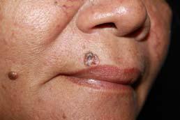
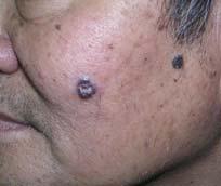
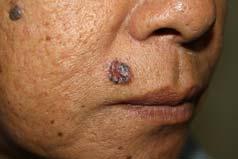
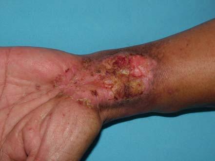
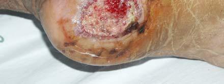
3 Blaschko s lines Inflammatory linear verrucous epidermal nevus (ILVEN). Linear, psoriasiform papules or plaques usually on one extremity 75% appear before 5 years 4 times more common in girls usually persists for years despite attempts at treatment Nevus comedonicus usually present by age of 10 closely arranged, grouped, often linear, slightly elevated papules center keratinous plugs Cutaneous horn firm, white to yellow, conical, markedly hyperkeratotic papule, plaque or nodule Most common in sun-exposed areas and arising from a hyperkeratotic actinic keratosis SCC is present at the base of lesion in up to 20% of patients Acanthosis nigricans 3


 Epidermoid")


4 Cutaneous cysts lining stratified squamous epithelium Epidermoid cyst Milium Trichilemmal cyst Vellus hair cyst Steatocystoma Ear Pit / Preauricular cyst non-stratified squamous epithelium Hidrocystoma no epithelium Mucocele Digital mucous cyst Ganglion Epidermoid (Epithelial) cyst sebaceous cyst is a misnomer most common cutaneous cysts occur anywhere but common on face and upper trunk young and middle-aged adults dermal nodules with central punctum Multiple cysts may associated with Gardner's syndrome (familial adenomatous polyposis) Epidermoid (Epithelial) cyst Inflamed epidermal cyst Milia small epidermoid cysts 1-2 mm white to yellow subepidermal papules 40-50% of infants will have milia on face, will resolve spontaneously in the first 1 month may secondary from blistering processes or superficial ulceration from trauma or resurfacing, topical corticosteroid-induced atrophy 4


5 Milia Trichilemmal (pilar) cyst 95% located on scalp Clinical = epidermal cyst Vellus Hair Cysts numerous tiny dome-shaped papules, ranging from skin-colored to darkly pigmented on trunk most commonly located on the trunk multiple may be inherited in AD Steatocystoma simplex/multiplex sebaceous cyst asymptomatic cysts in the dermis that drain oily fluid if punctured persist indefinitely chest, axillae, and groin Multiplex = autosomal dominant Steatocystoma multiplex Ear Pit / Preauricular cyst congenital defects 0.5 1% of normal population may be transmitted in AD usually unilateral and right-sided 5


 translucent, skin-colored to bluish cysts on")


 no epithelium Mucocele")


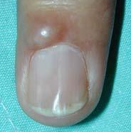
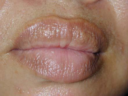
6 Cutaneous cysts lining stratified squamous epithelium Epidermoid cyst Milium Trichilemmal cyst Vellus hair cyst Steatocystoma Ear Pit / Preauricular cyst non-stratified squamous epithelium Hidrocystoma no epithelium Mucocele Digital mucous cyst Ganglion Apocrine hidrocystoma 1-3 mm (face, scalp) translucent, skin-colored to bluish cysts on face adenomas of apocrine sweat gland coils Eccrine hidrocystoma solitary or multiple* can enlarge with heat exposure or during the summer and regress with cooler temperatures cystic dilation of eccrine ducts due to retention of eccrine secretions Cutaneous cysts lining stratified squamous epithelium Epidermoid cyst Milium Trichilemmal cyst Vellus hair cyst Steatocystoma Ear Pit / Preauricular cyst non-stratified squamous epithelium Hidrocystoma (apocrine/eccrine) no epithelium Mucocele Digital mucous cyst Pseudocyst of the auricle Ganglion Mucocele Common - lower labial mucosa dome-shaped, bluish, translucent papules or nodules disruption of ducts of minor salivary glands Digital mucous cyst dorsal surface of finger distal phalanx depressed nail deformity 6


7 Pseudocyst of the auricle scaphoid fossa of ear in middle-aged men usually unilateral painless swelling? chronic trauma Adnexal Neoplasms Follicular lineage Nevus sebaceus Trichoepithelioma Trichofolliculoma Pilomatricoma sebaceous differentiation Sebaceous gland hyperplasia Fordyce s disease apocrine differentiation Syringoma eccrine differentiation Eccrine nevus Nevus sebaceus papillomatous yellow-orange linear plaque on the scalp or face associated with alopecia sebaceous glands are most prominent during early infancy and post-puberty scalp or face Trichoepithelioma skin-colored papule or small nodule face or upper trunk, lesions have a special predilection for the nose Trichofolliculoma a skin-colored papule with a dilated central pore no treatment is needed 7


")

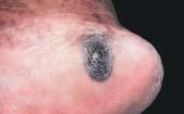
8 Pilomatricoma solitary skin-colored or bluish nodule head or upper trunk childhood and adolescence Adnexal Neoplasms follicular lineage Nevus sebaceus Trichofolliculoma Trichoepithelioma Pilomatricoma Sebaceous differentiation Sebaceous gland hyperplasia Fordyce s disease apocrine differentiation Syringoma eccrine differentiation Eccrine nevus Sebaceous gland hyperplasia yellowish, occasionally telangiectatic papules usually on central or upper face Sebaceous gland hyperplasia Adnexal Neoplasms ectopic sebaceous glands (Fordyce granules/spot) follicular lineage Nevus sebaceus Trichofolliculoma Trichoepithelioma Pilomatricoma sebaceous differentiation Sebaceous gland hyperplasia Fordyce s disease Apocrine differentiation Syringoma eccrine differentiation Eccrine nevus 8

 Vascular")


 Cherry angiomas (senile")
 Bright red,")

9 Syringoma small firm skin-color papules periorbital area (eyelids) Vascular Neoplasms Infantile hemangioma Pyogenic granuloma Cherry angioma Infantile Hemangioma most common benign tumors of childhood more common in females (2-5:1 ) and in premature rapid growth (proliferate phase) within first 5 months heal with telangiectasias, atrophy, fibro-fatty residuum, scarring 01/11/00 (2 mo.) 16/01/01 (4 mo.) Cherry angiomas (senile angiomas, de Morgan spots) Bright red, dome-shaped to polypoid papules 1-6 mm. during adult life on trunk and upper extremities 11/12/01 ( 1 y. 3 mo.) 22/04/03 (2 y. 6 mo.) 9
 Fibrous and")


10 Pyogenic Granuloma (Lobular capillary hemangioma) Fibrous and Fibrohistiocytic Proliferations of the Skin Cutaneous angiofibroma Fibrous papule Pearly penile papule Acral fibrokeratoma Dermatofibroma Reactive vascular hyperplasia most common in children and young adults rapidly growing, friable, red papule or polyp of skin or mucosa frequently ulcerates not involute spontaneously Electrocautery or CO 2 laser Dermatofibroma Hyperpigmented firm papule cm. Lipoma Lipoma : most common soft-tissue tumor Asymptomatic, soft, subcutaneous nodule arising at any site relative sparing of the head, hands, and feet Multiple lipomas are seen in Madelung s disease Gardner syndrome Proteus syndrome familial multiple lipomatosis adiposis dolorosa Bannayan Riley Ruvalcaba syndrome Neurofibromatosis Benign symmetric lipomatosis (Madelung s disease) Neurofibromas (60 90%) Skin-colored to tan violet papule or nodule May be pedunculated or have the buttonhole sign Predilection for the trunk and head Café-au-lait macules (>90%) Axillary and/or inguinal freckling (~80%) Plexiform neurofibroma (25%) 10


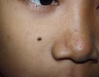
11 Development of clinical features in neurofibromatosis type 1. Becker s nevus Unilateral, hyperpigmented and often hypertrichotic patch or slightly elevated plaque Usually on shoulder of male patients Onset during adolescence Congenital nevomelanocytic nevus present at birth Small <1.5 cm, medium, giant > 20cm Risk of melanoma? Giant congenital melanocytic nevus Acquired melanocytic nevi Junctional dark brown macule with lighter brown rim. Compound light to medium brown papule. Intradermal soft light pink papule. Precancerous lesions (Squamous Cell Carcinoma in Situ) Bowen s disease Leukoplakia Actinic keratosis Erythroplasia of Queyrat 11


")
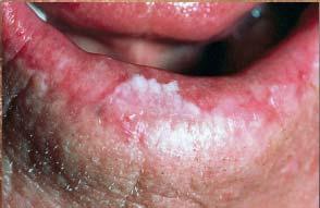
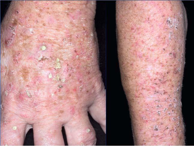
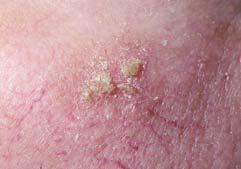
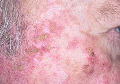
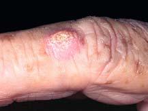
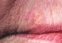
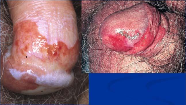

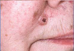
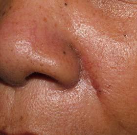
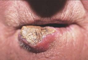
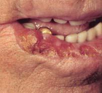
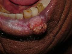
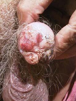
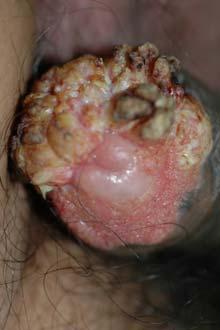
12 Squamous cell carcinoma in situ (Bowen s disease) Leukoplakia Actinic (solar) keratosis Actinic (solar) keratosis Erythroplasia of Queyrat of the penis มะเร งผ วหน ง Non-melanoma skin cancers (NMSC) Squamous cell carcinoma Basal cell carcinoma Malignant melanoma 12
 : trunk, extremities. Area M (middle risk) : cheeks, forehead, neck, scalp.")
, genitalia, hands and feet.")
 230 3.4 BCC:SCC ratio 4 : 1 1:1.")


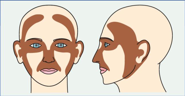
13 Risk factors for development of SCC and BCC Risk factors for development of SCC and BCC Risk factors for recurrence Area L (low risk) : trunk, extremities. Area M (middle risk) : cheeks, forehead, neck, scalp. Area H (high risk) : mask areas of face (central face, eyelids, eyebrows, periorbital, nose, lips, chin, mandible, preauricular and postauricular skin/sulci, ear, temple), genitalia, hands and feet. Risk factors for recurrence Basal cell carcinoma (BCC) Squamous cell carcinoma (SCC) Characteristic Lightly pigmented Darkly pigmented skin color skin color NMSC incidence (per 100,000) BCC:SCC ratio 4 : 1 1:1.1 BCC male:female ratio 1.5 : : 1 SCC male:female ratio 2:1-5:1 1.3 : 1 % of BCCs developing in head and neck region % of SCCs developing in head and neck region % of SCCs developing in scars and chronic nonhealing ulcers < NMSC incidence rates Increasing n/a NMSC mortality rates Decreasing Decreasing % of skin cancer deaths due to NMSC in persons <50 years of age % of skin cancer deaths due to NMSC in persons >85 years of age


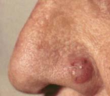
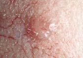
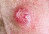
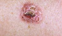
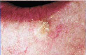
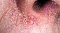
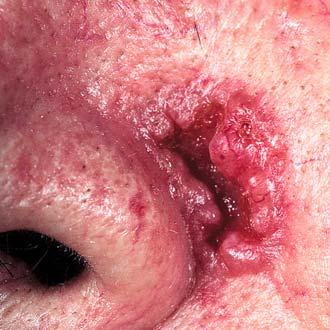
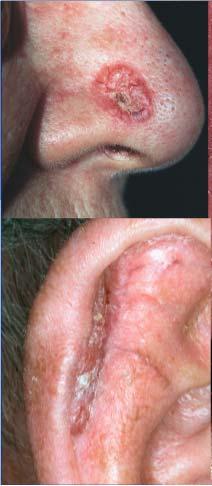

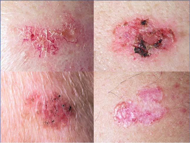
14 Basal cell carcinoma BCC (nodular) BCC (nodular) BCC (pigmented) BCC (superficial) 14
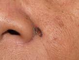
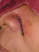


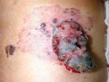

15 Indications for Mohs Micrographic Surgery BCC / Moh s Squamous cell carcinoma Squamous cell carcinoma Squamous cell carcinoma Malignant melanoma 15
 Site Radial growth")


 Yes More pagetoid,")


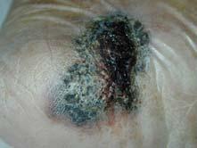
16 Type of melanoma Freq. (%) Site Radial growth Special features Superficial spreading melanoma Superficial spreading melanoma Any site, preference for lower extremities (female), trunk (male) Yes More pagetoid, less solar elastosis Nodular melanoma Any site, preference for trunk, head, neck No Nodule with vertical growth Lentigo maligna melanoma 5-15 Face, especially nose and cheeks Yes Slower growth over years on sun-damaged skin Acral lentiginous melanoma 5-10 Palms, soles, subungual Yes Most common melanoma in patients with darker skin types Superficial spreading melanoma Nodular melanoma Nodular melanoma Lentigo maligna melanoma 16


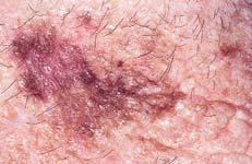
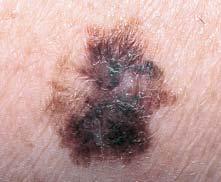
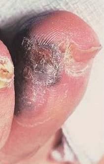
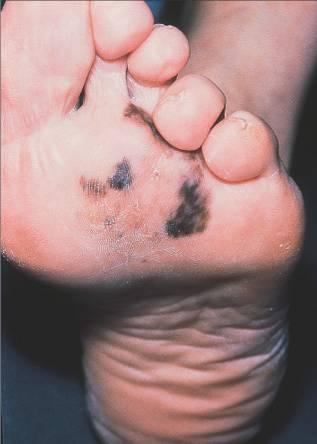

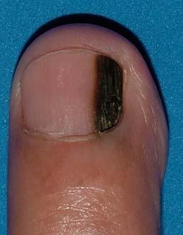
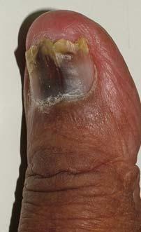

17 Lentigo maligna melanoma Acral lentiginous melanoma END 17
LUMPS AND BUMPS: AN ORGANIZED APPROACH TO DIAGNOSIS AND MANAGEMENT
 LUMPS AND BUMPS: AN ORGANIZED APPROACH TO DIAGNOSIS AND MANAGEMENT Tammy P. Than, M.S., O.D., F.A.A.O. The University of Alabama at Birmingham / School of Optometry 1716 University Blvd. Birmingham, AL
Benign versus Cancerous Lesions How to tell the difference FMF 2014 Christie Freeman MD, CCFP, DipPDerm, MSc
 1 Benign versus Cancerous Lesions How to tell the difference FMF 2014 Christie Freeman MD, CCFP, DipPDerm, MSc Benign lesions Seborrheic Keratoses: Warty, stuck-on Genetics and birthdays Can start in late
Lid Lesions: Relax or Refer
 Lid Lesions: Relax or Refer Blair Lonsberry, MS, OD, MEd., FAAO Professor of Optometry Pacific University College of Optometry blonsberry@pacificu.edu Agenda Benign vs. Malignant lesions Benign Eyelid
Clinical characteristics
 Skin Cancer Fernando Vega, MD Seattle Healing Arts Clinical characteristics Precancerous lesions Common skin cancers ACTINIC KERATOSIS Precancerous skin lesions Actinic keratoses Dysplastic melanocytic
Pathology of the skin. 2nd Department of Pathology, Semmelweis University
 Pathology of the skin 2nd Department of Pathology, Semmelweis University Histology of the skin Epidermis: Stratum corneum Stratum granulosum Stratum spinosum Stratum basale Dermis: papillary and reticular
Lumps and Bumps: The Dermatology of Lid Lesions
 Lumps and Bumps: The Dermatology of Lid Lesions Thomas J. Joly, MD, PhD Assistant Professor of Ophthalmology Eastern Virginia Medical School Ophthalmic Plastic Surgery Service Virginia Eye Consultants
Benign and malignant epithelial lesions: Seborrheic keratosis: A common benign pigmented epidermal tumor occur in middle-aged or older persons more
 Benign and malignant epithelial lesions: Seborrheic keratosis: A common benign pigmented epidermal tumor occur in middle-aged or older persons more common on the trunk; but extremities, head and neck are
Doctors of Optometry Course Notes
 Doctors of Optometry Course Notes OD19 1CE COPE: 43871-AS Eyelid Lumps and Bumps Sunday, February 26, 2017 2:40 pm 3:30 pm Regency C 3 rd Floor Presenter: Blair Lonsberry, OD, FAAO Dr. Lonsberry is a Full
Skin lesions The Good and the Bad. Dr Virginia Hubbard Ipswich Hospital NHS Trust Barts and the London School of Medicine and Dentistry
 Skin lesions The Good and the Bad Dr Virginia Hubbard Ipswich Hospital NHS Trust Barts and the London School of Medicine and Dentistry Case 1 32 year old woman Australian Lesion on back New hair growing
Identifying Benign and Malignant Skin Lesions. No Disclosures. Common Benign Lesions. Benign Lesions 2/25/2018. Stucco Keratoses.
 Dermatology in Primary Care Identifying Benign and Malignant Skin Lesions Christy Quire Baker, APRN, FNP-BC, DCNP Dermatology Certified Nurse Practitioner No Disclosures Common Benign Lesions Seborrheic
Learning Objectives. Tanning. The Skin. Classic Features. Sun Reactive Skin Type Classification. Skin Cancers: Preventing, Screening and Treating
 Learning Objectives Skin Cancers: Preventing, Screening and Treating Robert A. Baldor, MD, FAAFP Professor, Family Medicine & Community Health University of Massachusetts Medical School Distinguish the
IT S FUNDAMENTAL MY DEAR WATSON! A SHERLOCKIAN APPROACH TO DERMATOLOGY
 IT S FUNDAMENTAL MY DEAR WATSON! A SHERLOCKIAN APPROACH TO DERMATOLOGY Skin, Bones, and other Private Parts Symposium Dermatology Lectures by Debra Shelby, PhD, DNP, FNP-BC, FADNP, FAANP Debra Shelby,
Dermatological Manifestations in the Elderly. Sanjay Siddha Staff Dermatologist UHN & MSH
 Dermatological Manifestations in the Elderly Sanjay Siddha Staff Dermatologist UHN & MSH Disclosure No actual or potential conflicts of interest or commercial relationships to declare Objectives Recognize
Porokeratosis: Introduction
 Porokeratosis: Introduction Benign epidermal proliferation Distinct clinical & histologic features 5 clinical subtypes Erroneously named porokeratosis CLINICAL SUBTYPES 1. Porokeratosis of Mibelli 2. Disseminated
Samer Ghosn, MD Associate professor, Derpartment of Dermatology American University of Beirut Medical Center. Follicular lesions
 Samer Ghosn, MD Associate professor, Derpartment of Dermatology American University of Beirut Medical Center Follicular lesions Introduction Follicular lesions are important to recognize: For proper management
LENTIGO SIMPLEX. Epidemiology
 LENTIGO SIMPLEX Epidemiology The frequency of lentigo simplex in children and adults has not been determined. There does not appear to be a racial or gender predilection. Lentigo simplex is the most common
Appendageal skin tumors
 Appendageal skin tumors Ibrahim Khalifeh, M.D. Associate Professor Department of Pathology American University of Beirut Medical Center Beirut, Lebanon Appendageal tumors Neoplasms whose differentiation
المركب النموذج--- سبيتز وحمة = Type Spitz's Nevus, Compound SPITZ NEVUS 1 / 7
 SPITZ NEVUS 1 / 7 Epidemiology An annual incidence rate of 1.4 cases of Spitz nevus per 100,000 individuals has been estimated in Australia, compared with 25.4 per 100,000 individuals for cutaneous melanoma
Lumps and Bumps: An Organized Approach to Diagnosis and Management. Disclosure. Introduction. References. Structure of Skin.
 Lumps and Bumps: An Organized Approach to Diagnosis and Management Nothing to disclose Disclosure Tammy Pifer Than, MS, OD, FAAO Carl Vinson VAMC tammythan@bellsouth.net References Fitzpatrick's Color
Dermoscopy: Recognizing Top Five Common In- Office Diagnoses
 Dermoscopy: Recognizing Top Five Common In- Office Diagnoses Vu A. Ngo, DO Department of Family Medicine and Dermatology Choctaw Nation Health Services Authority Learning Objectives Introduction to dermoscopy
Objectives. 1. Recognizing benign skin lesions. 2.Know which patients will likely need surgical intervention.
 The Joy of Pediatric Skin Dr. Claire Sanger University of Kentucky Plastic & Reconstructive Surgery Objectives 1. Recognizing benign skin lesions 2.Know which patients will likely need surgical intervention.
Atlas of Eyelid and Conjunctival Tumors
 Atlas of Eyelid and Conjunctival Tumors Jerry A. Shields, M.D. Director, Ocular Oncology Service Wills Eye Hospital Professor of Ophthalmology Thomas Jefferson University Philadelphia, Pennsylvania Carol
 Epidermis Dermis Subcutaneous Hypodermis March 16, 2013 Telangiectasia's (spider veins) may develop anywhere within the body but can be easily seen in the skin, mucous membranes, and whites of
Living Beyond Cancer Skin Cancer Detection and Prevention
 Living Beyond Cancer Skin Cancer Detection and Prevention Cutaneous Skin Cancers Identification Diagnosis Treatment options Prevention What is the most common cancer in people? What is the most common
Pigmented lesions of the Oral cavity
 Oral medicine أ.م.د احسان عبد هللا كميل Pigmented lesions of the Oral cavity Pigmented oral lesions are a large group of disorders in which the dark or brown color is the essential clinical characteristic.
Case-Based Approach to Common Dermatologic Neoplasms
 Case-Based Approach to Common Dermatologic Neoplasms Patrick Retterbush, MD, FAAD Mohs Surgery & Dermatologic Oncology Associate Member of the American College of Mohs Surgery Private Practice: Lockman
Lagophthalmos. Lagophthalmos: signs. Lagophthalmos: clinical tips. Lagophthalmos: treatment plan. Madarosis
 Lagophthalmos Def: incomplete closure of the eyelid SX: FBS, irritation, red, burn, dry, chronic morning corneal irritation Lagophthalmos: signs 2-5 mm lid separation with slit lamp during blink can force
Non-melanocytic Patterns
 Non-melanocytic Lesions Non-melanocytic Patterns Michelle Tarbox, MD Assistant Professor of Dermatology and Dermatopathology Texas Tech University Health Sciences Center 2018 Seborrheic keratoses Acanthotic
Squamous papilloma Squamous acanthoma Keratoacanthoma Verruca vulgaris Condyloma acuminatum Focal epithelial hyperplasia Sino nasal papilloma
 Benign tumors Epithelial origin Squamous papilloma Squamous acanthoma Keratoacanthoma Verruca vulgaris Condyloma acuminatum Focal epithelial hyperplasia Sino nasal papilloma Squamous papilloma Exophytic
Cowden Syndrome PTEN Hamartoma Tumor Syndrome. ACCME/Disclosure. 1. Background. Outline
 MASSACHUSETTS GENERAL HOSPITAL HARVARD MEDICAL SCHOOL PATHOLOGY Cowden Syndrome PTEN Hamartoma Tumor Syndrome ACCME/Disclosure Vania Nosé, MD, PhD Professor of Pathology Director of Anatomic Pathology
Dermatopathology: The tumor is composed of keratinocytes which show atypia, increase mitoses and abnormal mitoses.
 Squamous cell carcinoma (SCC): A common malignant tumor of keratinocytes arising in the epidermis, usually from a precancerous condition: 1- UV induced actinic keratosis, usually of low grade malignancy.
Test your knowledge with multiple-choice cases. What are these speckled spots?
 Test your knowledge with multiple-choice cases Case 1 What are these speckled spots? A speckled, pigmented lesion is noticed on the upper arm of a 10-year-old girl. Her mother says the lesion has been
Describe the functions of the vertebrate integumentary system. Discuss the structure of the skin and how it relates to function.
 Chapter 5 Describe the functions of the vertebrate integumentary system. Discuss the structure of the skin and how it relates to function. Explain the basis for different skin colors. Describe the structure
Dermatology for the PCP Deanna G. Brown, MD, FAAD Susong Dermatology Consulting Staff at CHI Memorial
 Dermatology for the PCP Deanna G. Brown, MD, FAAD Susong Dermatology Consulting Staff at CHI Memorial Cutaneous Oncology for the PCP Deanna G. Brown, MD, FAAD Susong Dermatology Consulting Staff at CHI
 Page 1 of 15 Title Authored By Course No Contact Hours 2 Skin Cancer the Real Picture for Early Detection and Treatment Cheryl Sommer RN, MSN, ARNP SC120604 Purpose The purpose of this course is to provide
CASE REPORT SOLITARY SEBACEOUS NEVUS OF JADASSOHN COMPLICATED BY SQUAMOUS CELL CARCINOMA AND BASAL CELL CARCINOMA
 CASE REPORT Dennis H. Kraus, MD, Section Editor SOLITARY SEBACEOUS NEVUS OF JADASSOHN COMPLICATED BY SQUAMOUS CELL CARCINOMA AND BASAL CELL CARCINOMA Ahmad Ridzwan Arshad, FRCS, 1 Wan S. Azman, MS, 1 Ayadurai
Actinic keratosis (AK): Dr Sarma s simple guide
: Dr Sarma s simple guide") Actinic keratosis (AK): Dr Sarma s simple guide Actinic keratosis is a very common lesion that you will see in your day-to-day practice. First, let me explain the name Actinic keratosis. It means keratosis
Actinic keratosis (AK): Dr Sarma s simple guide Actinic keratosis is a very common lesion that you will see in your day-to-day practice. First, let me explain the name Actinic keratosis. It means keratosis
The Integumentary System. Mosby items and derived items 2010, 2006, 2002, 1997, 1992 by Mosby, Inc., an affiliate of Elsevier Inc.
 The Integumentary System The Skin Structure two primary layers called epidermis and dermis Epidermis Outermost and thinnest primary layer of skin Composed of several layers of stratified squamous epithelium
MECHANISMS OF HUMAN DISEASE: LABORATORY SESSION PATHOLOGY OF THE SKIN LAB. Friday, February 12, :30 am 11:00 am
 MECHANISMS OF HUMAN DISEASE: LABORATORY SESSION PATHOLOGY OF THE SKIN LAB Friday, February 12, 2012 9:30 am 11:00 am FACULTY COPY GOALS: Describe the basic clinical and morphologic features of various
MECHANISMS OF HUMAN DISEASE: LABORATORY SESSION PATHOLOGY OF THE SKIN LAB. Friday, February 13, :30 am 11:00 am
 MECHANISMS OF HUMAN DISEASE: LABORATORY SESSION PATHOLOGY OF THE SKIN LAB Friday, February 13, 2009 9:30 am 11:00 am FACULTY COPY GOALS: Describe the basic clinical and morphologic features of various
NEOPLASMS OF THE SURFACE EPITHELIUM (KERATINOCYTES)
") NEOPLASMS OF THE SURFACE EPITHELIUM (KERATINOCYTES) Papillary Lesions Precancerous Lesions Keratinocyte Proliferations Carcinomas Melanotic Lesions Melanomas Normal Mucosa Keratin layer Spinous layer Basal
NEOPLASMS OF THE SURFACE EPITHELIUM (KERATINOCYTES) Papillary Lesions Precancerous Lesions Keratinocyte Proliferations Carcinomas Melanotic Lesions Melanomas Normal Mucosa Keratin layer Spinous layer Basal
Malignant non-melanocytic lesions
 Malignant non-melanocytic lesions Course C023: Fundamentals of Dermoscopy March 4, 2019, 11:20 AM - 11:50 PM Room: 146B Jason B. Lee, MD Professor & Vice Chair Director of Dermatopathology & Pigmented
Periocular Malignancies
 Periocular Malignancies Andrew Gurwood, O.D., F.A.A.O., Dipl. Marc Myers, O.D., F.A.A.O. Drs. Myers and Gurwood have no financial interests to disclose. Course Description Discussion of the most common
Non-Melanocytic Pattern Dermoscopy
 Non-Melanocytic Pattern Dermoscopy I have no conflicts of interest to disclose Except that I LOVE dermoscopy Michelle Tarbox, MD Assistant Professor of Dermatology and Dermatopathology Texas Tech University
It can be helpful in some cases of actinic keratosis, Bowen s disease and squamous cell carcinoma
 Dermoscopy Introduction, Terminology and Structures (to be read in conjunction with the Diagnostic Dermoscopic Algorithm) Copyright to Cunliffe TP (Jan. 2017) All rights reserved Introduction Dermoscopy
SKIN SONOGRAPHY IN CHILDREN. CRISTIAN J. GARCIA MD Santiago, Chile
 SKIN SONOGRAPHY IN CHILDREN CRISTIAN J. GARCIA MD Santiago, Chile I HAVE NO DISCLOSURES OBJECTIVES RELEVANCE OF SKIN LESIONS IN CHILDREN ROLEN OF THE RADIOLOGIST CLINICAL CORRELATION US TECHNIQUE NORMAL
Contents. Part I Genodermatoses
 Contents Part I Genodermatoses 1 Hyperkeratotic Palms and Soles with Periorificial Keratosis............... 3 2 Indurated, Dark, Hairy Plaques, with Arthritis and Deafness.............. 9 3 Cleft Palate,
Congenital and Neonatal Lumps and Bumps. Diagnostico y manejo de las manchas y tumoraciones cutaneas congenitas en el neonato
 Congenital and Neonatal Lumps and Bumps Diagnostico y manejo de las manchas y tumoraciones cutaneas congenitas en el neonato Miriam Weinstein MD FRCPC Hospital for Sick Children, Toronto ALAPE Cartagena,
Birthmarks: When to worry, when to reassure
 Birthmarks: When to worry, when to reassure Aimee Smidt, MD, FAAD, FAAP Associate Professor, Depts of Dermatology and Pediatrics University of New Mexico School of Medicine November 2016 Goals and Objectives
Keratinocyte tumors. Actinic Keratosis. Squamous cell carcinoma in situ. Squamous Cell Carcinoma. (aka Bowen s disease)
") Actinic Keratosis Keratinocyte tumors Prepared by Kurt Schaberg Precancerous, risk of malignancy ~8-20% per year (progresses to SCC); Due to chronic sun exposure Rough scaly plaque; typically due to sun
Actinic Keratosis Keratinocyte tumors Prepared by Kurt Schaberg Precancerous, risk of malignancy ~8-20% per year (progresses to SCC); Due to chronic sun exposure Rough scaly plaque; typically due to sun
Basal cell carcinoma 5/28/2011
 Goal of this Presentation A practical approach to the diagnosis of cutaneous carcinomas and their mimics Thaddeus Mully, MD University of California San Francisco To review common non-melanoma skin cancers
Large majority caused by sun exposure Often sun exposure before age 20 Persons who burn easily and tan poorly are at greatest risk.
 Basics of Skin Cancer Detection and Treatment of Non- Melanoma Skin Cancers Large majority caused by sun exposure Often sun exposure before age 20 Persons who burn easily and tan poorly are at greatest
Brief Report. Shivanand Gundalli 1, Smita Kadadavar 1, Somil Singhania 1, Rutuja Kolekar 2 INTRODUCTION. Melanocytic Nevus
 Our Dermatology Online Histopathological spectrum of benign melanocytic nevi our experience in a tertiary care centre Shivanand Gundalli 1, Smita Kadadavar 1, Somil Singhania 1, Rutuja Kolekar 2 1 Department
Section 1: Personal information
 A survey on the use of ultrasound examination of the regional lymph nodes in the follow up of patients with high-risk cutaneous squamous cells carcinomas (SCCs). Note: In the definition of cutaneous squamous
Common Benign Lesions and Skin Cancers. 22nd May 2015 Dr Mark Foley
 Common Benign Lesions and Skin Cancers 22nd May 2015 Dr Mark Foley Thank you for downloading this file. This intended to supplement the presentation given at the NZ Wound Care Conference, it is not intended
ABCD rule. apocrine glands. arrector pili. ceruminous glands. contact dermatitis
 ABCD rule assessing moles: asymmetric, broder irregularity, color, diameter (larger than 6mm) apocrine glands arrector pili sweat glands in the pubic and underarm areas that secrete thicker sweat, that
Ex. 7: Integumentary
 Collin County Community College BIOL. 2401 Ex. 7: Integumentary. Skin or Integument Consists of three major regions Epidermis outermost superficial region Dermis middle region Hypodermis (superficial fascia)
Cutaneous Malignancies: A Primer COPYRIGHT. Marissa Heller, M.D.
 Cutaneous Malignancies: A Primer Marissa Heller, M.D. Associate Director of Dermatologic Surgery Department of Dermatology Beth Israel Deaconess Medical Center December 10, 2016 Skin Cancer Non-melanoma
Rash Decisions Approach to the patient with a skin condition
 National Conference for Nurse Practitioners April 25, 2014 Rash Decisions Approach to the patient with a skin condition Margaret A. Bobonich, DNP, FNP C, DCNP, FAANP Assistant Professor, Case Western Reserve
Malignant tumors of melanocytes: Part 1. Deba P Sarma, MD., Omaha
 Malignant tumors of melanocytes: Part 1 Deba P Sarma, MD., Omaha The melanocytic tumor is one of the most difficult and confusing areas in Dematopathology. It is true that most (95%) of such lesions are
Skin and Body Membranes Body Membranes Function of body membranes Cover body surfaces Line body cavities Form protective sheets around organs
 Skin and Body Membranes Body Membranes Function of body membranes Cover body surfaces Line body cavities Form protective sheets around organs Classification of Body Membranes Epithelial membranes Cutaneous
Skin Cancer 101: Diagnosis and Management of the Most Common Cancer
 Skin Cancer 101: Diagnosis and Management of the Most Common Cancer Sarah Patton, PA-C, MSHS Skin Surgery Center www.skinsurgerycenter.com Seattle/Bellevue, WA Skin cancer Skin cancer is by far the most
Chapter 6 Squamous Cell Carcinoma: Variants and Challenges
 Chapter 6 Squamous Cell Carcinoma: Variants and Challenges Michael B. Morgan EPIDEMIOLOGY: Second most common skin cancer, rare in the dark-skinned races. ETIOLOGY: Ultraviolet light, HPV infection. PATHOGENESIS:
Skin & Subcutaneous Tissues Dr. Tarek Said
 Skin & Subcutaneous Tissues Dr. Tarek Said Professor of Plastic Surgery Cairo University Lipoma -Solitary / Multiple / Diffuse -Subcutaneous -Subfacial (Palmar / Plantar) -Intermuscular -Submucous -Subserous
Skin and Body Membranes
 4 Skin and Body Membranes PowerPoint Lecture Slide Presentation by Jerry L. Cook, Sam Houston University ESSENTIALS OF HUMAN ANATOMY & PHYSIOLOGY EIGHTH EDITION ELAINE N. MARIEB Skin and Body Membranes
I have a skin lump doc! What s next? 12 th August 2017 Dr. Sue-Ann Ho Ju Ee
 I have a skin lump doc! What s next? 12 th August 2017 Dr. Sue-Ann Ho Ju Ee Some thoughts Is this skin cancer? How common is this? How likely is this in this patient? What happens next if it s something
comedo-like openings (clods, brown or orange & circles) milia-like cysts (dots or clods, white) 1/29/18 Dotted vessels are also commonly seen in SCC
 milia-like cysts (dots or clods, white) 1/29/18 Dotted vessels are also commonly seen in SCC") Brown circles Dotted vessels are also commonly seen in SCC Step1 1. Nevus (unequivocal) 2. DF/IDN 3. BCC 4. SCC Network Patchy network Peripheral network & central hypopigmentation DF: network with central
Brown circles Dotted vessels are also commonly seen in SCC Step1 1. Nevus (unequivocal) 2. DF/IDN 3. BCC 4. SCC Network Patchy network Peripheral network & central hypopigmentation DF: network with central
Chapter 5 The Integumentary System. Copyright 2009, John Wiley & Sons, Inc. 1
 Chapter 5 The Integumentary System Copyright 2009, John Wiley & Sons, Inc. 1 Introduction The organs of the integumentary system include the skin and its accessory structures including hair, nails, and
Disclosure. Objectives. PAFP CME Conference Lou Mancano MD, FAAFP Reading Health System November 18, 2016
 PAFP CME Conference Lou Mancano MD, FAAFP Reading Health System November 18, 2016 1 Disclosure The speaker has no conflict of interest, financial agreement, or working affiliation with any group or organization.
Epidermolytic hyperkeratosis and acantholytic dyskeratosis. Porokeratoma ORIGINAL ARTICLE
 ORIGINAL ARTICLE Sarah N. Walsh, MD,* Mark A. Hurt, MD,w and Daniel J. Santa Cruz, MDw Abstract: Cornoid lamellation is a specific disorder of epidermal maturation manifested by a vertical column of parakeratosis
Appendix : Dermoscopy
 Go Back to the Top To Order, Visit the Purchasing Page for Details APP Appendix : Dermoscopy Dermoscopy, also known as dermatoscopy, epiluminoscopy and epiluminescent microscopy, is an effective non-invasive
Journal of International Academy of Forensic Science & Pathology (JIAFP)
") Journal of International Academy of Forensic Science & Pathology (JIAFP) ISSN 2395-0722 MICROCYSTIC ADNEXAL CARCINOMA-A CASE REPORT WITH REVIEW OF LITERATURE Case Report Sulakshana M S 1,Natarajan M 2
Journal of International Academy of Forensic Science & Pathology (JIAFP) ISSN 2395-0722 MICROCYSTIC ADNEXAL CARCINOMA-A CASE REPORT WITH REVIEW OF LITERATURE Case Report Sulakshana M S 1,Natarajan M 2
Supplementary Online Content
 Supplementary Online Content Tschandl P, Rosendahl C, Akay BN, et al. Expert-level diagnosis of nonpigmented skin cancer by combined convolutional neural networks. JAMA Dermatol. Published online November
Chapter 5 The Integumentary System. Copyright 2009, John Wiley & Sons, Inc. 1
 Chapter 5 The Integumentary System Copyright 2009, John Wiley & Sons, Inc. 1 Introduction The organs of the integumentary system include the skin and its accessory structures including hair, nails, and
Associate Professor Amanda Oakley. Professor H. Peter Soyer. Academic Dermatologist The University of Queensland Brisbane. Dermatologist Hamilton
 Associate Professor Amanda Oakley Dermatologist Hamilton Professor H. Peter Soyer Academic Dermatologist The University of Queensland Brisbane 8:30-10:30 WS #3: Dermoscopy Workshop Part 1 11:00-13:00 WS
4Ps LUMPS AND BUMPS B.L.&T. BUMPS, LUMPS, AND TATTOOS. Most Common BUMP in the oral cavity Fibroma INTERDENTAL PAPILLAE LESIONS
 B.L.&T. BUMPS, LUMPS, AND TATTOOS LUMPS AND BUMPS DIFFERENTIAL DIAGNOSIS FOR LUMPS AND BUMPS Traumatic Fibroma Papilloma Epulis Fissuratum Inflammatory Papillary Hyperplasia Lesions of Attached Gingiva
Benign Cutaneous Neoplasms. Kathleen Haycraft, DNP, FNP/PNP-BC, DCNP
 Benign Cutaneous Neoplasms Kathleen Haycraft, DNP, FNP/PNP-BC, DCNP Objectives: 1. Identify common benign cutaneous neoplasms. 2. Identify the etiology, pathophysiology and treatment of common benign cutaneous
Exenteration. Introduction. The skin. Epidermal malignancies 8/3/2017. Neglected basal cell carcinoma
 Jeremiah Tao, MD, FACS Director, Oculoplastic and Orbital Surgery Associate Professor, UC Irvine Neglected basal cell carcinoma Exenteration Introduction Chief question with any eyelid lesion: Suspicious
Pathology. Skin Tumor. Bayan N. Mohammad 15/10/2015. Mohammad al-orjani. Page 0 of 23
 #7 35 Pathology Skin Tumor Bayan N. Mohammad 15/10/2015 Mohammad al-orjani Page 0 of 23 بسم هللا الرحمن الرحيم GREETINGS This lecture is about skin tumors, all the slides are included and every slide will
Unit 4 - The Skin and Body Membranes 1
 Unit 4 - The Skin and Body Membranes 1 I. Unit 4: Skin and Body Membranes A. Body Membranes 1. Function of body membranes a) Cover body surfaces b) Line body cavities c) Form protective sheets around organs
Glenn D. Goldman, MD. University of Vermont Medical Center. University of Vermont College of Medicine
 Glenn D. Goldman, MD University of Vermont Medical Center University of Vermont College of Medicine Recognize and identify the main types of skin cancer and their precursors Identify and understand new
Fundamentals of dermoscopy
 Fundamentals of dermoscopy Learning objectives Upon completion of this session, participants should be able to: describe the basic principles of dermoscopy identify features associated with pigmented and
PowerPoint Lecture Slide Presentation by Patty Bostwick-Taylor, Florence-Darlington Technical College Skin and Body Membranes
 PowerPoint Lecture Slide Presentation by Patty Bostwick-Taylor, Florence-Darlington Technical College Skin and Body Membranes 4 Body Membranes Function of body membranes Cover body surfaces Line body cavities
Skin and Body Membranes
 Essentials of Human Anatomy & Physiology Elaine N. Marieb Seventh Edition Chapter 4 Skin and Body Membranes Slides 4.1 4.32 Lecture Slides in PowerPoint by Jerry L. Cook Skin and Body Membranes Function
Principles of Anatomy and Physiology
 Principles of Anatomy and Physiology 14 th Edition CHAPTER 5 The Integumentary System Introduction The organs of the integumentary system include the skin and its accessory structures including hair, nails,
Ch. 4: Skin and Body Membranes
 Ch. 4: Skin and Body Membranes I. Body Membranes A. Function of body membranes 1. Cover body surfaces 2. Line body cavities 3. Form protective sheets around organs II. Classification of Body Membranes
Gross Appearance & Histology of Skin Cancer. Kyle Mannion M.D. January 21, 2005
 Gross Appearance & Histology of Skin Cancer Kyle Mannion M.D. January 21, 2005 Actinic Keratosis 5-20% will develop squamous/basal cell ca Almost solely from solar damage Usually develop during 4 th decade
Table of Contents. Preface xi. Acknowledgments xiii. Part I Overview of the Diagnostic Process 1. 1 Overview of Grading and Staging 3
 Table of Contents Preface xi Acknowledgments xiii Part I Overview of the Diagnostic Process 1 1 Overview of Grading and Staging 3 Identification of the process 3 Identification of tumor types 5 Grading
Benign Cutaneous Neoplams KATHLEEN HAYCRAFT, DNP, FNP/PNP-BC, DCNP, FAANP, REGION 7 DIRECTOR
 Benign Cutaneous Neoplams KATHLEEN HAYCRAFT, DNP, FNP/PNP-BC, DCNP, FAANP, REGION 7 DIRECTOR Objectives: 1. Identify common benign cutaneous neoplasms. 2. Identify the etiology, pathophysiology and treatment
Know who is at risk: LOOK! for ABCDs, rapidly changing lesions, do a biopsy when indicated
 Lindy P. Fox, MD Assistant Professor Director, Hospital Consultation Service Department of Dermatology University of California, San Francisco Applies to adults without history of malignancy or premalignant
- Selected Tumors of the Skin Appendages - Primary vs. Metastasis
 - Selected Tumors of the Skin Appendages - Primary vs. Metastasis Napa Valley 2018 Victor G. Prieto, MD, PhD Chair of Pathology UT MD Anderson Cancer Center vprieto@mdanderson.org Napa Valley in May Introduction
SEBACEOUS NEOPLASMS. Dr. Prachi Saraogi Clinical Fellow in Dermatology
 SEBACEOUS NEOPLASMS Dr. Prachi Saraogi Clinical Fellow in Dermatology Sebaceous neoplasms Sebaceous adenoma (Benign) Sebaceous carcinoma (Malignant) SEBACEOUS ADENOMA Benign tumours composed of incompletely
Integumentary System (Skin) Unit 6.3 (6 th Edition) Chapter 7.3 (7 th Edition)
 Unit 6.3 (6 th Edition) Chapter 7.3 (7 th Edition)") Integumentary System (Skin) Unit 6.3 (6 th Edition) Chapter 7.3 (7 th Edition) 1 Learning Objectives Identify the major components (anatomy) of skin Differentiate between the two types of skin glands Explain
Integumentary System (Skin) Unit 6.3 (6 th Edition) Chapter 7.3 (7 th Edition) 1 Learning Objectives Identify the major components (anatomy) of skin Differentiate between the two types of skin glands Explain
Pigmented Seborrheic Keratosis (Melanoacanthoma) of Nipple A case report with review of literature
 of Nipple A case report with review of literature") Case Report Pigmented Seborrheic Keratosis (Melanoacanthoma) of Nipple A case report with review of literature Aparna Narasimha, Harendra Kumar ML, Divyarani MN, Bhaskaran A* Department of Pathology and
Case Report Pigmented Seborrheic Keratosis (Melanoacanthoma) of Nipple A case report with review of literature Aparna Narasimha, Harendra Kumar ML, Divyarani MN, Bhaskaran A* Department of Pathology and
Introduction. Skin and Body Membranes. Cutaneous Membranes Skin 9/14/2017. Classification of Body Membranes. Classification of Body Membranes
 Introduction Skin and Body Membranes Body membranes Cover surfaces Line body cavities Form protective and lubricating sheets around organs Classified in 5 categories Epithelial membranes 3 types- cutaneous,
Pathology of the skin. Dr Fónyad László, 1sz. Patológiai és Kísérleti Rákkutató Intézet, SE
 Pathology of the skin Dr Fónyad László, 1sz. Patológiai és Kísérleti Rákkutató Intézet, SE The skin Biggest organ Kb. 1.8 nm Kb. 10 kg Most frequent site for tumor development (BCC) Pathology of the skin
PowerPoint Lecture Slide Presentation by Patty Bostwick-Taylor, Florence-Darlington Technical College Skin and Body Membranes
 PowerPoint Lecture Slide Presentation by Patty Bostwick-Taylor, Florence-Darlington Technical College Skin and Body Membranes 4 Body Membranes Function of body membranes Cover body surfaces Line body cavities
Integumentary System
 Integumentary System Physiology of Touch Skin: our most sensitive organ Touch: first sense to develop in embryos Most important but most neglected sense How many sensory receptors do we have? (We have
Integumentary System
 Integumentary System The integumentary system is commonly known as the Skin Largest organ of human body 10% total body weight and would cover over 20 square feet Functions of Skin 1. Protection Barrier