Paul K. Shitabata, M.D. Dermatopathology Institute
|
|
|
- Samantha Fisher
- 5 years ago
- Views:
Transcription

1 Paul K. Shitabata, M.D. Dermatopathology Institute

2

3 Technical Considerations Storage of slides at room temperature <11 month Michel s or Zeus solution and kept from light
4
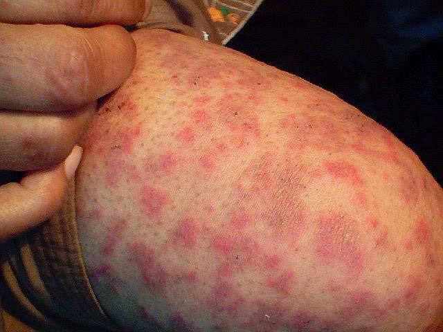
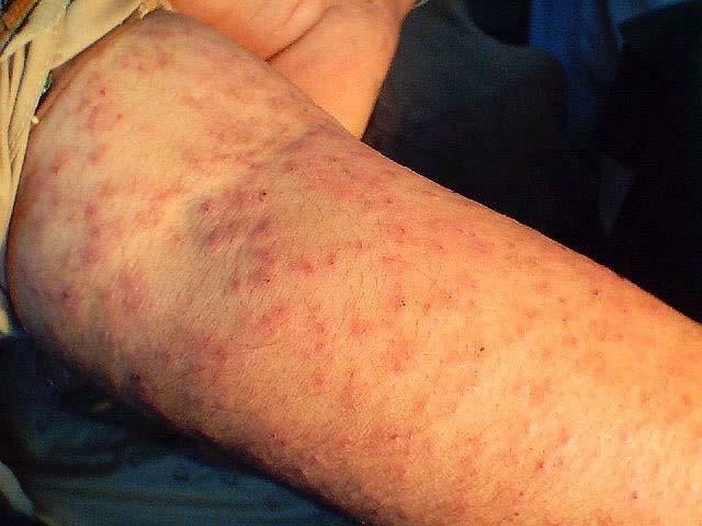
5 Specific Diseases Pemphigus or pemphigoid, skin Pemphigus or pemphigoid, oral Purpura/ vasculitis Stasis 1 st biopsy edge of lesion 2 nd 3 mm from lesion 1 st biopsy 3mm from lesion 2 nd at edge 10 mm from lesion Edge of lesion
6 Specific Diseases Dermatitis herpetiformis Porphyria/ Pseudoporphyria Biopsy normal skin 3mm from lesion Biopsy from edge of a fresh lesion with edge of normal skin
7 Disorders Excluded With Negative IF IgA pemphigus Pemphigus Bullous pemphigoid DLE SLE IgA vasculitis/henoch-schonlein Purpura
8 Disorders with Negative/Nonspecific IF Subcorneal pustulosis Hailey-Hailey disease Bullous impetigo Grover s disease Acantholytic PR Bullous insect bite Bullous drug eruption Lichen planopilaris Drug induced lichenoid photodermatitis Non-IgA associated vasculitis
9
10 Basement Membrane Components Bullous pemphigoid antigens (BP 220/BP180) Epiligrin (Laminin 5) Uncein Ladinin (LAD-1) EBA antigen (Noncollagenous domain of type VII collagen)
11 Location of Components Plasma membrane Lamina lucida Lamina densa BP antigen Laminin Type IV collagen EBA antigen Heparin sulfate
12 Component Epiligrin Uncein Disease Anti-epiligrin cicatricial pemphigoid Some junctional EB Overlap syndrome with features of CP and EBA Ladinin (LAD1) Chronic bullous disease of childhood Linear IgA disease EBA antigen EBA
13
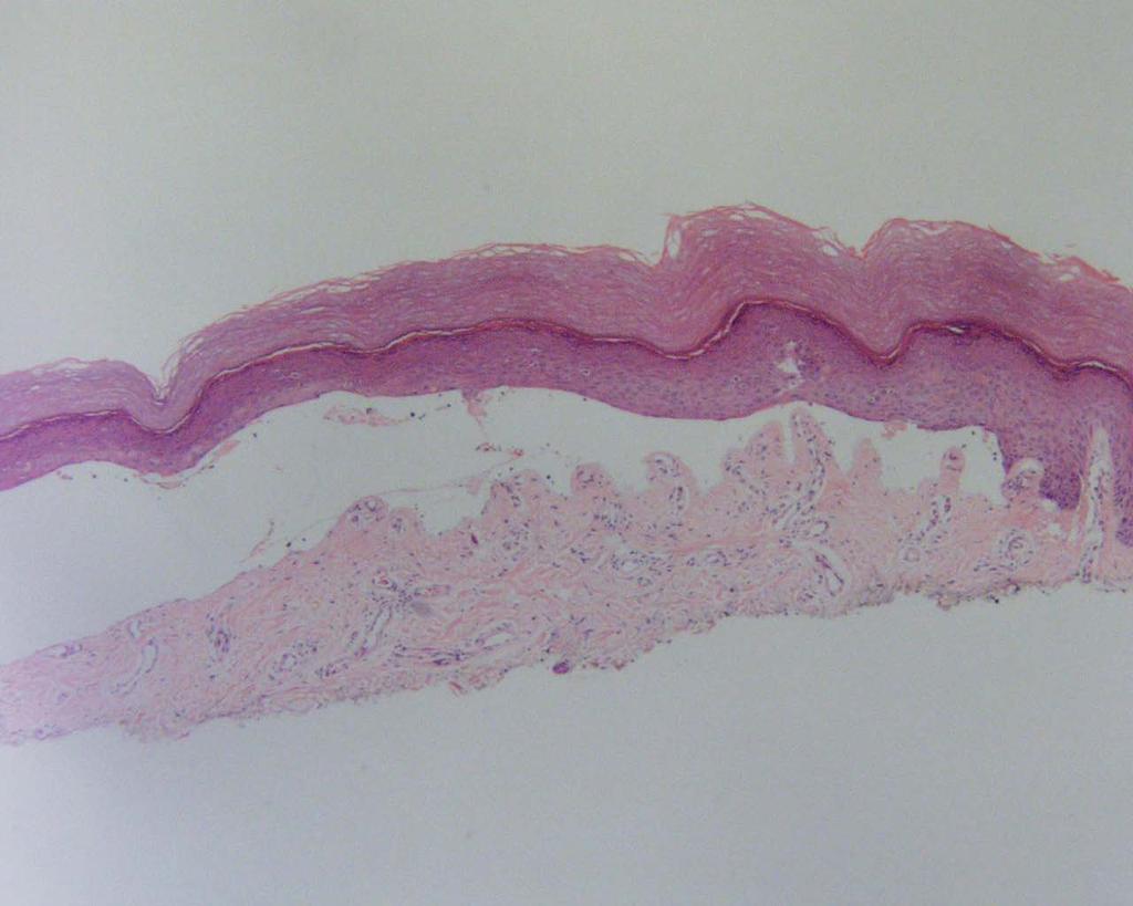
14
15
16
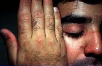
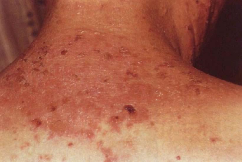
17 Dermatitis Herpetiformis
 scalp Erythematous papules and urticarialike plaques occur less frequently, bullae rare Burning, stinging, and intense pruritus, often precede new lesions Oral mucosa lesions rare")
18 Dermatitis Herpetiformis Flesh-colored to erythematous vesicles appear in a herpetiform pattern Symmetrically distributed over extensor surfaces including elbows, knees, buttocks, shoulders, and the posterior (nuchal) scalp Erythematous papules and urticarialike plaques occur less frequently, bullae rare Burning, stinging, and intense pruritus, often precede new lesions Oral mucosa lesions rare Palms and soles usually spared

19 Dermatitis Herpetiformis and Sprue Majority have some degree of gluten sensitive enteropathy although usually asymptomatic <10% severe 20-30% mild GI disease can be induced by increased gluten intake Gluten-free diet results in normalization of mucosal and skin lesions Resumption of a glutencontaining diet results in recurrence of skin lesions

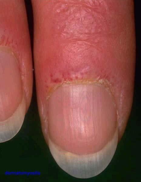
20 Serum Tests IgA endomysial Ab 80% of DH and all of atypical DH Gluten free diet leads to decreased levels

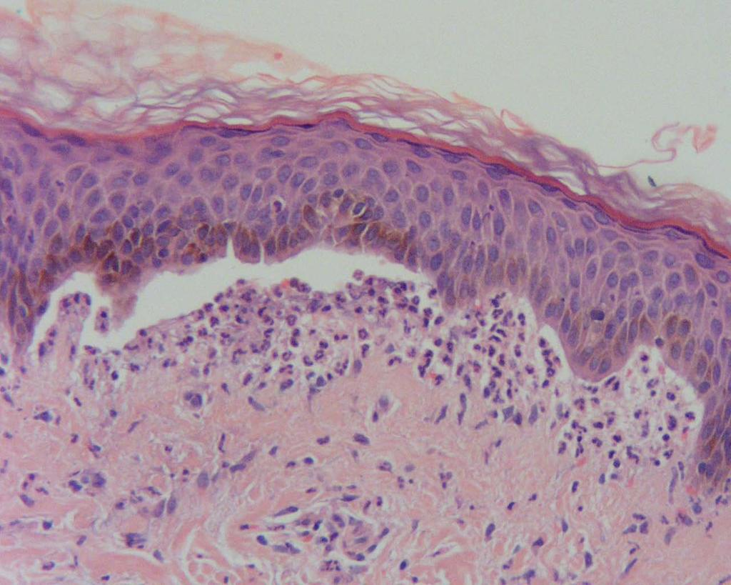
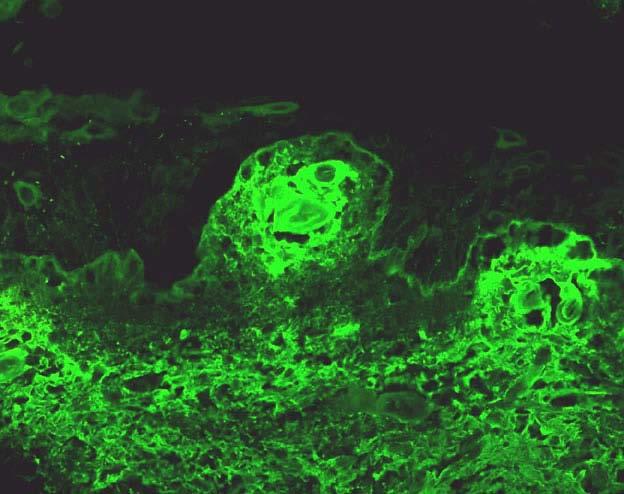
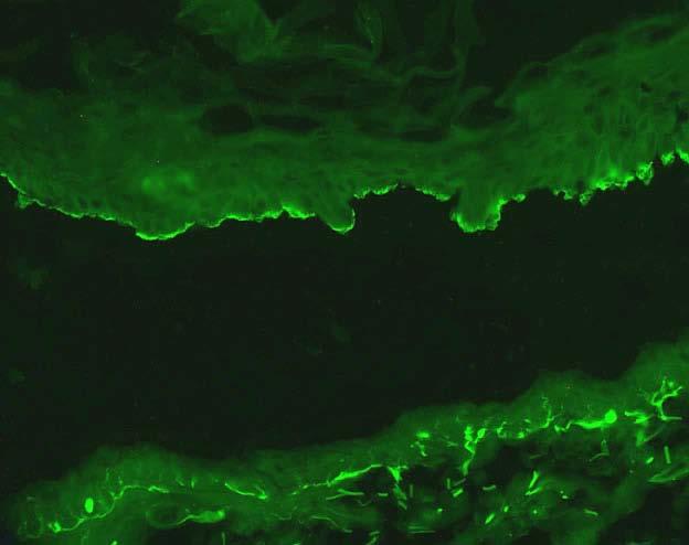
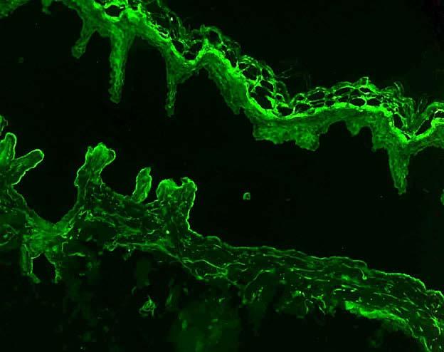
21 Dermatitis Herpetiformis Histopathology Biopsy from normal skin about 3 mm. from the lesion Neutrophils may degrade IgA DIF necessary, rule out Linear IgA disease and subepidermal bullous dermatoses
22 Clues in a monkey s gut! Anti-endomysial Ab bind to reticular structures in smooth muscle in primate esophagus 99% specific for gluten sensitive enteropathy Occur in >80% of DH cases >95% of DH cases with villous atrophy Not affected by dapsone but decreased with gluten free diet If gluten reintroduced, skin lesions precede AEmA and AEmA reappears before villous atrophy
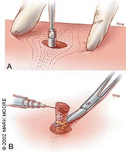
23 Location of Biopsy Skin blister 3 mm biopsy with both the edge of a fresh lesion and some adjacent normal skin Mucosa Perilesional with normal intact mucosa Screen Edge of fresh skin and include scale, if possible
24
25
26
27
28 Bullous Pemphigoid
29 Bullous Pemphigoid Tense bullae on erythematous base Negative Nikolsky Subepidermal bullous dermatosis with eosinophils DDX: Herpes gestationis, Bullous LE, Cicatricial pemphigoid
 Intra and extracellular with collagen-like domains Also called collagen XVII Extramembranous protion is antigenic epitope site for BP and")
30 Bullous Pemphigoid Antigen BPAg1 (220kd) Intracellular associated with hemidesmosomes Homology with desmoplakin 70% of BP pts have circulating Ab to this BPAg2 (180kd) Intra and extracellular with collagen-like domains Also called collagen XVII Extramembranous protion is antigenic epitope site for BP and HG

31 Cicatricial Pemphigoid Brunstig-Perry variant Scarring blisters on head and neck Mucosa rare Antiepiligrin variant Associated with malignancy including endometrial, lung, and stomach May be paraneoplastic blistering disease
32 Cicatricial Pemphigoid-Histopath/IF Subepithelial blister with mixed inflammatory cells Perilesional epithelium shows linear IgG and complement Technically difficult 70% have circulating Ab to BMZ material

33
34
35
36
37
38
39 Pemphigus Vulgaris

40 Pemphigus Vulgaris Mucous membranes, usually oral cavity with erosions Flaccid and fragile skin blister filled with clear fluid that arises on normal skin or erythematous base Vegetating PV frequently in intertriginous areas and scalp or face Nikolsky sign Asboe-Hansen sign

41 Pemphigus Vulgaris Histopathology Intradermal blister with suprabasal clefting and acantholysis May have preceding eosinophilic spongiosis DIF intercellular IgG IgG1 and IgG4 subclasses C3 and IgM less frequent IIF circulating IgG autoantibodies that bind to epidermis 80-90% of patients Ab titer correlates with disease course
42
43
44
45
46 Linear IgA Disease (Chronic Bullous Disease of Childhood)
47 Linear IgA Disease Vesiculobullous eruption on trunk, inner thigh, and pelvic region Asymmetrical unlike DH No gluten association Mucosal scarring Cluster of jewels sign (Discrete or herpetiform pattern) String of beads sign (Annular or polycyclic lesions)

48 Childhood lesions (Chronic bullous disease of childhood) Localized to the lower abdomen and anogenital areas with frequent involvement of the perineum Other sites of involvement include the feet, the hands, and the face, particularly the perioral area

49 Linear IgA Disease-Adults Trunk and the limbs are most commonly affected Perineum and the perioral area is less frequent

50 Linear IgA Disease-Clinical Drug related Vancomycin Penicillin Lithium Dilantin Diclophenac Lesions clear after cessation Rechallenge may have more severe changes

51 Linear IgA Disease-Histology and IF Neutrophil rich interface dermatitis Homogenous sharp linear band for IgA Linear granular variant No deposits within the dermal papillae, resembles DH Low level of HLAB8 and gluten sensitivity unlike DH
52
53
54
55
56
 DLE Biopsy of untreated skin lesion in exposed area that has been present for at least 3 months")
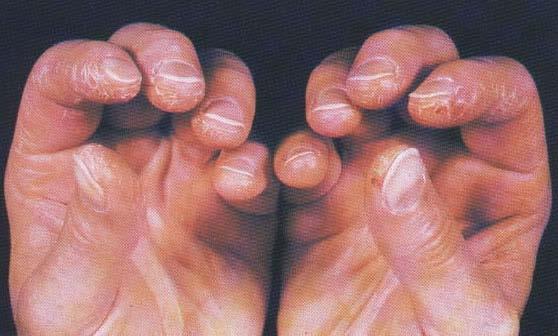
57 Lupus Band Test SLE Best specificity is to take biopsy of normal skin of sun-exposed forearm (Positive in 67%) Normal unexposed skin will be positive only in severe cases (35-40%) DLE Biopsy of untreated skin lesion in exposed area that has been present for at least 3 months
58 Lupus Band Test-Baseline Deposition of Ig at the DEJ in lesional and nonlesional skin IgM most frequent deposit IgA least frequent Granular pattern most frequent Sharp linear band not accepted
59 Baseline Sun exposed skin Non-sun exposed IgM continuous distribution over at least 50% width of biopsy with moderate intensity 25% of normal skin show weak interrupted linear granular IgM/C1q Interrupted IgM of moderate intensity If IgA present, high specificity
60 Sensitivity and Specificity SLE DLE 70-80% of patients with SLE in sun-exposed skin Non sun-exposed non lesional skin, only positive in SLE pts with severe extracutaneous disease and positive for DS DNA Positive in 90% interface dermatitis Negative in non-scarring cases Positive in 90% interface dermatitis

61 Lupus Band Test and Prognosis 70% of patients with active nephritis with LBT on normal skin C1q deposits-higher incidence of renal disease
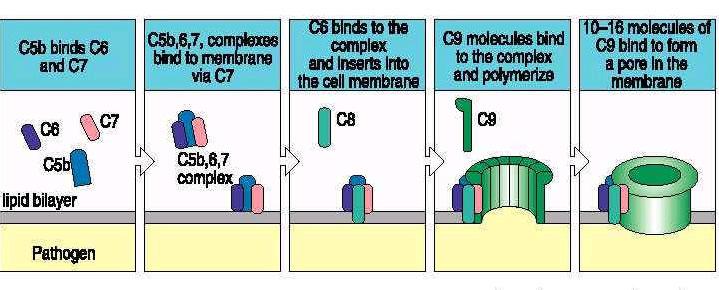
62
63
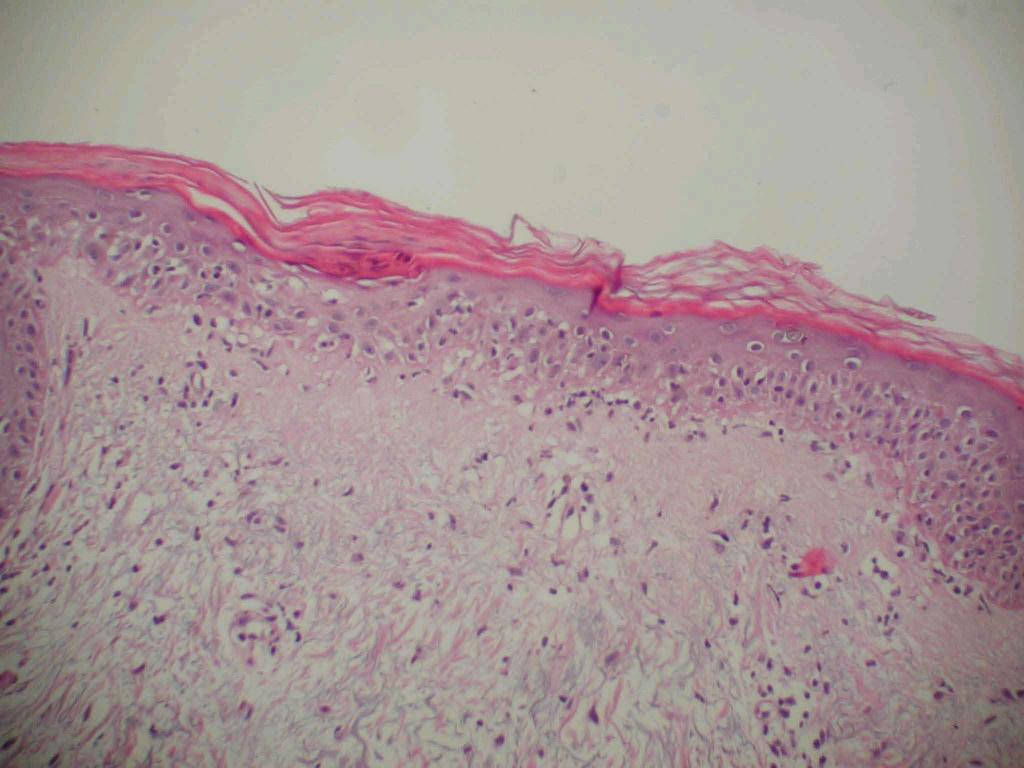
64
65
66
67 Dermatomyositis
 Photodistributed Facial")
68 Dermatomyositis Heliotrope rash and Gottron papules Malar erythema Poikiloderma Periungual telangiectases Poikiloderma may occur on exposed skin or the upper part of the back (Shawl sign) Photodistributed Facial erythema rarely

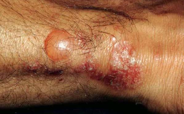
69 Dermatomyositis Scalp involvement in DM is relatively common (coup d sabre) Calcinosis of the skin or the muscle Unusual in adults 40% of children or adolescents Calcinosis cutis manifests as firm yellow nodules

70 Dermatomyositis Muscle enzyme levels abnormal CK, AST, LDH Myositis-specific antibodies (antisignal recognition protein and anti-ku) ANA positive Anti-Mi-1 is highly specific for DM, but it lacks sensitivity because only 25% Anti-Jo-1 is associated with pulmonary involvement, more common in patients with PM than DM

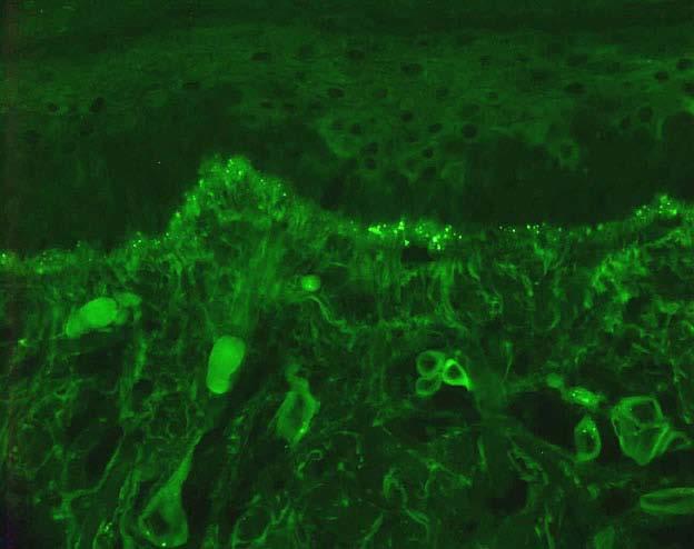
71 Dermatomyositis Histopathology Cell poor interface dermatitis with dermal mucinosis May be identical to DLE, SCLE, SLE DIF with Variable Lupus band Utilize C5b-9
72 C5b-9 and Disease Terminal complement, membrane attack complex (MAC) Formation of membrane pores allow circulating Ab access to nucleus and cytoplasm Represent activation of complement pathway within the BMZ
73
74 MAC Diseases and Patterns SLE Intense granular DEJ 80% SCLE Granular DEJ 60% Granular nuclear/cytoplasmic epidermal DLE DEJ 60% MCTD Granular nuclear/cytoplasmic epidermal 100% DEJ 100% Dermatomyositis DEJ 90% Endothelial cells Dermal papillary capillaries

75 Overlap Anti-Ro associated SLE, Dermatomyositis, and MCTD Endothelial C5b-9 Endothelial cell necrosis and denudement Reduction in vascular plexus Granular and cytoplasmic decoration keratinocytes Differentiate by LBT and clinical Non-lesional skin Usually negative or very weak
76
77
78
79 Epidermolysis Bullosa Acquisita

80 Epidermolysis Bullosa Acquisita Noninflammatory or mildly inflammatory form Extensor surfaces of hands, knuckles, elbows, knees, and ankles Blisters may be hemorrhagic Scar and milia Nail dystrophy and scarring alopecia rarely DDX: AD epidermolysis bullosa dystrophica in children PCT in adults
81 EBA-Pathogenesis IgG autoantibodies targeting non-collagenous domain of collagen VII in basement membrane Subset of clinically milder EBA IgA autoantibodies IgG autoantibodies to the collagenous domain, rather than the NC1 domain of collagen VII
82 EBA Histopathology Subepidermal blister with mixed inflammation DIF linear thick band of IgG, and to a lesser extent C3 at basement membrane zone Occasionally IgM or IgA IIF IgG circulating autoantibodies in the patient's serum that target the skin basement membrane component, type VII collagen. Bind to the dermal floor (lower part) on salt-split normal human skin substrate
83 Salt-Split Skin Assay IIF-utilize patient s serum Incubate normal skin with 1M NaCl Separates the epidermis from dermis Epidermal half Upper lamina lucida and hemidesmosomes BP antigen Dermal half Laminin 5 Lamina densa, anchoring fibrils
84
85
86 SSS Assay Split Epidermal Dermal Disease Bullous pemphigoid EBA Bullous lupus erythematosus Anti-epiligrin cicatricial pemphigoid Anti-p105 bullous pemphigoid
87
88
89
90
91 Porphyria Cutanea Tarda

92 Clinical Fragility of sun-exposed skin after trauma Erosions and bullae on the dorsal aspects of the hands, the forearms, and the face Healing of crusted erosions and blisters leaves scars, milia, and hyperpigmented and hypopigmented atrophic patches. Hypertrichosis Sclerodermal-like plaques-upper trunk Melasma-like facies Erythematous suffusion face, trunk Scarring alopecia and onocholysis Urine sample may have a tea- or wine-colored tint

 DIF with immunoglobulins and complement in and around the dermal capillaries and at the")
93 Porphyria Subepidermal bullae with minimal dermal inflammatory infiltrate festooning of dermal papillae Thickened upper dermal capillary walls and dermoepidermal basement membrane zones Elastosis and sclerosis Trapped basement membrane zone (caterpillar bodies) in epidermal roof (Ab to Collagen IV/laminin) DIF with immunoglobulins and complement in and around the dermal capillaries and at the basement membrane zone

94 Questions The trouble with facts is that there are so many of them. Samuel McChord Crothers The Gentle Reader
Background information of DIF
 Napa Dermatopathology Meeting 2018: Immunobullous Disease Whitney A. High, MD, JD, MEng whitney.high@ucdenver.edu Professor of Dermatology & Pathology Vice-Chairman, Dermatology Director of Dermatopathology
Autoimmune Diseases with Oral Manifestations
 Autoimmune Diseases with Oral Manifestations Martin S. Greenberg DDS, FDS RCSEd Professor Emeritus Department of Oral Medicine University of Pennsylvania Disclosure Statement I have no actual or potential
B. Autoimmune blistering diseases
 Go Back to the Top To Order, Visit the Purchasing Page for Details formation immediately above the basal layer. The dermal papillae, which are covered by basal cells in the single layer that is left in
Acquired and Inherited Bullous Diseases
 Acquired and Inherited Bullous Diseases Erin Wei MD Brigham and Women s Hospital, Department of Dermatology Instructor, Harvard Medical School Director, Bullous Disease Clinic No disclosures Conflict of
Immunobullous Diseases: Review and Update. May P. Chan, MD Associate Professor of Pathology and Dermatology University of Michigan
 Immunobullous Diseases: Review and Update May P. Chan, MD Associate Professor of Pathology and Dermatology University of Michigan Diagnosis of Immunobullous Diseases Clinical H&E DIF DIAGNOSIS IIF ELISA
Classification: 1. Infective: 2. Traumatic: 3. Idiopathic: Recurrent Aphthous Stomatitis (RAS) 4. Associated with systemic disease:
 4. Associated with systemic disease:") Classification: 1. Infective: 2. Traumatic: 3. Idiopathic: Recurrent Aphthous Stomatitis (RAS) 4. Associated with systemic disease: Hematological GIT Behcet s HIV 5. Associated with dermatological diseases:
Classification: 1. Infective: 2. Traumatic: 3. Idiopathic: Recurrent Aphthous Stomatitis (RAS) 4. Associated with systemic disease: Hematological GIT Behcet s HIV 5. Associated with dermatological diseases:
Epidermolysis Bullosa Acquisita
 Introduction Epidermolysis Bullosa Acquisita Pages with reference to book, From 192 To 194 Nasser Rashid Dar ( Departments of Dermatology, Combined Military Hospital, Peshawar. ) Ahsan Hameed, Ashfaq Ahmad
السكري للداء مرافقة فقاعات diabeticorum= Bullosis
 1 / 6 Bullosis diabeticorum Bullous disease of diabetes (bullosis diabeticorum) is a distinct, spontaneous, noninflammatory, blistering condition of acral skin unique to patients with diabetes mellitus.
Dr Saleem Taibjee. Consultant Dermatologist & Dermatopathologist
 Dr Saleem Taibjee saleem.taibjee@dchft.nhs.uk Consultant Dermatologist & Dermatopathologist Case S14-10797 and S15-4023 F50. Previous blistering, now marked milia on dorsum of hands. 4mm punch biopsy The
Erythema gyratumrepens-like eruption in a patient with epidermolysisbullosaacquisita associated with ulcerative colitis
 Erythema gyratumrepens-like eruption in a patient with epidermolysisbullosaacquisita associated with ulcerative colitis A. España C. Sitaru* M. Pretel L. Aguado J. Jimenez# Department of Dermatology, University
CLINCOPATHOLOGICAL CASE
 CLINCOPATHOLOGICAL CASE Generalized vesiculo-bullous and pustular eruption in an adult man Hassab El-Naby H, MD, El-Khalawany M, MD Department of Dermatology, Al-Azhar University, Cairo, Egypt CLINICAL
HEMORRHAGIC BULLOUS HENOCH- SCHONLEIN PURPURA: A CASE REPORT
 HEMORRHAGIC BULLOUS HENOCH- SCHONLEIN PURPURA: A CASE REPORT Nirmala Ponnuthurai, Sabeera Begum, Lee Bang Rom Paediatric Dermatology Unit, Institute of Paediatric, Hospital Kuala Lumpur, Malaysia Abstract
Cutanous Manifestation of Lupus Erythematosus. Presented By: Dr. Naif S. Al Shahrani Salman Bin Abdaziz university
 Cutanous Manifestation of Lupus Erythematosus Presented By: Dr. Naif S. Al Shahrani Salman Bin Abdaziz university A 50-year old lady, who is otherwise healthy, presented to the dermatology clinic with
Autoimmune bullous dermatoses
 Autoimmune bullous dermatoses Overview of serological diagnostics in autoimmune blister-forming diseases of the skin Pemphigoid diseases Pemphigus diseases Epidermolysis bullosa acquisita Dermatitis herpetiformis
Original Contribution
 Direct Immunofluorescence Test of Skin Biopsy Samples Results of 204 Cases Kabir AN, 1 Das RK, 2 Kamal M 3 Direct immunofluorescence (DIF) test of skin and renal biopsy specimens is being done on regular
A cross-sectional study of clinical, histopathological and direct immmunofluorescence diagnosis in autoimmune bullous diseases
 Original Article A cross-sectional study of clinical, histopathological and direct immmunofluorescence diagnosis in autoimmune bullous diseases Anchal Jindal, MD 1 Rushikesh Shah, MBBS 2 Neela Patel, MD
Bullous Pemphigoid with Lymphocytic Colitis: A Case Report and Short Literature Review
 Dermatol Ther (Heidelb) (2016) 6:437 441 DOI 10.1007/s13555-016-0135-4 CASE REPORT Bullous Pemphigoid with Lymphocytic Colitis: A Case Report and Short Literature Review Alexandra Sperl. Johann W. Bauer.
Clinicopathological correlation of blistering diseases of skin
 Bangladesh Med Res Counc Bull 2008; 34: 48-53 Copyright 2008 by Bangladesh Medical Research Council Clinicopathological correlation of blistering diseases of skin A.K.M. Nurul Kabir 1, Mohammed Kamal 1
Original Article ABSTRACT
 Original Article doi: 10.5146/tjpath.2015.01345 Utility of Direct Immunofluorescence Studies in Subclassification of Autoimmune Sub-Epidermal Bullous Diseases: A 2-Year Study in a Tertiary Care Hospital
A case of bullous pemphigoid following pemphigus foliaceus
 #2228 A case of bullous pemphigoid following pemphigus foliaceus Priyanka Vedak MD 1, Danielle Levine MD 1,3, Lyn Duncan MD 2,3, Hensin Tsao 1,3, Daniela Kroshinsky MD MPH 1,3 1. Department of Dermatology,
Actinic keratosis (AK): Dr Sarma s simple guide
: Dr Sarma s simple guide") Actinic keratosis (AK): Dr Sarma s simple guide Actinic keratosis is a very common lesion that you will see in your day-to-day practice. First, let me explain the name Actinic keratosis. It means keratosis
Actinic keratosis (AK): Dr Sarma s simple guide Actinic keratosis is a very common lesion that you will see in your day-to-day practice. First, let me explain the name Actinic keratosis. It means keratosis
Department of Dermatology, Nippon Medical School, 1-1-5, Sendagi, Bunkyo-ku, Tokyo , Japan 2
 Dermatology Research and Practice Volume 2010, Article ID 931340, 5 pages doi:10.1155/2010/931340 Case Report Paraneoplastic Pemphigus Presenting as Mild Cutaneous Features of Pemphigus Foliaceus and Lichenoid
To Correlate Clinical Diagnosis with Histopathology and DIF Pattern of Autoimmune Based Vesiculobullous Disorders In A Tertiary Teaching Hospital
 IOSR Journal of Dental and Medical Sciences (IOSR-JDMS) e-issn: 2279-0853, p-issn: 2279-0861.Volume 17, Issue 7 Ver. 2 (July. 2018), PP 01-06 www.iosrjournals.org To Correlate Clinical Diagnosis with Histopathology
Interesting Case Series. Linear IgA Bullous Dermatosis
 Interesting Case Series Linear IgA Bullous Dermatosis Sean Chen, BA, a Peter Mattei, MD, a Max Fischer, MD, MPH, a Joshua D. Gay, PA-C, b Stephen M. Milner, MBBS, BDS, FRCS (Ed), FACS, b and Leigh Ann
Introduction to Pemphigoid: Spectrum of Disease & Treatment
 Introduction to Pemphigoid: Spectrum of Disease & Treatment A Razzaque Ahmed, MD Center for Blistering Diseases Boston, MA A.Razzaque.Ahmed@tufts.edu centerforblisteringdiseases.com SPECTRUM OF PEMPHIGOID
DERMATOLOGY VOLUME 40 NUMBER 5 PART 1 MAY 1999
 CONTINUING MEDICAL EDUCATION The new pemphigus variants Journal of the American Academy of DERMATOLOGY VOLUME 40 NUMBER 5 PART 1 MAY 1999 Neha D. Robinson, MD, a Takashi Hashimoto, MD, b Masayuki Amagai,
Index. derm.theclinics.com. Note: Page numbers of article titles are in boldface type.
 Note: Page numbers of article titles are in boldface type. A Adhesion and migration, the diverse functions of the laminin a3 subunit, 79 87 Alopecia in epidermolysis bullosa, 165 169 Amblyopia and inherited
Current concepts of autoimmune bullous diseases Advances in pathogenesis. Luca Borradori
 Current concepts of autoimmune bullous diseases Advances in pathogenesis Luca Borradori Dept. of Dermatology Inselspital, University Hospital of Berne Switzerland Luca.Borradori@insel.ch Autoimmune bullous
Autoimmune bullous disorders 1)
") Clin Chem Lab Med 2006;44(2):144 149 2006 by Walter de Gruyter Berlin New York. DOI 10.1515/CCLM.2006.027 2006/39 Review Autoimmune bullous disorders 1) Rüdiger Eming* and Michael Hertl for the members
Clin Chem Lab Med 2006;44(2):144 149 2006 by Walter de Gruyter Berlin New York. DOI 10.1515/CCLM.2006.027 2006/39 Review Autoimmune bullous disorders 1) Rüdiger Eming* and Michael Hertl for the members
S003 CPC Self-Assessment
 S003 CPC Self-Assessment Alina G. Bridges, D.O. Associate Professor Program Director, Dermatopathology Fellowship Department of Dermatology, Division of Dermatopathology and Cutaneous Immunopathology Mayo
Indian Journal of Dermatopathology and Diagnostic Dermatology
 Volume 1 Issue 1 Jan-Jun 2014 Online full text at www.ijdpdd.com Indian Journal of Dermatopathology and Diagnostic Dermatology DSI IADVL Karnataka Branch Official Publication of Dermatopathology Society
Title. CitationBritish Journal of Dermatology, 155(5): Issue Date Doc URL. Rights. Type. File Information
: Issue Date Doc URL. Rights. Type. File Information") Title Childhood epidermolysis bullosa acquisita with autoa collagen : case report and review of the literature Author(s)Mayuzumi, M.; Akiyama, M.; Nishie, W.; Ukae, S.; Abe CitationBritish Journal of Dermatology,
Title Childhood epidermolysis bullosa acquisita with autoa collagen : case report and review of the literature Author(s)Mayuzumi, M.; Akiyama, M.; Nishie, W.; Ukae, S.; Abe CitationBritish Journal of Dermatology,
Department of Dermatology, Christian Medical College and Hospital, Ludhiana, Punjab, India.
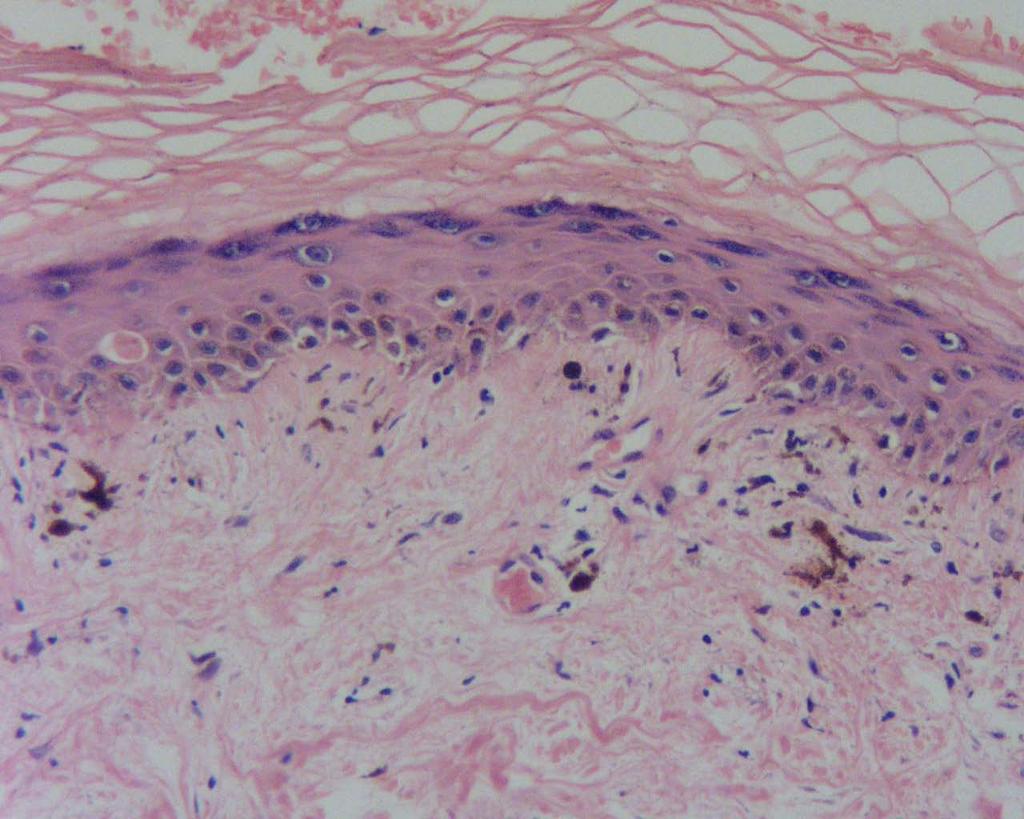
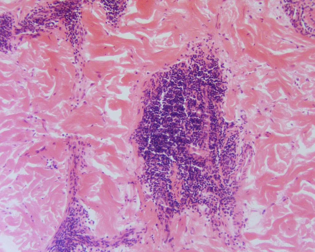
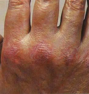
 Bullous pemphigoid mimicking granulomatous inflammation Abhilasha Williams, Emy Abi Thomas. Department of Dermatology, Christian Medical College and Hospital, Ludhiana, Punjab, India. Egyptian Dermatology
BSD SELF-ASSESSMENT CASES 21-24
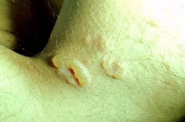
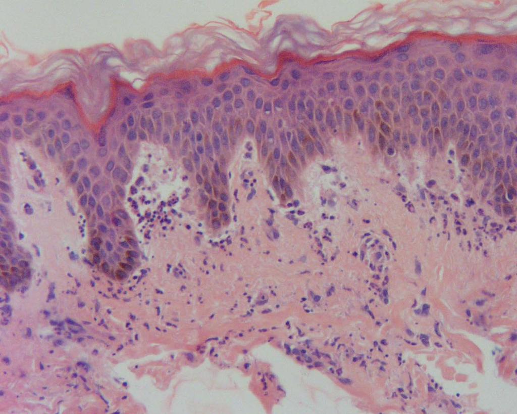
 BSD SELF-ASSESSMENT CASES 21-24 EDINBURGH, 2 JULY 2018 LASZLO IGALI CASE 21 CLINICAL HISTORY Female, 43 years, nodule on left side of face/jaw angle area. Slow growth over years. MACRO (NOT GIVEN) Skin
Figure 25.1 Figure 25.2
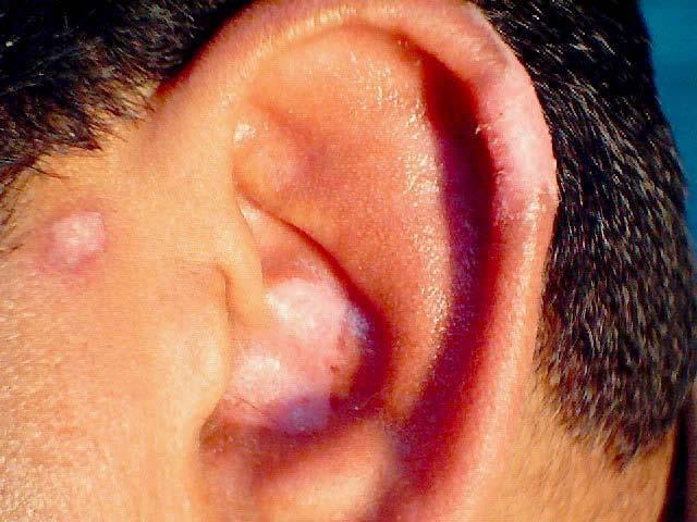
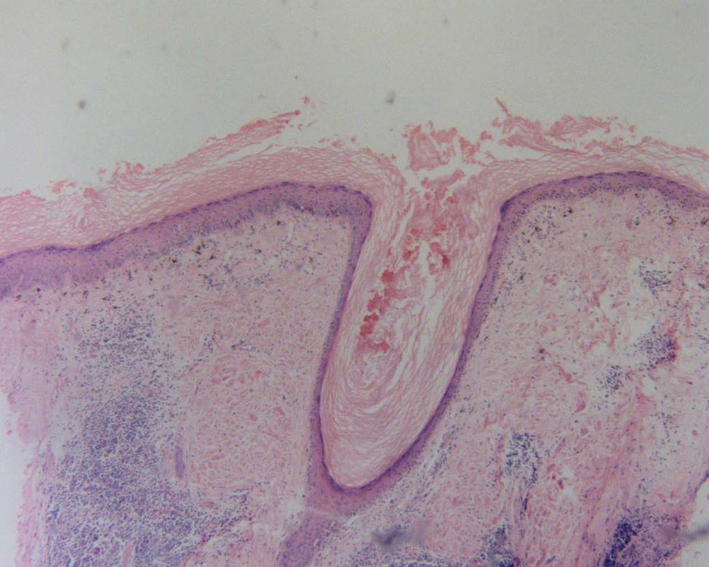
 CASE 25 Patient: A 75-year-old Thai man from Lamphun Chief Complaint: 6-month-history of itchy vesicles at both thighs and elbows, upper back and sacral area Present Illness: The patient presented with
atorvastatin 10mg, amlodipine 5mg and dilitazem 60mg. He had unexplained iron deficiency anaemia (hemoglobin-8.4gm/dl, ferritin- 4.73ng/ml, total iron
 Pemphigus herpetiformis : A rare clinical variant of pemphigus Shrestha P 1, Tajhya RB 2, Pokharel A 3 1,2 Consultant Dermatologist, Department of Dermatology, Vayodha Hospital Pvt. Ltd, Balkhu, Kathmandu,
A. Erythema multiforme and related diseases
 Go Back to the Top To Order, Visit the Purchasing Page for Details Chapter Erythema, Erythroderma (Exfoliative Dermatitis) Erythema is caused by telangiectasia or hyperemia in the papillary and reticular
Pemphigus in younger age group in Bangladeshi population
 ORIGINAL ARTICLE in younger age group in Bangladeshi population Abdul Wahab 1, MD, Lubna Khondker 1, MD, Jamal Uddin 1, MD, Ishrat Bhuiyan 2, MD Shirajul Islam Khan 3, MD, Zafrul Islam 1, MD, Rahmat Ali
Blistering disorders and their differential diagnosis and management
 and their differential diagnosis and management STEVE KOSSARD, MB BS, PhD, FACD The type and pattern of cutaneous blisters may provide an essential clue to the diagnosis of specific dermatological disorders.
MUCOCUTANEOUS LESIONS Normal structures in epithelium cell adhesion to each other and to underlying connective tissue:
 ORAL DERMATOSES AND MUCOSAL/GINGIVAL LESIONS MUCOCUTANEOUS LESIONS Normal structures in epithelium cell adhesion to each other and to underlying connective tissue: Diagram taken from: Oral and Maxillofacial
EPIDERMOLYSIS BULLOSA
 EPIDERMOLYSIS BULLOSA Definition Epidermolysis bullosa (EB) is a term used to describe a group of rare mainly hereditary, chronic, non-inflammatory diseases of skin and mucous membranes. EB is characterized
Immunofluorescence in Oral Dermatological Disorders- No Shiny Matter
 Journal of Academy of Dental Education Journal of Academy of Dental Education, 24-28, DOI: 10.18311/jade/2015-2016/15951 ISSN (Print): 2348-1595 ISSN (Online) : 2348-2621 Immunofluorescence in Oral Dermatological
Sarolta Kárpáti. Technology Transfer in Diagnostic Pathology, 5th Central European Regional Meeting May 1, 2010, Siófok
 Blistering diseases Sarolta Kárpáti SEMMELWEIS UNIVERSITY, BUDAPEST Technology Transfer in Diagnostic Pathology, 5th Central European Regional Meeting May 1, 2010, Siófok Blistering diseases Autoimmune
Vesiculobullous Diseases
 Vesiculobullous Diseases Larkin Community Hospital/NSU-COM Presenters: Yuri Kim, DO, Sam Ecker, DO, Jennifer David, DO, MBA Program Director: Stanley Skopit, DO, MSE, FAOCD, FAAD We have no relevant disclosures
Principi ed Aggiornamenti in Dermatologia Roma, 6-7 Aprile Grand rounds. Lorenzo Cerroni, Graz
 Principi ed Aggiornamenti in Dermatologia Roma, 6-7 Aprile 2018 Grand rounds Lorenzo Cerroni, Graz "Computer palms" Described in patient using computer keyboards for long periods; similar features described
Egyptian Dermatology Online Journal Vol. 8 No 2: 6, December Yasmeen J Bhat*, Iffat Hasan*, Atiya Yaseen*, Hina Altaf*, Shylla Mir**
 Pemphigoid gestationis in a multigravida Yasmeen J Bhat*, Iffat Hasan*, Atiya Yaseen*, Hina Altaf*, Shylla Mir** * Department of Dermatology, STD & Leprosy; Government Medical College, Srinagar ** Department
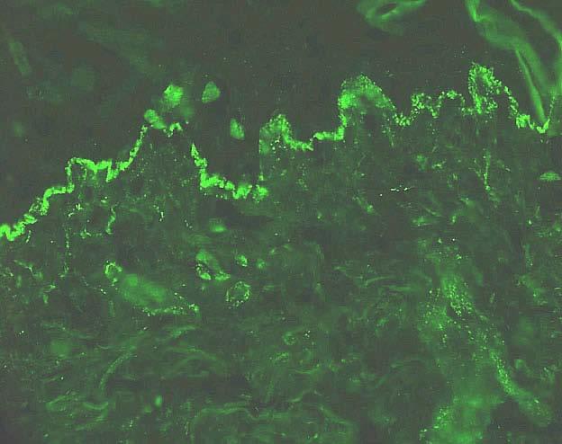
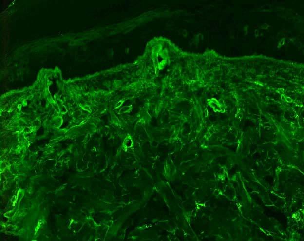
substance staining with IgG, C3 and IgA (trace) Linear deposition of IgG(+), IgA.M(trace) and C3(+++) at the DEJ
 Linear deposition of IgG(+), IgA.M(trace) and C3(+++) at the DEJ") Direct Immunofluorescence: Skin Diagnosis Findings Picture Pemphigus Vulgaris and it s Intracellular cement variants substance staining with IgG, C3 and IgA (trace) Bullous Pemphigoid and it s variants
Direct Immunofluorescence: Skin Diagnosis Findings Picture Pemphigus Vulgaris and it s Intracellular cement variants substance staining with IgG, C3 and IgA (trace) Bullous Pemphigoid and it s variants
LESIONS OF THE ORAL CAVITY ORAL CAVITY. Oral Cavity Subsites 4/10/2013 LIPS TEETH GINGIVA ORAL MUCOUS MEMBRANES PALATE TONGUE ORAL LYMPHOID TISSUES
 LESIONS OF THE ORAL CAVITY David I. Kutler, MD, FACS Associate Professor Division of Head and Neck Surgery Department of Otolaryngology HNS Weill Cornell Medical Center ORAL CAVITY LIPS TEETH GINGIVA ORAL
When your patient complains of
 WILLIAM LAWSON, MD, DDS Mount Sinai School of Medicine ABSTRACT: Certain clues can help you identify the cause of bullous oral lesions. Diffuse oral and labial bullous erosions, sometimes accompanied by
Skin Deep: Cutaneous Lupus. Dr Sarah Sasson Immunology Registrar, Liverpool Hospital 2016
 Skin Deep: Cutaneous Lupus Dr Sarah Sasson Immunology Registrar, Liverpool Hospital 2016 Introduction: Cutaneous lupus erythematosus LE is an autoimmune disease with a range of clinical manifestations
An Approach to Common and not so Common Rashes in the Office FMF 2014 Christie Freeman MD, CCFP, DipPDerm, MSc
 An Approach to Common and not so Common Rashes in the Office FMF 2014 Christie Freeman MD, CCFP, DipPDerm, MSc 1 Common Rashes Tinea Corporis: Annular- this is not the only criteria Advancing erythematous
Mucous membrane pemphigoid in a patient with hypertension treated with atenolol: a case report
 Kanjanabuch et al. Journal of Medical Case Reports 2012, 6:373 JOURNAL OF MEDICAL CASE REPORTS CASE REPORT Open Access Mucous membrane pemphigoid in a patient with hypertension treated with atenolol: a
Some skin conditions
 Some skin conditions Some skin conditions Acute Inflammatory Dermatoses Chronic Inflammatory Dermatoses Blistering (Bullous) Diseases Panniculitis Disorders of Epidermal Appendages -Urticaria -Acute eczematous
My Method for Approaching Skin Biopsies
 My Method for Approaching Skin Biopsies P A U L H A U N, MD, MS, F A A D A S S I S T A N T P R O F E S S O R D E R M A T O L O G Y A N D D E R M A T O P A T H O L O G Y D E P A R T M E N T O F D E R M
Pharmacologyonline 1: 1-6 (2010) Case Report Ravishankar and Hiremath CIPROFLOXACIN INDUCED BULLOUS PEMPHIGOID: A CASE REPORT
 Case Report Ravishankar and Hiremath CIPROFLOXACIN INDUCED BULLOUS PEMPHIGOID: A CASE REPORT") CIPROFLOXACIN INDUCED BULLOUS PEMPHIGOID: A CASE REPORT Ravishankar AC 1*, Hiremath SV 1 1 Dept of Pharmacology and Pharmacotherapeutics, JN Medical College, Belgaum, India. Summary Bullous pemphigoid
CIPROFLOXACIN INDUCED BULLOUS PEMPHIGOID: A CASE REPORT Ravishankar AC 1*, Hiremath SV 1 1 Dept of Pharmacology and Pharmacotherapeutics, JN Medical College, Belgaum, India. Summary Bullous pemphigoid
A RARE CASE OF LICHEN PLANUS PEMPHIGOIDES Ashok Jain 1, Anjali Dalal 2
 A RARE CASE OF LICHEN PLANUS PEMPHIGOIDES Ashok Jain 1, Anjali Dalal 2 HOW TO CITE THIS ARTICLE: Ashok Jain, Anjali Dalal. A Rare Case of Lichen Planus Pemphigoides. Journal of Evolution of Medical and
Importance of serological tests in diagnosis of autoimmune blistering diseases
 doi: 10.1111/1346-8138.12703 Journal of Dermatology 2015; 42: 3 10 REVIEW ARTICLE Importance of serological tests in diagnosis of autoimmune blistering diseases Ken ISHII Department of Dermatology, Toho
Recent Advances in the Molecular Pathology of Bullous Skin Disorders
 1 Bahrain Medical Bulletin, Vol. 27, No. 2, June 2005 Recent Advances in the Molecular Pathology of Bullous Skin Disorders John A McGrath* Maintenance of an intact epidermis depends on secure adhesion
Review Article Clinical Relevance of Autoantibodies in Patients with Autoimmune Bullous Dermatosis
 Clinical and Developmental Immunology Volume 2012, Article ID 369546, 9 pages doi:10.1155/2012/369546 Review Article Clinical Relevance of Autoantibodies in Patients with Autoimmune Bullous Dermatosis
Common Cutaneous Signs of Medical Illnesses
 Common Cutaneous Signs of Medical Illnesses DR COLIN THENG MBBS, MMED (FAM. MED), MRCP(UK), FAMS SENIOR CONSULTANT DERMATOLOGIST THE SKIN SPECIALISTS & LASER CLINIC MOUNT ALVERNIA MEDICAL CENTRE D, #07-61
Proceedings of the Southern European Veterinary Conference - SEVC -
 Close this window to return to IVIS www.ivis.org Proceedings of the Southern European Veterinary Conference - SEVC - Sep. 30-Oct. 3, 2010, Barcelona, Spain Next SEVC Conference: Sep. 30-Oct. 2, 2011 -
Dermatitis Herpetiformis (DH) in Association with H. pylori Infection: Description of a Case Report
 in Association with H. pylori Infection: Description of a Case Report") British Journal of Medicine & Medical Research 1(3): 163-169, 2011 SCIENCEDOMAIN international www.sciencedomain.org Dermatitis Herpetiformis (DH) in Association with H. pylori Infection: Description of
British Journal of Medicine & Medical Research 1(3): 163-169, 2011 SCIENCEDOMAIN international www.sciencedomain.org Dermatitis Herpetiformis (DH) in Association with H. pylori Infection: Description of
Retrospective 10 years review of 100 patients with psoriasis in the Kingdom of Saudi Arabia (KSA)
") Retrospective 10 years review of 100 patients with psoriasis in the Kingdom of Saudi Arabia (KSA) Ahmed Abdullah Alhumidi King saud university, Riyadh, kingdom of Saudi Arabia Abstract Background: This
Retrospective 10 years review of 100 patients with psoriasis in the Kingdom of Saudi Arabia (KSA) Ahmed Abdullah Alhumidi King saud university, Riyadh, kingdom of Saudi Arabia Abstract Background: This
BULLOUS SYSTEMIC lupus erythematosus
 OBSERVATION Bullous Systemic Lupus Erythematosus With Autoantibodies Recognizing Multiple Skin Basement Membrane Components, Bullous Pemphigoid Antigen 1, Laminin-5, Laminin-6, and Type VII Collagen Lawrence
AUTOIMMUNE BLISTERING DISEASES; WINDOW TO SYSTEMIC DISEASE
 AUTOIMMUNE BLISTERING DISEASES; WINDOW TO SYSTEMIC DISEASE Ron Feldman, MD, PhD Assistant Professor of Dermatology Emory University ron.j.feldman@emory.edu Disclosures I have no conflict of interest to
Bullous Eruption: A Manifestation of Lupus Erythematosus
 CONTINUING MEDICAL EDUCATION : A Manifestation of Lupus Erythematosus CPT Ronea Harris-Stith, USAF, MC; CPT Quenby L. Erickson, USAF, MC; Dirk M. Elston, MD; COL Kathleen David-Bajar, MC, USA GOAL To gain
A clinical syndrome, composed mainly of:
 Nephritic syndrome We will discuss: 1)Nephritic syndrome: -Acute postinfectious (poststreptococcal) GN -IgA nephropathy -Hereditary nephritis 2)Rapidly progressive GN (RPGN) A clinical syndrome, composed
Autoimmune Blistering Disease
 life. science. discovery. life. science. discovery. Autoimmune Blistering Disease - Diagnostic Methodology for Pemphigus and Pemphigoid - Pemphigus Epidermal cell-cell junction EBA Epidermal side Epidermal
Blistering skin conditions
 THEME weird skin stuff Belinda Welsh MBBS, MMed, FACD, is consultant dermatologist, St Vincent's Hospital, Melbourne and Sunbury Dermatology and Skin Cancer Clinic, Sunbury, Victoria. drbwelsh@bigpond.net.au
2018 Oregon Dental Conference Course Handout Denis Lynch, DDS, PhD
 2018 Oregon Dental Conference Course Handout Denis Lynch, DDS, PhD Course 9148: Diagnosis and Treatment of Recurrent Oral Ulcers Friday, April 6 9 am - 12 pm Diagnosis and Treatment of Recurrent Oral Ulcers
Inflammatory Dermatoses of the Vulva for the General/Gyn Pathologist with emphasis in the lichenoid pattern
 Inflammatory Dermatoses of the Vulva for the General/Gyn Pathologist with emphasis in the lichenoid pattern By Konstantinos Linos MD, FCAP, FASDP Bone, Soft Tissue and Dermatopathology Assistant Professor
=ﻰﻤاﻤﺤﻠا ﺔﻴﻘﻠﺤﻠا ﺔذﺒاﻨﻠا
 1 / 15 Erythema Annulare Centrifugum and Other Figurate Erythemas The figurate erythemas include a variety of eruptions characterized by annular and polycyclic lesions. Classification of this group has
Oral Medicine. Dr. Qianming Ian CHEN
 Oral Medicine Dr. Qianming Ian CHEN ORAL MEDICINE Oral medicine is the specialty of dentistry that is concerned with the oral health care of medically compromised patients and with the diagnosis and nonsurgical
Paul K. Shitabata, M.D. Dermatopathology Institute
 Paul K. Shitabata, M.D. Dermatopathology Institute Key Points Subtle dermatologic signs may suggest significant gastrointestinal and systemic disease Check family history Several dermatologic disorders
Immunofluorescence in Oral Pathology Part III: Pathology and Immunofluorescent Patterns in Intraepithelial Immunobullous Disorders
 10.5005/jp-journals-10015-1157 Roopa S Rao et al REVIEW ARTICLE Immunofluorescence in Oral Pathology Part III: Pathology and Immunofluorescent Patterns in Intraepithelial Immunobullous Disorders Roopa
SWISS SOCIETY OF NEONATOLOGY. Neonatal blistering - a butterfly child
 SWISS SOCIETY OF NEONATOLOGY Neonatal blistering - a butterfly child August 2010 2 Kaelin S, Weibel L, Arlettaz Mieth R, Neonatal Intensive Care Unit (KS, AMR), Department of Dermatology (WL), University
Update in deposition diseases
 Genoa, Italy Update in deposition diseases Prof. Franco Rongioletti, Section of Dermatology, Chair of Dermatopathology, University of Genoa,Italy Cutaneous deposition disorders Endogenous Exogenous Cutaneous
INFLAMMATORY DISEASES PART I. Immunopathology Part I
 INFLAMMATORY DISEASES PART I Immunopathology Part I Nonspecific & T Cell Mediated Mucosal Inflammatory Lesions Nonspecific and Idiopathic Mucositis Hypersensitivity and Autoimmune T cell mediated Immunoglobulin
A Study of 75 Cases of Pemphigus in Saurashtra Region of India
 Original Article A Study of 75 Cases of in Saurashtra Region of India Deval Vora, MD 1 Vijay Popat, MD 2 Viral Bhanvadia, MD 2 Dimple A. Mehta, MD 2 Bharat Bhetariya, MD 2 Meet Kumar, MD 2 1. Department
Case No. 5; Slide No. B13/8956/2
 Interface diseases Case No. 5; Slide No. B13/8956/2 Histological findings Severe hydropic vacuolation of epidermal and follicular basal cells/ interface dermatitis Multifocally apoptotic keratinocytes
IN THE NAME OF GOD. Dr.kheirandish DDS,MSC Oral and maxillofacial pathology
 IN THE NAME OF GOD Dr.kheirandish DDS,MSC Oral and maxillofacial pathology Dermatologic Diseases Chapter 16 ECTODERMAL DYSPLASIA o Two or more ectodermally derived anatomic structures fail to develop o
Emergency Dermatology Dr Melissa Barkham
 Emergency Dermatology Dr Melissa Barkham Spotlight Seminar 30 th September 2010 Why is this important? Urgent recognition and treatment of dermatologic emergencies can be life saving and prevent long term
COPYRIGHTED MATERIAL. Introduction CHAPTER 1. Introduction
 CHAPTER 1 Introduction OVERVIEW The clinical features of skin lesions are related to the underlying pathological processes. Broadly skin conditions fall into three clinical groups: (a) those with a well-defined
Rash Decisions Approach to the patient with a skin condition
 National Conference for Nurse Practitioners April 25, 2014 Rash Decisions Approach to the patient with a skin condition Margaret A. Bobonich, DNP, FNP C, DCNP, FAANP Assistant Professor, Case Western Reserve
BSD Self Assessment Workshop 7 th July 2013 CASE 27 RAC6123
 BSD Self Assessment Workshop 7 th July 2013 CASE 27 RAC6123 M55. 4/7 tender lesions on knee, legs and arms. Also iritis/ weight loss/headache, synovitis.?vasculitis. Sarcoidosis. Biopsy from left elbow
CPC. Chutika Srisuttiyakorn, M.D. Kobkul Aunhachoke, M.D. Phramongkutklao Hospital Bangkok, Thailand
 CPC Chutika Srisuttiyakorn, M.D. Kobkul Aunhachoke, M.D. Phramongkutklao Hospital Bangkok, Thailand A 53 year-old woman with fever, facial swelling and rashes on face, trunk and upper extremities for 3
Epidermolysis Bullosa Acquisita: A Retrospective Clinical Analysis of 30 Cases
 Acta Derm Venereol 2011 Epub ahead of print CLINICAL REPORT Epidermolysis Bullosa Acquisita: A Retrospective Clinical Analysis of 30 Cases Jong Hoon Kim 1, Yeon Hee Kim 2 and Soo-Chan Kim 1 1 Department
What's New in Oncodermatopathology: Immunotherapy Reactions
 What's New in Oncodermatopathology: Immunotherapy Reactions Emily Y. Chu, M.D., Ph.D. Assistant Professor of Dermatology & Pathology and Laboratory Medicine Hospital of the University of Pennsylvania March
Mucinoses Diverse group of disorders which have in common deposition of basophilic, finely granular and stringy material in the connective tissues of
 Cutaneous Mucinoses Nathan C. Walk, M.D. Mucinoses Diverse group of disorders which have in common deposition of basophilic, finely granular and stringy material in the connective tissues of the dermis.
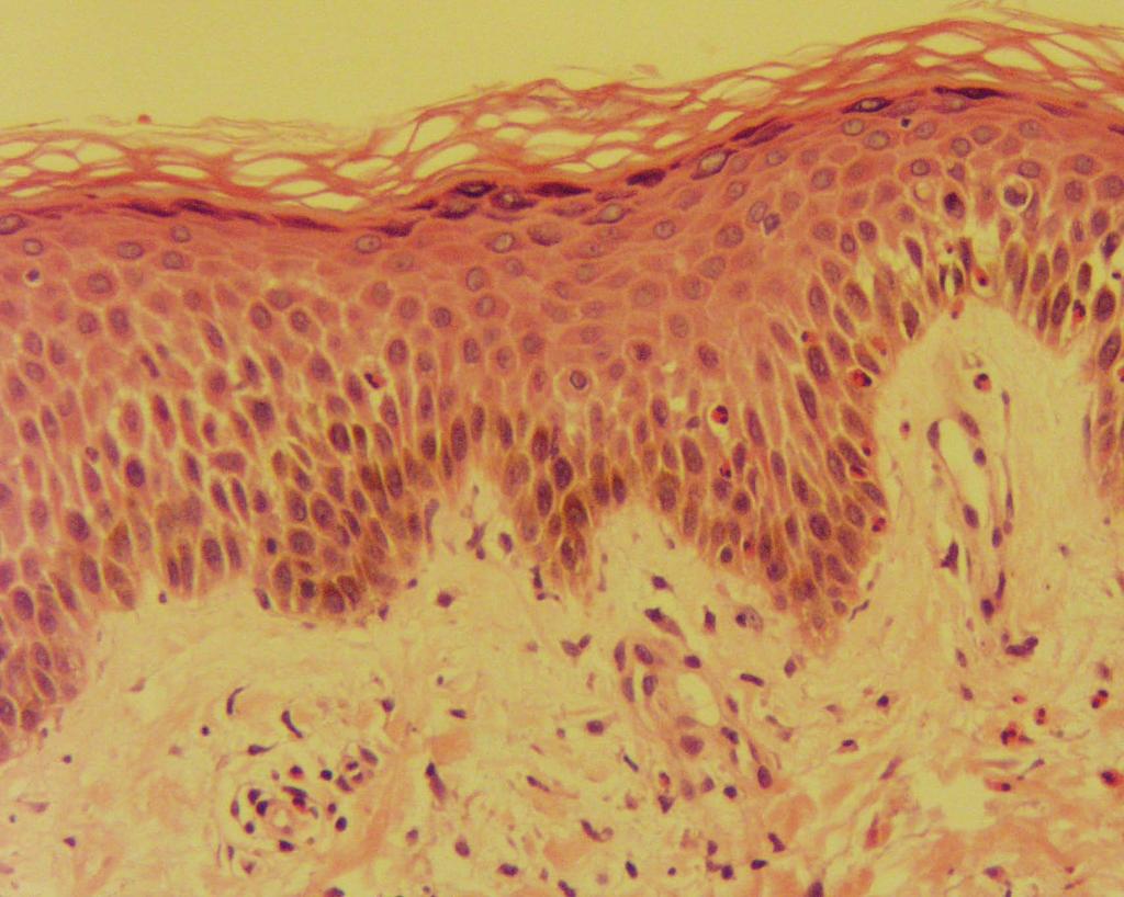
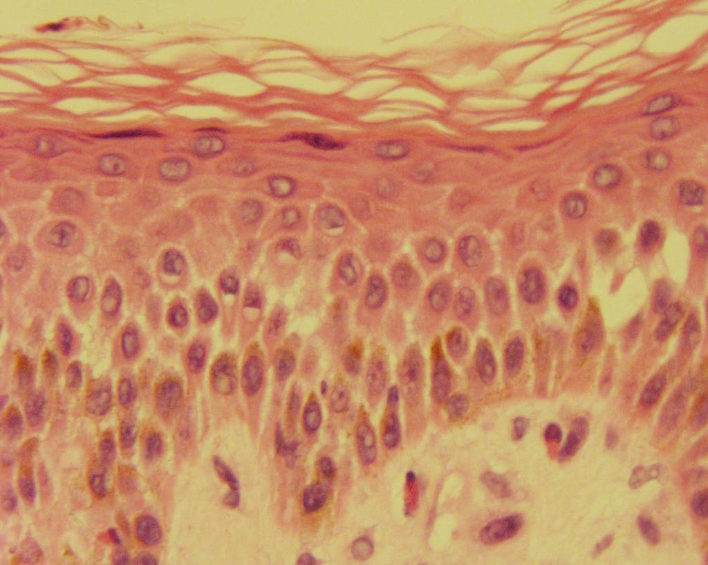
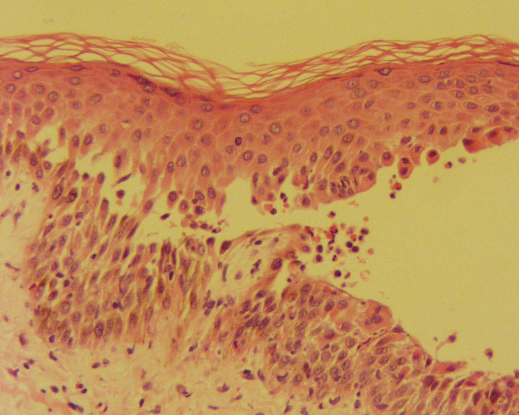
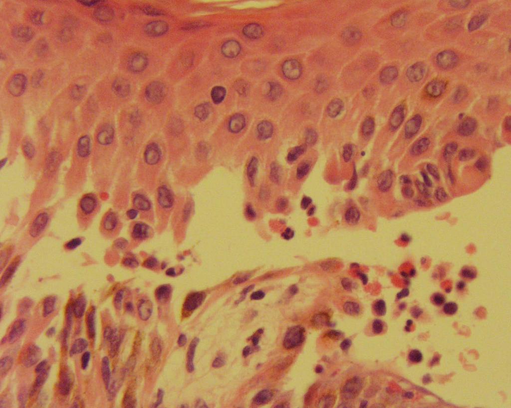
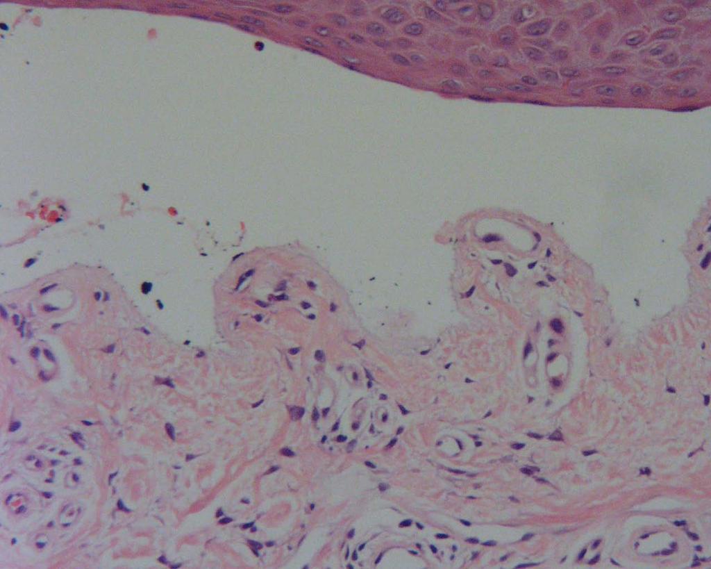
Comparative microanatomy of the normal skin with that of immunobullous condition
 Original article: Comparative microanatomy of the normal skin with that of immunobullous condition 1Dr BananiKundu, 2 DrAnirban Sadhu, 3 DrRudradev Meyur, 4 Dr SauravKundu, 5 Dr Alpana De, 6Dr SatabdiSarkar
Grover s disease: A case report.
 320 Case report Thai J Dermatol, October-December 2011 ABSTRACT: Grover s disease: A case report. Supicha Chavanich MD, Praneet Sajjachareonpong MD. CHAVANICH C, SAJJACHAREONPONG P. GROVER S DISEASE: A
المركب النموذج--- سبيتز وحمة = Type Spitz's Nevus, Compound SPITZ NEVUS 1 / 7
 SPITZ NEVUS 1 / 7 Epidemiology An annual incidence rate of 1.4 cases of Spitz nevus per 100,000 individuals has been estimated in Australia, compared with 25.4 per 100,000 individuals for cutaneous melanoma
Cutaneous manifestations of Systemic Lupus Erythematosus
 IOSR Journal of Dental and Medical Sciences (IOSR-JDMS) e-issn: 2279-0853, p-issn: 2279-0861.Volume 17, Issue 5 Ver. 11 (May. 2018), PP 01-08 www.iosrjournals.org Cutaneous manifestations of Systemic Lupus
Clinical Mucosal Immunology
 Clinical Mucosal Immunology Natural caries immunology in human It is sure, there is natural anti-caries immunity in human. Persons with low caries frequency have high anti-s.mutans IgG antibody level in
Hair and Scalp Changes in Cutaneous and Systemic Lupus Erythematosus
 Am J Clin Dermatol https://doi.org/10.1007/s40257-018-0363-8 REVIEW ARTICLE Hair and Scalp Changes in Cutaneous and Systemic Lupus Erythematosus Siriorn Udompanich 1 Kumutnart Chanprapaph 1 Poonkiat Suchonwanit
Index. derm.theclinics.com. Note: Page numbers of article titles are in boldface type.
 Note: Page numbers of article titles are in boldface type. A Abatacept for DLE, 493 for SLE, 497 Ablative therapies, localized, for cutaneous T-cell lymphoma, 502 506. See also Cutaneous T-cell lymphoma,
Index. in tissue, 87-89
 ANA titers, 79-84 Adult atopic dermatitis, 239 Allergens, contact dermatitis, 253 Allergic contact dermatitis Acontact sensitivity, cellular participants in, 258 factors influencing incidence of, 254 'hardening',
Spongiotic Dermatitis
 Prepared by Kurt Schaberg Introduction to Inflammatory Dermpath Spongiotic Dermatitis intraepidermal intercellular edema (spongiosis) - presence of widened intercellular spaces between keratinocytes, with
Citation The Journal of Dermatology, 37(8), available at
, available at") NAOSITE: Nagasaki University's Ac Title Two cases of blaschkitis with promi Author(s) Utani, Atsushi Citation The Journal of Dermatology, 37(8), Issue Date 2010-08 URL Right http://hdl.handle.net/10069/25634
NAOSITE: Nagasaki University's Ac Title Two cases of blaschkitis with promi Author(s) Utani, Atsushi Citation The Journal of Dermatology, 37(8), Issue Date 2010-08 URL Right http://hdl.handle.net/10069/25634
A study on the effectiveness of Tzanck smear to diagnose the vesiculobullous lesions in comparison with histopathology
 Original Research Article A study on the effectiveness of Tzanck smear to diagnose the vesiculobullous lesions in comparison with histopathology B. Lakshminarayana 1, Rammohan 2*, G. Saisoumya 3, J. Tulasi