Skin Cancer A Personal Approach. Dr Matthew Strack Dunedin New Zealand
|
|
|
- Edwina Patterson
- 5 years ago
- Views:
Transcription
1 Skin Cancer A Personal Approach Dr Matthew Strack Dunedin New Zealand
2 Outline Dermoscopy Instruments and setup Photochemosurgery Clinical Aim: Leave with 2-3 ideas
3
4
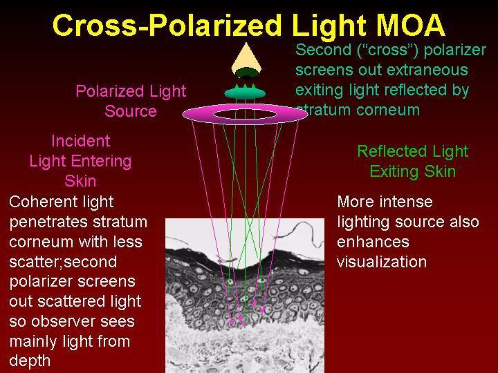
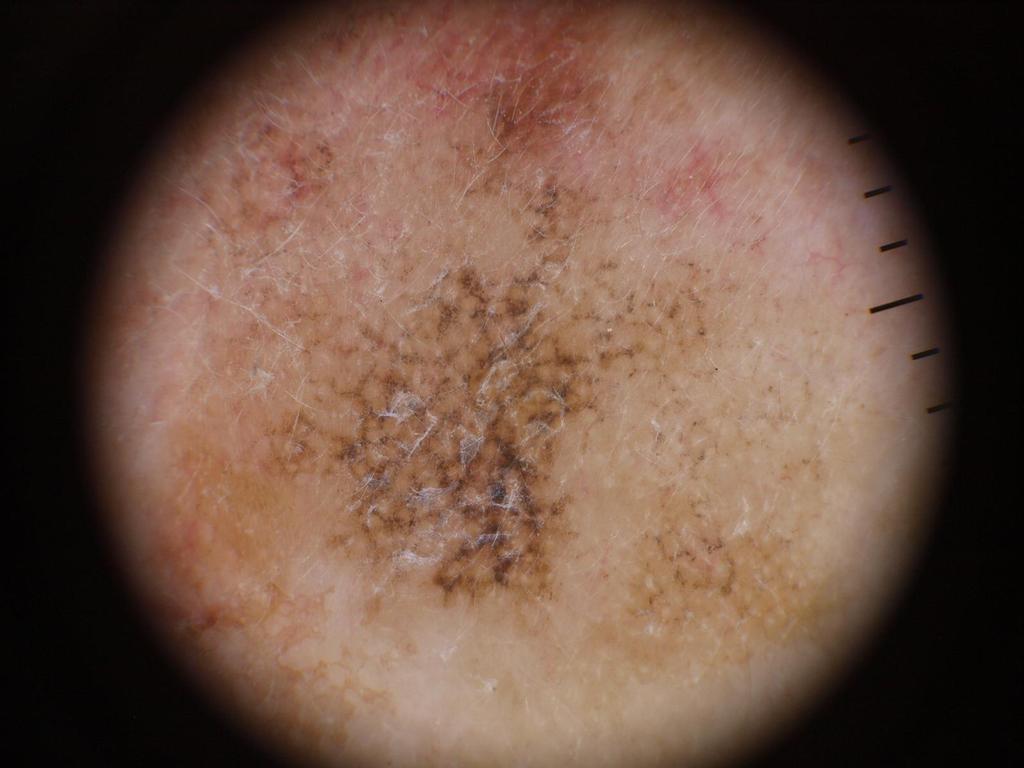
5 JLE

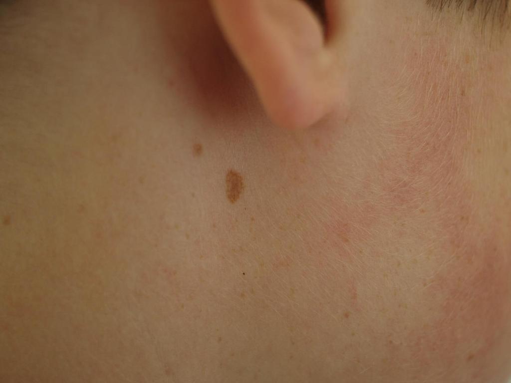
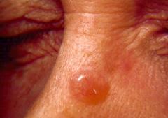
6 Benign Junctional Nevus

7 Management Resect Reassure Refer Rebook

8 My struggle with Dermoscopy The Scream Edvard Munch 1895
9 Terms Dermatoscopy Epiluminescent Microscopy Skin Surface Microscopy Dermoscopy


10 Dermoscopy - Brilliant for: Small Angiomas Seborrhoeic Keratosies
11 Dermatologists: In total, 4691 lesions were examined. The overall mean NNT was 6.3, with a range of for each of nine consultant dermatologists. The mean NNT was 7.6 for female and 4.8 for male patients. Clin Exp Dermatol Jan;37(1):6-9. doi: /j x. Epub 2011 Oct 7. The number of benign moles excised for each malignant melanoma: the number needed to treat. Sidhu S, Bodger O, Williams N, Roberts DL.
12 Benign Nevus: Melanoma Excision Ratios by Time NSCS = Non Specialized Skin Clinic SCS = Specialized Skin Clinic Accuracy in melanoma detection: A 10-year multicenter survey Giuseppe Argenziano, MD et al, Journal of the American academy of Dermatology,

13 General Practice, New Zealand All excisions, semirural practice, 3 year period B Wood, NZFP, 2006;v6, n4, pp256-8
14
15
16
17
18
19
20
21
22
23
24
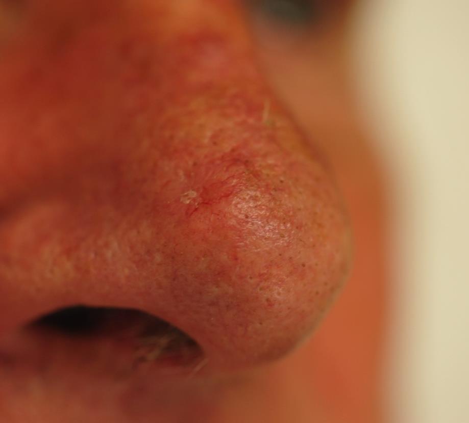
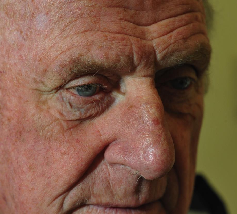
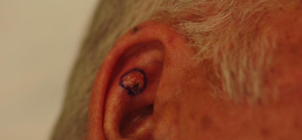
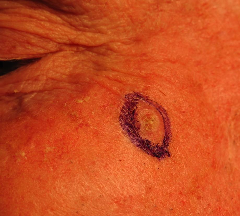
25
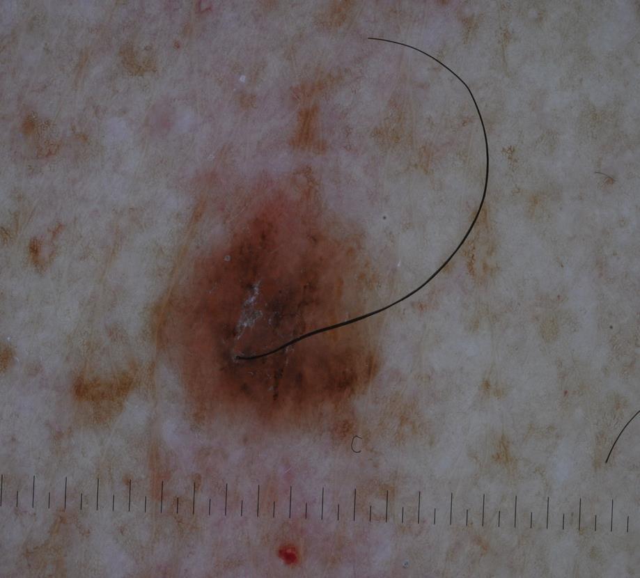
26 jrm
27

28 Melanoma in situ
29
30
31 Dysplastic nevus
32 92y M
33 Biopsies Lower: Pigmented Keratosis Upper: BCC

34 Algorithms You see but you do not observe
35 ABCD rule Stolz method Criteria Asymmetry In perpendicular axes: contour, colours and structures Borders 8 segments: abrupt ending of pigment pattern Colours White, red, light-brown (tan), dark-brown, blue-grey, black Score X Factor = Results Differential structural components Total score Benign <4.76 Suspicious Melanoma >5.45
36 Blum's modified ABC-point list A B C D E lesion asymmetry of outer shape or differential structures inside the in at least 1 axis the abrupt cutoff of network at the border in at least one quarter of circumference 3 or more colors 3 or more differential structures noticed change (evolution) in the last 3 months
37 Menzies method Negative features (benign lesions): Symmetrical pattern (colours, structure) Single colour Positive features (melanoma): Blue-white veil Multiple brown dots Pseudopods Radial streaming Scar-like depigmentation Multiple (5-6) colours Multiple blue/grey dots Broadened network
38 Seven-point rule Three or more indicates melanoma Atypical pigment network (2) Blue-whitish veil (2) Atypical vascular pattern (2) Irregular streaks (1) Irregular dots/globules (1) Irregular blotches (1) Regression structures (1)


39 ABCD


40 Did you look twice?
41 More Cases
42
43
44
45
46
47
48
49
50
51
52
53
54
55 RMS
56
57 jcd
58
59 HB
60
61 PJM
62
63
64 MAP
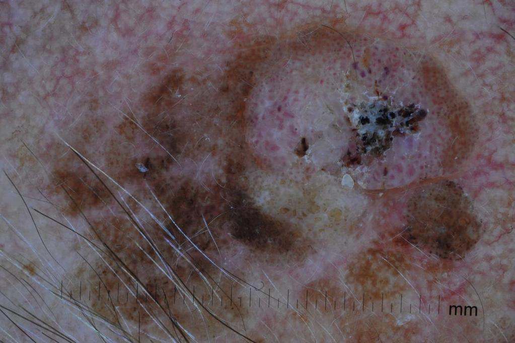
65

66 Pigmented Lesion Assessment

67 Pigmented Lesion Assessment

68 The Dance of Life Aasgardstrand
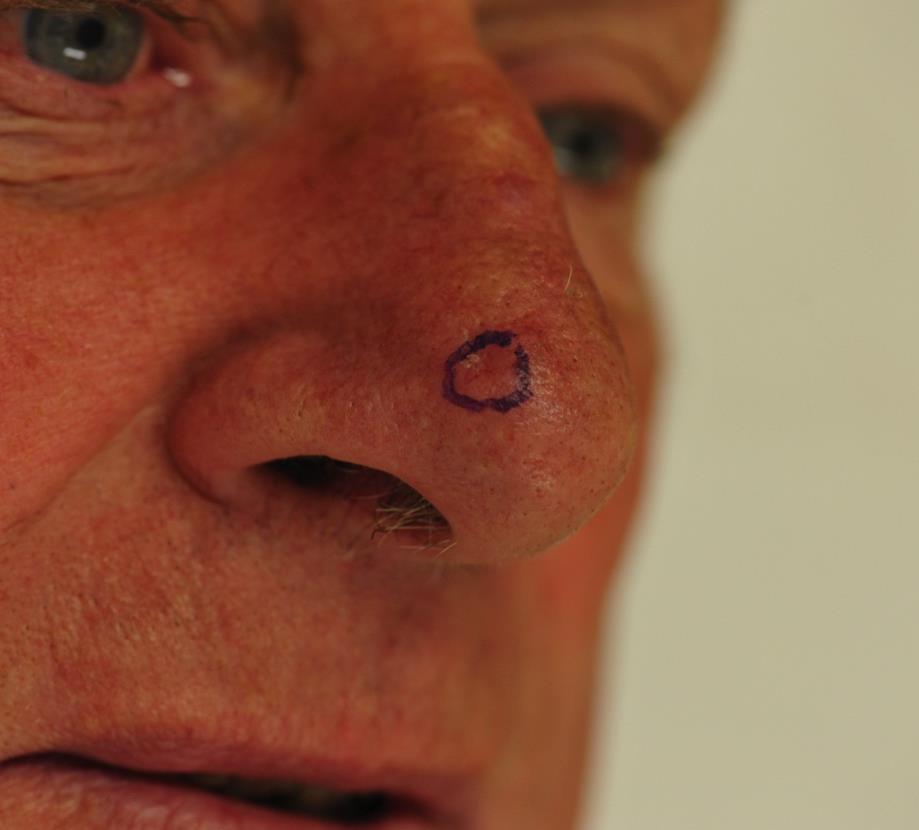
69 1:5-10
70
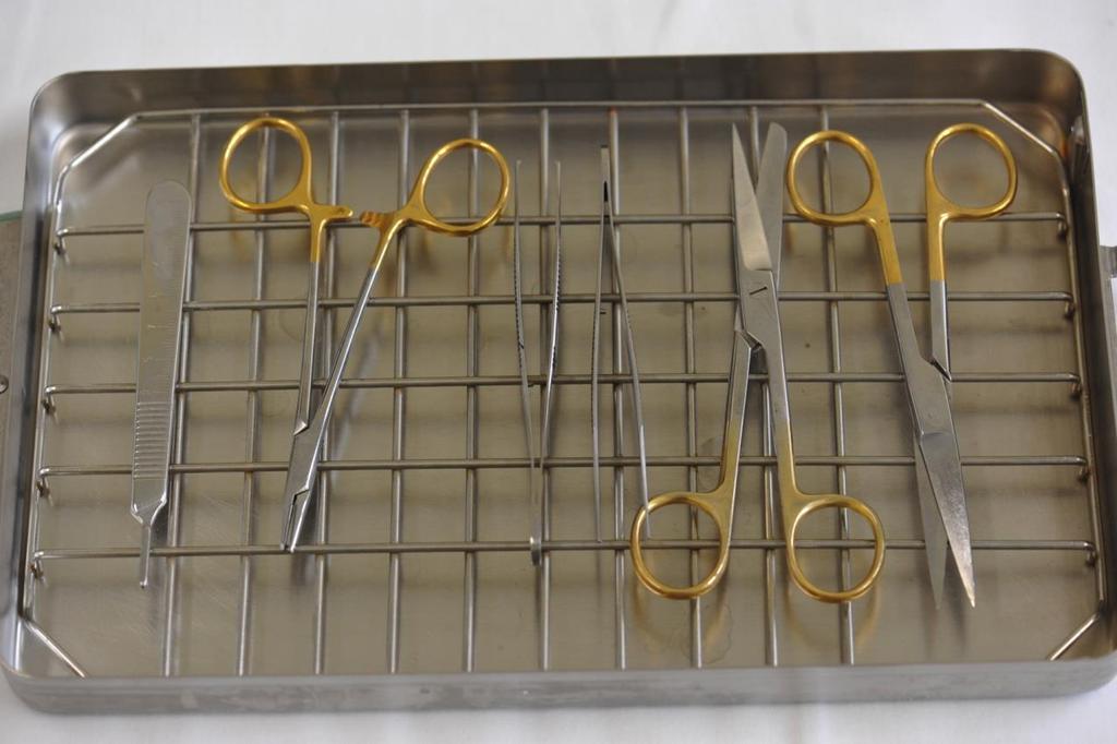
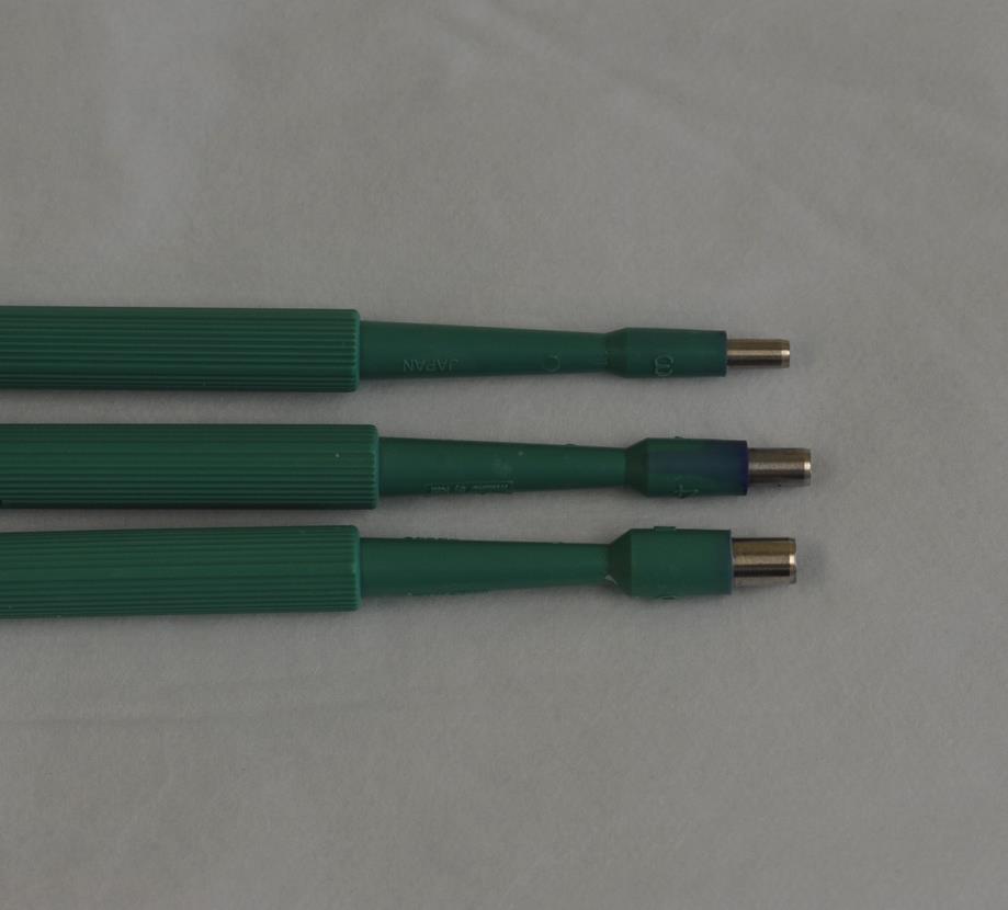
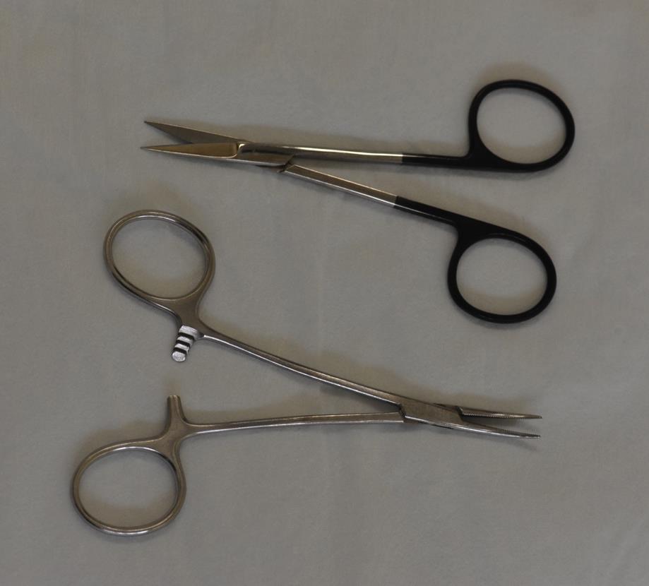
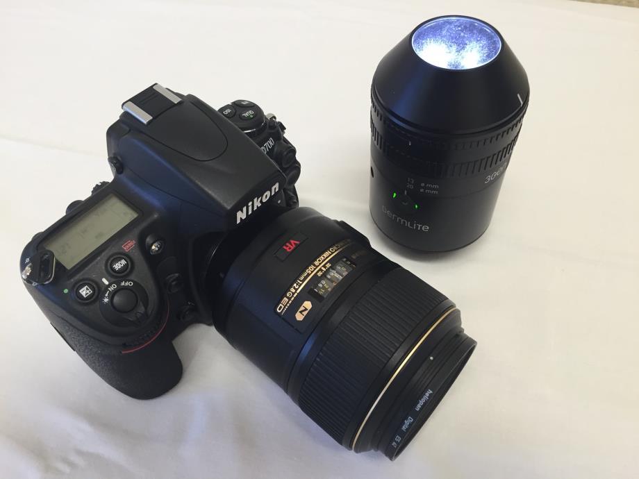
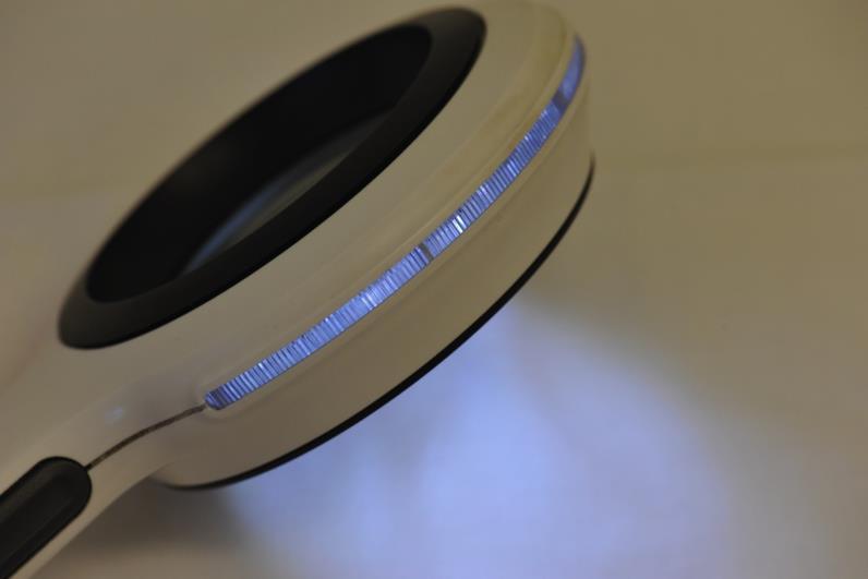
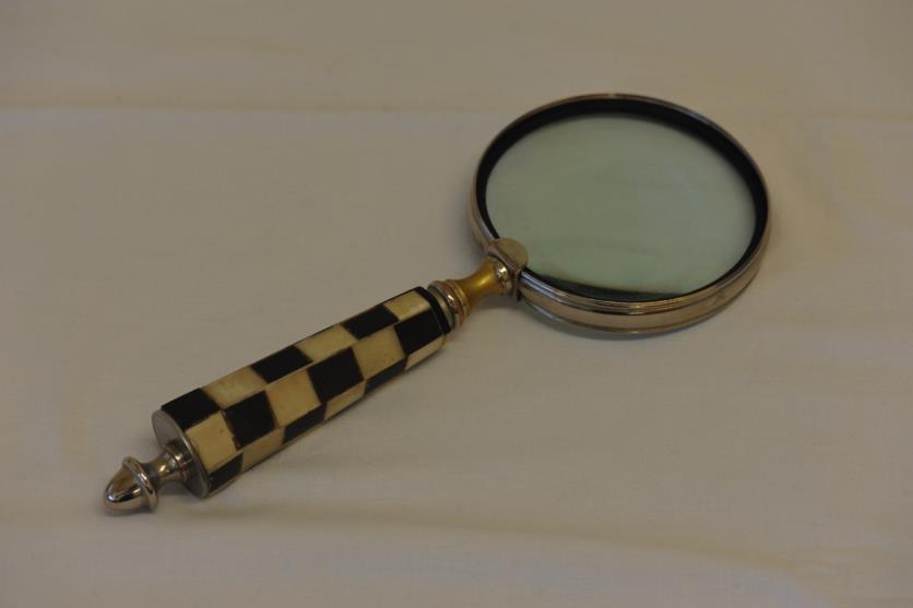
71
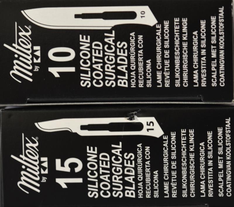
72 Lesion was benign Seborrhoeic Keratosis
73 91y M
74
75 Melanoma In-Situ 0.6mm Diagnosis


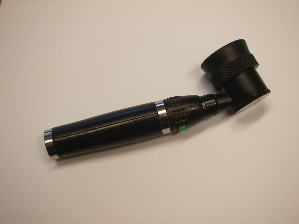
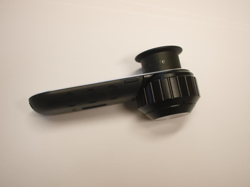
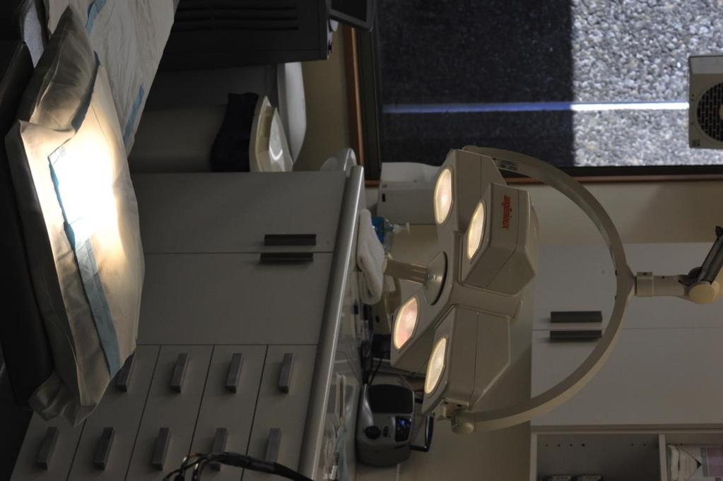
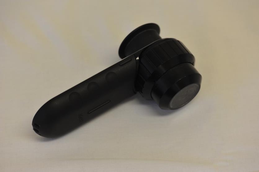
76 73y M My Diagnosis: Seborrhoeic Keratosis Benign Solar Lentigo

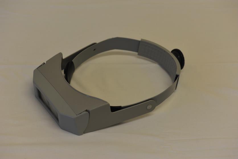
77
78 Instruments
79 Blades

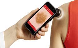
80 Sutures Ask for free samples!
81

82 Curettage Relies on physical differences between affected and normal tissue Diathermy - hemostasis Useful for BCC Histology will report incomplete removal
83 Lighting


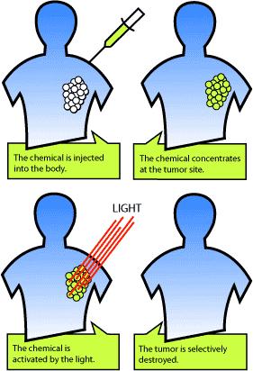
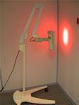
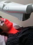
84 Eyewear Try before you buy!


85 Local Anaesthetic 2.2ml cartridge 27g 22g 18g
86 Most important things: Use enough Time Reassurance
87 Photography
88 Consent Lighting System
89 Examinaton
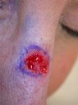
90 Examination
91 Photochemosurgery for Basal Cell Carcinoma
92
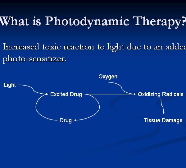
93 Photodynamic Therapy

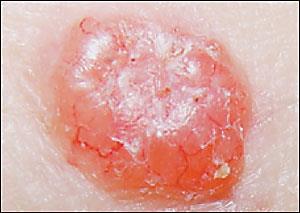
94 d
95
96
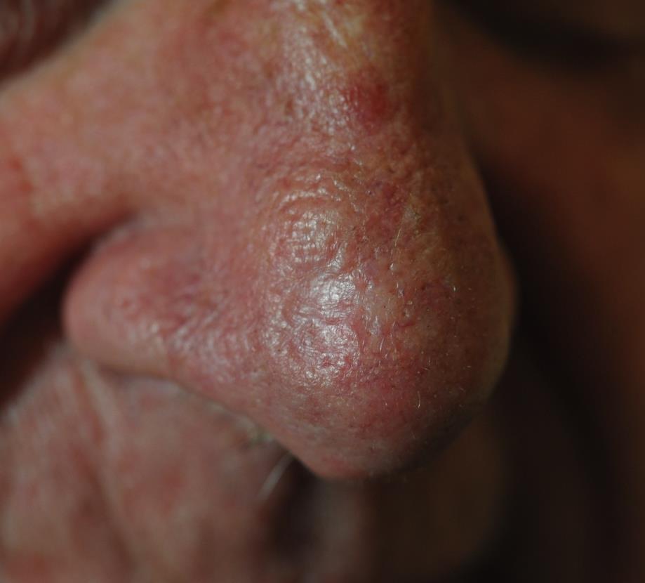
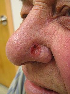
97 BCC, pre Photochemosurgery
98 Post Photochemosurgery
99 Basal Cell Carcinoma Commonest Skin Cancer Rarely Life Threatening Treatment ranges from easy to very challenging Many treatment options
100 Treatment Options Surgery Excison Curetage Mohs Radiotherapy Photochemosurgery (Photodynamic Therapy) Topical No Treatment rare
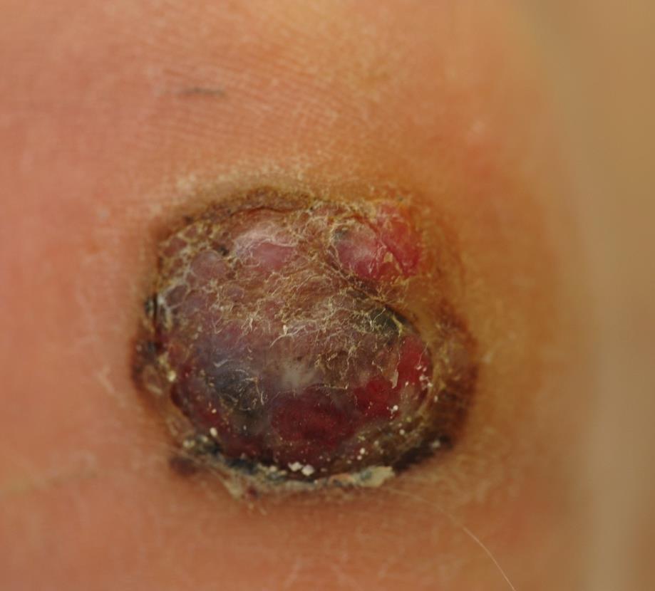
101 Waxy with capillaries
102
103
104 BCC

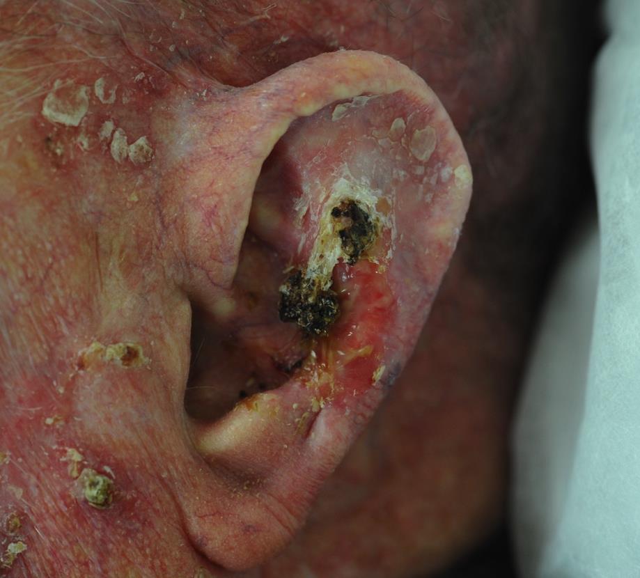
105 BCC post Imiquimod
106 Bowen s Disease Squamous Cell Carcinoma in Situ Prognosis unclear? 10% progress to SCC Main Treatment Options: Cryotherapy Topical 5 Flourouracil Scaring may be due to lesion not treatment
107 Post 5FU

108 Cutaneous Horn?Cause Often Painful


109 What I think of:
110 What s Under a Cutaneous Horn Solar Keratosis Basal Cell Carcinoma Squamous Cell Carcinoma This Case Nonspecific Change
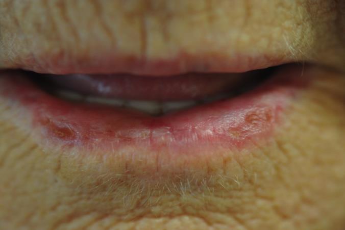
111 BCC

112 Subungual Pigmented Lesions
113
114 Lab Write brief details of history Diagnosis Differential Diagnosis Consider ringing pathologist Consider second opinion

115 Trapdoor Problem with a Flap 94y F, result painful and not happy with appearance

116 Squamous Cell Carcionma

117 84y F Summer Winter
118 Actinic Chelitis High risk for SCC Topical treatment OK Follow up mandatory

119 94y M, confused


120 66y M, change over 2 months
121

122 Wart, present 6/12

123 Wart GP x1 first seen >1y ago Nurse x2 Podiatrist x1
124 Take Home Messages #1 Have equipment you enjoy using Get chaperone consent in writing When in doubt refer but you can sometimes reassess Biopsies are your friend If you don t have loupes get some on loan Have your eyes checked
125 Take Home Messages #2 Consider Polarizing Dermoscope Printed Handouts Last Point: call me Follow up Surgery incl Sutures Cryotherapy

126
22/04/2015. Dermoscopy of Melanoma. Ilsphi Browne. Overview
 Dermoscopy of Melanoma Ilsphi Browne Overview The device Dermoscopic criteria (terminology) Colour Patterns Global features Local features Approach to diagnosing pigmented lesions Other uses in general
Mole mapping and monitoring. Dr Stephen Hayes. Associate Specialist in Dermatology, University Hospital Southampton
 Mole mapping and monitoring Dr Stephen Hayes Associate Specialist in Dermatology, University Hospital Southampton Outline of presentation The melanoma epidemic Benefits of early detection Risks of the
Regression 2/3/18. Histologically regression is characterized: melanosis fibrosis combination of both. Distribution: partial or focal!
 Regression Margaret Oliviero MSN, ARNP Harold S. Rabinovitz MD Histologically regression is characterized: melanosis fibrosis combination of both Distribution: partial or focal! Dermatoscopic terminology
Dermoscopy: Recognizing Top Five Common In- Office Diagnoses
 Dermoscopy: Recognizing Top Five Common In- Office Diagnoses Vu A. Ngo, DO Department of Family Medicine and Dermatology Choctaw Nation Health Services Authority Learning Objectives Introduction to dermoscopy
Clinical characteristics
 Skin Cancer Fernando Vega, MD Seattle Healing Arts Clinical characteristics Precancerous lesions Common skin cancers ACTINIC KERATOSIS Precancerous skin lesions Actinic keratoses Dysplastic melanocytic
Disclosure. Objectives. PAFP CME Conference Lou Mancano MD, FAAFP Reading Health System November 18, 2016
 PAFP CME Conference Lou Mancano MD, FAAFP Reading Health System November 18, 2016 1 Disclosure The speaker has no conflict of interest, financial agreement, or working affiliation with any group or organization.
Finding Melanoma. Is not easy!
 Finding Melanoma Is not easy! Finding Melanoma Victoria mean depth at diagnosis is 1.5 mm. Melanoma 1.5mm Has Stage 1B Mortality 10% Melanoma Spotting a killer! Spotting a killer Visual Clues What are
Dermoscopy. Enhanced Diagnostic Ability: Pigmented Lesions. Ted Rosen, MD Baylor College of Medicine Houston, Texas
 Dermoscopy Enhanced Diagnostic Ability: Pigmented Lesions Ted Rosen, MD Baylor College of Medicine Houston, Texas Faculty Disclosure Statement No conflicts relevant to this workshop! Sir William Osler
6/17/2018. Breaking Bad (Part 1) Dermoscopy of Brown(ish) Things. Bad?
 Dermoscopy of Brown(ish) Things. Bad?") Breaking Bad (Part 1) Dermoscopy of Brown(ish) Things Jennie T. Clarke, MD ssociate Professor of Dermatology University of Utah School of Medicine Bad? 1 Brown(ish) Things Bad Melanoma Pigmented basal
Breaking Bad (Part 1) Dermoscopy of Brown(ish) Things Jennie T. Clarke, MD ssociate Professor of Dermatology University of Utah School of Medicine Bad? 1 Brown(ish) Things Bad Melanoma Pigmented basal
Skin lesions The Good and the Bad. Dr Virginia Hubbard Ipswich Hospital NHS Trust Barts and the London School of Medicine and Dentistry
 Skin lesions The Good and the Bad Dr Virginia Hubbard Ipswich Hospital NHS Trust Barts and the London School of Medicine and Dentistry Case 1 32 year old woman Australian Lesion on back New hair growing
INTRODUCTION HOUSEKEEPING June 11 th Dr John Adams Dermatologist/Dermoscopist MOLEMAP NZ/Australia MOLESAFE USA
 INTRODUCTION HOUSEKEEPING June 11 th 2015 Dr John Adams Dermatologist/Dermoscopist MOLEMAP NZ/Australia MOLESAFE USA Program Skin cancer statistics. Dermoscopy description and usefulness. Patient /lesion
Clinical and Dermoscopic Features of Thin Nodular Melanoma
 Clinical and Dermoscopic Features of Thin Nodular Melanoma A study of the International Dermoscopy Society Coordinator: Dr. Alexander J. Stratigos and colleagues, alstrat2@gmail.com ** Extended to May
VACAVILLE DERMATOLOGY
 Connecting the Dots on those Spots NANDAN V. KAMATH, M.D. VACAVILLE DERMATOLOGY Sources All of the photos were taken with permission from the Dermnet NZ website - Dermnet New Zealand after communicating
Dermoscopy. Sir William Osler. Dermoscopy. Dermoscopy. Melanoma USA Primary Care Update Faculty Disclosure Statement
 Diagnostic Ability: Pigmented Lesions Ted Rosen, MD Baylor College of Medicine Houston, Texas Enhanced 2010 Primary Care Update Faculty Disclosure Statement Ted Rosen, MD Speakers Bureau: Abbott, Amgen,
Dermoscopy-a BRIEF introduction
 Dermoscopy-a BRIEF introduction Aim of presentation -to tell you what dermoscopy is -to show some of what it can do -point the interested learner to further resources Overview of dermoscopy Dermoscopy
Malignant non-melanocytic lesions
 Malignant non-melanocytic lesions Course C023: Fundamentals of Dermoscopy March 4, 2019, 11:20 AM - 11:50 PM Room: 146B Jason B. Lee, MD Professor & Vice Chair Director of Dermatopathology & Pigmented
It can be helpful in some cases of actinic keratosis, Bowen s disease and squamous cell carcinoma
 Dermoscopy Introduction, Terminology and Structures (to be read in conjunction with the Diagnostic Dermoscopic Algorithm) Copyright to Cunliffe TP (Jan. 2017) All rights reserved Introduction Dermoscopy
MODULE 1. LOCAL AND GENERAL CRITERIA IN PIGMENTED MELANOCYTIC LESIONS.
 DERMOSCOPY TEACHING PROGRAMME Dermoscopy Teaching Programme Module 1 MODULE 1. LOCAL AND GENERAL CRITERIA IN PIGMENTED MELANOCYTIC LESIONS. Dermoscopy is a non-invasive in vivo technique that provides
Know who is at risk: LOOK! for ABCDs, rapidly changing lesions, do a biopsy when indicated
 Lindy P. Fox, MD Assistant Professor Director, Hospital Consultation Service Department of Dermatology University of California, San Francisco Applies to adults without history of malignancy or premalignant
Benign versus Cancerous Lesions How to tell the difference FMF 2014 Christie Freeman MD, CCFP, DipPDerm, MSc
 1 Benign versus Cancerous Lesions How to tell the difference FMF 2014 Christie Freeman MD, CCFP, DipPDerm, MSc Benign lesions Seborrheic Keratoses: Warty, stuck-on Genetics and birthdays Can start in late
Know who is at risk: LOOK! for ABCDs, rapidly changing lesions, do a biopsy when indicated
 Lindy P. Fox, MD Associate Professor Director, Hospital Consultation Service Department of Dermatology University of California, San Francisco Applies to adults without history of malignancy or premalignant
STUDY. Epiluminescence Microscopy for the Diagnosis of Doubtful Melanocytic Skin Lesions
 STUDY Epiluminescence Microscopy for the Diagnosis of Doubtful Melanocytic Skin Lesions Comparison of the ABCD Rule of Dermatoscopy and a New 7-Point Checklist Based on Pattern Analysis Giuseppe Argenziano,
Identifying Skin Cancer. Mary S. Stone MD Professor of Dermatology and Pathology University of Iowa Carver College of Medicine March, 2018
 Identifying Skin Cancer Mary S. Stone MD Professor of Dermatology and Pathology University of Iowa Carver College of Medicine March, 2018 American Cancer Society web site Skin Cancer Melanoma Non-Melanoma
50 interactive dermoscopic case discussions Dr Stephen Hayes
 50 interactive dermoscopic case discussions Dr Stephen Hayes Annotations will be found on your memory drive, as will 100 case discussions and other learning material Melanoma 2mm thick Ugly duckling-one
Common Benign Lesions and Skin Cancers. 22nd May 2015 Dr Mark Foley
 Common Benign Lesions and Skin Cancers 22nd May 2015 Dr Mark Foley Thank you for downloading this file. This intended to supplement the presentation given at the NZ Wound Care Conference, it is not intended
Total body photography in high risk patients
 Total body photography in high risk patients Doug Grossman, MD, PhD Department of Dermatology Huntsman Cancer Institute University of Utah Summer AAD F032 Practical Considerations for Patients with Melanoma
Fundamentals of dermoscopy
 Fundamentals of dermoscopy Learning objectives Upon completion of this session, participants should be able to: describe the basic principles of dermoscopy identify features associated with pigmented and
Dermoscopy Quiz 3-Point Checklist Algorithm
 Dermoscopy Quiz 3-Point Checklist Algorithm GLOBAL PATTERN Globular LOCAL CRITERIA Aggregated globules Milia-like cysts 3 POINT CHECK LIST Symmetrical No abnormal net Slight Blue-white veil BENIGN MELANOCYTIC
Melanoma and Dermoscopy. Disclosure Statement: ABCDE's of melanoma. Co-President, Usatine Media
 Melanoma and Dermoscopy Richard P. Usatine, MD, FAAFP Professor, Family and Community Medicine Professor, Dermatology and Cutaneous Surgery Medical Director, University Skin Clinic University of Texas
Nonmelanoma skin cancers
 Skin cancer Philip Clarke Nonmelanoma skin cancers Treatment options Background Australia has one of the highest skin cancer rates in the world. Early detection and treatment of skin cancer is vital to
Acral and Mucosal Dermoscopy
 Acral and Mucosal Dermoscopy Caroline C. Kim, MD Assistant Professor, Department of Dermatology Harvard Medical School Director, Pigmented Lesion Clinic Associate Director, Cutaneous Oncology Program Beth
Dermatological Manifestations in the Elderly. Sanjay Siddha Staff Dermatologist UHN & MSH
 Dermatological Manifestations in the Elderly Sanjay Siddha Staff Dermatologist UHN & MSH Disclosure No actual or potential conflicts of interest or commercial relationships to declare Objectives Recognize
SKIN CANCER. Most common cancer diagnosis 40% of all cancers
 SKIN CANCER Most common cancer diagnosis 40% of all cancers OBJECTIVES Review common and uncommon cancers of the skin. Special emphasis on melanoma and dysplastic nevus Review pathology/tnm/staging, which
The impact of GP sub-specialisation and dermatoscopy use on diagnostic accuracy for melanomas in Australia
 The impact of GP sub-specialisation and dermatoscopy use on diagnostic accuracy for melanomas in Australia Cliff Rosendahl, Gail Williams, Diann Eley, Tobias Wilson, Greg Canning, Jeffrey Keir, Ian McColl,
Hauora Maori. Map. Non-Melanoma Skin Lesion. Clinically Seborrhoeic Keratosis. Management Options. Punch Biopsy. Refer to Public Skin Lesion Service
 Care map information Information resources for patients and carers Updates to this Care Map Hauora Maori Pacific Skin lesion history Clinical examination and diagnostic test Suspicious of Melanoma Non-Melanoma
Dr Stephen Hayes Associate Specialist in Dermatology University Hospital Southampton
 South East Dermatology Transformation and Sustainability Network Guildford, 19 th April 2018 Dermoscopy as an effective skin lesion triage tool in GP surgeries Dr Stephen Hayes Associate Specialist in
Multiple Primary Melanoma in a Thai Male: A Case Report
 Case Report Multiple Primary Melanoma in a Thai Male: A Case Report J Med Assoc Thai 2014; 97 (Suppl. 2): S234-S238 Full text. e-journal: http://www.jmatonline.com Kittisak Payapvipapong MD*, Pinyapat
Graph-based Pigment Network Detection in Skin Images
 Graph-based Pigment Network Detection in Skin Images M. Sadeghi a,b, M. Razmara a, M. Ester a, T. K. Lee a,b,c, M. S. Atkins a a Simon Fraser University, 8888 University Drive, Burnaby, BC, Canada, V5A1S6;
Introduction to Dermoscopy. Nicholas Compton, MD June 16, 2010
 Introduction to Dermoscopy Nicholas Compton, MD June 16, 2010 Overview What is dermoscopy Brief history Types of dermoscopy General approach to lesion of interest 2 step algorithm 3-point checklist Practice
Cutaneous Malignancies: A Primer COPYRIGHT. Marissa Heller, M.D.
 Cutaneous Malignancies: A Primer Marissa Heller, M.D. Associate Director of Dermatologic Surgery Department of Dermatology Beth Israel Deaconess Medical Center December 10, 2016 Skin Cancer Non-melanoma
Appendix : Dermoscopy
 Go Back to the Top To Order, Visit the Purchasing Page for Details APP Appendix : Dermoscopy Dermoscopy, also known as dermatoscopy, epiluminoscopy and epiluminescent microscopy, is an effective non-invasive
Histopathological and SIAscopic Correlation of Pigmented Skin Lesions
 Histopathological and SIAscopic Correlation of Pigmented Skin Lesions Professor Sujatha Fernando MBBS(Hon), MSc(London, Distinction), FRSTM&H, FRCPA, FIAC, FACTM Senior Consultant in Anatomical Pathology,
Skin Cancer. Dr Elizabeth Ogden Associate Specialist in Dermatology East and North Herts Dr Elizabeth Ogden
 Skin Cancer Dr Elizabeth Ogden Associate Specialist in Dermatology East and North Herts 13.10.16 Skin Cancer Melanoma mole cancer - is a true cancer which can metastasize and kill Non Melanoma skin cancer
STUDY. Scott W. Menzies, MB,BS, PhD; Karin Westerhoff, MD; Harold Rabinovitz, MD; Alfred W. Kopf, MD; William H. McCarthy, MBBS, MEd; Brian Katz
 STUDY Surface Microscopy of Pigmented Basal Cell Carcinoma Scott W. Menzies, MB,BS, PhD; Karin Westerhoff, MD; Harold Rabinovitz, MD; Alfred W. Kopf, MD; William H. McCarthy, MBBS, MEd; Brian Katz Objectives:
Skin Cancer. 5 Warning Signs. American Osteopathic College of Occupational and Preventive Medicine OMED 2012, San Diego, Monday, October 8, 2012 C-1
 Skin Cancer AMERICAN OSTEOPATHIC COLLEGE OF OCCUPATIONAL & PREVENTIVE MEDICINE OMED 2012 October 8, 2012 E. Robert Wanat II, D.O., M.P.H. Learning Objectives: Identify the 3 Basic Types of Skin Cancer
Learning Objectives. Tanning. The Skin. Classic Features. Sun Reactive Skin Type Classification. Skin Cancers: Preventing, Screening and Treating
 Learning Objectives Skin Cancers: Preventing, Screening and Treating Robert A. Baldor, MD, FAAFP Professor, Family Medicine & Community Health University of Massachusetts Medical School Distinguish the
Key factors in successfully integrating dermoscopy into your clinical practice
 Key factors in successfully integrating dermoscopy into your clinical practice S051 Dilemmas and challenges in skin cancer therapies and management Monday, March 4 th 2019 (9AM-12PM) Room 209A 10:56-11:09AM
Basics in Dermoscopy
 Basics in Dermoscopy Manal Bosseila Professor of Dermatology, Cairo University Member of European Academy Dermatology & Venereology EADV Member of International Dermoscopy Society IDS Member of Aesthetic
Optical Diagnostic Devices for Evaluating Skin Lesions Suspected of Malignancy
 2.01.42 Optical Diagnostic Devices for Evaluating Skin Lesions Suspected of Malignancy Section 2.0 Medicine Subsection Effective Date November 26, 2014 Original Policy Date November 26, 2014 Next Review
Algorithmic reproduction of asymmetry and border cut-off parameters according to the ABCD rule for dermoscopy
 JEADV ISSN 1468-3083 Blackwell Publishing Ltd ORIGINAL ARTICLE Algorithmic reproduction of asymmetry and border cut-off parameters according to the ABCD rule for dermoscopy G Pellacani,* C Grana, S Seidenari
Identifying Benign and Malignant Skin Lesions. No Disclosures. Common Benign Lesions. Benign Lesions 2/25/2018. Stucco Keratoses.
 Dermatology in Primary Care Identifying Benign and Malignant Skin Lesions Christy Quire Baker, APRN, FNP-BC, DCNP Dermatology Certified Nurse Practitioner No Disclosures Common Benign Lesions Seborrheic
Technicians & Nurses Program
 ASCRS ASOA Symposium & Congress Technicians & Nurses Program May 6-10, 2016 New Orleans Evaluation and Treatment of Eyelid Malignancies Richard C. Allen MD PhD FACS Professor Section of Ophthalmology Dept.
IT S FUNDAMENTAL MY DEAR WATSON! A SHERLOCKIAN APPROACH TO DERMATOLOGY
 IT S FUNDAMENTAL MY DEAR WATSON! A SHERLOCKIAN APPROACH TO DERMATOLOGY Skin, Bones, and other Private Parts Symposium Dermatology Lectures by Debra Shelby, PhD, DNP, FNP-BC, FADNP, FAANP Debra Shelby,
LUMPS AND BUMPS: AN ORGANIZED APPROACH TO DIAGNOSIS AND MANAGEMENT
 LUMPS AND BUMPS: AN ORGANIZED APPROACH TO DIAGNOSIS AND MANAGEMENT Tammy P. Than, M.S., O.D., F.A.A.O. The University of Alabama at Birmingham / School of Optometry 1716 University Blvd. Birmingham, AL
I have a skin lump doc! What s next? 12 th August 2017 Dr. Sue-Ann Ho Ju Ee
 I have a skin lump doc! What s next? 12 th August 2017 Dr. Sue-Ann Ho Ju Ee Some thoughts Is this skin cancer? How common is this? How likely is this in this patient? What happens next if it s something
What is Dermoscopy? Early Dermoscopes. Deciphering Dermoscopy: Terminology, Features & Algorithms 6/17/2018
 Deciphering Dermoscopy: Terminology, Features & Algorithms Where did it come from and why do we use it? Jennie T. Clarke, MD Associate Professor of Dermatology University of Utah School of Medicine What
Dermatopathology: The tumor is composed of keratinocytes which show atypia, increase mitoses and abnormal mitoses.
 Squamous cell carcinoma (SCC): A common malignant tumor of keratinocytes arising in the epidermis, usually from a precancerous condition: 1- UV induced actinic keratosis, usually of low grade malignancy.
Dermoscopy in everyday practice. What and Why? When in doubt cut it out? Trilokraj Tejasvi MD
 Dermoscopy in everyday practice Trilokraj Tejasvi MD Assistant Professor, Department of Dermatology, Director Teledermatology services, University of Michigan, Faculty Associate, GLOBAL REACH, Michigan
Large majority caused by sun exposure Often sun exposure before age 20 Persons who burn easily and tan poorly are at greatest risk.
 Basics of Skin Cancer Detection and Treatment of Non- Melanoma Skin Cancers Large majority caused by sun exposure Often sun exposure before age 20 Persons who burn easily and tan poorly are at greatest
The GP s approach to the patient who is worried about sun, skin and moles. Dr Stephen Hayes
 The GP s approach to the patient who is worried about sun, skin and moles Dr Stephen Hayes Associate Specialist in Dermatology, University Hospital Southampton Dr Stephen Hayes DECLARATION OF INTERESTS
Cover Page. The handle holds various files of this Leiden University dissertation.
 Cover Page The handle http://hdl.handle.net/1887/22172 holds various files of this Leiden University dissertation. Author: Rhee, Jasper Immanuel van der Title: Clinical characteristics and management of
Management of Atypical Pigmented Lesions
 Management of Atypical Pigmented Lesions Jennifer A. Stein MD, PhD Associate Director, Pigmented Lesion Section Ronald O. Perelman Department of Dermatology NYU Langone Medical Center July 29, 2017 1-4
Apps and Telemedicine H. Peter Soyer Dermatology Research Centre
 Apps and Telemedicine H. Peter Soyer Dermatology Research Centre p.soyer@uq.edu.au https://twitter.com/hpsoyer William Gibson The future is already here it's just not very evenly distributed Vision 3D
Living Beyond Cancer Skin Cancer Detection and Prevention
 Living Beyond Cancer Skin Cancer Detection and Prevention Cutaneous Skin Cancers Identification Diagnosis Treatment options Prevention What is the most common cancer in people? What is the most common
Skin Malignancies. Presented by Dr. Douglas Paauw
 Skin Malignancies Presented by Dr. Douglas Paauw Disclosure: Dr. Paauw has no significant financial interest in any of the products or manufacturers mentioned. How Common Is Skin Cancer? *½ of all White
Glenn D. Goldman, MD. University of Vermont Medical Center. University of Vermont College of Medicine
 Glenn D. Goldman, MD University of Vermont Medical Center University of Vermont College of Medicine Recognize and identify the main types of skin cancer and their precursors Identify and understand new
Page: 1 of 16. Optical Diagnostic Devices for Evaluating Skin Lesions Suspected of Malignancy
 Last Review Status/Date: December 2014 Page: 1 of 16 Skin Lesions Suspected of Malignancy Description There is interest in non-invasive devices that will improve the diagnosis of malignant skin lesions.
Dermoscopy. Synonyms. Dermoscopy. Definition. Dermoscopy opens up a world of colour and structure that can t be seen with the naked eye
 Synonyms Dermoscopy Australasian College of Dermatologists G.P Training Module Dermoscopy Dermatoscopy Epiluminescence microscopy Skin surface microscopy Incident light microscopy Oil immersion microscopy
Interesting Case Series. Aggressive Tumor of the Midface
 Interesting Case Series Aggressive Tumor of the Midface Adrian Frunza, MD, Dragos Slavescu, MD, and Ioan Lascar, MD, PhD Bucharest Emergency Clinical Hospital, Bucharest University School of Medicine,
STUDY. Identification of Clinically Featureless Incipient Melanoma Using Sequential Dermoscopy Imaging
 STUDY Identification of Clinically Featureless Incipient Melanoma Using Sequential Dermoscopy Imaging Harald Kittler, MD; Pascale Guitera, MD; Elisabeth Riedl, MD; Michelle Avramidis, MD; Ligia Teban,
DERMATOLOGY PRACTICAL & CONCEPTUAL. Gabriel Salerni 1,2, Teresita Terán 3, Carlos Alonso 1,2, Ramón Fernández-Bussy 1 ABSTRACT
 DERMATOLOGY PRACTICAL & CONCEPTUAL www.derm101.com The role of dermoscopy and digital dermoscopy follow-up in the clinical diagnosis of melanoma: clinical and dermoscopic features of 99 consecutive primary
Dermoscopy, the use of a handheld
 ONLINE EXCLUSIVE Dermoscopy in family medicine: A primer Dermoscopy allows you to see deeper into the skin than with the naked eye. Here s how you can make use of it to spot malignant conditions sooner.
Dermoscopy STFM Richard Usatine, MD 5/2/16. Disclosure Statement: Some Dermatoscopes. Dermoscopy Video. Thanks to Dr.
 Disclosure Statement: Dermoscopy STFM 2016 Richard P. Usatine, MD, FAAFP Professor, Family and Community Medicine Professor, Dermatology and Cutaneous Surgery Medical Director, Clinic University of Texas
Glenn D. Goldman, MD. Fletcher Allen Health Care. University of Vermont College of Medicine
 Glenn D. Goldman, MD Fletcher Allen Health Care University of Vermont College of Medicine Recognize and identify the main types of skin cancer Understand how and why Mohs surgery is utilized for the treatment
Talking to Your Clients About Skin Cancer. Objectives 9/9/2017. Amanda Friedrichs, MD, FAAD AMTA National Conference September 14, 2017
 Talking to Your Clients About Skin Cancer Amanda Friedrichs, MD, FAAD AMTA National Conference September 14, 2017 Objectives Provide general information about skin cancer and how skin cancers commonly
Phoebe Rich MD Adjunct Professor OHSU Portland, Oregon
 Nail Tips for Diagnosis and Management of Nail Disorders Winter Clinical Dermatology Conference 2017 Hawaii Phoebe Rich MD Adjunct Professor OHSU Portland, Oregon Objectives diagnostic clues for benign
Features Causing Confusion between Basal Cell Carcinoma and Squamous Cell Carcinoma in Clinical Diagnosis
 TH Ryu, et al pissn 1013-9087ㆍeISSN 2005-3894 Ann Dermatol Vol. 30, No. 1, 2018 https://doi.org/10.5021/ad.2018.30.1.64 ORIGINAL ARTICLE Features Causing Confusion between Basal Cell Carcinoma and Squamous
Description of Some Dermatoscopic Features of Acral Pigmented Lesions in Iranian Patients: A Preliminary Study
 ORIGINAL REPORT Description of Some Dermatoscopic Features of Acral Pigmented Lesions in Iranian Patients: A Preliminary Study Reza Nemati Ahmadabad 1, Hayede Ghaninezhad 1, Homayoon Moslehi 2, Sahar Azizahari
Diagnostics Assessment Programme
 Diagnostics Assessment Programme Diagnostics Consultation Document: VivaScope 1500 and 3000 imaging systems for detecting and monitoring skin cancer lesions Evaluation Report NATIONAL INSTITUTE FOR HEALTH
Fluorescence spectroscopy and microscopy of cutaneous tumours correlation between micro- and macro- spectral measurements
 Fluorescence spectroscopy and microscopy of cutaneous tumours correlation between micro- and macro- spectral measurements E. Borisova 1, L. Avramov 1, M. Lomova 2, O. Semyachkina-Glushkovskaya 2, D. Gorin
Revised Pattern Analysis: a method for the accurate diagnosis of pigmented skin lesions
 Dermatoscopy for Students A concise outline of: Revised Pattern Analysis: a method for the accurate diagnosis of pigmented skin lesions And Chaos and Clues: a decision algorithm for routine practice to
Periocular Malignancies
 Periocular Malignancies Andrew Gurwood, O.D., F.A.A.O., Dipl. Marc Myers, O.D., F.A.A.O. Drs. Myers and Gurwood have no financial interests to disclose. Course Description Discussion of the most common
Toby Maurer, MD University of California, San Francisco. Lifetime risk of an American developing melanoma
 Distinguishing Pigmented Skin Lesions and Melanoma Toby Maurer, MD University of California, San Francisco Epidemiology of Melanoma Lifetime risk of an American developing melanoma 1935: 1 in 1500 1980:
CANCER INSIGHT. FOR GPs. Summer 2018 WHAT YOU NEED TO KNOW ABOUT SKIN CANCER VISIT. our Skin Cancer Recognition Toolkit at
 CANCER INSIGHT FOR GPs Summer 2018 WHAT YOU NEED TO KNOW ABOUT SKIN CANCER VISIT our Skin Cancer Recognition Toolkit at www.doctors.net. uk/sct Cancer Research UK, Angel Building, 407 St John Street, London
Case Report Micromelanomas: A Review of Melanomas 2mmand a Case Report
 Case Reports in Oncological Medicine, Article ID 206260, 4 pages http://dx.doi.org/10.1155/2014/206260 Case Report Micromelanomas: A Review of Melanomas 2mmand a Case Report Sharad P. Paul 1,2,3 1 Skin
Toby Maurer, MD University of California, San Francisco. Lifetime risk of an American developing melanoma
 Distinguishing Pigmented Skin Lesions and Melanoma Toby Maurer, MD University of California, San Francisco Epidemiology of Melanoma Lifetime risk of an American developing melanoma 1935: 1 in 1500 1980:
General information about skin cancer
 Skin Cancer General information about skin cancer Key points Skin cancer is a disease in which malignant (cancer) cells form in the tissues of the skin. There are different types of cancer that start in
EARLY ONLINE RELEASE
 EARLY ONLINE RELEASE Note: This article was posted on the Archives Web site as an Early Online Release. Early Online Release articles have been peer reviewed, copyedited, and reviewed by the authors. Additional
INCREASE IN incidence and mortality rates for
 Skin Research and Technology 2005; 11: 236 241 Copyright & Blackwell Munksgaard 2005 Printed in Denmark. All rights reserved Skin Research and Technology Pigment distribution in melanocytic lesion images:
Melanoma. Consultation on draft guideline - stakeholder comments. Comments to be submitted before 5pm on Friday 13 March 2015
 Please note: Please fill in both the stakeholder organisation and name of commentator fields. We cannot accept forms with attachments such as research articles, letters or leaflets. Stakeholder organisation(s)
Optical Diagnostic Devices for Evaluating Skin Lesions Suspected of Malignancy. Original Policy Date
 MP 2.01.29 Optical Diagnostic Devices for Evaluating Skin Lesions Suspected of Malignancy Medical Policy Section Medicine Issue 12:2013 Original Policy Date 12:2013 Last Review Status/Date Reviewed with
Diagnostics guidance Published: 11 November 2015 nice.org.uk/guidance/dg19
 VivaScope 1500 and 3000 imaging systems for detecting skin cancer lesions Diagnostics guidance Published: 11 November 2015 nice.org.uk/guidance/dg19 NICE 2018. All rights reserved. Subject to Notice of
Dermatology pilots. Ram Patel GPwSI Dermatology Gateway lead for Dermatology.
 Dermatology pilots Ram Patel GPwSI Dermatology Gateway lead for Dermatology Assessing needs Extent of skin disease: Study of 1500 people age 15 years and over 54% reported a skin condition 14% seek advice
Disclosures. I have no conflicts of interest to disclose
 Disclosures I have no conflicts of interest to disclose Lindy P. Fox, MD Associate Professor Director, Hospital Consultation Service Department of Dermatology University of California, San Francisco 2
Abrupt Intralesional Color Change on Dermoscopy as a New Indicator of Early Superficial Spreading Melanoma in a Japanese Woman
 Published online: June 24, 2015 1662 6567/15/0072 0123$39.50/0 This is an Open Access article licensed under the terms of the Creative Commons Attribution-NonCommercial 3.0 Unported license (CC BY-NC)
Multispectral Digital Skin Lesion Analysis. Summary
 Subject: Multispectral Digital Skin Lesion Analysis Page: 1 of 8 Last Review Status/Date: March 2016 Multispectral Digital Skin Lesion Analysis Summary There is interest in noninvasive devices that will
Conjunctival Melanoma: A New Clinical and Therapeutical Approach
 149 Conjunctival Melanoma: A New Clinical and Therapeutical Approach M. Rodríguez-Martín a J. Rodríguez-Martín c N. Merino de Paz a P. Contreras Ferrer a P. Rocha Cabrera c B. Rodríguez Martín d G. Gordillo
Non-melanocytic Patterns
 Non-melanocytic Lesions Non-melanocytic Patterns Michelle Tarbox, MD Assistant Professor of Dermatology and Dermatopathology Texas Tech University Health Sciences Center 2018 Seborrheic keratoses Acanthotic
Contrast with Australian Guidelines A/Pr Pascale Guitera,
 Contrast with Australian Guidelines A/Pr Pascale Guitera, Dermatologist, Sydney University NO CONFLICT OF INTEREST Sydney Melanoma Diagnostic Centre, RPAH 2011 2008 225 pages 16 pages http://www.cancer.org.au/file/healthprofessionals/clinica