Basal cell carcinoma 5/28/2011
|
|
|
- Kerry McGee
- 5 years ago
- Views:
Transcription
1 Goal of this Presentation A practical approach to the diagnosis of cutaneous carcinomas and their mimics Thaddeus Mully, MD University of California San Francisco To review common non-melanoma skin cancers and some of their mimics To discuss their histopathologic features To examine diagnostic strategies and ancillary studies that aid in rendering the correct diagnosis in difficult cases Skin Cancer Prevalence Approximately 1,000,000 nonmelanoma skin cancers are diagnosed each year in the United States Of these roughly 80% are basal cell carcinomas 20% are squamous cell carcinomas Small percentage of rare carcinomas High likelihood of encountering skin carcinomas in daily practice Basal cell carcinoma The most common non-melanoma skin cancer Associated with sun-exposure Most commonly seen on the head and neck May be seen in children associated with genetic disorders: Basal cell nevus (Gorlin) and xeroderma pigmentosum 1


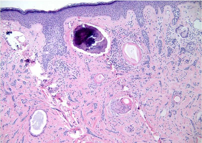
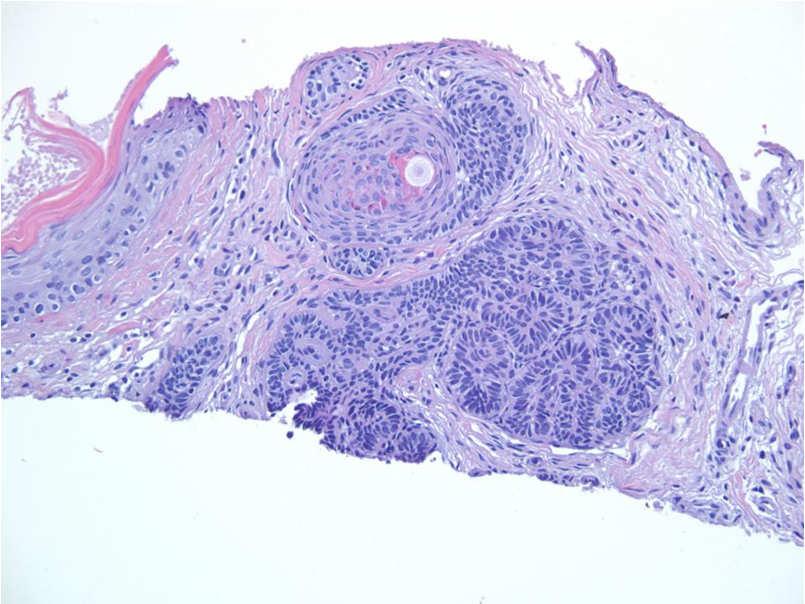
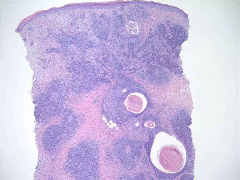
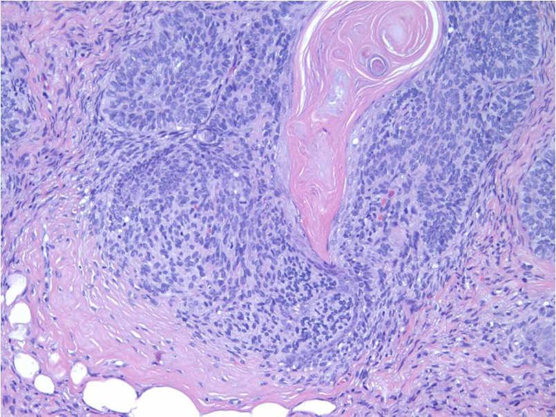
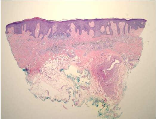
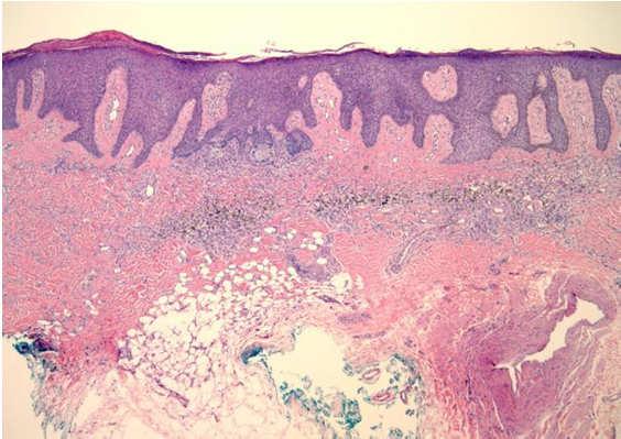
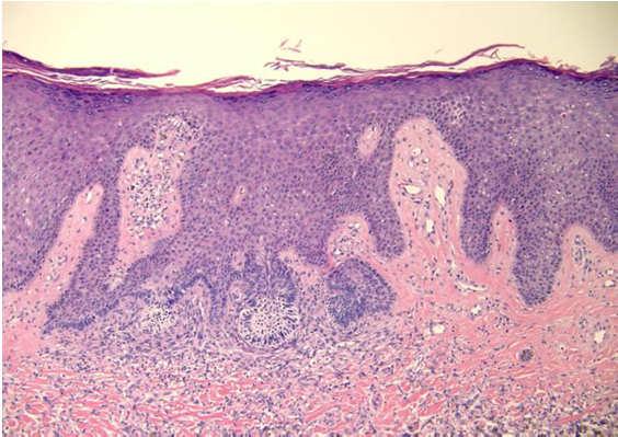
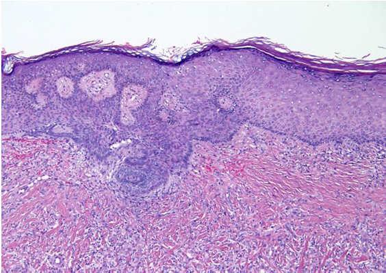
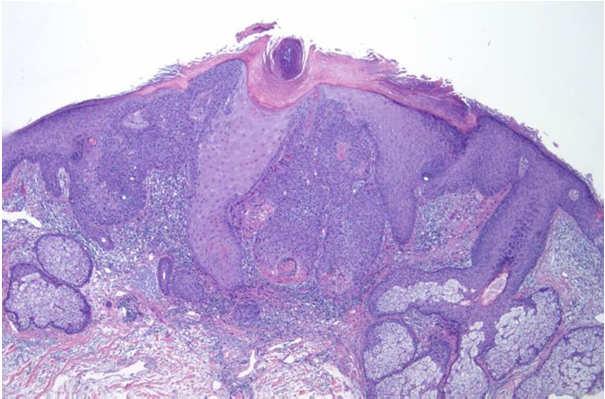
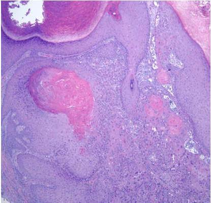
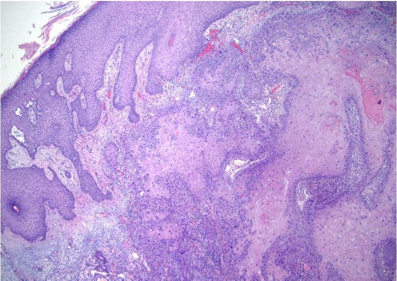
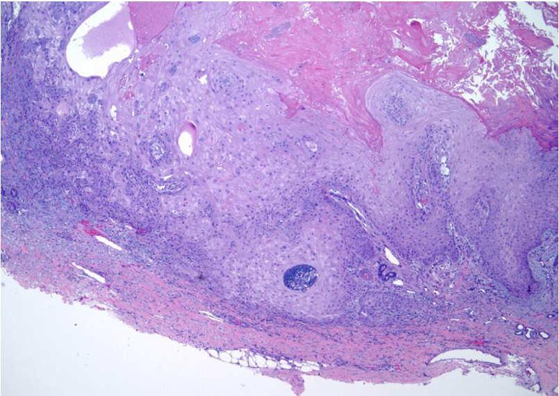
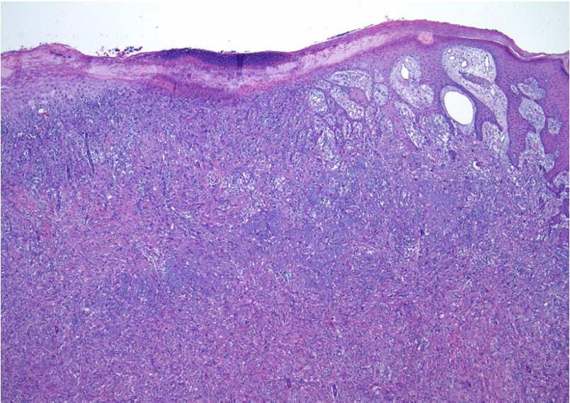
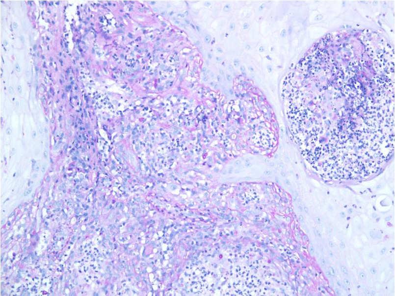
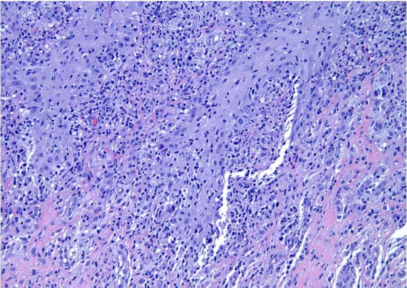
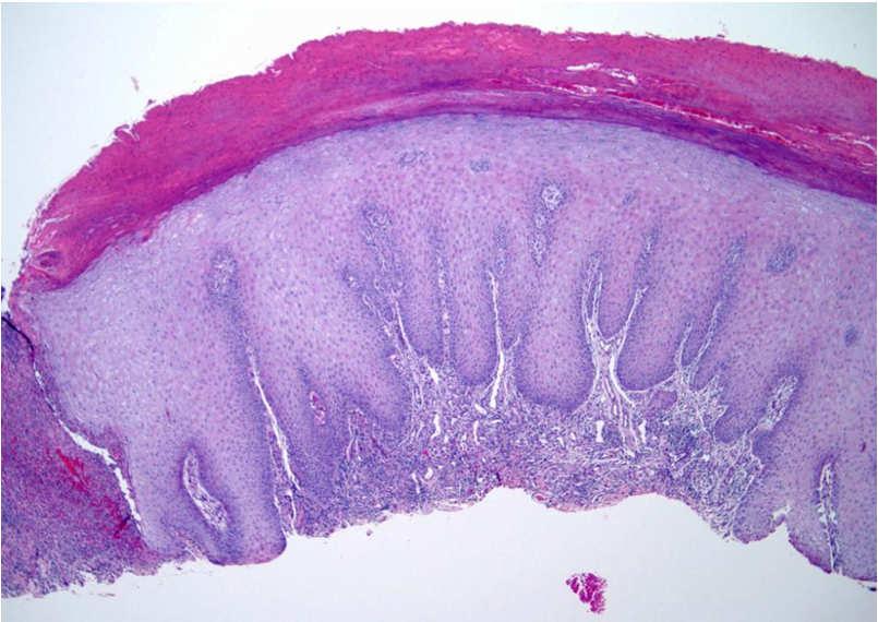
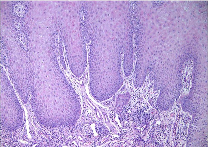
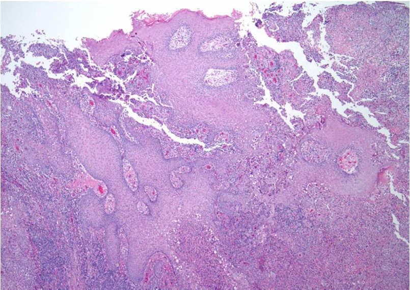
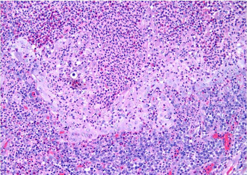
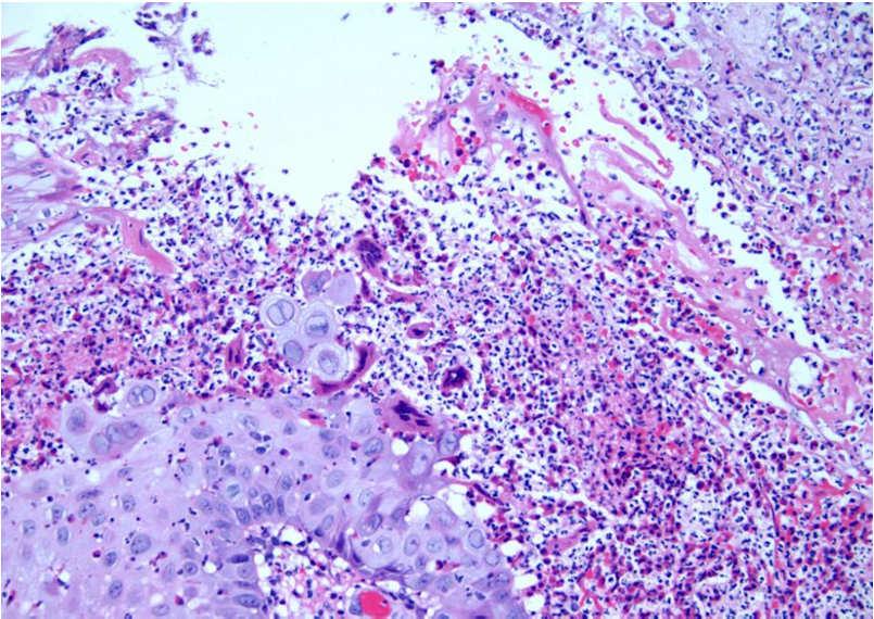
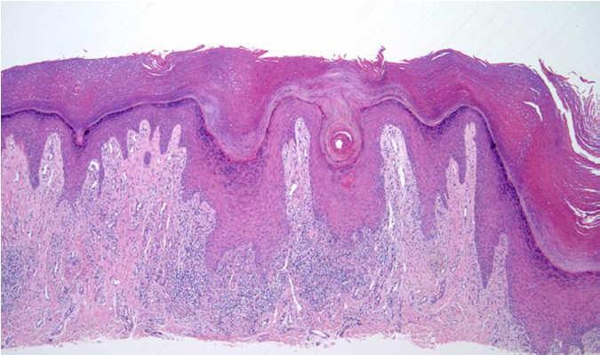
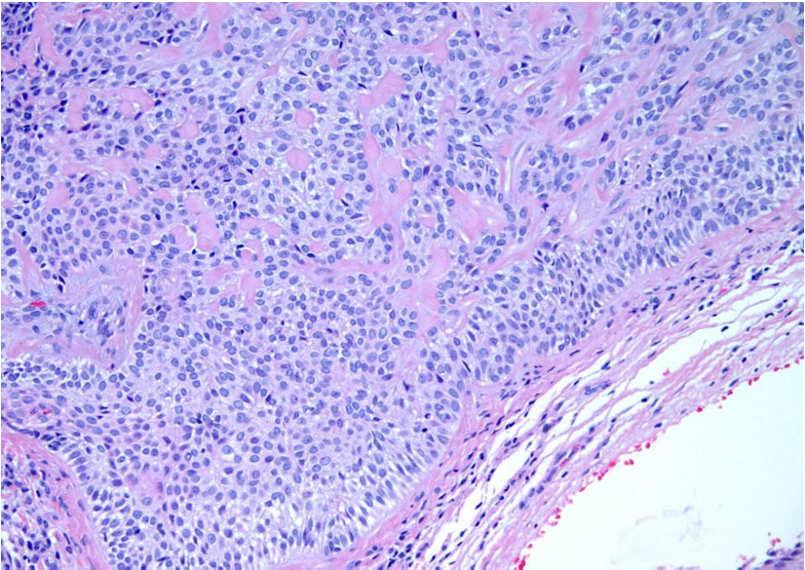
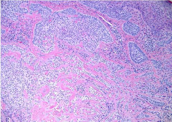
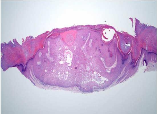
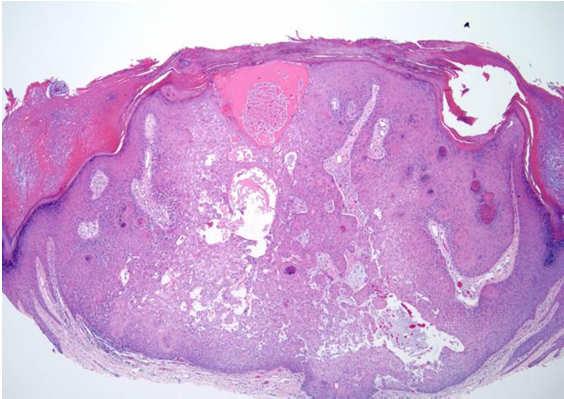
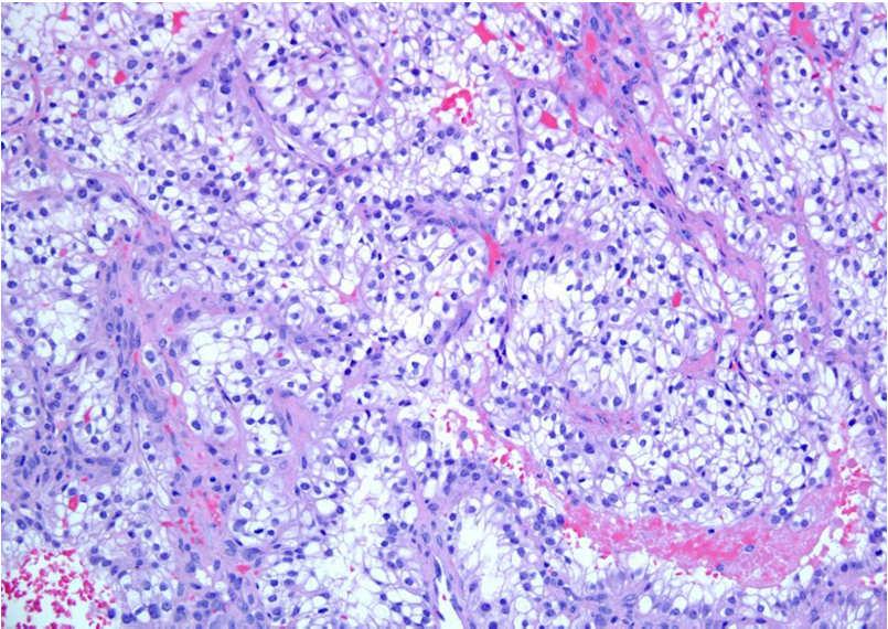
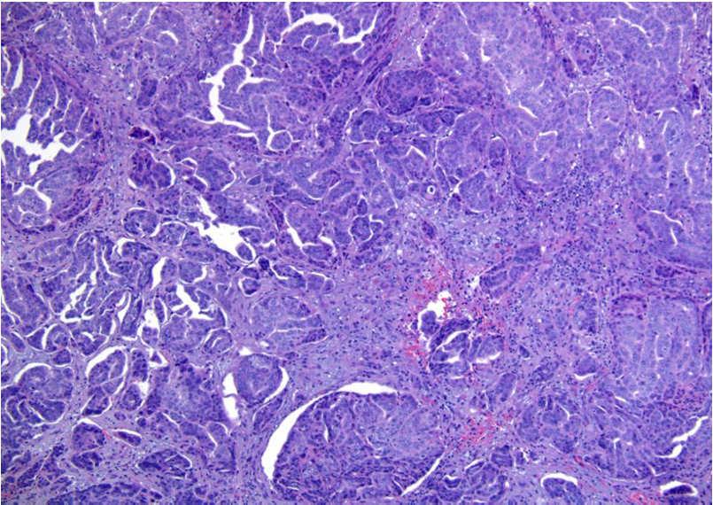
2 Basal cell carcinoma Accurate diagnosis unusually not a problem Classic criteria Basaloid cells Peripheral palisading Clefting from the surrounding stroma Mitotic figures and individually necrotic tumor cells Palisading and atypia Palisading 2
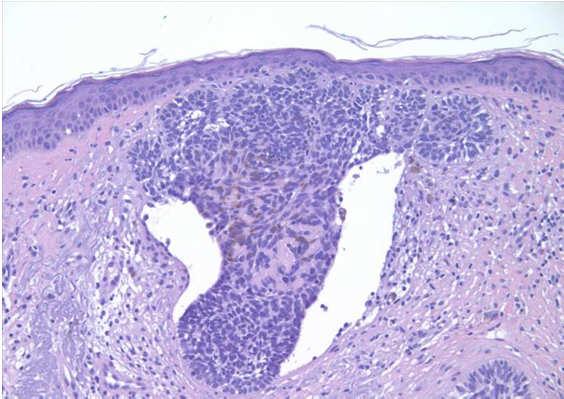
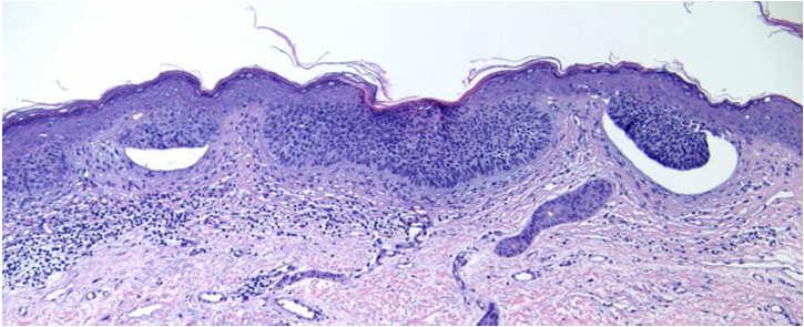
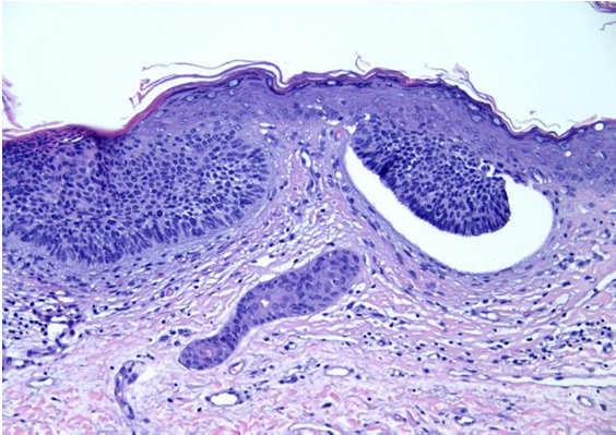
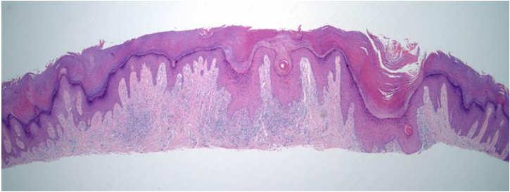
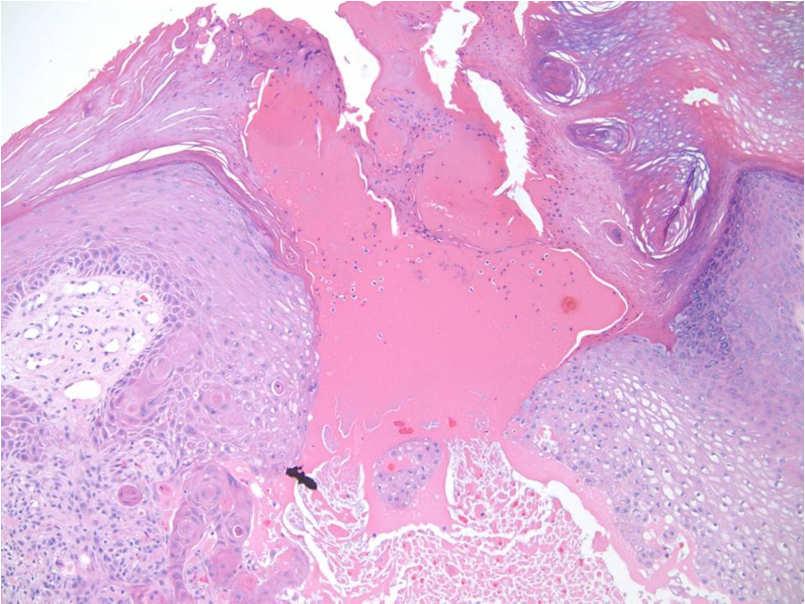
3 Clefting Superficial BCC Superficial BCC Micronodular BCC 3


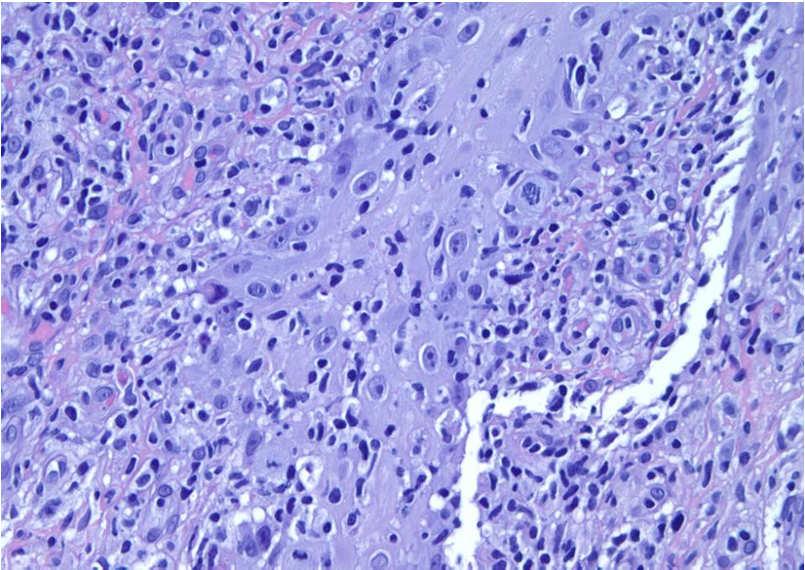
 Stromal involvement of")
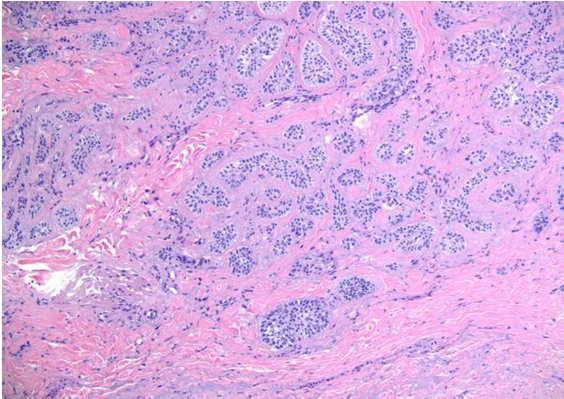
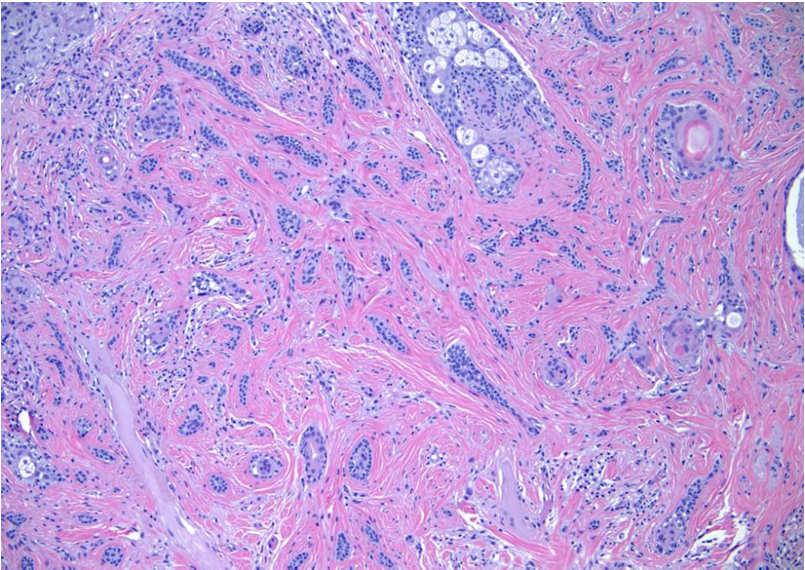
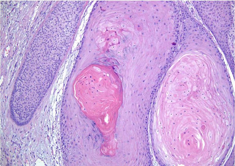
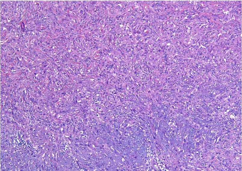
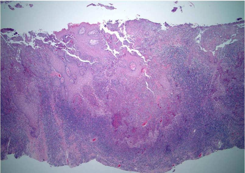
4 Fibromyxoid stoma Mitosis and necrosis BCC reporting Histologic suptype (to guide further therapy i.e. aggressive subtypes area treated with surgery, topical treatment an option for superficial variants) Involvement of margins (biopsy and excision) Stromal involvement of margin is considered positive 4

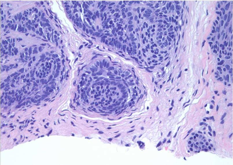
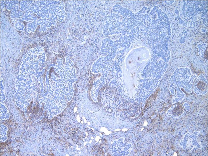
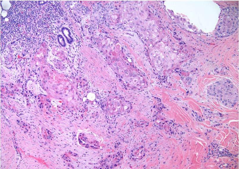
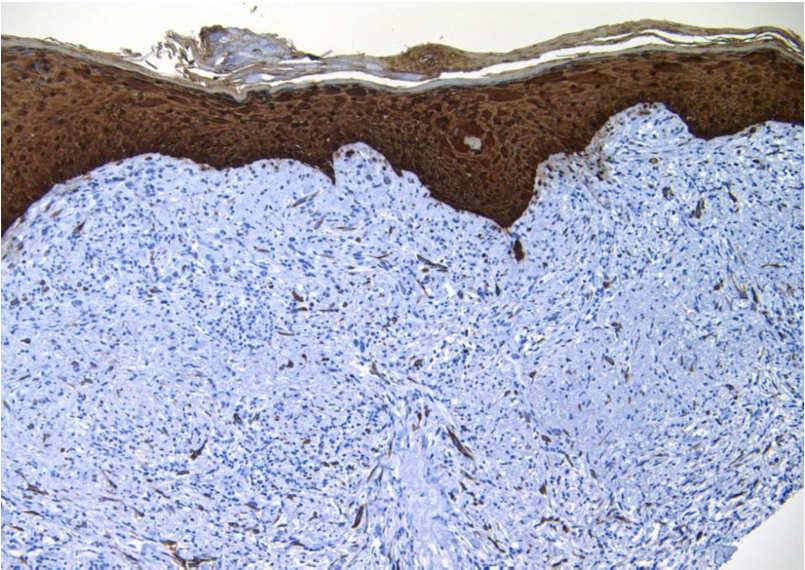
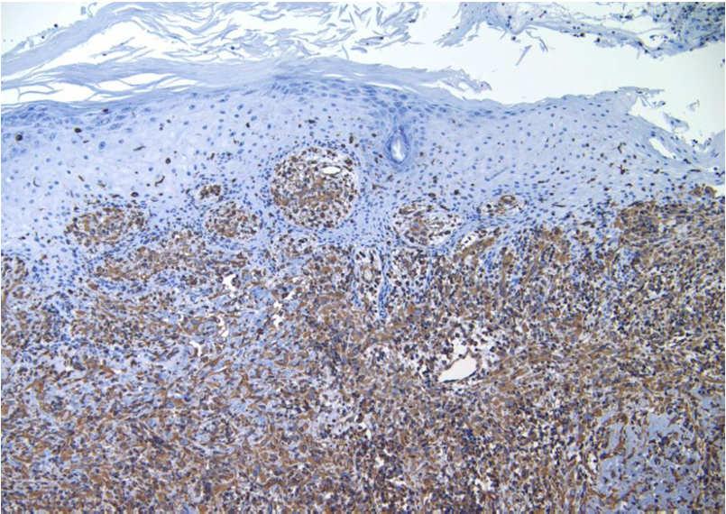
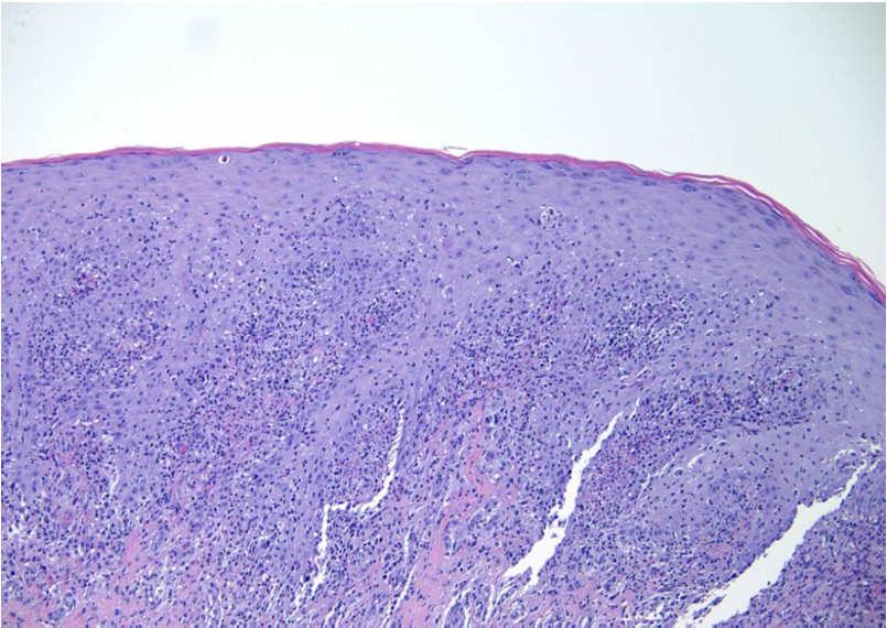
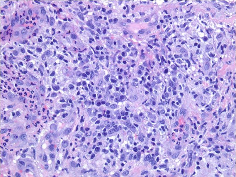
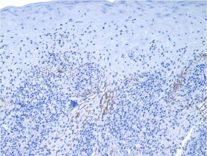
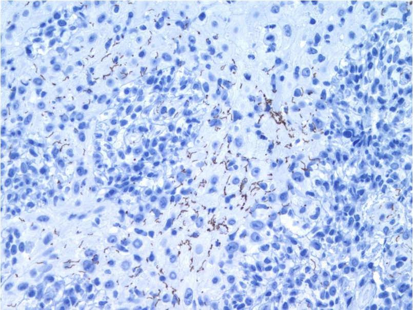
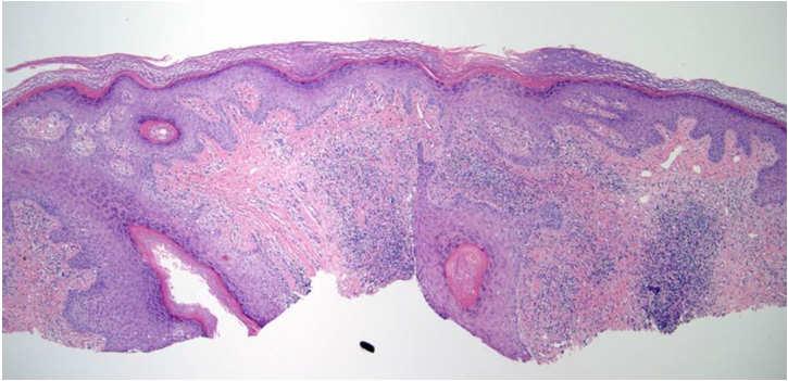
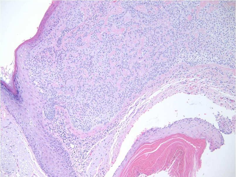
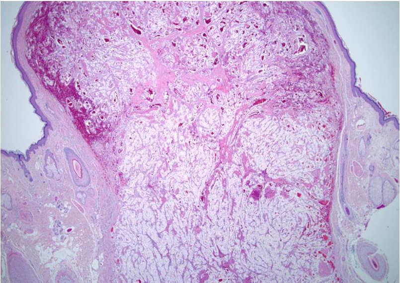
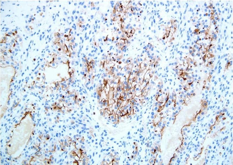
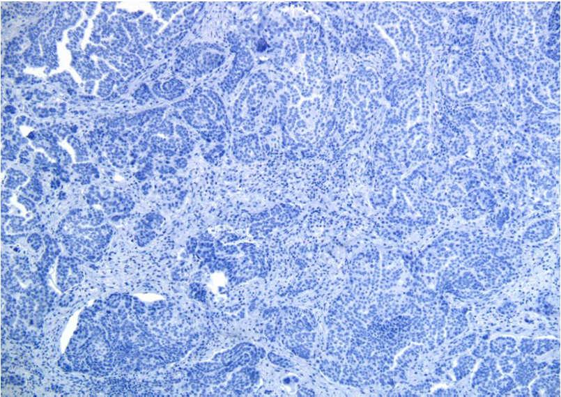
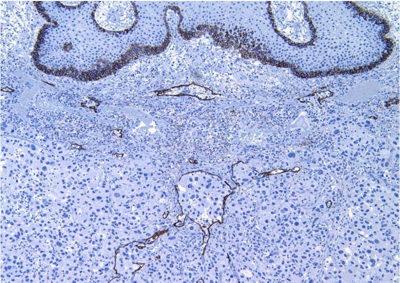
5 BCC reporting Other notable features such as perineural infiltration Other associated lesions (i.e. nevus sebaceus) Diagnostic difficulties in BCC Unusual subtypes that mimic other carcinomas and benign adnexal neoplasms Small biopsies from the head and neck (sampling error) Carcinoma may detached from its stroma while in formalin Squamous metaplasia in ulcerated BCC s Immunostaining Ber EP4 Ber-EP4 helpful to visualize aggregates of basal cell carcinoma that are subtle or obscured by inflammation BCC is positive for androgen receptor Cytokeratin 20 Stains resident Merkel cells Merkel cells are typically lost in basal cell carcinomas 5


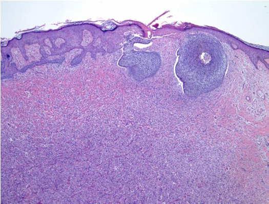
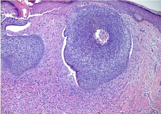
6 Benign basaloid proliferations Trichoepitheliomas Trichoblastomas Follicular hamartomas Follicular induction above dermatofibromas Benign basaloid Typically show a more organized growth pattern 50/50 rule of thumb Stroma/stroma clefting Different immunophenotype Retain CK20 positive Merkel cells Androgen receptor negative Stroma is CD10 positive 6

7 7


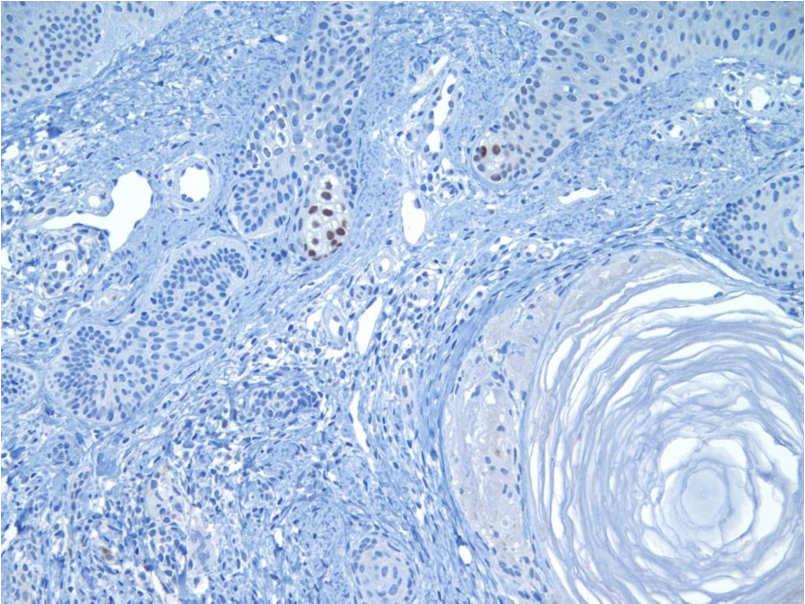
8 CK20 Androgen Receptor 8

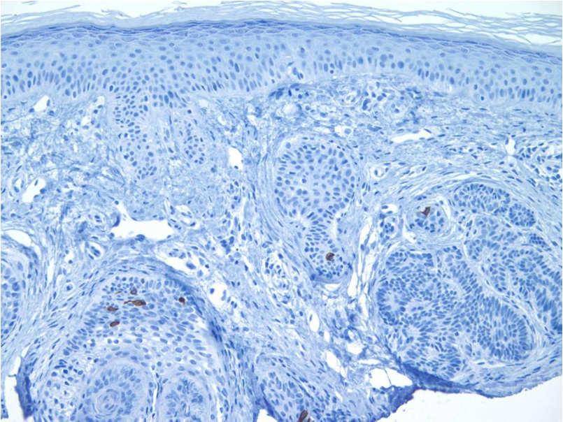
9 Androgen Recptor Cytokeratin 20 9
10 Follicular induction above a DF 10
11 11
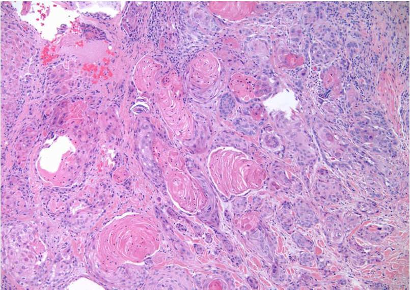
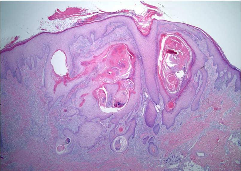
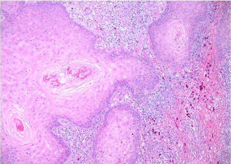
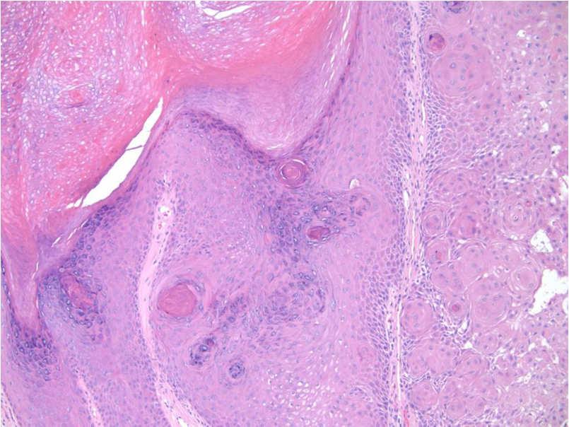
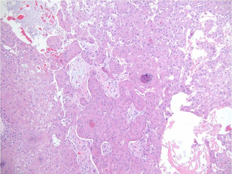
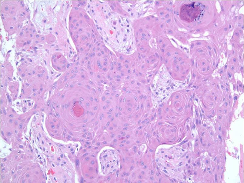
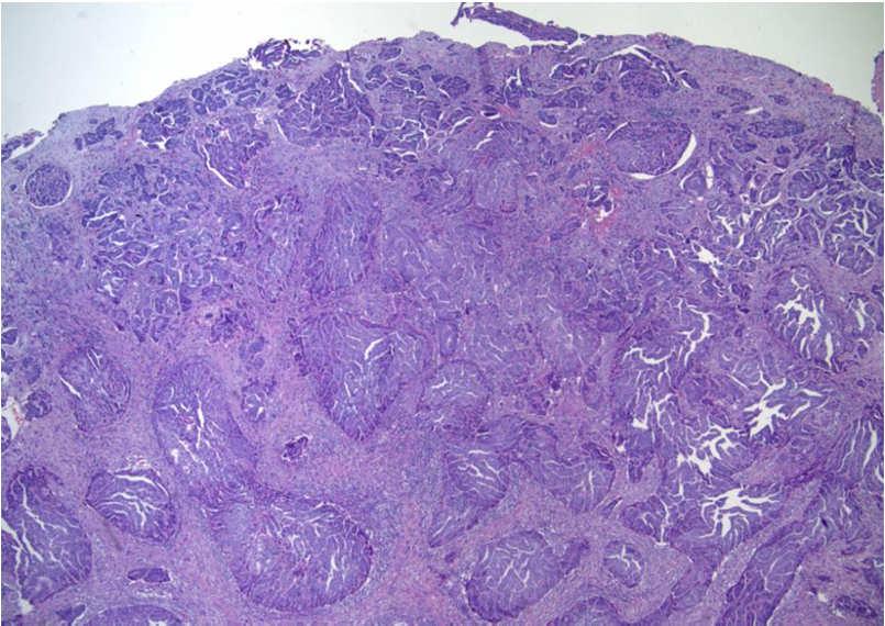
12 BCC vs B9 basaloid neoplasm Squamous cell carcinoma Basal Cell CA AR positive CK 20 negative CD10 positive tumor cells Basaloid neoplasm AR negative CK20 positive (Merkel cells) CD10 positive stromal cells Second most common non-melanoma skin cancer Great variety of clinical and histopathologic presentations Squamous cell carcinoma risk Sun exposure Viral Toxins Radiation Ulcers Transplant patients (know the population you serve) SCC Diagnosis usually straightforward Full thickness keratinocytic atypia of the epidermis with parakeratosis (implying disordered maturation) Extension of irregular aggregates of tumor cells into the dermis Increased mitoses, often atypical Keratin pearls 12


13 SCC Most squamous cell carcinomas arise in association with solar keratoses Gray areas between AK and SCC Look for presence of a granular layer and adnexal sparing in AK 13
14 14
15 15

16 KA in tattoo 16


17 p53 Small biopsies Difficulties Tremendous heterogeneity of histopathology Poorly differentiated lesions and subtle variants Wide variety of reactive conditions that mimic SCC 17

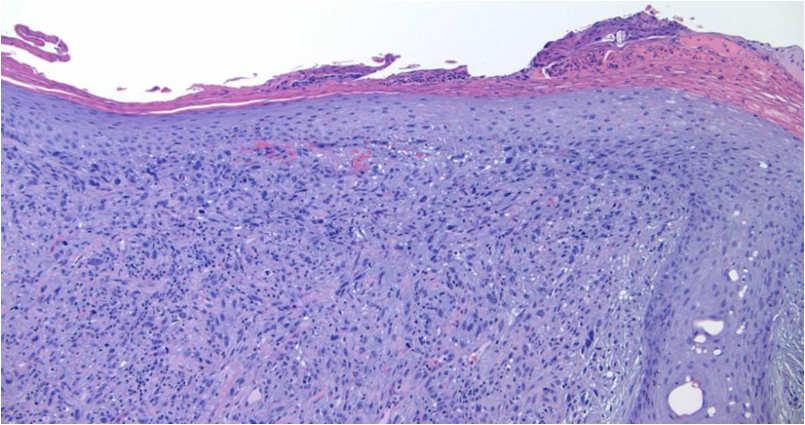
18 Spindled SCC 18


19 19

20 Ancillary studies Levels of small biopsies Keratin, p16, p53, p63, ki-67 stains Our panel for spindle cell lesions includes S100 Procollagen and CD10 High molecular weight keratin 903 Mimics of SCC Reactive Primary inflammatory Infectious Benign neoplastic Pseudoepitheliomatous Hyperplasia Irregular squamous epithelial hyperplasia that may involve the epidermis, follicles, and acrosyringia It simulates the architecture of invasive carcinoma but fails to demonstrate significant cytologic atypia and increased mitotic activity 20


21 Pseudoepitheliomatous hyperplasia Result of trauma, chronic inflammation, or infection with a wide range of agents Recognition of the histopathology in context of the appropriate clinical setting is important Wound healing One of the most common places to see this type is reaction is adjacent to a biopsy site The question is whether or not carcinoma remains When in doubt comparison to the prior biopsy can be helpful 21


22 Chondrodermatitis Occurs on the sun-damaged ears of older individuals Clinical impression is frequently carcinoma Characterized by marked epidermal hyperplasia, hypergranulosis, and an underlying proliferation of small blood vessels May also see epidermal ulceration, and underlying degenerated cartilage 22


23 P.E.H Infectious Classically seen above deep fungal and mycobacterial infections Very often see a suppurative and granulomatous infiltrate underlying PEH May also see neutrophilic microabsecces in keratoacanthomas Special stains/culture study vital 23


24 Infectious causes May also see P.E.H. at the edge of chronic ulcers In AIDS patients HSV and syphilis may lead to chronic ulcers that can be misdiagnosed as carcinoma Know your patient population 24

25 25
26 26
27 27
28 28


29 PEH and other neoplasia May be seen in association with lymphoproliferative disorders Lymphoproliferative disorders 29


30 5/28/2011 CD56 Epstein-Barr virus in situ Hypertrophic Lichen Planus Commonly seen on the shins of older individuals We commonly see squamous and basal cell carcinomas on the shin May present at a hyperkeratotic plaque and clinically masquerade as squamous cell carcinoma 30

31 31


32 Benign Keratoses Many benign squamous lesions can mimic squamous cell carcinoma in the right setting Often these lesions present clinically as a carcinoma and are located on sundamaged skin Desmoplastic Trichilemmoma First described in 1990 by Santa Cruz et al and thought to be a secondary change in a pre-existing trichilemmoma Characterized by thin cords of epithelial cells that infiltrate through a sclerotic stroma thereby simulating a carcinoma Most commonly seen on the face of older individuals 32
33 33


34 CD34 Inverted follicular keratosis A benign keratosis thought to arise from either hair follicles or represent a traumatized seborrheic keratosis or verruca May be seen anywhere on the body Characterized by an endophytic lesion with acanthosis, hypergranulosis and squamous eddies that can mimic invasive squamous cell carcinoma 34
35 35

36 36


37 Metastatic carcinoma Renal cell carcinoma may mimic clear cell hidradenoma New work shows a very different immunophenotype between primary adnexal neoplasms and metastatic adenocarcinoma 37
38 38

39 39
40 40
Cutaneous Adnexal Tumors
 Cutaneous Adnexal Tumors Lesions with Predominant Follicular Differentiation Special Emphasis on Basal Cell Carcinoma 2014-04-01 Prof. Dr. med. Katharina Glatz Pathologie Cutaneous Adnexal Tumors Hair
04/09/2018. Squamous Cell Neoplasia and Precursor Lesions. Agenda. Squamous Dysplasia. Squamo-proliferative lesions. Architectural features
 Squamous Cell Neoplasia and Precursor Lesions Jennifer L. Hunt, MD, MEd Aubrey J. Hough Jr, MD, Endowed Professor of Pathology Chair of Pathology and Laboratory Medicine University of Arkansas for Medical
Common Benign Lesions and Skin Cancers. 22nd May 2015 Dr Mark Foley
 Common Benign Lesions and Skin Cancers 22nd May 2015 Dr Mark Foley Thank you for downloading this file. This intended to supplement the presentation given at the NZ Wound Care Conference, it is not intended
Benign and malignant epithelial lesions: Seborrheic keratosis: A common benign pigmented epidermal tumor occur in middle-aged or older persons more
 Benign and malignant epithelial lesions: Seborrheic keratosis: A common benign pigmented epidermal tumor occur in middle-aged or older persons more common on the trunk; but extremities, head and neck are
Squamous Cell Neoplasia and Precursor Lesions
 Squamous Cell Neoplasia and Precursor Lesions Jennifer L. Hunt, MD, MEd Aubrey J. Hough Jr, MD, Endowed Professor of Pathology Chair of Pathology and Laboratory Medicine University of Arkansas for Medical
MECHANISMS OF HUMAN DISEASE: LABORATORY SESSION PATHOLOGY OF THE SKIN LAB. Friday, February 12, :30 am 11:00 am
 MECHANISMS OF HUMAN DISEASE: LABORATORY SESSION PATHOLOGY OF THE SKIN LAB Friday, February 12, 2012 9:30 am 11:00 am FACULTY COPY GOALS: Describe the basic clinical and morphologic features of various
Pathology of the skin. 2nd Department of Pathology, Semmelweis University
 Pathology of the skin 2nd Department of Pathology, Semmelweis University Histology of the skin Epidermis: Stratum corneum Stratum granulosum Stratum spinosum Stratum basale Dermis: papillary and reticular
Histopathology: skin pathology
 Histopathology: skin pathology These presentations are to help you identify, and to test yourself on identifying, basic histopathological features. They do not contain the additional factual information
Desmoplastic Melanoma R/O BCC. Clinical Information. 74 y.o. man with lesion on left side of neck r/o BCC
 R/O BCC Sabine Kohler, M.D. Professor of Pathology and Dermatology Dermatopathology Service Stanford University School of Medicine Clinical Information 74 y.o. man with lesion on left side of neck r/o
Case Report A Rare Cutaneous Adnexal Tumor: Malignant Proliferating Trichilemmal Tumor
 Case Reports in Medicine Volume 2015, Article ID 742920, 4 pages http://dx.doi.org/10.1155/2015/742920 Case Report A Rare Cutaneous Adnexal Tumor: Malignant Proliferating Trichilemmal Tumor Omer Alici,
Chapter 6 Squamous Cell Carcinoma: Variants and Challenges
 Chapter 6 Squamous Cell Carcinoma: Variants and Challenges Michael B. Morgan EPIDEMIOLOGY: Second most common skin cancer, rare in the dark-skinned races. ETIOLOGY: Ultraviolet light, HPV infection. PATHOGENESIS:
MECHANISMS OF HUMAN DISEASE: LABORATORY SESSION PATHOLOGY OF THE SKIN LAB. Friday, February 13, :30 am 11:00 am
 MECHANISMS OF HUMAN DISEASE: LABORATORY SESSION PATHOLOGY OF THE SKIN LAB Friday, February 13, 2009 9:30 am 11:00 am FACULTY COPY GOALS: Describe the basic clinical and morphologic features of various
IT S FUNDAMENTAL MY DEAR WATSON! A SHERLOCKIAN APPROACH TO DERMATOLOGY
 IT S FUNDAMENTAL MY DEAR WATSON! A SHERLOCKIAN APPROACH TO DERMATOLOGY Skin, Bones, and other Private Parts Symposium Dermatology Lectures by Debra Shelby, PhD, DNP, FNP-BC, FADNP, FAANP Debra Shelby,
Appendageal skin tumors
 Appendageal skin tumors Ibrahim Khalifeh, M.D. Associate Professor Department of Pathology American University of Beirut Medical Center Beirut, Lebanon Appendageal tumors Neoplasms whose differentiation
Whitney A. High, MD, JD, MEng
 ADS Dermatopathology Meeting 2014 Selected Adnexal Tumors Whitney A. High, MD, JD, MEng Associate Professor, Dermatology & Pathology Director of Dermatopathology (Dermatology) University of Colorado School
Journal of International Academy of Forensic Science & Pathology (JIAFP)
") Journal of International Academy of Forensic Science & Pathology (JIAFP) ISSN 2395-0722 MICROCYSTIC ADNEXAL CARCINOMA-A CASE REPORT WITH REVIEW OF LITERATURE Case Report Sulakshana M S 1,Natarajan M 2
Journal of International Academy of Forensic Science & Pathology (JIAFP) ISSN 2395-0722 MICROCYSTIC ADNEXAL CARCINOMA-A CASE REPORT WITH REVIEW OF LITERATURE Case Report Sulakshana M S 1,Natarajan M 2
Dermoscopy: Recognizing Top Five Common In- Office Diagnoses
 Dermoscopy: Recognizing Top Five Common In- Office Diagnoses Vu A. Ngo, DO Department of Family Medicine and Dermatology Choctaw Nation Health Services Authority Learning Objectives Introduction to dermoscopy
UNIVERSITY OF MEDICINE AND PHARMACY OF CRAIOVA FACULTY OF MEDICINE DOCTORAL THESIS SUMMARY
 UNIVERSITY OF MEDICINE AND PHARMACY OF CRAIOVA FACULTY OF MEDICINE DOCTORAL THESIS SUMMARY CLINICAL, HISTOPATHOLOGICAL AND IMMUNOHISTOCHEMICAL STUDY OF THE EPITHELIAL PRECANCEROUS LESIONS PRECURSORS OF
Benign versus Cancerous Lesions How to tell the difference FMF 2014 Christie Freeman MD, CCFP, DipPDerm, MSc
 1 Benign versus Cancerous Lesions How to tell the difference FMF 2014 Christie Freeman MD, CCFP, DipPDerm, MSc Benign lesions Seborrheic Keratoses: Warty, stuck-on Genetics and birthdays Can start in late
Keratinocyte tumors. Actinic Keratosis. Squamous cell carcinoma in situ. Squamous Cell Carcinoma. (aka Bowen s disease)
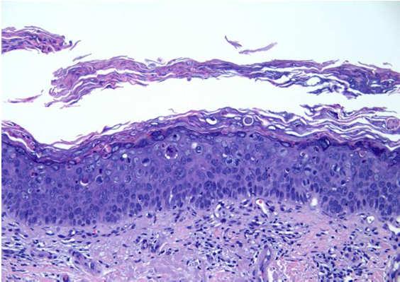
") Actinic Keratosis Keratinocyte tumors Prepared by Kurt Schaberg Precancerous, risk of malignancy ~8-20% per year (progresses to SCC); Due to chronic sun exposure Rough scaly plaque; typically due to sun
Actinic Keratosis Keratinocyte tumors Prepared by Kurt Schaberg Precancerous, risk of malignancy ~8-20% per year (progresses to SCC); Due to chronic sun exposure Rough scaly plaque; typically due to sun
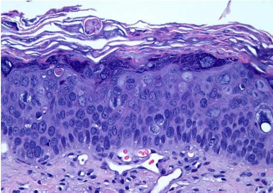
Dermatopathology: The tumor is composed of keratinocytes which show atypia, increase mitoses and abnormal mitoses.
 Squamous cell carcinoma (SCC): A common malignant tumor of keratinocytes arising in the epidermis, usually from a precancerous condition: 1- UV induced actinic keratosis, usually of low grade malignancy.
Actinic keratosis (AK): Dr Sarma s simple guide
: Dr Sarma s simple guide") Actinic keratosis (AK): Dr Sarma s simple guide Actinic keratosis is a very common lesion that you will see in your day-to-day practice. First, let me explain the name Actinic keratosis. It means keratosis
Actinic keratosis (AK): Dr Sarma s simple guide Actinic keratosis is a very common lesion that you will see in your day-to-day practice. First, let me explain the name Actinic keratosis. It means keratosis
أملس عضلي غرن = Leiomyosarcoma. Leiomyosarcoma 1 / 5
 Leiomyosarcoma 1 / 5 EPIDEMIOLOGY Exact incidence is unknown, but older studies suggest that leiomyosarcomas comprise approximately 3 percent of soft-tissue sarcomas. Superficial leiomyosarcoma occurs
American College of Mohs Surgery. Diagnostic Quality Control Program (Review of Answers)
") American College of Mohs Surgery Diagnostic Quality Control Program 2010 (Review of Answers) Question 1 A flesh-colored plaque on the right ala of an otherwise healthy 57 year-old male is referred for
American College of Mohs Surgery Diagnostic Quality Control Program 2010 (Review of Answers) Question 1 A flesh-colored plaque on the right ala of an otherwise healthy 57 year-old male is referred for
Malignant tumors of melanocytes: Part 1. Deba P Sarma, MD., Omaha
 Malignant tumors of melanocytes: Part 1 Deba P Sarma, MD., Omaha The melanocytic tumor is one of the most difficult and confusing areas in Dematopathology. It is true that most (95%) of such lesions are
When Immunostains Can Get You in Trouble: Gynecologic Pathology p16: Panacea or Pandora s Box?
 When Immunostains Can Get You in Trouble: Gynecologic Pathology p16: Panacea or Pandora s Box? Teri A. Longacre, MD Stanford Medicine Stanford California pi6 in Gynecologic Pathology: Panacea or Pandora
10/2/17. A 45-year-old female presents with a lesion on the cheek, rule out basal cell carcinoma. Overview
 10/2/17 Overview Desmoplastic trichoepithelioma vs. morpheaform BCC vs. microcystic adnexal carcinoma Trichilemmomas & Cowden s syndrome Cutaneous Adnexal Tumours with Clinical Impact Rajiv M. Patel, M.D.
David B. Troxel, MD. Common Medicolegal Situations: Misdiagnosis of Melanoma
 Common Medicolegal Situations: Misdiagnosis of Melanoma David B. Troxel, MD Medical Director, The Doctors Company, Napa, California Clinical Professor Emeritus, University of California at Berkeley Past
Dysplasia, Mimics and Other Controversies
 Dysplasia, Mimics and Other Controversies Mary S. Richardson, MD Dept. of Pathology Medical University of South Carolina Charleston, SC Notice of Faculty Disclosure In accordance with ACGME guidelines,
Skin lesions The Good and the Bad. Dr Virginia Hubbard Ipswich Hospital NHS Trust Barts and the London School of Medicine and Dentistry
 Skin lesions The Good and the Bad Dr Virginia Hubbard Ipswich Hospital NHS Trust Barts and the London School of Medicine and Dentistry Case 1 32 year old woman Australian Lesion on back New hair growing
Head & Neck Squamous Carcinoma: Artifacts, Challenges, and Controversies. Agenda
 Head & Neck Squamous Carcinoma: Artifacts, Challenges, and Controversies Jennifer L. Hunt, MD, MEd Aubrey J. Hough Jr, MD, Endowed Professor of Pathology Chair of Pathology and Laboratory Medicine University
Papillary Lesions of the breast
 Papillary Lesions of the breast Emad Rakha Professor of Breast Pathology The University of Nottingham Papillary lesions of the breast are a heterogeneous group of disease, which are characterised by neoplastic
Clinical characteristics
 Skin Cancer Fernando Vega, MD Seattle Healing Arts Clinical characteristics Precancerous lesions Common skin cancers ACTINIC KERATOSIS Precancerous skin lesions Actinic keratoses Dysplastic melanocytic
LARYNGEAL DYSPLASIA. Tomas Fernandez M; 3 rd year ENT resident, Son Espases University Hospital
 LARYNGEAL DYSPLASIA Tomas Fernandez M; 3 rd year ENT resident, Son Espases University Hospital INTRODUCTION Laryngeal cancer constitutes 1-2% of all malignancies diagnosed worldwide Survival is related
- Selected Tumors of the Skin Appendages - Primary vs. Metastasis
 - Selected Tumors of the Skin Appendages - Primary vs. Metastasis Napa Valley 2018 Victor G. Prieto, MD, PhD Chair of Pathology UT MD Anderson Cancer Center vprieto@mdanderson.org Napa Valley in May Introduction
Porokeratosis: Introduction
 Porokeratosis: Introduction Benign epidermal proliferation Distinct clinical & histologic features 5 clinical subtypes Erroneously named porokeratosis CLINICAL SUBTYPES 1. Porokeratosis of Mibelli 2. Disseminated
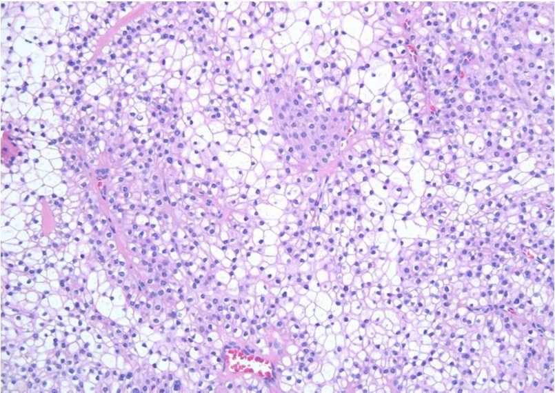
Case Report Clear Cell Basal Cell Carcinoma
 SAGE-Hindawi Access to Research Volume 2011, Article ID 386921, 4 pages doi:10.4061/2011/386921 Case Report Clear Cell Basal Cell Carcinoma Deba P. Sarma, 1 Daniel Olson, 1 Jennifer Olivella, 1 Tracey
Salivary Glands 3/7/2017
 Salivary Glands 3/7/2017 Goals and objectives Focus on the entities unique to H&N Common board type facts Information for your future practice Salivary Glands Salivary Glands Major gland. Paratid. Submandibular.
Pathology. Skin Tumor. Bayan N. Mohammad 15/10/2015. Mohammad al-orjani. Page 0 of 23
 #7 35 Pathology Skin Tumor Bayan N. Mohammad 15/10/2015 Mohammad al-orjani Page 0 of 23 بسم هللا الرحمن الرحيم GREETINGS This lecture is about skin tumors, all the slides are included and every slide will
SEBACEOUS NEOPLASMS. Dr. Prachi Saraogi Clinical Fellow in Dermatology
 SEBACEOUS NEOPLASMS Dr. Prachi Saraogi Clinical Fellow in Dermatology Sebaceous neoplasms Sebaceous adenoma (Benign) Sebaceous carcinoma (Malignant) SEBACEOUS ADENOMA Benign tumours composed of incompletely
Simulators of melanoma
 Simulators of melanoma Philip E. LeBoit, M.D. Depts. of Pathology and Dermatology University of California, San Francisco Simulators of melanoma Simulators of melanoma in situ Melanocytic Non-melanocytic
Case 18. M75. Excision of mass on scalp. Clinically SCC. The best diagnosis is:
 Case 18 M75. Excision of mass on scalp. Clinically SCC. The best diagnosis is: A. Pilomatrical carcinoma B. Adnexal carcinoma NOS C. Metastatic squamous cell carcinoma D.Primary squamous cell carcinoma
BSD 2015 Case 19. Female 21. Nodule on forehead. The best diagnosis is:
 BSD 2015 Case 19 Female 21. Nodule on forehead. The best diagnosis is: A. mixed tumour of skin B. porocarcinoma C. nodular hidradenoma D. metastatic adenocarcinoma BSD 2015 Case 19 Female 21 Nodule on
5/21/2018. Prostate Adenocarcinoma vs. Urothelial Carcinoma. Common Differential Diagnoses in Urological Pathology. Jonathan I.
 Common Differential Diagnoses in Urological Pathology Jonathan I. Epstein Prostate Adenocarcinoma vs. Urothelial Carcinoma 1 2 NKX3.1 NKX3.1 3 4 5 6 Proposed ISUP Recommendations Option to use PSA as a
المركب النموذج--- سبيتز وحمة = Type Spitz's Nevus, Compound SPITZ NEVUS 1 / 7
 SPITZ NEVUS 1 / 7 Epidemiology An annual incidence rate of 1.4 cases of Spitz nevus per 100,000 individuals has been estimated in Australia, compared with 25.4 per 100,000 individuals for cutaneous melanoma
Case year female. Routine Pap smear
 Case 1 57 year female Routine Pap smear Diagnosis? 1. Atypical glandular cells of unknown significance (AGUS) 2. Endocervical AIS 3. Endocervical adenocarcinoma 4. Endometrial adenocarcinoma 5. Adenocarcinoma
Samer Ghosn, MD Associate professor, Derpartment of Dermatology American University of Beirut Medical Center. Follicular lesions
 Samer Ghosn, MD Associate professor, Derpartment of Dermatology American University of Beirut Medical Center Follicular lesions Introduction Follicular lesions are important to recognize: For proper management
Cutaneous Malignancies: A Primer COPYRIGHT. Marissa Heller, M.D.
 Cutaneous Malignancies: A Primer Marissa Heller, M.D. Associate Director of Dermatologic Surgery Department of Dermatology Beth Israel Deaconess Medical Center December 10, 2016 Skin Cancer Non-melanoma
ISPUB.COM. Seborrheic Keratosis: A Pictorial Review of the Histopathologic Variations. D Sarma, S Repertinger
 ISPUB.COM The Internet Journal of Dermatology Volume 7 Number 2 Seborrheic Keratosis: A Pictorial Review of the Histopathologic Variations D Sarma, S Repertinger Citation D Sarma, S Repertinger.. The Internet
NEOPLASMS OF THE SURFACE EPITHELIUM (KERATINOCYTES)
") NEOPLASMS OF THE SURFACE EPITHELIUM (KERATINOCYTES) Papillary Lesions Precancerous Lesions Keratinocyte Proliferations Carcinomas Melanotic Lesions Melanomas Normal Mucosa Keratin layer Spinous layer Basal
NEOPLASMS OF THE SURFACE EPITHELIUM (KERATINOCYTES) Papillary Lesions Precancerous Lesions Keratinocyte Proliferations Carcinomas Melanotic Lesions Melanomas Normal Mucosa Keratin layer Spinous layer Basal
Periocular Malignancies
 Periocular Malignancies Andrew Gurwood, O.D., F.A.A.O., Dipl. Marc Myers, O.D., F.A.A.O. Drs. Myers and Gurwood have no financial interests to disclose. Course Description Discussion of the most common
Objectives. Salivary Gland FNA: The Milan System. Role of Salivary Gland FNA 04/26/2018
 Salivary Gland FNA: The Milan System Dr. Jennifer Brainard Section Head Cytopathology Cleveland Clinic Objectives Introduce the Milan System for reporting salivary gland cytopathology Define cytologic
Large majority caused by sun exposure Often sun exposure before age 20 Persons who burn easily and tan poorly are at greatest risk.
 Basics of Skin Cancer Detection and Treatment of Non- Melanoma Skin Cancers Large majority caused by sun exposure Often sun exposure before age 20 Persons who burn easily and tan poorly are at greatest
In the third part of the present study tumours which previous were described as basal cell tumours but now have been reclassified as trichoblastomas
 170 6. SUMMARY Immunhistochemical investigations for identifying the histogenesis of basaloid neoplasias and hyperplasias in the mamma parenchyma of the bitch, for the use of the human nuclear protein
Malignant non-melanocytic lesions
 Malignant non-melanocytic lesions Course C023: Fundamentals of Dermoscopy March 4, 2019, 11:20 AM - 11:50 PM Room: 146B Jason B. Lee, MD Professor & Vice Chair Director of Dermatopathology & Pigmented
Papillary Lesions of the Breast A Practical Approach to Diagnosis. (Arch Pathol Lab Med. 2016;140: ; doi: /arpa.
 Papillary Lesions of the Breast A Practical Approach to Diagnosis (Arch Pathol Lab Med. 2016;140:1052 1059; doi: 10.5858/arpa.2016-0219-RA) Papillary lesions of the breast Span the spectrum of benign,
Identifying Skin Cancer. Mary S. Stone MD Professor of Dermatology and Pathology University of Iowa Carver College of Medicine March, 2018
 Identifying Skin Cancer Mary S. Stone MD Professor of Dermatology and Pathology University of Iowa Carver College of Medicine March, 2018 American Cancer Society web site Skin Cancer Melanoma Non-Melanoma
A dinical and histopathologic entity associated with an increased risk of nonmelanoma skin cancer
 PUVA keratosis A dinical and histopathologic entity associated with an increased risk of nonmelanoma skin cancer M. C. G. van Praag, MD, a J. N. Bouwes Bavinck, MD, a W. Bergman, MD, PhD, a F. R. Rosendaal,
Problem diagnoses. Current issues in Anatomic pathology. Problem Diagnoses in Tumors of the Oral Cavity 5/29/2009
 Current issues in Anatomic pathology Problem Diagnoses in Tumors of the Oral Cavity Richard Jordan DDS PhD FRCPath Professor of Oral Pathology & Pathology Director, UCSF Oral Pathology Diagnostic Laboratory
Cutaneous metastases. Thaddeus Mully. University of California, San Francisco Professor, Departments of Pathology and Dermatology
 Cutaneous metastases Thaddeus Mully University of California, San Francisco Professor, Departments of Pathology and Dermatology DISCLOSURE OF RELATIONSHIPS WITH INDUSTRY Thaddeus Mully Course C005 Essential
Regression 2/3/18. Histologically regression is characterized: melanosis fibrosis combination of both. Distribution: partial or focal!
 Regression Margaret Oliviero MSN, ARNP Harold S. Rabinovitz MD Histologically regression is characterized: melanosis fibrosis combination of both Distribution: partial or focal! Dermatoscopic terminology
Lumps and Bumps: The Dermatology of Lid Lesions
 Lumps and Bumps: The Dermatology of Lid Lesions Thomas J. Joly, MD, PhD Assistant Professor of Ophthalmology Eastern Virginia Medical School Ophthalmic Plastic Surgery Service Virginia Eye Consultants
History A 89 year old gentleman presenting with a scalp/forehead nodule. Patient had squamous cell carcinoma 18 m at same site, excised. Outside diagn
 Case III History A 89 year old gentleman presenting with a scalp/forehead nodule. Patient had squamous cell carcinoma 18 m at same site, excised. Outside diagnoses: Squamous cell carcinoma. R/O: SCC, Melanoma,
Lymphoma and Pseudolymphoma
 Lymphoma and Pseudolymphoma Laura B. Pincus, MD Co-Director, Cutaneous Lymphoma Clinic Associate Professor Dermatology and Pathology University of California, San Francisco I HAVE NO RELEVANT RELATIONSHIPS
Supplementary Online Content
 Supplementary Online Content Tschandl P, Rosendahl C, Akay BN, et al. Expert-level diagnosis of nonpigmented skin cancer by combined convolutional neural networks. JAMA Dermatol. Published online November
Atlas of Eyelid and Conjunctival Tumors
 Atlas of Eyelid and Conjunctival Tumors Jerry A. Shields, M.D. Director, Ocular Oncology Service Wills Eye Hospital Professor of Ophthalmology Thomas Jefferson University Philadelphia, Pennsylvania Carol
64 y.o. F with CLL and leg tumour
 64 y.o. F with CLL and leg tumour Case History Excision with split-skin grafting Histology moderately differentiated squamous cell carcinoma with large areas of necrosis and brisk mitotic activity.
Case Report A Case of Cystic Basal Cell Carcinoma Which Shows a Homogenous Blue/Black Area under Dermatoscopy
 Volume 20, Article ID 450472, 4 pages doi:0.55/20/450472 Case Report A Case of Cystic Basal Cell Carcinoma Which Shows a Homogenous Blue/Black Area under Dermatoscopy Akihiro Yoneta, Kohei Horimoto, Keiko
Diagnostic difficulties with lesions of the oral mucosa
 BDIAP London, November 2010 School of Clinical Dentistry University of Sheffield Diagnostic difficulties with lesions of the oral mucosa Paul M Speight Dept Oral & Maxillofacial Pathology University of
Important Decisions in Dermatopathology: The Clinico- Pathologic Correlation. Dermatopathology Specialists Needed. Changing Trends
 Important Decisions in Dermatopathology: The Clinico- Pathologic Correlation Uma Sundram, MD, PhD Departments of Pathology and Dermatology Stanford University May 29, 2008 Dermatopathology Specialists
Desmoplastic trichoepithelioma; a new case and review of the literature with emphasis on its differential diagnosis
 Hong Kong J. Dermatol. Venereol. (2014) 22, 135-139 Case Report Desmoplastic trichoepithelioma; a new case and review of the literature with emphasis on its differential diagnosis M Concha-Rogazy, S Alvarez-Veliz,
SESSION 1: GENERAL (BASIC) PATHOLOGY CONCEPTS Thursday, October 16, :30am - 11:30am FACULTY COPY
 PATHOLOGY CONCEPTS Thursday, October 16, :30am - 11:30am FACULTY COPY") SESSION 1: GENERAL (BASIC) PATHOLOGY CONCEPTS Thursday, October 16, 2008 9:30am - 11:30am FACULTY COPY GOAL: Describe the basic morphologic (structural) changes which occur in various pathologic conditions.
SESSION 1: GENERAL (BASIC) PATHOLOGY CONCEPTS Thursday, October 16, 2008 9:30am - 11:30am FACULTY COPY GOAL: Describe the basic morphologic (structural) changes which occur in various pathologic conditions.
Skin is a multilayered organ that covers and protects the body.
 Section 1: Skin is a multilayered organ that covers and protects the body. K What I Know W What I Want to Find Out L What I Learned Essential Questions What are the four tissue types that are found in
BASAL CELL CARCINOMA WITH ECCRINE DIFFERENTIATION: A RARE ENTITY Divvya B 1, Rehana Tippoo 2, P. Viswanathan 3, B. Krishnaswamy 4, A.
 BASAL CELL CARCINOMA WITH ECCRINE DIFFERENTIATION: A RARE ENTITY Divvya B 1, Rehana Tippoo 2, P. Viswanathan 3, B. Krishnaswamy 4, A. Anvar Ali 5 HOW TO CITE THIS ARTICLE: Divvya B, Rehana Tippoo, P. Viswanathan,
Acantholytic Anaplastic Extramammary Paget s Disease: A Case Report and Review of the Literature
 Ann Dermatol Vol. 23, Suppl. 2, 2011 http://dx.doi.org/10.5021/ad.2011.23.s2.s226 CASE REPORT Acantholytic Anaplastic Extramammary Paget s Disease: A Case Report and Review of the Literature Yu-Jin Oh,
Know who is at risk: LOOK! for ABCDs, rapidly changing lesions, do a biopsy when indicated
 Lindy P. Fox, MD Assistant Professor Director, Hospital Consultation Service Department of Dermatology University of California, San Francisco Applies to adults without history of malignancy or premalignant
SKIN SERVICES REVIEW Changes to Medicare Benefits Schedule for 1 November 2016
 Attachment A SKIN SERVICES REVIEW Changes to Medicare Benefits Schedule for 1 November 2016 Deleted items 31200-31215, 31230-31240 31255-31335 Colour Coding for new / updated items: MUCOSAL BIOPSY AND
Skin Cancer of the Nose: Common and Uncommon
 Skin Cancer of the Nose: Common and Uncommon Mark Russell, M.D. Associate Professor of Dermatology, Otolaryngology, and Pathology University of Virginia Objectives Review clinical presentations of select
CLEAR CELL BASAL CELL CARCINOMA- AN UNUSUAL ENTITY IN AN INDIAN MALE
 Indian Journal of Medical s ISSN: 2319 3832(Online) CLEAR CELL BASAL CELL CARCINOMA- AN UNUSUAL ENTITY IN AN INDIAN MALE * Cheena Garg 1, Arjun Agarwal 2, Bushra Siddiqui 3, Shahbaz Faridi 4, 1 Department
Living Beyond Cancer Skin Cancer Detection and Prevention
 Living Beyond Cancer Skin Cancer Detection and Prevention Cutaneous Skin Cancers Identification Diagnosis Treatment options Prevention What is the most common cancer in people? What is the most common
Lichenoid Tissue Reaction in Malignant Melanoma A Potential Diagnostic Pitfall
 natomic Pathology / LICHENOID TISSUE RECTION IN MLIGNNT MELNOM Lichenoid Tissue Reaction in Malignant Melanoma Potential Diagnostic Pitfall CPT Scott R. Dalton, MC, US, 1,3 Capt Matt. aptista, USF, MC,
Technicians & Nurses Program
 ASCRS ASOA Symposium & Congress Technicians & Nurses Program May 6-10, 2016 New Orleans Evaluation and Treatment of Eyelid Malignancies Richard C. Allen MD PhD FACS Professor Section of Ophthalmology Dept.
Malignant Peripheral Nerve Sheath Tumor
 C H A P T E R 120 Malignant Peripheral Nerve Sheath Tumor Currently, malignant peripheral nerve sheath tumor (MPNST) is the most commonly used generic name for the neoplasms known in the past as neurosarcoma,
CASE REPORT SOLITARY SEBACEOUS NEVUS OF JADASSOHN COMPLICATED BY SQUAMOUS CELL CARCINOMA AND BASAL CELL CARCINOMA
 CASE REPORT Dennis H. Kraus, MD, Section Editor SOLITARY SEBACEOUS NEVUS OF JADASSOHN COMPLICATED BY SQUAMOUS CELL CARCINOMA AND BASAL CELL CARCINOMA Ahmad Ridzwan Arshad, FRCS, 1 Wan S. Azman, MS, 1 Ayadurai
PATHOLOGY OF THE SKIN 2. Tumours of the skin
 PATHOLOGY OF THE SKIN 2. Tumours of the skin Máirín E. McMenamin MB MRCPI FRCPath Dip (Dermatopathol) RCPath St. James s Hospital and University of Dublin, Trinity College Tumour (Neoplasia) Benign or
Diploma examination. Dermatopathology: First paper. Tuesday 21 March Candidates must answer FOUR questions ONLY. Time allowed: Three hours
 Dermatopathology: First paper Tuesday 21 March 2017 1. Discuss the role of fluorescent in-situ hybridization (FISH) and emerging molecular techniques in the diagnosis of cutaneous melanocytic lesions,
SKIN CANCER. Most common cancer diagnosis 40% of all cancers
 SKIN CANCER Most common cancer diagnosis 40% of all cancers OBJECTIVES Review common and uncommon cancers of the skin. Special emphasis on melanoma and dysplastic nevus Review pathology/tnm/staging, which
Microcystic Squamous Cell Carcinoma of the Lung A Clinicopathologic Study of Three Cases
 Anatomic Pathology / Microcystic SCC of the Lung Microcystic Squamous Cell Carcinoma of the Lung A Clinicopathologic Study of Three Cases Annikka Weissferdt, MD, and Cesar A. Moran, MD Key Words: Squamous
Glenn D. Goldman, MD. Fletcher Allen Health Care. University of Vermont College of Medicine
 Glenn D. Goldman, MD Fletcher Allen Health Care University of Vermont College of Medicine Recognize and identify the main types of skin cancer Understand how and why Mohs surgery is utilized for the treatment
The Role of Bcl-2, CD10 and CD34 Expression in Differentiation between Basal Cell Carcinoma and Trichoepithelioma
 Open Journal of Pathology, 2014, 4, 116-124 Published Online July 2014 in SciRes. http://www.scirp.org/journal/ojpathology http://dx.doi.org/10.4236/ojpathology.2014.43018 The Role of Bcl-2, CD10 and CD34
الطلاوة = Leukoplakia LEUKOPLAKIA
 LEUKOPLAKIA Leukoplakia is a clinical term that refers to a predominantly white lesion of the oral mucosa that cannot be rubbed off or characterized by any other definable lesion or known disease. 130
04/10/2018. Intraductal Papillary Neoplasms Of Breast INTRADUCTAL PAPILLOMA
 Intraductal Papillary Neoplasms Of Breast Savitri Krishnamurthy MD Professor of Pathology Deputy Division Head The University of Texas MD Anderson Cancer Center 25 th Annual Seminar in Pathology Pittsburgh,
Pathology of the skin. Dr Fónyad László, 1sz. Patológiai és Kísérleti Rákkutató Intézet, SE
 Pathology of the skin Dr Fónyad László, 1sz. Patológiai és Kísérleti Rákkutató Intézet, SE The skin Biggest organ Kb. 1.8 nm Kb. 10 kg Most frequent site for tumor development (BCC) Pathology of the skin
Mesothelioma: diagnostic challenges from a pathological perspective. Naseema Vorajee August 2016
 Mesothelioma: diagnostic challenges from a pathological perspective Naseema Vorajee August 2016 Naseema.vorajee@nhls.ac.za Pleural diseases (whether neoplastic, reactive or infective) may have similar
Know who is at risk: LOOK! for ABCDs, rapidly changing lesions, do a biopsy when indicated
 Lindy P. Fox, MD Associate Professor Director, Hospital Consultation Service Department of Dermatology University of California, San Francisco Applies to adults without history of malignancy or premalignant
Introduction. Results. Discussion. Histopathologic and immunohistochemical findings. Results. conclusions,
 1/5 2/5 Carcinoma distinctive carcinoma. form erysipeloides (CE), metastasis. which clinically Itfrom has resembles been termed erysipelas, is an uncommon, but may extend It164 toclassically back, presents
Diagnosis of Lentigo Maligna Melanoma. Steven Q. Wang, M.D. Memorial Sloan-Kettering Cancer Center Basking Ridge, NJ
 Diagnosis of Lentigo Maligna Melanoma Steven Q. Wang, M.D. Memorial Sloan-Kettering Cancer Center Basking Ridge, NJ Conflict of Interest: None Topics Epidemiology and Natural History Clinical and Histologic
Interesting Case Series. Aggressive Tumor of the Midface
 Interesting Case Series Aggressive Tumor of the Midface Adrian Frunza, MD, Dragos Slavescu, MD, and Ioan Lascar, MD, PhD Bucharest Emergency Clinical Hospital, Bucharest University School of Medicine,
Nonmelanoma skin cancers
 Skin cancer Philip Clarke Nonmelanoma skin cancers Treatment options Background Australia has one of the highest skin cancer rates in the world. Early detection and treatment of skin cancer is vital to