Epi-on Iontophoresis CXL latest clinical data Prof Paolo Vinciguerra
|
|
|
- Katherine Jefferson
- 5 years ago
- Views:
Transcription
1 Epi-on Iontophoresis CXL latest clinical data Prof Paolo Vinciguerra Associate Professor Ophthalmology Department of Biomedical Sciences, Humanitas University Director Eye Centre, Humanitas Clinical and Research Center, IRCCS Rozzano Milano
2 Financial interest Consultant Nidek, Oculus, Schwind, Paolo Vinciguerra M.D.


3 Epi OffàCONS Pain Risk of Infections Slow visual acuity recovery Vinciguerra et al Ophthalmology (2013) Problems with thin corneas(swelling solutions)


 Baiocchi et")
 Alhamad et al J Cataract Refract")
")
4 Epithelium and CXL (1) Epithelium with riboflavin 85% UV 1 Epi-on with scratch provides good impregnation and results 2 Reduced efficacy (1/5) if epi-on 3 Epi induces increase of Max Stress e Young s modulus of 35.9% e 15.4% more than epion 4 1) Baiocchi et al. J Cataract Refract Surg (2009) 2) Alhamad et al J Cataract Refract Surg (2012) 3) Wollensak et al. J Cataract Refract Surg (2009 4) Tao et al. Biomed Res Int (2013)
5 Epithelium and UV (2) Kolazsvari et al: Epithelium blocks only UV wavelenght < 300 nm 1!!! Bottos et al: epithelium reduces CXL principally because of the reduced impregnation with riboflavin 1)Kolozsvári L et al. Invest Ophthalmol Vis Sci ) Bottós et al. Arq Bras Oftalmol (2011)

6 CXL and epithelium (3) Zhang et al Epithelial cells are not enriched with riboflavin IOVS 2012
7 What makes CXL unpredictable? The obtained concentration of riboflavin in to the cornea Poor riboflavin concentration leads to: ú Corneal opacity ú Superficial demarcation line ú Weak biomechanical outcomes ú Weaker refractive improvement ú Unsafe corneal stability
8 Ways to improve riboflavin penetration in to the cornea Epi off Increase concentration Ipotonic solution Without dextran Increase soaking time Corneal suction ring to get a constant riboflavin layer on the cornea IONTOPHORESIS

9 Iontophoresis Iontophoresis is a non-invasive technique in which a small electric current is applied to enhance ionized drug penetration into tissue. The drug is applied with an electrode carrying the same charge as the drug, and the ground electrode, which is of the opposite charge, is placed elsewhere on the body to complete the circuit. The drug serves as a conductor of the current through the tissue Riboflavin is a perfect"candidate" for iontophoresis as it has a small molecular weight (476 Da), negatively charged at physiological ph and high solubility in H2O.

.")
10 Iontophoresis technique An anular suction ring of 9 mm in diameter, acts as special electrode, is placed on the cornea and connected to a DC generator powered by batteries. DC will emit a current of 1 ma Low suction is created by a syringe connected to the ring Ring is then filled with o,5 ml the solution of Riboflavin (TEB). Another electrode is placed on the forehead The duration of the iontophoresis is 5 minutes "RICROLIN + for iontophoresis in combination with UV-A irradiation at 10 mw/cm2 for 9 minutes


 10000\" 5000\" 0\" CXL)Standard\" CXL)TE\"")
11 Iontophoresis Research Article Imaging Mass Spectrometry by Matrix-Assisted Laser Desorption/Ionization and Stress-Strain Measurements in Iontophoresis Transepithelial Corneal Collagen Cross-Linking Paolo Vinciguerra, 1 Rita Mencucci, 2 Vito Romano, 3 Eberhard Spoerl, 4 Fabrizio I Camesasca, 1 Eleonora Favuzza, 2 Claudio Azzolini, 5 Rodolfo Mastropasqua, 6 and Riccardo Vinciguerra 1,5 Stress&in&kPa& 1600" 1400" 1200" 1000" 800" 600" 400" 200" 0" 25000" 20000" 15000" 2" 4" 6" 8" 10" 12" 14" Strain&in&%& CXL+Standard" CXL+Ionto"10"mW" CXL+Ionto"3mW" CXL+TE" Young's"Modulus"" in"mpa" Increase in stress strain and Young s modulus Lower than epi-off Good Riboflavin concentration (lower than epi-off) 10000" 5000" 0" CXL)Standard" CXL)TE" CXL)Ionto"3mW" CXL)Ionto"10"mW"
12 TE B3 B10 IONTO 10 GRID Number * 1/1** Impregnation Ricrolin TE Ricrolin TE B solution Ricrolin TE B solution Ricrolin TE B solution Ricrolin TE B solution Imregnation Time 30 min 30 min 30 min 10 min 30 min Irradiation Power 3mW/cm 2 3mW/cm 2 10mW/cm 2 10mW/cm 2 3mW/cm 2/ 10mW/cm 2 Irradiation Time 30 min 30 min 10 min 10 min 30/10 min Iontophoresis NO NO NO YES NO Grid-removal epithelium NO NO NO NO YES *one cornea excluded because too thick **one cornea treated with 3mW/cm 2 and one with 10 mw/cm 2
13 Stress strain analysis UNIVERSITY OF DRESDEN, Prof Spoerl Vertical strips of 5 mm wide were cut from the cornea (superior to inferior) and clamped in the stress-strain device. The distance of the clamps were 7 mm, the load 5 N and the preload 20 mn. The thickness of the corneas were measured with a pachymeter (PACH-PEN-XL). Static stress-strain measurements of the corneas were performed using a microcomputer-controlled biomaterial tester with a pre-stress of 10x10 3 Pa" The stress stain curves were fitted with an exponential function and the Young's modulus were calculated. Corneal pachymetry The thicknesses of the corneas were measured with an ultrasound pachymeter.

14
15 Control Standard epi off Ionto 3 mw Ionto 10 mw Number 4 4* 4 4 Impregnation Ricrolin Ricrolin Ricrolin TE B solution Ricrolin TE B solution Impregnation Time min 30 min 10 min Irradiation Power 0mW/cm 2 3mW/cm 2 10mW/cm 2 10mW/cm 2 Irradiation Time 0 min 30 min 9 min 9 min Iontophoresis NO NO YES YES removal epithelium YES YES NO NO *one cornea excluded from the analysis for abnormal data

16 Stress Strain analysis


17 EPI ON EPI OFF With Vinciguerra Ring Epithelial disruptor grid Cxl ionto the UV stimulated fluorescence is greater than epi off /on & grid removal epithelium


18 IONTOPHORESIS: WHAT DO WE KNOW?

19 CLINICAL STUDY Istituto Clinico Humanitas
20 JRS 2016 SURGICAL TECHNIQUE Transepithelial Iontophoresis Corneal Collagen Cross-linking for Progressive Keratoconus: Initial Clinical Outcomes Paolo Vinciguerra, MD; J. Bradley Randleman, MD; Vito Romano, MD; Emanuela F. Legrottaglie, MD; Pietro Rosetta, MD; Fabrizio I. Camesasca, MD; Raffaele Piscopo, MD; Claudio Azzolini, MD; Riccardo Vinciguerra, MD ABSTRACT PURPOSE: To report initial clinical results of transepithelial corneal collagen cross-linking with iontophoresis (I-CXL). METHODS: Twenty eyes of 20 patients diagnosed as having progressive keratoconus who underwent I-CXL were included in this prospective non-randomized clinical study. Corrected distance visual acuity (CDVA), spherical equivalent and cylinder refraction, various corneal topography and Scheimpflug tomography parameters, aberrometry, anterior segment optical coherence tomography, and endothelial cell count were assessed at baseline and at 1, 3, 6, and 12 months postoperatively. RESULTS: CDVA improved significantly at 3, 6, and 12 months postoperatively (logmar difference of ± 0.01, ± 0.03, and ± 0.06, respectively; P <.05). Aberrometry remained stable during follow-up and a trend toward improvement was noted. All topographic parameters (including maximum keratometry) were stable during the follow-up, but exhibited a positive non-significant trend toward improvement. Minimum corneal thickness values were stable for up to 12 months postoperatively. None of the patients showed a progression of keratoconus. Endothelial cell counts did not change significantly (P >.05). CONCLUSIONS: Preliminary results up to 1 year postoperatively indicate the efficacy of I-CXL in stabilizing the progression of this degenerative disease combined with significant improvement of CDVA. I-CXL, which spares the corneal epithelium, has the potential to become a valid alternative for halting the progression of keratoconus while reducing postoperative patient pain, risk of infection, and treatment time in select patients; however, the relative efficacy of this technique compared to standard epithelium-off techniques remains to be determined. [J Refract Surg. 2014;30(11): ] C orneal collagen cross-linking (CXL) is able to change the biomechanical properties of corneas and is currently the only treatment that can potentially slow or block the progression of ectatic disease. 1,2 Long-term followup studies on CXL mostly refer to the standard technique, which entails epithelial debridement to allow riboflavin penetration in the corneal stroma. 1,3 Epithelial removal causes pain 4 and a higher risk of corneal infection, 5 as well as visual loss for the first few months after treatment. 2,3 To avoid these drawbacks, transepithelial corneal collagen cross-linking (TE-CXL) was developed. The transepithelial protocol currently used employs a specially formulated riboflavin solution (Ricrolin TE; SOOFT, Montegiorgio, Italy) in which two enhancers (ie, trometamol and sodium ethylenediaminetetraacetic acid) are added to help riboflavin penetration in the corneal stroma. 6 However, results of TE-CXL are limited and have not achieved the same efficacy as standard CXL, frequently due to inadequate riboflavin penetration The use of enhancers may not be the only way to increase riboflavin penetration through the epithelium. In other specialties (ie, dermatology), iontophoresis has been adopted for a long time. It is a non-invasive technique in which a small electric current is applied to enhance an ionized drug s penetration. Preclinical results have shown that CXL with iontophoresis (I-CXL) is able to increase the concentration of riboflavin in the corneal stroma when compared to TE-CXL with From the Eye Center, Humanitas Clinical and Research Center, Rozzano, Italy (PV, EFL, PR, FIC, RP, RV); the Department of Ophthalmology, Emory University, Atlanta, Georgia (JBR); the Department of Ophthalmology, Second University of Naples, Naples, Italy (VR); and the Department of Surgical and Morphological Sciences, Section of Ophthalmology, School of Medicine, University of Insubria, Varese, Italy (CA, RV). Submitted: August 15, 2014; Accepted: September 15, 2014; Posted online: November 5, 2014 Dr. Paolo Vinciguerra is a consultant for Nidek, Inc. and Oculus Optikgeräte GmbH. The remaining authors have no financial or proprietary interest in the materials presented herein. Dr. Randleman did not participate in the editorial review of this manuscript. Correspondence: Paolo Vinciguerra, MD, Humanitas Clinical and Research Center, Via Manzoni 56, Rozzano (Milan), Italy. paolo.vinciguerra@humanitas.it doi: / x Copyright SLACK Incorporated
21 Materials and Methods 20 eyes of 20 patients with progressive keratoconus Progression proved with differential maps Age >18 years Follow up 12 months


22 -HD-OCT after impregnation -HD-OCT after irradiation
23 IntraOp OCT Epithelium off Group Iontophoresis Group! 9 mm epithelium removal! 8 mm annular suction ring Intraop HD-OCT! 30 m riboflavin imbibition! 5 min iontophoresis imbibition (1mA/min) Early Postop HD-OCT! 9 m Irradiation at 10 mw/cm2 Vinciguerra et al J Refract Surg (2013)
.")
24 High Fluence Iontophoretic Corneal Collagen Cross-linking: In Vivo OCT Imaging of Riboflavin Penetration To the Editor: We read with interest the excellent article by Malhotra et al. 1 regarding in vivo estimation of riboflavin penetration using anterior segment optical coherence tomography (OCT). The article evaluates the effect of complete versus grid-like epithelial removal on riboflavin penetration during collagen cross-linking (CXL) in vivo using hand-held OCT. Twenty eyes of 20 patients were imaged intraoperatively at 30 and 60 minutes after starting the procedure. Results showed h h fl i b d di eratively using high-resolution OCT. The epithelium was removed completely in the central 9-mm zone in 3 eyes (epi-off group), whereas riboflavin penetration through intact epithelium was promoted by an iontophoresis device in the remaining 3 eyes (iontophoresis group). The iontophoresis device for corneal application (8 mm in diameter) is placed on the cornea using an annular suction ring (low suction created by a syringe connected on the suction annulus). The device is filled with approximately 0.5 ml solution from the open proximal side, until the electrode (stainless steel mesh) is covered (Figure 1A). The device is connected to a constant current generator (I-ON XL, Sooft, Italy) set at 1 ma (the total dose of 5 ma min is monitored by the generator). h d h f h h fl i b d ( i


25 Normal cornea Epi off a2er 30 min imbibi1on and irradia1on IONTO riboflavin penetra1on a2er 5 minutes of impregna1on 80 micron 88 micron


26 IONTO riboflavin penetration follow up post cxl ionto 1 mos post cxl ionto Sometimes demarcation line is evident only after 3 mos post Cxl iontophoresis 3 mos post cxl ionto 520 μ Demarcation line 196 μ in cornea

27 Standard Demarcation line in Epi off CXL 328μ in cornea

28 Cxl Epi on demarcation line variable depth and less homogeneous 83µ

29 Demarcation line 392 micron

30 Parameters!!

31 Results Significant improvement of of BCVA at 3, 6 and 12 months of follow up reduction of HOA, AK.


32 Risultati Reduction of HOA and AK.

33 Numbers Pachimetria Stable pachymetry Endothelial cell count stable
34 RB Patients are not only numbers

35 GF Patients


36 Pre cxl ionto 64,90 D Differential tangential map Tangential map 1 mos post cxl ionto 3 mos post cxl ionto 61,05 D 56,60 D Reduction of curvature 1-3 mos post-op


37 Pre cxl ionto Differential posterior elevation map 3 mos post cxl ionto Posterior elevation map Elevation back reduced


38 Endothelial cell count almost unchanged Pre Post


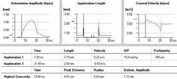

39 Pre cxl CORVIS 1 mos post cxl ionto Biomechanical response improved Note reduced deformation amplitude


")

40 DAPI: nuclear staining (indice indiretto apoptosi) CTL Gruppo1 Gruppo2 Group3 x5


41 TUNEL: indice apotosi CTL Gruppo1 Gruppo 2 Gruppo 3 x10


42 WHAT S NEW? COMPARISON WITH STANDARD EPI-OFF
43 JRS 2016 Transepithelial Iontophoresis Versus Standard Corneal Collagen Cross-linking: 1-Year Results of a Prospective Clinical Study Paolo Vinciguerra, MD; Vito Romano, MD; Pietro Rosetta, MD; Emanuela F. Legrottaglie, MD; Raffaele Piscopo, MD; Claudia Fabiani, MD; Claudio Azzolini, MD; Riccardo Vinciguerra, MD ABSTRACT PURPOSE: To compare 1-year transepithelial corneal collagen cross-linking with iontophoresis (I-CXL) outcomes with standard CXL (S-CXL) epithelium-off for progressive keratoconus. METHODS: Forty eyes of 40 patients with progressive keratoconus were included in this comparative, prospective clinical study. Corrected distance visual acuity (CDVA), spherical equivalent, cylinder refraction, corneal topography, Scheimpflug tomography, aberrometry, and endothelial cell count were assessed at baseline and at 1, 3, 6, and 12 months of follow-up. RESULTS: Patients received either I-CXL (20 eyes) or S-CXL (20 eyes). Functional parameters (visual acuity and aberrometry) showed a significant improvement (P <.05) after 6 and 12 months of follow-up in both groups. In the I-CXL group, the CDVA showed a rapid recovery of vision after 3 months (P =.01).AQ1 Morphological parameters showed a significant reduction of maximum keratometry in the S-CXL group by ± 1.51 D after 12 months, whereas the I-CXL group curvature was stable (-0.31 ± 1.87 D). Minimum pachymetry values were stable even after 12 months of follow-up in the I-CXL group, whereas a significant corneal thinning 12 months following treatment was recorded in the S-CXL group (P <.001). None of the patients had continuous progression of keratoconus or had to repeat CXL procedures. Endothelial cell counts did not change significantly (P >.05). CONCLUSIONS: The 1-year outcomes suggest that I-CXL might be comparable to S-CXL in stabilizing the progression of the degenerative ectatic disease. Additionally, quicker improvement of functional parameters was reported in the I-CXL group. [J Refract Surg. 201X;X(X):XX-XX.] C orneal collagen cross-linking (CXL) is currently the only treatment able to slow or halt the progression of ectatic disease. 1-4 Long-term follow-up studies on CXL refer to the standard technique (S-CXL), which entails epithelial debridement to allow riboflavin (hydrophilic) penetration in the corneal stroma; otherwise the corneal epithelium (lipophilic) reduces its permeability. 5 Nevertheless, epithelial removal causes postoperative pain, 6 delayed visual recovery, 1,7,8 and increased risks of infection. Transepithelial cross-linking (TE-CXL) was introduced to avoid these threats. The original dextran-containing solutions have been reported to be ineffective for TE-CXL, 9-11 but other formulations of riboflavin (with chemical enhancers) 12 showed equivocal results in clinical studies Conversely, preliminary results have shown that transepithelial cross-linking with iontophoresis (I-CXL) is able to increase the riboflavin concentration inside the stroma compared to other TE-CXL techniques together with histological changes Pilot clinical findings using I-CXL have also reported encouraging results. 19 In this study, we compared 1-year results of two groups of patients with keratoconus who were treated with I-CXL and S-CXL (epithelium-off Dresden protocol). From Humanitas Clinical and Research Center, Milan, Italy (PV, PR, EFL, RP, CF); Humanitas University, Rozzano, Milan, Italy (PV); the Department of Corneal and External Eye Diseases, St. Paul s Eye Unit, Royal Liverpool University Hospital, Liverpool, United Kingdom (VR); and the Department of Surgical Sciences, Division of Ophthalmology, University of Insubria, Varese, Italy (CA, RV) Vinciguerra, Romano, Rosetta, et al.; licensee SLACK Incorporated. This is an Open Access article distributed under the terms of the Creative Commons Attribution 4.0 International ( This license allows users to copy and distribute, to remix, transform, and build upon the article, for any purpose, even commercially, provided the author is attributed and is not represented as endorsing the use made of the work. Submitted: March 6, 2016; Accepted: June 23, 2016 Dr. Paolo Vinciguerra is a consultant for Nidek and Oculus Optikgeräte, GmbH. The remaining authors have no financial or proprietary interest in the materials presented herein. Correspondence: Paolo Vinciguerra, MD, Istituto Clinico Humanitas, Via Manzoni 56, Rozzano, Milan, Italy. paolo.vinciguerra@humanitas.it doi: / x Journal of Refractive Surgery 1
44 Aim To compare 1-year transepithelial corneal collagen cross-linking with iontophoresis (I- CXL) outcomes with standard CXL (S-CXL) epithelium-off for progressive keratoconus.
45 Study design Prospective comparative ú 20 eyes of 20 patients Ionto (I-CXL) ú 20 eyes of 20 patients Stadard 3 mw (S-CXL) 12 months of follow up Corrected distance visual acuity (CDVA), spherical equivalent, cylinder refraction, corneal topography, Scheimpflug tomography, aberrometry, and endothelial cell count were assessed.
46 Results BCVA ú I-CXL e S-CXL were comparable, both inducing a significant increase in BCAV ú I-CXL induces a faster recovery (already at month 3) HOA ú I-CXL is able to significantly reduce HOA and Coma (month 6 and 12) ú S-CXL group showed an improvement only in coma after 6 and 12 months
47 Visual acuity linear regression
48 Coma linear regression
49 HOA linear regression
50 Results Kmax ú S-CXL induced a significant improvement of numerous topographic indices during the followup (such as CKI) ú Kmax was reduced significantly by ± 1.51 D after 12 months after S-CXL. ú I-CXL only showed a significant improvement of corneal symmetry index after 12 months of follow-up. ú The reduction of ± 1.87 of Kmax did not reach statistical significance in I-CXL group.
51 However, linear regression analysis for Kmax was not significant in either group
52 Results Pachymetry ú The main result of this analysis is that there is a statistically significant thinning of the minimum corneal thickness in the S-CXL group (P =.0001), whereas this did not occur after I-CXL. ú Even after 12 months of follow- up; this difference was statistically significant
53 Discussion These results highlight the clinical efficacy of I-CXL to overcome the problems of TE-CXL: the penetration of riboflavin through the epithelium. It is known from preclinical reports that the biomechanical effect, riboflavin penetration, and distribution of I-CXL are higher than in TE-CXL but lower when compared to S-CXL. We will continue the follow-up of the patients to determine whether this stiffening effect, even if reduced, will be enough to halt the ectatic disease in the long term.
54 Conclusions At 12 months follow up ú I-CXL is not inferior to S-CXL ú Faster recovery of BCVA ú More reduction of HOA and Coma ú Does not induce thinning Only 12 months! Follow up continues!
55 Conclusions Iontophoresis is a safe technique It appears effective in arresting the progression of the disease Significant imrpovements of functional parameters Reduction of pain
Efficacy of transepithelial corneal cross-linking using iontophoresis
 Efficacy of transepithelial corneal cross-linking using iontophoresis Experimental studies on eye-bank donor eyes and Preliminary results on the clinical trial NCT02117999 Marco Lombardo, MD, PhD Senior
Prof.Paolo Vinciguerra, M.D. 1, 2 Riccardo Vinciguerra, M.D Humanitas University 1. Humanitas Clinical and Research Center IRCS 2
 Prof.Paolo Vinciguerra, M.D. 1, 2 Riccardo Vinciguerra, M.D. 1-3 1 Humanitas University 1 Humanitas Clinical and Research Center IRCS 2 Columbus, Ohio State University 3 University of Insubria, Varese
Results INTRA PROTOCOL ANALYSIS
 Results INTRA PROTOCOL ANALYSIS 0.35 0.3 BCVA logmar Pre op Post op 6m Post op 1y 0.25 0.2 0.15 0.1 0.05 0.3 0.21 0.17 0.15 0.2 0.17 0.15 0.16 0.22 0.21 0 0.18 0.13 0.14 0.15 0.2 3/30 9/10 18/5 30/3 TE
Transepithelial cross-linking
 Transepithelial cross-linking Collection of scientific studies PROCEDURE FOR TRANSEPITHELIAL CROSS-LINKING (TE-CXL) Instill one drop of pilocarpine 2% 30 minutes before UV-A treatment. Place the patient
CORNEAL IONTOPHORESIS APPLIED TO CXL PROCEDURE
 CORNEAL IONTOPHORESIS APPLIED TO CXL PROCEDURE IONTOPHORESIS IONTOPHORESIS(from iòntos=ion andphòresis= to moveacross, ionsmovingacross) consistsin the oneway movement of charged moleculesthroughoutthe
TRANSEPITHELIAL CORNEAL CROSS-LINKING BY IONTOPHORESIS
 TRANSEPITHELIAL CORNEAL CROSS-LINKING BY IONTOPHORESIS FOR THE PATIENT IL CROSS-LINKING TRANSEPITHELIAL CORNEALE TRANSEPITELAILE CROSS-LINKING BY MEDIANTE IONTOPHORESIS IONTOFORESI WHAT IS TRANSEPITHELIAL
Long-term Results of Cross-linking in Children with Keratoconus
 Long-term Results of Cross-linking in Children with Keratoconus Beatrice Frueh,MD Professor of Ophthalmology Financial Disclosure No finantial interests 8-year-old Progression in 6 months Keratoplasty
TRANSEPITHELIAL CORNEAL CROSS-LINKING BY IONTOPHORESIS
 TRANSEPITHELIAL CORNEAL CROSS-LINKING BY IONTOPHORESIS FOR THE OPHTHALMOLOGIST Guidelines for transepithelial cross-linking (absorption through IONTOPHORESIS) INGREDIENTS 100 ml contain (mg/100 ml): Riboflavin
AT A GLANCE EPI-ON VERSUS EPI-OFF: WHERE DO WE STAND? Strides have been made in epi-on techniques, but more are needed.
 EPI-ON VERSUS EPI-OFF: WHERE DO WE STAND? Strides have been made in epi-on techniques, but more are needed. Epi-on techniques are still considered less efficacious BY COSIMO MAZZOTTA, MD, PhD The technique
PRELIMINARY RESULTS IN TRANS EPITHELIAL CORNEAL CROSSLINKING
 PRELIMINARY RESULTS IN TRANS EPITHELIAL CORNEAL CROSSLINKING Authors: Diana Mihu, Adriana Stănilă, Mihaela Florescu, ValericaProştean Ophthalmology Clinic Sibiu Ocular Surface Research Center Sibiu, 2
Transepithelial cross-linking
 Transepithelial cross-linking Collection of scientific studies November 29 th, 211 - UPDATE PROCEDURE FOR TRANSEPITHELIAL CROSS-LINKING (TE-CXL) Instill one drop of pilocarpine 2% 3 minutes before UV-A
Simultaneous Topography-guided Surface Ablation with Collagen Cross-linking for Keratoconus
 IJKECD Case series Simultaneous Topography-guided Surface Ablation with Collagen 10.5005/jp-journals-10025-1124 Cross-linking for Keratoconus Simultaneous Topography-guided Surface Ablation with Collagen
Riboflavin, Oxygen and Light. Sabine Kling, PhD, Farhad Hafezi, MD,PhD
 Riboflavin, Oxygen and Light Sabine Kling, PhD, Farhad Hafezi, MD,PhD Corneal Cross-linking Increasing corneal stiffness to stop the progression of keratoconus 365 nm, 3mW/cm 2 30 min UV-light de-epithelialization
Keratoconus is a noninflammatory process in which the
 CLINICAL SCIENCE Epithelium-Off Corneal Collagen Cross-linking Versus Transepithelial Cross-linking for Pediatric Keratoconus Adriano Magli, MD,* Raimondo Forte, MD,* Achille Tortori, MD, Luigi Capasso,
Topo-Guided Custom Ablation (TGCA) and Corneal Collagen Cross-Linking (CCL) in treatment of advanced keratoectasia
 and Corneal Collagen Cross-Linking (CCL) in treatment of advanced keratoectasia") Topo-Guided Custom Ablation (TGCA) and Corneal Collagen Cross-Linking (CCL) in treatment of advanced keratoectasia Alekandar Stojanovic, MD University Hospital North Norway Tromsø, Norway Jia Zhang, MD
Topo-Guided Custom Ablation (TGCA) and Corneal Collagen Cross-Linking (CCL) in treatment of advanced keratoectasia Alekandar Stojanovic, MD University Hospital North Norway Tromsø, Norway Jia Zhang, MD
Epithelium On Versus Epithelium Off
 ONLINE SURVEY Point/Counterpoint: Epithelium On Versus Epithelium Off Surgeons discuss whether to remove the outer corneal layer during CXL. By Parag A. Majmudar, MD; Rebecca McQuaid, MSc; Arthur B. Cummings,
CORNEAL IONTOPHORESIS APPLIED TO CXL PROCEDURE
 CORNEAL IONTOPHORESIS APPLIED TO CXL PROCEDURE IONTOPHORESIS IONTOPHORESIS(from iòntos=ion andphòresis= to moveacross, ionsmovingacross) consistsin the oneway movement of charged moleculesthroughoutthe
Corneal Cross-Linking in Keratoconus Using the Standard and Rapid Treatment Protocol: Differences in Demarcation Line and 12-Month Outcomes METHODS
 Cornea Corneal Cross-Linking in Keratoconus Using the Standard and Rapid Treatment Protocol: Differences in Demarcation Line and 12-Month Outcomes Sara Brittingham, Christoph Tappeiner, and Beatrice E.
NEW VISION EYE CENTER
 Topographic Aberrometric Guided PRK For Keratoconus With Accelerated Corneal Cross-Linking Using Schwind AMARIS 750S Laser NEW VISION EYE CENTER Dr. Safwan Al Bayati FRCS (Glasgow) FICMS OPTH Consultant
Management of Unpredictable Post-PRK Corneal Ectasia with Intacs Implantation
 Management of Unpredictable Post-PRK Corneal Ectasia with Intacs Implantation Mohammad Naser Hashemian, MD 1 Mahdi AliZadeh, MD 2 Hassan Hashemi, MD 1,3 Firoozeh Rahimi, MD 4 Abstract Purpose: To present
POST-LASIK ECTASIA MANAGEMENT
 POST-LASIK ECTASIA MANAGEMENT A. John Kanellopoulos MD 1,2 1: Laservision.gr Clinical & Research Eye Institute, Athens, Greece 2: NYU Medical School Department of Ophthalmology, NY, NY Financial interests:
Corneal Crosslinking Without Epithelial Removal
 1 Corneal Crosslinking Without Epithelial Removal R. Doyle Stulting, MD, PhD 1 Jonathan Woolfson, MD 1 Bill Trattler, MD 2 Roy Rubinfeld, MD, MA 3 1 Woolfson Eye Institute, Atlanta, GA 2 Center for Excellence
Thirty-month results after the treatment of post-lasik ectasia with allogenic lenticule addition and corneal cross-linking: a case report
 Li et al. BMC Ophthalmology (2018) 18:294 https://doi.org/10.1186/s12886-018-0967-z CASE REPORT Open Access Thirty-month results after the treatment of post-lasik ectasia with allogenic lenticule addition
Intrastromal corneal ring
 Intrastromal corneal ring Kyriakidou Nantia M.D. Diathlasis Day Care Unit Scienti1ic Workshop of Diathlasis Day Care Unit 18-19 November, 2016 The Met Hotel Thessaloniki, Greece DAY CARE UNIT DIATHLASIS,
Corneal collagen crosslinking for keratoconus or corneal ectasia without epithelial debridement
 (2015) 29, 764 768 2015 Macmillan Publishers Limited All rights reserved 0950-222X/15 www.nature.com/eye CLINICAL STUDY Corneal collagen crosslinking for keratoconus or corneal ectasia without epithelial
Bilateral Keratectasia 34 Years after Corneal Transplant
 24 Bilateral Keratectasia 34 Years after Corneal Transplant Xavier Valldeperas a, b Martina Angi b, c Vito Romano d Mario R. Romano b, e a Department of Ophthalmology, Hospital Universitari Germans Trias
Photochemical corneal collagen cross-linkage using riboflavin and ultraviolet A for keratoconus and keratectasia
 Photochemical corneal collagen cross-linkage using riboflavin and ultraviolet A for keratoconus and keratectasia Issued: September 2013 guidance.nice.org.uk/ipg466 NICE has accredited the process used
Article. Reference. Collagen crosslinking with ultraviolet-a and hypoosmolar riboflavin solution in thin corneas. HAFEZI, Farhad, et al.
 Article Collagen crosslinking with ultraviolet-a and hypoosmolar riboflavin solution in thin corneas HAFEZI, Farhad, et al. Abstract Corneal collagen crosslinking (CXL) with riboflavin and ultraviolet-a
Cataract and Refractive Surgery Patients: Still Two Different Breeds?
 Cataract and Refractive Surgery Patients: Still Two Different Breeds? Fabrizio I. Camesasca, MD Department of Ophthalmology IRCCS Istituto Clinico Humanitas Rozzano Milano, Italy Financial Disclosure I
Interventional procedures guidance Published: 25 September 2013 nice.org.uk/guidance/ipg466
 Photochemical corneal collagen cross-linkage using riboflavin and ultraviolet A for keratoconus and keratectasia Interventional procedures guidance Published: 25 September 2013 nice.org.uk/guidance/ipg466
Corneal molding and riboflavin-uva collagen cross-linking in keratoconus
 Corneal molding and riboflavin-uva collagen cross-linking in keratoconus Antonio Calossi Department of Optics and Optometry, University of Turin, Italy Studio Optometrico Calossi, Certaldo (Florence),
Our experience with Athens protocol - simultaneous topo-guided photorefractive keratectomy followed by corneal collagen cross linking for keratoconus
 International Journal of Research in Medical Sciences Shah S et al. Int J Res Med Sci. 2016 Jul;4(7):2639-2644 www.msjonline.org pissn 2320-6071 eissn 2320-6012 Research Article DOI: http://dx.doi.org/10.18203/2320-6012.ijrms20161924
Interpretation of corneal tomography
 Interpretation of corneal tomography Presented by Chameen Samarawickrama - Westmead Hospital - Liverpool Hospital - University of Sydney - University of New South Wales The University of Sydney Page 1
Keratoconus Clinic. Optometric Co-management Opportunities
 Keratoconus Clinic Optometric Co-management Opportunities The Bochner Eye Institute established the first Keratoconus Clinic in Canada in 2008. The consultation and advanced imaging are OHIP covered. All
FEP Medical Policy Manual
 FEP Medical Manual 9.03.05 Corneal Topography/Computer-Assisted Corneal Topography/ Photokeratoscopy Last Review: September 2016 Next Review: September 2017 Related Policies 9.03.28 Corneal Collagen Cross-linking
SCHWIND CAM Perfect Planning wide range of applications
 SCHWIND CAM Perfect Planning wide range of applications ORK-CAM PresbyMAX PTK-CAM 2 SCHWIND CAM the system solution The modular design of the SCHWIND CAM offers customised treatment planning for a uniquely
Efficacy and safety of transepithelial collagen cross linking for progressive keratoconus
 Open Access Original Article Efficacy and safety of transepithelial collagen cross linking for progressive keratoconus Sameer Shahid Ameen 1, Mohammad Asim Mehboob 2, Kashif Ali 3 ABSTRACT Objective: To
Structural Modifications and Tissue Response After Standard Epi-Off and Iontophoretic Corneal Crosslinking With Different Irradiation Procedures
 Cornea Structural Modifications and Tissue Response After Standard Epi-Off and Iontophoretic Corneal Crosslinking With Different Irradiation Procedures Leonardo Mastropasqua, 1 Manuela Lanzini, 1 Claudia
Case Report Outcome of Two Corneal Collagen Crosslinking Methods in Bullous Keratopathy due to Fuchs Endothelial Dystrophy
 Case Reports in Medicine, Article ID 463905, 5 pages http://dx.doi.org/10.1155/2014/463905 Case Report Outcome of Two Corneal Collagen Crosslinking Methods in Bullous Keratopathy due to Fuchs Endothelial
Corneal Collagen Crosslinking for Post-LASIK Ectasia: An Australian Study. 18 rigid gas permeable contact lenses, and intrastromal corneal
 original clinical study Corneal Collagen Crosslinking for ost-lasik Ectasia: An Australian Study Jessica Y. Tong, BMed, MD,* Deepa Viswanathan, hd, FRCS,* Christopher Hodge, hd, BAppSc(Orth), Gerard Sutton,
This is an Open Access document downloaded from ORCA, Cardiff University's institutional repository:
 This is an Open Access document downloaded from ORCA, Cardiff University's institutional repository: http://orca.cf.ac.uk/44020/ This is the author s version of a work that was submitted to / accepted
COLLAGEN CROSSLINKING FOR KERATOCONUS CAN CHANGE SCLERAL SHAPE Gregory DeNaeyer OD 1 and Donald R Sanders MD, PhD 2
 COLLAGEN CROSSLINKING FOR KERATOCONUS CAN CHANGE SCLERAL SHAPE Gregory DeNaeyer OD 1 and Donald R Sanders MD, PhD 2 1 Optometrist at Arena Eye Surgeons 2 Director, Center for Clinical Research and President
Medical Affairs Policy
 Medical Affairs Policy Service: Corneal Treatments and Specialized Contact Lenses (Corneal remodeling, Corneal transplant, Corneal collagen crosslinking, Intrastromal Rings- INTACS, Keratoconus treatments,
First Clinical Impressions on the Integrated Corneal Tomography and Corneal Deformation with Scheimpflug Imaging
 CASE SERIES First Clinical Impressions on 10.5005/jp-journals-10025-1151 the Integrated Corneal Tomography First Clinical Impressions on the Integrated Corneal Tomography and Corneal Deformation with Scheimpflug
LASIK has been the primary type of corneal refractive surgery
 Cornea Higher-Order Aberrations of Anterior and Posterior Corneal Surfaces in Patients With Keratectasia After LASIK Naoyuki Maeda, 1 Tomoya Nakagawa, 1 Ryo Kosaki, 1 Shizuka Koh, 1 Makoto Saika, 2 Takashi
CXL. The Road Ahead. Now that corneal cross-linking has received FDA approval, will clinical practice outpace evidence-based protocols?
 CXL The Road Ahead Now that corneal cross-linking has received FDA approval, will clinical practice outpace evidence-based protocols? By Gabrielle Weiner, Contributing Writer For almost 20 years, researchers
ML-00043B FULL PRESCRIBING INFORMATION: CONTENTS*
 HIGHLIGHTS OF PRESCRIBING INFORMATION These highlights do not include all the information needed to use PHOTREXA VISCOUS and PHOTREXA safely and effectively. See full prescribing information for PHOTREXA
Corneal densitometry using Pentacam based scheimpflug imaging system: Indian rural population
 Original article: Corneal densitometry using Pentacam based scheimpflug imaging system: Indian rural population Dr Nikhil Mahajan*, Prof. Swati Tomar** **Professor,*Resident Department of Ophthalmology,
CLINICAL SCIENCES. Intraoperative and Postoperative Effects of Corneal Collagen Cross-linking on Progressive Keratoconus
 CLINICAL SCIENCES Intraoperative and Postoperative Effects of Corneal Collagen Cross-linking on Progressive Keratoconus Paolo Vinciguerra, MD; Elena Albè, MD; Silvia Trazza, BS; Theo Seiler, MD; Daniel
In the early 1990s, photorefractive keratectomy (PRK) and
 and") CLINICAL SCIENCE Collagen Crosslinking After Radial Keratotomy Uri Elbaz, MD, Sonia N. Yeung, MD, PhD, FRCSC, Setareh Ziai, MD, FRCSC, Alejandro D. Lichtinger, MD, Noa Avni Zauberman, MD, MHA, Yakov Goldich,
CLINICAL SCIENCE Collagen Crosslinking After Radial Keratotomy Uri Elbaz, MD, Sonia N. Yeung, MD, PhD, FRCSC, Setareh Ziai, MD, FRCSC, Alejandro D. Lichtinger, MD, Noa Avni Zauberman, MD, MHA, Yakov Goldich,
Title: Two-photon fluorescence microscopy of corneal riboflavin absorption
 Title: Two-photon fluorescence microscopy of corneal riboflavin absorption through an intact epithelium. Authors: Daniel M Gore FRCOphth, 1* Paul French PhD, 2 David O Brart MD FRCS, 3 Chris Dunsby PhD,
Transepithelial corneal collagen crosslinking: Bilateral study
 ARTICLE Transepithelial corneal collagen crosslinking: Bilateral study Massimo Filippello, MD, PhD, Edoardo Stagni, MD, David O Brart, MD, FRCS, FRCOphth PURPOSE: To evaluate the efficacy of transepithelial
National Institute for Health and Clinical Excellence
 National Institute for Health and Clinical Excellence [IP724/2] [Photochemical corneal cross linkage using riboflavin and ultraviolet A for keratoconus and keratectasia] Consultation table IPAC date: [Thursday
Refractive Surgery Dilemma
 Refractive Surgery Dilemma Section Editor: lireza aradaran-rafii, MD CSE PRESENTTION 33-year-old man seeking refractive surgery presented with refractive error of -1.75-4.0 20 in the right and -0.75-2.5
In Practice. Surgical Procedures Diagnosis New Drugs
 In Practice Surgical Procedures Diagnosis New Drugs 32 35 Bowman + Bulk = Better Results Mid-stromal lamellar keratoplasty (MSLK) offers a new approach to the management of advanced keratoconus that can
Transepithelial Corneal Cross-linking Using an Enhanced Riboflavin Solution
 ORIGINAL ARTICLE Transepithelial Corneal Cross-linking Using an Enhanced Riboflavin Solution Zisis Gatzioufas, MD, PhD; Frederik Raiskup, MD, PhD, FEBO; David O Brart, FRCS, FRCOphth, MD; Eberhard Spoerl,
Effects of Intracorneal Ring Segment on Corneal Biomechanics in Keratoconic Eyes. Abstract
 Effects of Intracorneal Ring Segment on Corneal Biomechanics in Keratoconic Eyes Javad Amoozadeh, MD 1 Nima Mirzaee Rad, MD 2 Amir Houshang Beheshtnejad, MD 3 Ahmad Kheirkhah, MD 1 Hassan Hashemi, MD 4,5
Corneal haze is a condition resulting from several
 CLINICAL SCIENCE Automated Detection and Classification of Corneal Haze Using Optical Coherence Tomography in Patients With Keratoconus After Cross-Linking Ahmad R. Dhaini, PhD,* Maamoun Abdul Fattah,
Corneal Remodeling. Medical Coverage Policy. Related Coverage Resources. Table of Contents. Coverage Policy. Corneal Crosslinking
 Medical Coverage Policy Effective Date... 8/15/2018 Next Review Date... 8/15/2019 Coverage Policy Number... 0141 Corneal Remodeling Table of Contents Coverage Policy... 1 Overview... 3 General Background...
Collagen Cross-linking for the Treatment of Keratoconus in Pediatric Patients
 Rana Hanna et al Review Article 10.5005/jp-journals-10025-1106 Collagen Cross-linking for the Treatment of Keratoconus in Pediatric Patients 1 Rana Hanna, 2 Eran Berkwitz, 3 Jamyl Habib Castillo, 4 Beatrice
Corneal Hydrops Secondary to Intrastromal Corneal Ring Intrusion into the Anterior Chamber 7 Years after Implantation: A Case Report
 Ophthalmol Ther (2017) 6:373 379 DOI 10.1007/s40123-017-0105-7 CASE REPORT Corneal Hydrops Secondary to Intrastromal Corneal Ring Intrusion into the Anterior Chamber 7 Years after Implantation: A Case
Simultaneous Conventional Photorefractive Keratectomy and Corneal Collagen Cross-linking for Pellucid Marginal Corneal Degeneration
 ORIGINAL ARTICLE Simultaneous Conventional Photorefractive Keratectomy and Corneal Collagen Cross-linking for Pellucid Marginal Corneal Degeneration George D. Kymionis, MD, PhD; Michael A. Grentzelos,
Abdel Rahman ElSebaey, MD, PhD.
 Surface Ablation Refractive Surgery Abdel Rahman ElSebaey, MD, PhD. Menoufia University History Correction of optical defects of human eye started 1200 AD. Spherical error corrected by spectacle on 13
Equivalence of Biomechanical Changes Induced by Rapid and Standard Corneal Cross-linking, Using Riboflavin and Ultraviolet Radiation METHODS
 Cornea Equivalence of Biomechanical Changes Induced by Rapid and Standard Corneal Cross-linking, Using Riboflavin and Ultraviolet Radiation Silvia Schumacher, Lydia Oeftiger, and Michael Mrochen PURPOSE.
PresbyMax Outcomes in Myopia, Hyperopia, Emmetropia and Patients post Lasik
 PresbyMax Outcomes in Myopia, Hyperopia, Emmetropia and Patients post Lasik SCHWIND eye-tech-solutions Lunch Symposium Prof. Jorge L. Alió MD, PhD. UNIVERSIDAD MIGUEL HERNÁNDEZ VISSUM INSTITUTO OFTALMOLÓGICO
Riboflavin/ultraviolet A corneal collagen cross-linking (CXL)
") Cornea A Comparison of Different Corneal Iontophoresis Protocols for Promoting Transepithelial Riboflavin Penetration Daniel M. Gore, 1 David P. O Brart, 2 Paul French, 3 Chris Dunsby, 3,4 and Bruce D.
Cornea A Comparison of Different Corneal Iontophoresis Protocols for Promoting Transepithelial Riboflavin Penetration Daniel M. Gore, 1 David P. O Brart, 2 Paul French, 3 Chris Dunsby, 3,4 and Bruce D.
Hun Lee 1,2, David Sung Yong Kang 3, Byoung Jin Ha 3, Jin Young Choi 3, Eung Kweon Kim 2,4, Kyoung Yul Seo 2 and Tae-im Kim 2*
 Lee et al. BMC Ophthalmology (2016) 16:139 DOI 10.1186/s12886-016-0320-3 RESEARCH ARTICLE Open Access Changes in posterior corneal elevations after combined transepithelial photorefractive keratectomy
Cataract Surgery in the Patient with a History of LASIK or PRK
 Cataract Surgery in the Patient with a History of LASIK or PRK #56996-RS April 2018 Sebastian Lesniak, MD Matossian Eye Associates None Disclosures Bio Matossian Eye Associates, Hopewell NJ, 7/2015 Present
Ocular Studies of EMF Exposure at the MMW M. Kojima 1,2,3), Y. Suzuki 4)
, Y. Suzuki 4)") Ocular Studies of EMF Exposure at the MMW M. Kojima 1,2,3), Y. Suzuki 4) 1. Division of Vision Research for Environmental Health, Medical Research Institute, Kanazawa Medical University 2. Department of
Ocular Studies of EMF Exposure at the MMW M. Kojima 1,2,3), Y. Suzuki 4) 1. Division of Vision Research for Environmental Health, Medical Research Institute, Kanazawa Medical University 2. Department of
Retrospective Testing of the Score for the Detection of Ectasia Susceptibility: A Case Report of Ectasia 7 Years after LASIK
 IJKECD 10.5005/jp-journals-10025-1055 Case Retrospective Report Testing of the Score for the Detection of Ectasia Susceptibility: Case Report of Ectasia 7 Years after LSIK Retrospective Testing of the
Preliminary Results of UV-A Riboflavin Crosslinking in Progressive Cases of Keratoconus, in Pakistani Population
 Original Article Preliminary Results of UV-A Riboflavin Crosslinking in Progressive Cases of Keratoconus, in Pakistani Population Muhammad Dawood Khan, Sameer Shahid Ameen, Omar Ishtiaq, Muhammad Khizar
Recent concerns regarding the depth of tissue ablation with
 Volume Estimation of Excimer Laser Tissue Ablation for Correction of Spherical Myopia and Hyperopia Damien Gatinel, 1 Thanh Hoang-Xuan, 1 and Dimitri T. Azar 1,2 PURPOSE. To determine the theoretical volumes
Original Article High myopia as a risk factor for post-lasik ectasia: a case report
 Original Article High myopia as a risk factor for post-lasik ectasia: a case report Mona Harissi-Dagher, MD, a,b Sonja A. F. Frimmel, c and Samir Melki, MD, PhD a,d Author affiliations: a Massachusetts
L. Spadea, R. Ferrante, F. Romani, A. Di Gregorio
 University of L Aquila Eye Clinic Head: Prof. Leopoldo Spadea ULTRAFAST EXCIMER LASER FOR TRANS-EPITHELIAL CUSTOMIZED PHOTOREFRACTIVE SURGERIES: CLINICAL RESULTS WITH 6 MONTHS FOLLOW UP L. Spadea, R. Ferrante,
Topography-Guided. Relevant Literature. March 10th / New York, NY. Become a Cornea Diagnostics & Topography - Guided Treatment Designing Expert!
 Topography-Guided Become a Cornea Diagnostics & Topography - Guided Treatment Designing Expert! Relevant Literature March 10th / New York, NY Course Director A. John Kanellopoulos, MD Clinical Professor
Contoura TM Vision Correction
 Contoura TM Vision Correction Fernando Faria Correia, Financial Disclosures: Alcon/Wavelight Cairo (Egypt) 26/01/2018 Topography-guided ablations Topography guided ablations Evolution from complicated
Central corneal thickness (CCT) measurement is a critical
 measurement is a critical") ARTICLE Topographic Paracentral Corneal Thickness With Pentacam and Orbscan: Effect of Acoustic Factor Javier González-Pérez, O.D., M.Sc., Ph.D., Jose M. González-Méijome, O.D., Ph.D., María T. Rodríguez
ARTICLE Topographic Paracentral Corneal Thickness With Pentacam and Orbscan: Effect of Acoustic Factor Javier González-Pérez, O.D., M.Sc., Ph.D., Jose M. González-Méijome, O.D., Ph.D., María T. Rodríguez
NIH Public Access Author Manuscript Cornea. Author manuscript; available in PMC 2014 May 29.
 NIH Public Access Author Manuscript Published in final edited form as: Cornea. 2013 December ; 32(12): 1544 1548. doi:10.1097/ico.0b013e3182a7f39d. Repeatability of corneal epithelial thickness measurements
Photrexa Viscous, Photrexa and the KXL System for Corneal Cross-Linking
 Photrexa Viscous, Photrexa and the KXL System for Corneal Cross-Linking First and Only FDA Approved Therapeutic treatment for progressive keratoconus and corneal ectasia following refractive surgery The
THE PENTACAM AXL. Improving Cataract Surgery Outcomes. Optical biometry and anterior segment tomography in one device
 Insert to November/December 2016 Sponsored by OCULUS THE PENTACAM AXL Improving Cataract Surgery Outcomes Optical biometry and anterior segment tomography in one device A New Way to Calculate IOL Power
Influence of pterygium size on corneal higher-order aberration evaluated using anterior-segment optical coherence tomography
 Minami et al. BMC Ophthalmology (2018) 18:166 https://doi.org/10.1186/s12886-018-0837-8 RESEARCH ARTICLE Open Access Influence of pterygium size on corneal higher-order aberration evaluated using anterior-segment
PRE-DESCEMET S ENDOTHELIAL KERATOPLASTY (PDEK) DR ASHVIN AGARWAL
 DR ASHVIN AGARWAL") PRE-DESCEMET S ENDOTHELIAL KERATOPLASTY (PDEK) DR ASHVIN AGARWAL Endothelial keratoplasty (EK) has evolved at a brisk pace and the volume of data accumulated over the past 10 years has demonstrated that
PRE-DESCEMET S ENDOTHELIAL KERATOPLASTY (PDEK) DR ASHVIN AGARWAL Endothelial keratoplasty (EK) has evolved at a brisk pace and the volume of data accumulated over the past 10 years has demonstrated that
Grand Rounds CASE CONSULTATION. Edited by Alfredo Castillo, MD & Jaime Aramberri, MD
 CASE CONSULTATION Grand Rounds Edited by Alfredo Castillo, MD & Jaime Aramberri, MD A 23-year-old patient with a stable myopia for 3 years attended for refractive surgery assessment. Corrected visual acuity
FUCH S DYSTROPHY & CATARACT SURGERY TREATMENT ALGORITHM
 FUCH S DYSTROPHY & CATARACT SURGERY TREATMENT ALGORITHM ΙΟΑΝΝΙS Α. MALLIAS, MD, PHD Director of the Dept. of Ophthalmology, Mediterraneo Hospital, Glyfada, Athens, Greece Clinical Fellow in Cornea and
Hun Lee 1,2, David Sung Yong Kang 3, Byoung Jin Ha 3, Jin Young Choi 3, Eung Kweon Kim 2,4, Kyoung Yul Seo 2 and Tae-im Kim 2*
 Lee et al. BMC Ophthalmology (2017) 17:270 DOI 10.1186/s12886-017-0666-1 RESEARCH ARTICLE Open Access Visual rehabilitation in moderate keratoconus: combined corneal wavefrontguided transepithelial photorefractive
Pearls for the Refractive Technician Fadiah Alkhawaldeh, IMBA, COT, ROUB
 Pearls for the Refractive Technician Fadiah Alkhawaldeh, IMBA, COT, ROUB Cleveland Clinic Cole Eye Institute OOS, Columbus, OH February, 2014 alkhawf@ccf.org NO FINANCIAL DISCLOSURES A Puzzle of an Eye
Comparison of Corneal Shape Changes and Aberrations Induced By FS-LASIK and SMILE for Myopia
 ORIGINAL ARTICLE Comparison of Corneal Shape Changes and Aberrations Induced By FS-LASIK and SMILE for Myopia Anders Gyldenkerne, MS; Anders Ivarsen, MD, PhD; Jesper Ø. Hjortdal, MD, PhD ABSTRACT PURPOSE:
Description of iatrogenic corneal ectasia in patients without traditional risk factors
 ARTICLE Description of iatrogenic corneal ectasia in patients without traditional risk factors Julio Ortega-Usobiaga, MD, PhD 1 ; Rosario Cobo-Soriano, MD, PhD 1 ; Fernando Llovet-Osuna, MD, PhD 1 ; Stephan
Integration of Scheimpflug-Based Corneal Tomography and Biomechanical Assessments for Enhancing Ectasia Detection
 BIOMECHANICS ELECTRONICALLY REPRINTED FROM VOL. 33, NO. 7, 2017 Integration of Scheimpflug-Based Corneal Tomography and Biomechanical Assessments for Enhancing Ectasia Detection Renato Ambrósio, Jr., MD,
INTRODUCTION J. DAWCZYNSKI, E. KOENIGSDOERFFER, R. AUGSTEN, J. STROBEL. Department of Ophthalmology, University Hospital Jena, Jena - Germany
 European Journal of Ophthalmology / Vol. 17 no. 3, 2007 / pp. 363-367 Anterior segment optical coherence tomography for evaluation of changes in anterior chamber angle and depth after intraocular lens
3/23/2016. Diagnostic Services Taylor Pannell CRA, OCT-C. Services Available. Important info for the Tech to know. Visual Fields
 Services Available Diagnostic Services Taylor Pannell CRA, OCT-C Static and Kinetic Visual Fields Pachymetry Anterior and Posterior Segment OCT Fundus Photos FAF,FA,ICG Slit Lamp Photography Confocal HRT
Dott. Luca Avoni Ospedale Maggiore di Bologna Banca delle Cornee dell Emilia Romagna SIBO, GENOVA 21 /04/2012
 Dott. Luca Avoni Ospedale Maggiore di Bologna Banca delle Cornee dell Emilia Romagna SIBO, GENOVA 21 /04/2012 SURGEON-DISSECTED PRECUT TISSUE FOR DESCEMET S STRIPPING AUTOMATED ENDOTHELIAL KERATOPLASTY
Clinical Outcomes of an Optimized Prolate Ablation Procedure for Correcting Residual Refractive Errors Following Laser Surgery
 pissn: 111-8942 eissn: 292-9382 Korean J Ophthalmol 217;31(1):16-24 ht tps://doi.org/1.3341/k jo.217.31.1.16 Original Article Clinical Outcomes of an Optimized Prolate Ablation Procedure for Correcting
Immersion Vs Contact Biometery for Axial Length Measurement before Phacoemulsification
 Original Article Immersion Vs Contact Biometery for Axial Length Measurement before Phacoemulsification with Foldable IOL Irum Abbas, Atif Mansoor Ahmad, Tahir Mahmood Pak J Ophthalmol 2009, Vol. 25 No.
Ocular Jeopardy Marc R. Bloomenstein OD, FAAO
 I. Introduction a. Introduction of topics b. Various ocular related topics II. Lipiflow a. Meibomian gland dysfunction i. Define the disease ii. Prevalence iii. Current treatment options 1. Lid scrubs
RECENT ADVANCES IN ANTERIOR SEGMENT IMAGING OF THE EYE. by Eszter Szalai, M.D. Supervisor: László Módis, M.D., Ph.D.
 SHORT THESIS FOR THE DEGREE OF DOCTOR OF PHILOSOPHY (Ph.D.) RECENT ADVANCES IN ANTERIOR SEGMENT IMAGING OF THE EYE by Eszter Szalai, M.D. Supervisor: László Módis, M.D., Ph.D. UNIVERSITY OF DEBRECEN DOCTORAL
Anterior segment imaging
 CET CONTINUING Sponsored by 1 CET POINT Anterior segment imaging Sundeep Vaswani, BSc (Hons), MCOptom 39 The anterior segment of the eye encompasses all structures from the front surface of the cornea
Pardon the Objection: Cases Marc R. Bloomenstein OD, FAAO Scot Morris, OD Derek Cunningham, OD Kathy Mastrota, OD
 I. Introduction a. Treating the anterior segment different for each patient b. How can new technology help your patients? c. The anterior segment is going to be the hot bed for new technology II. Lipiflow
Biomechanics of Corneal Ring Implants. Albert Daxer, MD, MSc, PhD*
 BASIC INVESTIGATION Biomechanics of Corneal Ring Implants Albert Daxer, MD, MSc, PhD* Purpose: To evaluate the biomechanics of corneal ring implants by providing a related mathematical theory and biomechanical
The pinnacle of refractive performance.
 The pinnacle of refractive performance. WaveLight REFRACTIVE PORTFOLIO Advancing REFRACTIVE SURGERY Contoura Vision sets a new standard in LASIK outcomes More than 98% of patients would choose it again.