Adnexal Masses and Problem Solving Pelvic MRI
|
|
|
- Piers Payne
- 6 years ago
- Views:
Transcription

1 28th Congress of the Hungarian Society of Radiologists RCR Session Budapest June 2016 Adnexal Masses and Problem Solving Pelvic MRI DrSarah Swift St James s University Hospital Leeds, UK
2 Objectives Characterisation of adnexal masses with MRI When Why How
3 Adnexal masses Increased use of imaging to assess patients electively and emergently CT as the primary investigation KUB, CTU, CTC Increased detection of adnexal masses

4 Adnexal masses Multidisciplinary meetings frequently ask Where is it? What is it? Does it need to come out? If so how?
5 Differential diagnosis of adnexal masses 2 clinical scenarios Adnexal mass in the symptomatic patient Will be resected Question is how big or small operation? Incidental finding of an adnexal mass Aim of further imaging is to characterise benign lesions which can be managed conservatively


6 US and MR for adnexal masses Determine which patients may benefit from MRI Clinical and biochemical features crucial Sohaibet al, ClinRad 2005


7 False positive diagnoses of malignancy by US Complex cystic lesions endometrioticor haemorrhagic cysts and dermoids Solid masses - leiomyomataand fibromata


8 MRI correct diagnosis of benign lesion Haemorrhage, clot, fat and non-perfused debris can be readily identified on MRI MRI performed better than US in cases where CA 125 was normal or only slightly elevated
9 Ultrasound Looks benign Suspicious US Looks malignant Premenopausal, normal tumour markers, low clinical index of suspicion Premenopausal, normal markers intermediate or low clinical suspicion High clinical suspicion for malignancy Elevated CA 125 MRI has little to add MRI for characterisation CT for staging
10 Ultrasound Looks benign Suspicious US Looks malignant Premenopausal, normal tumour markers, low clinical index of suspicion Premenopausal, normal markers intermediate or low clinical suspicion High clinical suspicion for malignancy Elevated CA 125 MRI has little to add MRI for characterisation CT for staging
11 Ultrasound Looks benign Suspicious US Looks malignant Premenopausal, normal tumour markers, low clinical index of suspicion Premenopausal, normal markers intermediate or low clinical suspicion High clinical suspicion for malignancy Elevated CA 125 MRI has little to add MRI for characterisation CT for staging
12 Ultrasound Looks benign Suspicious US Looks malignant Premenopausal, normal tumour markers, low clinical index of suspicion Premenopausal, normal markers intermediate or low clinical suspicion High clinical suspicion for malignancy Elevated CA 125 MRI has little to add MRI for characterisation CT for staging Can also apply this to CT findings
13 Problem solving MRI T1W T2W Fat Saturation Contrast Enhancement Diffusion Weighted Imaging Simple fluid Fat Blood Non-perfused debris Perfused solid material


14 Mature Fat T2W T1W T1W+Fat Sat Dermoid


15 Big lesions may be difficult to fully assess with Ultrasound


16 Dermoids be careful Malignant change Squamous carcinoma Skin elements, wall of the lesion Poor prognosis


17 2 years later


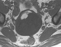
18 Blood T2W T1W T1W + Fat Sat Variable appearance depending on age


19 Blood of differing ages Shading of contents on T2W MRI Characteristic of endometrioma


20 HaemorrhagicEndometriosis Shading of contents on T2W = Endometrioma Haemorrhagiclesion with incomplete septations plical folds = Haematosalpinx
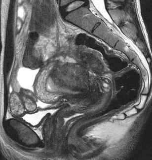


21 Fibrotic Endometriosis Tethering of adjacent structures


22 Endometriosis be careful Solid looking mass on CT


23 MRI shading on T2W, blood on T1W, solid enhancing focus, cystic ademomyosis Endometrioidcarcinoma arising in an endometrioma


24 Solid elements? Give contrast Non enhancing solid foci Benign fibrous lesion T1W Solid enhancing focus Likely malignant T2W T1W +Gad


25 Solid elements? Give contrast Non enhancing solid foci Benign fibrous lesion T2W Solid enhancing focus Likely malignant T1W + Gad


26 Patterns of solid enhancement Serous borderline ovarian tumours Cystic and surface papillary types Younger age group Better prognosis Fertility sparing surgery Important diagnosis


27 Patterns of solid enhancement Serous borderline ovarian tumours Cystic and surface papillary types Younger age group Better prognosis Fertility sparing surgery Important diagnosis
28 Solid Adnexal Masses Is it uterine or adnexal? What is the signal intensity on T2W? Does it enhance?
29 Three key signs Acute angle Claw Feeding vessels Obtuse angle U Courtesy of Dr John Spencer


30 Solid Adnexal Masses Is it uterine or adnexal? What is the T2 SI? Does it enhance?


31 Solid Adnexal Masses Is it uterine or adnexal? What is the T2 SI? Does it enhance?


32 Solid Adnexal Masses Is it uterine or adnexal? What is the T2 SI? Does it enhance?
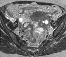
33 Solid Adnexal Masses Is it uterine or adnexal? What is the T2 SI? T2W b = 800 Does it enhance? T1W T1 + Gad


34 Solid Adnexal Masses Low T2 signal and lack of enhancement suggest fibroid lineage Adenofibromacan look heterogeneous on T2 and show variable enhancement Low b = 800 value on DWI gives confidence to call a lesion benign Associated with free fluid in Meigs Syndrome T2W b = 800 T1W T1 + Gad


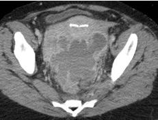
35 Solid Adnexal Masses be careful Change in bowel habit Palpable mass CT bilateral solid adnexal masses


36 Solid Adnexal Masses be careful Change in bowel habit Palpable mass CT bilateral solid adnexal masses


37 Solid Adnexal Masses be careful Change in bowel habit Palpable mass CT bilateral solid adnexal masses Sigmoid Carcinoma Ovarian metastases


38 Ovarian metastases Krukenberg tumours Original description of bilateral solid masses Not always the case Variable appearance Indistinguishable from primary ovarian tumour Important differential diagnosis
39 Adnexal Masses in the Acute Setting Symptomatic adnexal masses will be resected How? Cancer surgery Simple excision of the mass Role of further imaging Try to characterise the mass Allow decision making about patient management


40 Acute pain, known adnexal lesion


41 Acute pain, known adnexal lesion T1W T2W T1W+ Fat Sat Torsion


42 Intermittent pain, pelvic mass
43 Torsion of adnexal masses In adults there is usually an underlying benign lesion Commonly dermoid Symptoms can be intermittent and non-specific Intramural haemorrhage is a recognised finding on MRI


44 Abdominal pain, fever


45 Abdominal pain, fever


46 Associated with longstanding IUD Spreads by contiguity Crosses tissue planes Mimics malignancy Actinomycosis


47 Actinomycosis 6 months later Important diagnosis Conservative management Removal of IUD Antibiotic therapy

48 Adnexal Masses and Problem MRI excellent problem solving tool particularly where the prior probability of malignancy is low Solving MRI Be careful to look for neoplastic change


49 Adnexal Masses and Problem Metastatic disease to the ovaries is not always solid Solving MRI Pain may reflect a complication of benign pathology or an inflammatory process
50 Thank you
51


52 Solid Adnexal Masses Is it uterine or adnexal? What is the T2 SI? Does it enhance?


53 Solid Adnexal Masses Is it uterine or adnexal? What is the T2 SI? Does it enhance?
A Practical Approach to Adnexal Masses
 A Practical Approach to Adnexal Masses Darcy J. Wolfman, MD Section Chief of Genitourinary Imaging American Institute for Radiologic Pathology Clinical Associate Johns Hopkins Community Radiology Division
CA125 in the diagnosis of ovarian cancer: the art in medicine
 CA125 in the diagnosis of ovarian cancer: the art in medicine Dr Marcia Hall Consultant Medical Oncology Mount Vernon Cancer Centre Hillingdon Hospital Wexham Park Hospital Epidemiology Ovarian cancer
2/24/19. Ovarian pathology: IOTA ADNEXAL MASSES. Content. IOTA terms for description of an adnexal mass. IOTA terms for description of an adnexal mass
 Content Ovarian pathology: IOTA ADNEXAL MASSES X SIMPLE COMPLEX Dr DESCRIBE WHAT YOU SEE FRANZCOG, MPH, DDU, COGU Sonologist Clinically useful Benign Malignant Communication between clinicians/research
بسم هللا الرحمن الرحيم. Prof soha Talaat
 بسم هللا الرحمن الرحيم Ovarian tumors The leading indication for gynecologic surgery. Preoperative characterization of complex solid and cystic adnexal masses is crucial for informing patients about possible
MRI in Cervix and Endometrial Cancer
 28th Congress of the Hungarian Society of Radiologists RCR Session Budapest June 2016 MRI in Cervix and Endometrial Cancer DrSarah Swift St James s University Hospital Leeds, UK Objectives Cervix and endometrial
Squamous cell carcinoma arising in a dermoid cyst of the ovary: a case series
 DOI: 10.1111/j.1471-0528.2007.01478.x www.blackwellpublishing.com/bjog Gynaecological oncology Squamous cell carcinoma arising in a dermoid cyst of the ovary: a case series JL Hurwitz, a A Fenton, a WG
MR diagnostics of adnexal masses
 MR diagnostics of adnexal masses Poster No.: C-1499 Congress: ECR 2017 Type: Educational Exhibit Authors: O. Nikolic, J. Ostojic, M. Basta Nikolic, A. Spasic, D. Donat, S. Stojanovic; Novi Sad/RS Keywords:
Characterizing Adnexal Masses: Pearls and Pitfalls 20 th Annual Summer Practicum SCBT-MR Jackson Hole August 11, 2010
 Characterizing Adnexal Masses: Pearls and Pitfalls 20 th Annual Summer Practicum SCBT-MR Jackson Hole August 11, 2010 Evan S. Siegelman MD University of Pennsylvania Medical Center Adnexal Masses: Pearls
Value of MRI in Characterizing Adnexal Masses
 The Journal of Obstetrics and Gynecology of India (July August 2015) 65(4):259 266 DOI 10.1007/s13224-015-0730-9 PHOTO ESSAY Value of MRI in Characterizing Adnexal Masses Alpana Karnik 1 Raina Anil Tembey
Diane DeFriend Derriford Hospital, Plymouth
 Diane DeFriend Derriford Hospital, Plymouth Ultrasound US remains primary imaging modality for investigation of an adnexal mass Aim to characterise Benign Malignant Indeterminate 90% adnexal masses characterised
MR Imaging of the Adnexal Masses: A Review
 Page54 Review of Literature NJR 2011;1(1):54 60; Available online at www.nranepal.org MR Imaging of the Adnexal Masses: A Review I Ahmad 1, S Kirmani 1, M Rashid 2, K Ahmad 3 1 Department of Radiodiagnosis,
General history. Basic Data : Age :62y/o Date of admitted: Married status : Married
 General history Basic Data : Age :62y/o Date of admitted:940510 Married status : Married General history Chief Complain : bilateral ovarian cyst incidentally being found out during pap smear. Present Illness
Newcastle HPB MDM updated radiology imaging protocol recommendations. Author Dr John Scott. Consultant Radiologist Freeman Hospital
 Newcastle HPB MDM updated radiology imaging protocol recommendations Author Dr John Scott. Consultant Radiologist Freeman Hospital This document is intended as a guide to aid radiologists and clinicians
3 cell types in the normal ovary
 Ovarian tumors 3 cell types in the normal ovary Surface (coelomic epithelium) the origin of the great majority of ovarian tumors 90% of malignant ovarian tumors Totipotent germ cells Sex cord-stromal cells
IOTA and Models for Screening for Ovarian Cancer
 IOTA and Models for Screening for Ovarian Cancer Hennie Botha MARCH 2017 T H IG PY R O C F O SP EA KE R Silent Killer to Whispering Disease Listening to your body.. new, persistent, and increases in severity
Clinical summary. Male 30 year-old with past history of non-seminomous germ cell tumour. Presents with retroperitoneal lymphadenopathy on CT.
 Clinical summary Male 30 year-old with past history of non-seminomous germ cell tumour. Presents with retroperitoneal lymphadenopathy on CT. For restaging PET/CT. PET/CT findings No significant FDG uptake
Imaging evaluation of ovarian masses.
 Imaging evaluation of ovarian masses. Poster No.: C-0988 Congress: ECR 2012 Type: Educational Exhibit Authors: M. Forment Navarro, C. La Parra Casado, A. Vera, C. Martínez 1 2 2 2 2 2 1 Rubio, M. Mazón
3 cell types in the normal ovary
 Ovarian tumors 3 cell types in the normal ovary Surface (coelomic epithelium) the origin of the great majority of ovarian tumors (neoplasms) 90% of malignant ovarian tumors Totipotent germ cells Sex cord-stromal
The Adnexal Mass. Handout NCUS 3/18/2017 Suzanne Dixon, MD
 The Adnexal Mass Handout NCUS 3/18/2017 Suzanne Dixon, MD Objectives: Pelvic mass differential Characteristics of the normal ovary Standard terminology for ovarian masses Benign vs. malignant features
Triage of Ovarian Masses. Andreas Obermair Brisbane
 Triage of Ovarian Masses Andreas Obermair Brisbane Why Triage? In ovarian cancer, best outcomes for patients can be achieved when patients are treated in tertiary centres by a multidisciplinary team led
Gynecologic Ultrasound. Sujata Ghate, MD Associate Professor of Radiology Duke University Medical Center
 Gynecologic Ultrasound Sujata Ghate, MD Associate Professor of Radiology Duke University Medical Center Objectives Understand work-up of endometrial abnormalities Show examples of uterine and endometrial
CASE STUDY. Presented by: Jessica Pizzo. CFCC Sonography student Class of 2018
 CASE STUDY Presented by: Jessica Pizzo CFCC Sonography student Class of 2018 Case Presentation April 4, 2017 56 yr old woman presented to ED with lower abdominal pain & swelling, along with constipation.
Mature Cystic Teratomas and the most common complications
 Mature Cystic Teratomas and the most common complications Poster No.: C-2230 Congress: ECR 2015 Type: Authors: Keywords: DOI: Educational Exhibit S. C. S. Silva 1, D. N. Silva 1, D. Garrido 1, I. C. S.
JMSCR Vol 05 Issue 06 Page June 2017
 www.jmscr.igmpublication.org Impact Factor 5.84 Index Copernicus Value: 83.27 ISSN (e)-2347-176x ISSN (p) 2455-0450 DOI: https://dx.doi.org/10.18535/jmscr/v5i6.29 MRI in Clinically Suspected Uterine and
Patient Information. Age: 8 y/o Sex: Female. Date of Admission: Date of Discharge:
 Patient Information Age: 8 y/o Sex: Female Date of Admission: 92-10-08 Date of Discharge: 92-10-18 Chief Complaint Severe admominal pain and vomiting with dysuria since last afternoon Present Illness Lower
CLINICAL PRESENTATION AND RADIOLOGY QUIZ QUESTION
 Donald L. Renfrew, MD Radiology Associates of the Fox Valley, 333 N. Commercial Street, Suite 100, Neenah, WI 54956 2/5/2011 Radiology Quiz of the Week # 6 Page 1 CLINICAL PRESENTATION AND RADIOLOGY QUIZ
Ovaries: In Sickness and Health. Mr N Pisal Consultant Gynaecologist The Portland Hospital
 Ovaries: In Sickness and Health Mr N Pisal Consultant Gynaecologist The Portland Hospital Topics for discussion How to assess ovarian function? AMH PCOS Ovarian pain Ovarian cysts Ovarian screening Menopause
Assessment of adnexal masses. Ultrasound workup of adnexal masses. symptoms. symptoms. Age. Serum tumor markers 10/1/2018
 Assessment of adnexal masses Ultrasound workup of adnexal masses Kevin Robinson, DO Department of Radiology Michigan State University October 4, 2018 Patients symptoms Age Menstrual status Serum tumor
Determining the nature of a sonographically indeterminate adnexal mass has great clinical importance. Decisions regarding the type and extent of surge
 Note: This copy is for your personal, non-commercial use only. To order presentation-ready copies for distribution to your colleagues or clients, contact us at www.rsna.org/rsnarights. John A. Spencer,
Dr Claire Smith, Consultant Radiologist St James University Hospital Leeds
 Dr Claire Smith, Consultant Radiologist St James University Hospital Leeds Imaging in jaundice and 2ww pathway Image protocol Staging Limitations Pancreatic cancer 1.2.4 Refer people using a suspected
Recommendations for cross-sectional imaging in cancer management, Second edition
 www.rcr.ac.uk Recommendations for cross-sectional imaging in cancer management, Second edition Renal and adrenal tumours Faculty of Clinical Radiology www.rcr.ac.uk Contents Renal cell carcinoma 3 Clinical
CLINICAL PRESENTATION AND RADIOLOGY QUIZ QUESTION
 Donald L. Renfrew, MD Radiology Associates of the Fox Valley, 333 N. Commercial Street, Suite 100, Neenah, WI 54956 2/12/2011 Radiology Quiz of the Week # 7 Page 1 CLINICAL PRESENTATION AND RADIOLOGY QUIZ
Case 1. Gynaecology Case Presentation. Objectives. Disclosures 22/10/ year old female Clinical history: Assess right ovarian cyst
 Gynaecology Case Presentation Organ Imaging 2016 University of Toronto Sarah Johnson 39 year old female Clinical history: Assess right ovarian cyst Clinically diagnosed endometriosis Started fertility
 INTRAUTERINE DEVICE = IUD INTRAUTERINE DEVICE = IUD CONGENITAL DISORDERS Pyometra = pyometrea is a uterine infection, it is accumulation of purulent material in the uterine cavity. Ultrasound is usually
Category Term Definition Comments 1 Major Categories 1a
 Working Lexicon Categories, Terms & Definitions Category Term Definition Comments 1 Major Categories 1a Physiologic Category (consistent with normal ovarian physiology) Follicle Simple 3 cm in premenopausal
Gynaecological cancers. Mr Vivek Nama MD MRCOG Consultant Gynaecological Oncologist
 Gynaecological cancers Mr Vivek Nama MD MRCOG Consultant Gynaecological Oncologist Gynaecological cancers Why do we need 2 week wait? Early/timely diagnosis of cancer Possibly less invasive treatment and
Malignant Transformation of Endometriosis: Magnetic Resonance Imaging Aspects
 Malignant Transformation of Endometriosis: Magnetic Resonance Imaging Aspects Poster No.: C-0084 Congress: ECR 2014 Type: Scientific Exhibit Authors: E. A. Yukhno, I. Trofimenko, G. Trufanov; St. Petersburg/RU
Malignant Transformation of Endometriosis: Magnetic Resonance Imaging Aspects
 Malignant Transformation of Endometriosis: Magnetic Resonance Imaging Aspects Poster No.: C-0084 Congress: ECR 2014 Type: Scientific Exhibit Authors: E. A. Yukhno, I. Trofimenko, G. Trufanov; St. Petersburg/RU
Female Genital Tract Lab. Dr. Nisreen Abu Shahin Assistant Professor of Pathology University of Jordan
 Female Genital Tract Lab Dr. Nisreen Abu Shahin Assistant Professor of Pathology University of Jordan Ovarian Pathology A 20-year-old female presented with vague left pelvic pain. Pelvic exam revealed
Top Tips for Gynaecological Ultrasound. Catherine Kirkpatrick Consultant Sonographer Dublin Oct 2018
 Top Tips for Gynaecological Ultrasound Catherine Kirkpatrick Consultant Sonographer Dublin Oct 2018 We can all scan a pelvis so what can we do to improve? Uterus, endometrium and ovaries, got it covered!
Pregnancy With Huge Ovarian Cyst
 BMH Med. J. 2018;5(3):74-78 Case Report Pregnancy With Huge Ovarian Cyst Suja Ann Ranji, Usha Payyodi, Ani Praveen, Rajesh MC, Jini Chandran Baby Memorial Hospital, Kozhikode 673004 Address for Correspondence:
ISUOG Basic Training Typical Ultrasound Appearances of Common Pathologies in the Adnexae
 ISUOG Basic Training Typical Ultrasound Appearances of Common Pathologies in the Adnexae Learning objectives At the end of the lecture series you will be able to: Compare the differences between typical
CLINICAL PRESENTATION AND RADIOLOGY QUIZ QUESTION
 Donald L. Renfrew, MD Radiology Associates of the Fox Valley, 333 N. Commercial Street, Suite 100, Neenah, WI 54956 8/20/2011 Radiology Quiz of the Week # 34 Page 1 CLINICAL PRESENTATION AND RADIOLOGY
Endometrioma With Calcification Simulating a Dermoid on Sonography
 Case Report Endometrioma With Calcification Simulating a Dermoid on Sonography Kiran A. Jain, MD Several investigators have explored the sonographic diagnostic criteria of endometriomas. Endometriomas
Title of Guideline (must include the word Guideline (not protocol, policy, procedure etc)
") Title of Guideline (must include the word Guideline (not protocol, policy, procedure etc) Author: Contact Name and Job Title Directorate & Speciality Assessment, referral and initial management of ultrasound
Title of Guideline (must include the word Guideline (not protocol, policy, procedure etc) Author: Contact Name and Job Title Directorate & Speciality Assessment, referral and initial management of ultrasound
Ovarian pathology: A practical approach to imaging diagnosis and management
 Ovarian pathology: A practical approach to imaging diagnosis and management Award: Certificate of Merit Poster No.: C-1865 Congress: ECR 2015 Type: Educational Exhibit Authors: A. Castan, A. Mir Torres,
The Incidental Renal lesion
 The Incidental Renal lesion BACKGROUND Increase in abdominal CT/US in last 15 years Resulted in detection of many (small) renal lesions 50% > 50yrs has at least 1 lesion majority simple cysts Renal lesions
CAN TV U/S REDUCE THE
 CAN TV U/S REDUCE THE NEED FOR SURGERY IN GYNECOLOGY? Steven R. Goldstein, M.D. Professor of Obstetrics & Gynecology New York University it School of fmedicine i Director of Gynecologic Ultrasound Co-Director
ENDOMETRIOSIS AS A COMMON CAUSE OF PELVIC PAIN
 ENDOMETRIOSIS AS A COMMON CAUSE OF PELVIC PAIN M.Basta Nikolić, S. Stojanović, O. Nikolić, T. Mrđanin, D. Donat, V. Žigić Center for Radiology, Clinical Center of Vojvodina Novi Sad Chronic pelvic pain
Terminology Estimate the risk of malignancy in adnexal masses - Overview
 Understanding the IOTA (International Ovarian Tumor Analysis) terminology & Classification Using the IOTA simple rules to estimate the risk of malignancy in women with adnexal masses Elisabeth Epstein,
MANAGEMENT RECOMMENDATIONS
 1 MANAGEMENT RECOMMENDATIONS 1. Adrenal masses!!!!!!! page 2 2. Liver Masses!!!!!!! page 3 3. Obstetric US Soft Markers for Aneuploidy!! pages 4-6 4. Ovarian and Adnexal Cysts!!!!! pages 7-10 5. Pancreatic
IN THE NAME OF GOD POV: CYSTIC OVARIAN LESION
 IN THE NAME OF GOD POV: CYSTIC OVARIAN LESION CASE 1 20 years old girl with AUB and pelvic pain from 2 weeks ago Impression :Simple unilocular 6 cm ovarian cyst Next step? Almost certainly benign so FU
Please complete prior to the webinar. HOSPITAL REGISTRY WEBINAR FEMALE REPRODUCTIVE SYSTEM EXERCISES CASE 1: FEMALE REPRODUCTIVE
 Please complete prior to the webinar. HOSPITAL REGISTRY WEBINAR FEMALE REPRODUCTIVE SYSTEM EXERCISES PHYSICAL EXAMINATION CASE 1: FEMALE REPRODUCTIVE 3/5 Patient presents through the emergency room with
Does Ovarian Cysts affect your Fertility?
 Does Ovarian Cysts affect your Fertility? Ovarian cysts are sac like structures (filled with liquid or semi-solid) develop in the ovary. Ovarian cysts are very common, mostly painless and harmless with
Investigations and management of severe endometriosis
 Investigations and management of severe endometriosis Dr Jim Tsaltas Head of Gynaecological Endoscopy and Endometriosis Surgery Monash Health Monash University Dept of O&G Melbourne IVF Freemasons Hospital
EDUCATIONAL COMMENTARY CA 125. Learning Outcomes
 EDUCATIONAL COMMENTARY CA 125 Learning Outcomes Upon completion of this exercise, participants will be able to: discuss the use of CA 125 levels in monitoring patients undergoing treatment for ovarian
OVARIES. MLS Basic histological diagnosis MLS HIST 422 Semester 8- batch 7 L13 Dr: Ali Eltayb.
 OVARIES MLS Basic histological diagnosis MLS HIST 422 Semester 8- batch 7 L13 Dr: Ali Eltayb. OBJECTIVES Recognize different disease of ovaries Classify ovarian cyst Describe the pathogenesis, morphology
The shading sign: is it exclusive of endometriomas?
 Abdominal Imaging ª Springer Science+Business Media New York 2015 Abdom Imaging (2015) DOI: 10.1007/s00261-015-0465-1 The shading sign: is it exclusive of endometriomas? João Lopes Dias, 1,2 Filipe Veloso
Characterization of adrenal lesions on CT and MRI: all that a radiologist must know
 Characterization of adrenal lesions on CT and MRI: all that a radiologist must know Poster No.: C-2476 Congress: ECR 2013 Type: Educational Exhibit Authors: N. Benzina, S. MAJDOUB, C. H. ZARRAD, H. Zaghouani,
Essentials of Clinical MR, 2 nd edition. 73. Urinary Bladder and Male Pelvis
 73. Urinary Bladder and Male Pelvis Urinary bladder carcinoma is best locally staged with MRI. It is important however to note that a thickened wall (> 5 mm) is a non-specific finding seen in an underfilled
Endometriosis - MRI findings with anatomic-pathologic correlation
 Endometriosis - MRI findings with anatomic-pathologic correlation Poster No.: C-2551 Congress: ECR 2015 Type: Educational Exhibit Authors: E. Matos, A. T. Almeida, A. Sanches; Vila Nova de Gaia/PT Keywords:
My Patient Has Pelvic Pain. David A. Kenny DO
 My Patient Has Pelvic Pain David A. Kenny DO Definition Of apparent pelvic origin Present most of the time for at least six months Severe enough to cause functional disability Requiring surgical or medical
Erratum to: Fertility-sparing for young patients with gynecologic cancer: How MRI can guide patient selection prior to conservative management
 Abdominal Radiology ª Springer Science+Business Media, LLC 2017 Published online: 11 November 2017 Abdom Radiol (2017) 42:2966 2973 DOI: 10.1007/s00261-017-1205-5 Erratum to: Fertility-sparing for young
Pre-operative Ultrasound of Lymph Nodes in Thyroid Cancer
 Pre-operative Ultrasound of Lymph Nodes in Thyroid Cancer AACE - Advances in Medical and Surgical Management of Thyroid Cancer - 2018 Robert A. Levine, MD, FACE, ECNU Thyroid Center of New Hampshire Geisel
Gynaecology Cancer Red Flags. Dr Dina Bisson Consultant Obstetrician and Gynaecologist Southmead Hospital North Bristol NHS Trust 27 April 2017
 Gynaecology Cancer Red Flags Dr Dina Bisson Consultant Obstetrician and Gynaecologist Southmead Hospital North Bristol NHS Trust 27 April 2017 Gynaecological Cancers Endometrial Cancer Ovarian Cancer Cervical
Icd-10 code for cystic right adnexal lesion
 Icd-10 code for cystic right adnexal lesion The Borg System is 100 % Icd-10 code for cystic right adnexal lesion Jun 28, 2013. Findings: The right ovary measures 1.7 x 3.6 x 2.0 cm. There are. ICD-9-CM:
The many faces of Endometriosis
 The many faces of Endometriosis Beryl Benacerraf M.D Harvard Medical School What is Endometriosis? Endometriosis is defined as the presence of normal endometrial tissue occurring outside of the endometrial
SCBT MR ADNEXAL INCIDENTALOMAS. Susan M. Ascher, MD Georgetown University School of Medicine Washington, DC
 2 0 1 5 SCBT MR ADNEXAL INCIDENTALOMAS Susan M. Ascher, MD Georgetown University School of Medicine Washington, DC Managing incidentalomas can feel like http://www.writeraccess.com/blog/wp-content/uploads/2014/07/herding-cats-blog.jpg
Clinical guideline Published: 27 April 2011 nice.org.uk/guidance/cg122
 Ovarian cancer: recognition and initial management Clinical guideline Published: 27 April 2011 nice.org.uk/guidance/cg122 NICE 2018. All rights reserved. Subject to Notice of rights (https://www.nice.org.uk/terms-and-conditions#notice-ofrights).
Approach to imaging of the ovaries
 First encounter Approach to imaging of the ovaries Mariam Moshiri MD Associate professor Body Imaging Most common first encounter is via ultrasound Many of clinicians order US imaging for various female
Ovarian Lesion Benign vs Malignant?
 Ovarian Lesion Benign vs Malignant? Michele Keenan 1,2 Bernice Dunne 2 Mary Moran 1 Therese Herlihy 1 1. Radiography and Diagnostic Imaging, School of Medicine, University College Dublin, Ireland 2. Midland
Thyroid nodules - medical and surgical management. Endocrinology and Endocrine Surgery Manchester Royal Infirmary
 Thyroid nodules - medical and surgical management JRE Davis NR Parrott Endocrinology and Endocrine Surgery Manchester Royal Infirmary Thyroid nodules - prevalence Thyroid nodules common, increase with
3 Summary of clinical applications and limitations of measurements
 CA125 (serum) 1 Name and description of analyte 1.1 Name of analyte Cancer Antigen 125 (CA125) 1.2 Alternative names Mucin-16 1.3 NLMC code To follow 1.4 Description of analyte CA125 is an antigenic determinant
MRI features of primary and metastatic mucinous ovarian tumors
 MRI features of primary and metastatic mucinous ovarian tumors Poster No.: C-0551 Congress: ECR 2014 Type: Authors: Keywords: DOI: Educational Exhibit P.-E. LAURENT, J. Thomassin-Piana, A. JALAGUIER; Marseille/
Adnexal Masses in Menopausal Women Surgery or Surveillance?
 Adnexal Masses in Menopausal Women Surgery or Surveillance? FREDTALK IDEASWORTHSPREADING Disclosure I am a member of Vermillion s Speakers Bureau I am NOT a paid consultant for Vermillion Inc. nor do I
Sonographic features of tubo-ovarian abscess mimicking an endometrioma. adnexal masses
 Sonographic features of tubo-ovarian abscess mimicking an endometrioma and review of cystic adnexal masses Artur Velcani 1*, Patrick Conklin 1, Neil Specht 1 1. Department of Radiology, St.Vincent's Medical
X-ray Corner. Imaging of The Pancreas. Pantongrag-Brown L
 X-ray Corner 125 Imaging of The Pancreas Modern imaging modalities commonly used in pancreas include ultrasound (US), CT, and MRI. Pancreas is a retroperitoneal organ which makes it difficult to visualize
The key contribution of MRI in adnexal mass evaluation is in: 1. Identifying benign features. 2. Identifying malignant features.
 19 th Annual Women s Imaging Conference University of Toronto - 2016 Disclosures : None phyllis.glanc@sunnybrook.ca Sunnybrook Health Science Centre University of Toronto, Dept Medical Imaging, Obstetrics
Pathology of Ovarian Tumours. Dr. Jyothi Ranganathan MD ( Path) AFMC Pune PDCC (Cytopathology) PGI Chandigarh
 AFMC Pune PDCC (Cytopathology) PGI Chandigarh") Pathology of Ovarian Tumours Dr. Jyothi Ranganathan MD ( Path) AFMC Pune PDCC (Cytopathology) PGI Chandigarh Outline Incidence Risk factors Classification Pathology of tumours Tumour markers Prevention
Pathology of Ovarian Tumours Dr. Jyothi Ranganathan MD ( Path) AFMC Pune PDCC (Cytopathology) PGI Chandigarh Outline Incidence Risk factors Classification Pathology of tumours Tumour markers Prevention
Title of Guideline (must include the word Guideline not protocol, policy, procedure etc)
") Title of Guideline (must include the word Guideline not protocol, policy, procedure etc) Author: Contact Name and Job Title Directorate & Speciality Assessment, referral and initial management of ultrasound
Title of Guideline (must include the word Guideline not protocol, policy, procedure etc) Author: Contact Name and Job Title Directorate & Speciality Assessment, referral and initial management of ultrasound
The Role of Imaging for Gynecologic Emergencies
 Objectives The Role of Imaging for Gynecologic Emergencies M. Jonathon Solnik, MD, FACOG FACS Associate Professor of Obstetrics & Gynaecology Head of Gynaecology & Minimally Invasive Surgery University
Adnexal Masses in Pregnancy: Added Value of Magnetic Resonance Imaging in Guiding Patient Management Our Initial Experience
 Poovini Soundararajan et al. RESEARCH ARTICLE 10.5005/jp-journals-10006-1590 Adnexal Masses in Pregnancy: Added Value of Magnetic Resonance Imaging in Guiding Patient Management Our Initial Experience
Armed Forces Institute of Pathology.
 Armed Forces Institute of Pathology www.radpath.com Armed Forces Institute of Pathology Breast Disease www.radpath.org Armed Forces Institute of Pathology Interpretation of Breast MRI Leonard M. Glassman
ADRENAL MEDULLARY DISORDERS: PHAEOCHROMOCYTOMAS AND MORE
 ADRENAL MEDULLARY DISORDERS: PHAEOCHROMOCYTOMAS AND MORE DR ANJU SAHDEV READER AND CONSULTANT RADIOLOGIST QUEEN MARY UNIVERSITY AND ST BARTHOLOMEW S HOSPITAL BARTS HEALTH, LONDON, UK DISCLOSURE OF CONFLICT
UTERINE LEIOMYOSARCOMA. About Uterine leiomyosarcoma
 UTERINE LEIOMYOSARCOMA Uterine Lms, Ulms Or Just Lms Rare uterine malignant tumour that arises from the smooth muscular part of the uterine wall. Diagnosis Female About Uterine leiomyosarcoma Uterine LMS
OVARIAN MASSES : MANAGEMENT CHALLENGE
 SRU 2015 OVARIAN MASSES : MANAGEMENT CHALLENGE phyllis.glanc@sunnybrook.ca Sunnybrook Health Science Centre University of Toronto, Dept Medical Imaging, Obstetrics & Gynecology Thank you on behalf of the
Adnexal Mass Management: Risk Stratification and Management Practice for Best Patient Outcomes
 Transcript Details This is a transcript of a continuing medical education (CME) activity accessible on the ReachMD network. Additional media formats for the activity and full activity details (including
cysts is possible if imaging findings are correlated with appropriate clinical findings [1]. The
![cysts is possible if imaging findings are correlated with appropriate clinical findings [1]. The](/thumbs/73/68677649.jpg "cysts is possible if imaging findings are correlated with appropriate clinical findings [1]. The") Pictorial Essay Imaging of Peritoneal Inclusion Cysts Kiran. Jain1 lthough fairly common, peritoneal inclusion cysts are less well-recognized entities on imaging of the female pelvis. Peritoneal inclusion
Pictorial Essay Imaging of Peritoneal Inclusion Cysts Kiran. Jain1 lthough fairly common, peritoneal inclusion cysts are less well-recognized entities on imaging of the female pelvis. Peritoneal inclusion
Detailed Program of the second BREAST IMAGING AND INTERVENTIONS PROGRAM am am : Clinician s requirements from breast imaging
 Detailed Program of the second BREAST IMAGING AND INTERVENTIONS PROGRAM 2012 Day one, 2 nd November BREAST IMAGING AND INTERVENTIONS PROGRAM 2012 9.00 AM 9.10 am Introduction 9.10 am - 9.30 am : Clinician
Ovarian Clear Cell Carcinoma
 Ovarian Clear Cell Carcinoma Rouba Ali-Fehmi, MD Professor of Pathology The Karmanos Cancer Institute, Wayne State University School of Medicine 50 year old woman with chief complaint of shortness of breath
Appendix 5. EFSUMB Newsletter. Gastroenterological Ultrasound
 EFSUMB Newsletter 87 Examinations should encompass the full range of pathological conditions listed below A log book listing the types of examinations undertaken should be kept Training should usually
Case 9551 Primary ovarian Burkitt lymphoma
 Case 9551 Primary ovarian Burkitt lymphoma Monteiro V, Cunha TM, Saldanha T Section: Genital (Female) Imaging Published: 2011, Nov. 20 Patient: 23 year(s), female Authors' Institution V Monteiro 1, TM
Ovarian Masses: role of MRI in the differential diagnosis. A systematic approach.
 Ovarian Masses: role of MRI in the differential diagnosis. A systematic approach. Poster No.: C-0597 Congress: ECR 2017 Type: Educational Exhibit Authors: I. Mussetto, F. Rosa, J. Matos, G. Ficarra, D.
Intraperitoneal cysts in infancy and childhood An overview and sonographic differentiation
 Intraperitoneal cysts in infancy and childhood An overview and sonographic differentiation M. Mearadji International Foundation for Pediatric Imaging Aid Rotterdam, The Netherlands Intraperitoneal cysts
PAPILLARY THYROID CARCINOMA PRESENTING AS A LATERAL NECK MASS MASS. Dr. Pamela Hanson DO PGY3
 PAPILLARY THYROID CARCINOMA PRESENTING AS A LATERAL NECK MASS MASS Dr. Pamela Hanson DO PGY3 MK CASE PRESENTATION 28 yo Female presented to the ENT Clinic in October 2016, with the complaint of chronic
Borderline Ovarian Tumours. Andreas Obermair Brisbane
 Borderline Ovarian Tumours Andreas Obermair Brisbane Definition First described in 1929 Cellular features of malignancy Cellular atypia Mitotic activity No stromal invasion An entity per se??? (or precursor
Case Scenario 1. Pathology report Specimen from mediastinoscopy Final Diagnosis : Metastatic small cell carcinoma with residual lymphatic tissue
 Case Scenario 1 Oncology Consult: Patient is a 51-year-old male with history of T4N3 squamous cell carcinoma of tonsil status post concurrent chemoradiation finished in October two years ago. He was hospitalized
Update on RECIST and Staging of Common Pediatric Tumors Ethan A. Smith, MD
 Update on RECIST and Staging of Common Pediatric Tumors Ethan A. Smith, MD Section of Pediatric Radiology C.S. Mott Children s Hospital University of Michigan ethans@med.umich.edu Disclosures No relevant
Cervical Cancer: 2018 FIGO Staging
 Cervical Cancer: 2018 FIGO Staging Jonathan S. Berek, MD, MMS Laurie Kraus Lacob Professor Stanford University School of Medicine Director, Stanford Women s Cancer Center Senior Scientific Advisor, Stanford
A Tale of Two Ovaries: Cross-Sectional Imaging Spectrum of Ovarian Emergencies
 A Tale of Two Ovaries: Cross-Sectional Imaging Spectrum of Ovarian Emergencies Presenting author A J Baxi Co-authors A M Nagar, MBBS D Rajderkar, MD V Ojili, MD Contact:ojili@uthscsa.edu Disclaimer: We