Electroencephalography
|
|
|
- Barnaby Mason
- 5 years ago
- Views:
Transcription
1 The electroencephalogram (EEG) is a measure of brain waves. It is a readily available test that provides evidence of how the brain functions over time. The EEG is used in the evaluation of brain disorders. Most commonly it is used to show the type and location of the activity in the brain during a seizure. It also is used to evaluate people who are having problems associated with brain function. These problems might include confusion, coma, tumors, long-term difficulties with thinking or memory, or weakening of specific parts of the body (such as weakness associated with a stroke). An EEG is also used to determine brain death. It may be used to prove that someone on life-support equipment has no chance of recovery. Electroencephalography is the recording of electrical activity along the scalp. EEG measures voltage fluctuations resulting from ionic current flows within the neurons of the brain. In clinical contexts, EEG refers to the recording of the brain's spontaneous electrical activity over a short period of time, usually minutes, as recorded from multiple electrodes placed on the scalp.

 ECG 0.01 300 0.")

2 Frequency & Amplitude Ranges for Bioelectric Signals Signal Frequency range (Hz) Amplitude range(mv) ECG EEG EOG EMG
3 Diagnostic applications generally focus on the spectral content of EEG, that is, the type of neural oscillations that can be observed in EEG signals. In neurology, the main diagnostic application of EEG is in the case of epilepsy, as epileptic activity can create clear abnormalities on a standard EEG study. A secondary clinical use of EEG is in the diagnosis of coma, encephalopathy, and brain death. A third clinical use of EEG is for studies of sleep and sleep disorders where recordings are typically done for one full night, sometimes more. EEG used to be a first-line method for the diagnosis of tumors, stroke and other focal brain disorders, but this use has decreased with the advent of anatomical imaging techniques with high (<1 mm) spatial resolution such as MRI (magnetic resonance imaging) and CT (computed tomography). Despite limited spatial resolution, EEG continues to be a valuable tool for research and diagnosis, especially when millisecond-range temporal resolution (not possible with CT or MRI) is required.
4 The cerebral cortex is composed of neurons that are interconnected to each other in networks and also receive inputs from other areas of the brain. Electrical activity in the form of nerve impulses being sent and received to and from cortical neurons is always present, even during sleep. Biologically, medically and legally, the absence of cortical activity signifies death. The electrical activity you are measuring reflects both the intrinsic activity of neurons in the cerebral cortex and the information sent to it by subcortical structures and the sense receptors. This composite activity is called an electroencephalogram or EEG. An EEG electrode will mainly detect the activity in the brain region just under it. Nevertheless, the electrodes receive the activity from thousands of neurons. In fact, one square millimeter of cortex has more than 100,000 neurons. It is only when the input to a region is synchronized with electrical activity occurring at the same time that you begin to distinguish simple periodic waveforms in the EEG.
5 Source of EEG activity The brain's electrical charge is maintained by billions of neurons. Neurons are electrically charged (or "polarized") by membrane transport proteins that pump ions across their membranes. Neurons are constantly exchanging ions with the extracellular milieu, for example to maintain resting potential and to propagate action potentials. Ions of similar charge repel each other, and when many ions are pushed out of many neurons at the same time, they can push their neighbors, who push their neighbors, and so on, in a wave. This process is known as volume conduction. When the wave of ions reaches the electrodes on the scalp, they can push or pull electrons on the metal on the electrodes. Since metal conducts the push and pull of electrons easily, the difference in push or pull voltages between any two electrodes can be measured by a voltmeter. Recording these voltages over time gives us the EEG. The electric potential generated by single neuron is far too small to be picked up by EEG or MEG. EEG activity therefore always reflects the summation of the synchronous activity of thousands or millions of neurons that have similar spatial orientation. Scalp EEG activity shows oscillations at a variety of frequencies. Several of these oscillations have characteristic frequency ranges, spatial distributions and are associated with different states of brain functioning (e.g., waking and the various sleep stages). These oscillations represent synchronized activity over a network of neurons.
, an invention described \"as one of the most")
6 German physiologist and psychiatrist Hans Berger ( ) recorded the first human EEG in Expanding on work previously conducted on animals by Richard Caton and others, Berger also invented the electroencephalogram (giving the device its name), an invention described "as one of the most surprising, remarkable, and momentous developments in the history of clinical neurology". His discoveries were first confirmed by British scientists E. D. Adrian and B. H. C. Matthews in 1934 and developed by them. Hans Berger The first human EEG recording obtained by Hans Berger in The upper tracing is EEG, and the lower is a 10 Hz timing signal.


7 In conventional scalp EEG, the recording is obtained by placing electrodes on the scalp with a conductive gel or paste, usually after preparing the scalp area by light abrasion to reduce impedance due to dead skin cells. Many systems typically use electrodes, each of which is attached to an individual wire. Some systems use caps or nets into which electrodes are embedded; this is particularly common when high-density arrays of electrodes are needed. Epileptic spike and wave discharges monitored with EEG Computer Electroencephalograph Neurovisor-BMM 40 Portable recording device for EEG


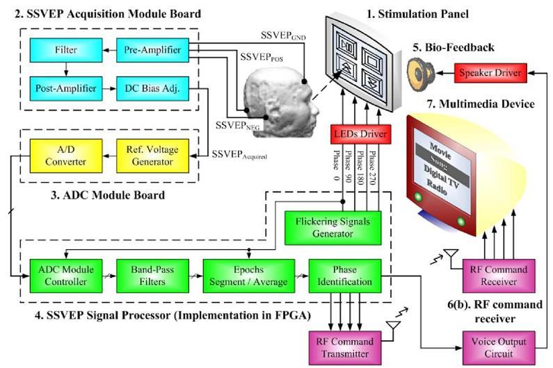
8 Brain Computer Interface Preprocessing Signal Processing Feature Extraction by FD (CEM) Classification (Detection) Signal Acquisition Application Interface Feedback
9


10 EEG signals are created by measuring the difference in electrical currents across neuron membranes. Many naturally occurring signals in the human body effect EEG signals. Frequency analysis helps to separate the different signals. Electrode placement can effect signals received.
11 Where to place the electrodes? NASION REF FP1 FP2 F7 F8 F3 FZ F4 T3 C3 CZ C4 T4 GND GND P3 PZ P4 T5 T6 O1 O2 The electrodes are placed according to the international standard system. The electrode position of 16 channels placed as shown in Fig. of frontal area (F3 and F4), central area (C3, Cz and C4), parietal area(p3, Pz and P4) The grounding electrode and referencing electrode are placed at forehead and right ear lobe respectively. EEG signal are digitized at 1024 samples/sec, resolution 16bit/sample. Signals are analog bandpass filtered between 1.5 and 100 Hz.

12 EEG is a difference in potential between two electrodes.
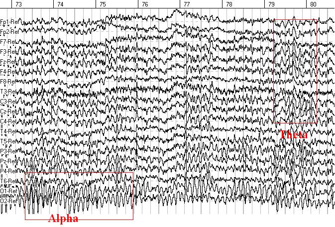
13 Delta waves: 1-5 Hz mv 1 second of Characteristic EEG signals Theta waves: 4-8 Hz Beta waves: Hz 10 mv 5-10 mv Alpha/Mu waves: 8-13 Hz Gamma waves: Hz mv

14 Amplitude is the voltage in microvolts measured from the peak of the wave to the trough of the wave. Varies from 10 mv to 100 mv with average around mv. Spectrums reflect the amount of energy in a certain frequency range of EEG. The presence of large-amplitude delta-activity may indicate infarct or other lesion. Slow (0-4 Hz) and high (more 20 Hz) frequency bands of EEG may pick up artifacts, such as eye movements and muscle activity, and therefore should be evaluated with caution. The presence of large amplitude spikes and waves may indicate the presence of epilepsy.
15 EEG is a mixture of waves at different frequencies and amplitudes. At each time interval several sine-waves at different frequencies may be present in the signal.


16 Eyes closed condition Eyes opened condition


17 Electrode circuit EEG sensing Interference Amplifier Filter
. EEG Amplifier Circuit")
18 Each electrode is connected to one input of a differential amplifier (one amplifier per pair of electrodes); a common system reference electrode is connected to the other input of each differential amplifier. These amplifiers amplify the voltage between the active electrode and the reference (typically db of voltage gain). EEG Amplifier Circuit

19 A differential amplifier is a type of electronic amplifier that amplifies the difference between two voltages but does not amplify the particular voltages.

20 Design Block Diagram Amplifier Schematic
21

22 Biological artifacts Electrical signals detected along the scalp by an EEG, but that originate from non-cerebral origin are called artifacts. EEG data is almost always contaminated by such artifacts. The amplitude of artifacts can be quite large relative to the size of amplitude of the cortical signals of interest. This is one of the reasons why it takes considerable experience to correctly interpret EEGs clinically. Some of the most common types of biological artifacts include: Eye-induced artifacts (includes eye blinks, eye movements) ECG (cardiac) artifacts EMG (muscle activation)-induced artifacts Glosso-kinetic artifacts Environmental artifacts IV drip In addition to artifacts generated by the body, many artifacts originate from outside the body. Movement by the patient, or even just settling of the electrodes, may cause electrode pops, spikes originating from a momentary change in the impedance of a given electrode. Poor grounding of the EEG electrodes can cause significant 50 or 60 Hz artifact, depending on the local power system's frequency. A third source of possible interference can be the presence of an IV (intravenous) drip; such devices can cause rhythmic, fast, low-voltage bursts, which may be confused for spikes.

23 EMG and eye blink artifacts in EEG These types of artifacts can be detected by visual inspection. Eye blinks can be excluded from data analysis. EMG should be taken into account during spectral analysis.
24 EMG artifact starts as low as 12 Hz and ranges to 300 Hz. Most of the spectrum lies between Hz. Posterior electrodes can pick up EMG from occipitalis, trapezius and supraspinal muscles. To avoid this type of artifact one can relax or position the head properly or change slightly the position of electrode. ECG artifacts occur from the electrodes that pick up activity from underlying pulsating blood vessels in the scalp. EKG artifact gets more prevalent with aging.


25 Drowsy level detection via EEG measurements provides safe car driving to alert tired drivers. A real-time drowsiness monitoring can prevent traffic accidents effectively.

.")
26 Electrocorticography Electrocorticography (ECoG), or intracranial EEG (ieeg), is the practice of using electrodes placed directly on the exposed surface of the brain to record electrical activity from the cerebral cortex. ECoG may be performed either in the operating room during surgery (intraoperative ECoG) or outside of surgery (extraoperative ECoG). Because a craniotomy (a surgical incision into the skull) is required to implant the electrode grid, ECoG is an invasive procedure.
. The acronym ENG is often used. An ENG is similar to an EMG, but the later is used to visualize muscular activity.")
27 Electroneurogram An electroneurogram is a method used to visualize directly recorded electrical activity of neurons in the central nervous system (brain, spinal cord) or the peripheral nervous system (nerves, ganglions). The acronym ENG is often used. An ENG is similar to an EMG, but the later is used to visualize muscular activity. An EEG is a particular type of ENG in which several electrodes are placed around the head and the general activity of the brain is recorded, without having very high resolution to distinguish between the activity of different groups of neurons. An ENG is usually obtained by placing an electrode in the neural tissue. The electrical activity generated by neurons is recorded by the electrode and transmitted to an acquisition system, which usually allows to visualize the activity of the neuron. Each vertical line in an ENG represents one neuronal action potential. Depending on the precision of the electrode used to record neural activity, an ENG can contain the activity of a single neuron to thousands of neurons. Researchers adapt the precision of their electrode to either focus on the activity of a single neuron or the general activity of a group of neurons, both strategies having their advantages.
The Sonification of Human EEG and other Biomedical Data. Part 3
 The Sonification of Human EEG and other Biomedical Data Part 3 The Human EEG A data source for the sonification of cerebral dynamics The Human EEG - Outline Electric brain signals Continuous recording
CHAPTER 6 INTERFERENCE CANCELLATION IN EEG SIGNAL
 116 CHAPTER 6 INTERFERENCE CANCELLATION IN EEG SIGNAL 6.1 INTRODUCTION Electrical impulses generated by nerve firings in the brain pass through the head and represent the electroencephalogram (EEG). Electrical
EEG in the ICU: Part I
 EEG in the ICU: Part I Teneille E. Gofton July 2012 Objectives To outline the importance of EEG monitoring in the ICU To briefly review the neurophysiological basis of EEG To introduce formal EEG and subhairline
Practical 3 Nervous System Physiology 2 nd year English Module. Dept. of Physiology, Carol Davila University of Medicine and Pharmacy
 Electroencephalography l h (EEG) Practical 3 Nervous System Physiology 2 nd year English Module Dept. of Physiology, Carol Davila University of Medicine and Pharmacy What is EEG EEG noninvasively records
EEG History. Where and why is EEG used? 8/2/2010
 EEG History Hans Berger 1873-1941 Edgar Douglas Adrian, an English physician, was one of the first scientists to record a single nerve fiber potential Although Adrian is credited with the discovery of
Introduction to Electrophysiology
 Introduction to Electrophysiology Dr. Kwangyeol Baek Martinos Center for Biomedical Imaging Massachusetts General Hospital Harvard Medical School 2018-05-31s Contents Principles in Electrophysiology Techniques
PD233: Design of Biomedical Devices and Systems
 PD233: Design of Biomedical Devices and Systems (Lecture-7 Biopotentials- 2) Dr. Manish Arora CPDM, IISc Course Website: http://cpdm.iisc.ac.in/utsaah/courses/ Electromyogram (EMG) Skeletal muscles are
Restoring Communication and Mobility
 Restoring Communication and Mobility What are they? Artificial devices connected to the body that substitute, restore or supplement a sensory, cognitive, or motive function of the nervous system that has
EEG- A Brief Introduction
 Fatemeh Hadaeghi EEG- A Brief Introduction Lecture Notes for BSP, Chapter 4 Master Program Data Engineering 1 4 Introduction Human brain, as the most complex living structure in the universe, has been
EEG, ECG, EMG. Mitesh Shrestha
 EEG, ECG, EMG Mitesh Shrestha What is Signal? A signal is defined as a fluctuating quantity or impulse whose variations represent information. The amplitude or frequency of voltage, current, electric field
Brain Computer Interface. Mina Mikhail
 Brain Computer Interface Mina Mikhail minamohebn@gmail.com Introduction Ways for controlling computers Keyboard Mouse Voice Gestures Ways for communicating with people Talking Writing Gestures Problem
EEG Instrumentation, Montage, Polarity, and Localization
 EEG Instrumentation, Montage, Polarity, and Localization 2 Krikor Tufenkjian The Source of EEG The source of the EEG potentials recorded from the scalp is the excitatory and inhibitory postsynaptic potentials
states of brain activity sleep, brain waves DR. S. GOLABI PH.D. IN MEDICAL PHYSIOLOGY
 states of brain activity sleep, brain waves DR. S. GOLABI PH.D. IN MEDICAL PHYSIOLOGY introduction all of us are aware of the many different states of brain activity, including sleep, wakefulness, extreme
Outline of Talk. Introduction to EEG and Event Related Potentials. Key points. My path to EEG
 Outline of Talk Introduction to EEG and Event Related Potentials Shafali Spurling Jeste Assistant Professor in Psychiatry and Neurology UCLA Center for Autism Research and Treatment Basic definitions and
This presentation is the intellectual property of the author. Contact them for permission to reprint and/or distribute.
 Modified Combinatorial Nomenclature Montage, Review, and Analysis of High Density EEG Terrence D. Lagerlund, M.D., Ph.D. CP1208045-16 Disclosure Relevant financial relationships None Off-label/investigational
13 Electroencephalography
 13 Electroencephalography 13.1 INTRODUCTION The first recording of the electric field of the human brain was made by the German psychiatrist Hans Berger in 1924 in Jena. He gave this recording the name
ANALYSIS OF BRAIN SIGNAL FOR THE DETECTION OF EPILEPTIC SEIZURE
 ANALYSIS OF BRAIN SIGNAL FOR THE DETECTION OF EPILEPTIC SEIZURE Sumit Kumar Srivastava 1, Sharique Ahmed 2, Mohd Maroof Siddiqui 3 1,2,3 Department of EEE, Integral University ABSTRACT The electroencephalogram
An Overview of BMIs. Luca Rossini. Workshop on Brain Machine Interfaces for Space Applications
 An Overview of BMIs Luca Rossini Workshop on Brain Machine Interfaces for Space Applications European Space Research and Technology Centre, European Space Agency Noordvijk, 30 th November 2009 Definition
Brain and Cognition. Cognitive Neuroscience. If the brain were simple enough to understand, we would be too stupid to understand it
 Brain and Cognition Cognitive Neuroscience If the brain were simple enough to understand, we would be too stupid to understand it 1 The Chemical Synapse 2 Chemical Neurotransmission At rest, the synapse
Sleep-Wake Cycle I Brain Rhythms. Reading: BCP Chapter 19
 Sleep-Wake Cycle I Brain Rhythms Reading: BCP Chapter 19 Brain Rhythms and Sleep Earth has a rhythmic environment. For example, day and night cycle back and forth, tides ebb and flow and temperature varies
Introduction to EEG del Campo. Introduction to EEG. J.C. Martin del Campo, MD, FRCP University Health Network Toronto, Canada
 Introduction to EEG J.C. Martin, MD, FRCP University Health Network Toronto, Canada What is EEG? A graphic representation of the difference in voltage between two different cerebral locations plotted over
LESSON 1.3 WORKBOOK. How can we study the behaving brain?
 LESSON 1.3 WORKBOOK How can we study the behaving brain? We are in the middle of a technological revolution when it comes to how closely we can look at the behaving brain. Scientists and doctors now have
Seizure onset can be difficult to asses in scalp EEG. However, some tools can be used to increase the seizure onset activity over the EEG background:
 This presentation was given during the Dianalund Summer School on EEG and Epilepsy, July 24, 2012. The main purpose of this introductory talk is to show the possibilities of improved seizure onset analysis
Lóska Ádám
 Emotiv Epoc EEG Based Brain-Computer Interface Lóska Ádám 2011.05.28. Consultant: Mészáros Tamás Overview... 2 ElectroEncephalography... 3 Brief history of EEG... 3 EEG studies... 3 Area of interest...
Localization a quick look
 Localization a quick look Covering the basics Differential amplifiers Polarity convention 10-20 electrode system Basic montages: bipolar and referential Other aspects of displaying the EEG Localization
Physiological and Physical Basis of Functional Brain Imaging 6. EEG/MEG. Kâmil Uludağ, 20. November 2007
 Physiological and Physical Basis of Functional Brain Imaging 6. EEG/MEG Kâmil Uludağ, 20. November 2007 Course schedule 1. Overview 2. fmri (Spin dynamics, Image formation) 3. fmri (physiology) 4. fmri
STRUCTURAL ORGANIZATION OF THE NERVOUS SYSTEM
 STRUCTURAL ORGANIZATION OF THE NERVOUS SYSTEM STRUCTURAL ORGANIZATION OF THE BRAIN The central nervous system (CNS), consisting of the brain and spinal cord, receives input from sensory neurons and directs
Biomedical Research 2013; 24 (3): ISSN X
: ISSN X") Biomedical Research 2013; 24 (3): 359-364 ISSN 0970-938X http://www.biomedres.info Investigating relative strengths and positions of electrical activity in the left and right hemispheres of the human brain
Biomedical Research 2013; 24 (3): 359-364 ISSN 0970-938X http://www.biomedres.info Investigating relative strengths and positions of electrical activity in the left and right hemispheres of the human brain
A Brain Computer Interface System For Auto Piloting Wheelchair
 A Brain Computer Interface System For Auto Piloting Wheelchair Reshmi G, N. Kumaravel & M. Sasikala Centre for Medical Electronics, Dept. of Electronics and Communication Engineering, College of Engineering,
Electroencephalogram (EEG) Hsiao-Lung Chan Dept Electrical Engineering Chang Gung University
 Hsiao-Lung Chan Dept Electrical Engineering Chang Gung University") Electroencephalogram (EEG) Hsiao-Lung Chan Dept Electrical Engineering Chang Gung University chanhl@mail.cgu.edu.tw Cerebral function examination Electroencephalography (EEG) Near infrared ray spectroscopy
Electroencephalogram (EEG) Hsiao-Lung Chan Dept Electrical Engineering Chang Gung University chanhl@mail.cgu.edu.tw Cerebral function examination Electroencephalography (EEG) Near infrared ray spectroscopy
Quick Notes. BioCapture : Acquiring EEG data
 Electroencephalography (EEG) is a recording used to measure the synaptic electrical activity of the brain. The BioCapture system uses electrodes on the scalp to monitor the average behavior of millions
The AASM Manual for the Scoring of Sleep and Associated Events
 The AASM Manual for the Scoring of Sleep and Associated Events Summary of Updates in Version 2.1 July 1, 2014 The American Academy of Sleep Medicine (AASM) is committed to ensuring that The AASM Manual
: Biomedical Signal Processing
 : Biomedical Signal Processing 0. Introduction: Biomedical signal processing refers to the applications of signal processing methods, such as Fourier transform, spectral estimation and wavelet transform,
EEG in Medical Practice
 EEG in Medical Practice Dr. Md. Mahmudur Rahman Siddiqui MBBS, FCPS, FACP, FCCP Associate Professor, Dept. of Medicine Anwer Khan Modern Medical College What is the EEG? The brain normally produces tiny
Introduction to the EEG technique
 Introduction to the EEG technique Part 1: neural origins of the EEG Niko Busch Charité University Medicine Berlin The History of the EEG 18th cent. Physiologists discover elctrical properties of living
BIOPAC Systems, Inc BIOPAC Inspiring people and enabling discovery about life
 BIOPAC Systems, Inc. 2016 BIOPAC Inspiring people and enabling discovery about life 1 BIOPAC s Guide to EEG for Research: Mobita Wireless EEG Housekeeping Attendees are on Mute Headset is Recommended!
Lesson 5 EEG 1 Electroencephalography: Brain Rhythms
 Physiology Lessons for use with the Biopac Science Lab MP40 PC running Windows XP or Mac OS X 10.3-10.4 Lesson 5 EEG 1 Electroencephalography: Brain Rhythms Lesson Revision 2.23.2006 BIOPAC Systems, Inc.
11/18/13 ECG SIGNAL ACQUISITION HARDWARE DESIGN. Origin of Bioelectric Signals
 ECG SIGNAL ACQUISITION HARDWARE DESIGN Origin of Bioelectric Signals 1 Cell membrane, channel proteins Electrical and chemical gradients at the semi-permeable cell membrane As a result, we get a membrane
A Study of Smartphone Game Users through EEG Signal Feature Analysis
 , pp. 409-418 http://dx.doi.org/10.14257/ijmue.2014.9.11.39 A Study of Smartphone Game Users through EEG Signal Feature Analysis Jung-Yoon Kim Graduate School of Advanced Imaging Science, Multimedia &
Students will be able to determine what stage of sleep someone is in by analyzing their EEG.
 Outline 2Lesson Unit1.2 OVERVIEW Rationale: This lesson is intended to engage students with the concept of the neural circuit. The lesson and unit as a whole use sleep, a behavior everyone is familiar
Introduction to Computational Neuroscience
 Introduction to Computational Neuroscience Lecture 10: Brain-Computer Interfaces Ilya Kuzovkin So Far Stimulus So Far So Far Stimulus What are the neuroimaging techniques you know about? Stimulus So Far
Intracranial Studies Of Human Epilepsy In A Surgical Setting
 Intracranial Studies Of Human Epilepsy In A Surgical Setting Department of Neurology David Geffen School of Medicine at UCLA Presentation Goals Epilepsy and seizures Basics of the electroencephalogram
Polysomnography Artifacts and Updates on AASM Scoring Rules. Robin Lloyd, MD, FAASM, FAAP 2017 Utah Sleep Society Conference
 Polysomnography Artifacts and Updates on AASM Scoring Rules Robin Lloyd, MD, FAASM, FAAP 2017 Utah Sleep Society Conference x Conflict of Interest Disclosures for Speakers 1. I do not have any relationships
Est-ce que l'eeg a toujours sa place en 2019?
 Est-ce que l'eeg a toujours sa place en 2019? Thomas Bast Epilepsy Center Kork, Germany Does EEG still play a role in 2019? What a question 7T-MRI, fmri, DTI, MEG, SISCOM, Of ieeg course! /HFO, Genetics
Asian Epilepsy Academy (ASEPA) & ASEAN Neurological Association (ASNA) EEG Certification Examination
 & ASEAN Neurological Association (ASNA) EEG Certification Examination") Asian Epilepsy Academy (ASEPA) & ASEAN Neurological Association (ASNA) EEG Certification Examination EEG Certification Examination Aims To set and improve the standard of practice of Electroencephalography
Asian Epilepsy Academy (ASEPA) & ASEAN Neurological Association (ASNA) EEG Certification Examination EEG Certification Examination Aims To set and improve the standard of practice of Electroencephalography
Biomedical Signal Processing
 DSP : Biomedical Signal Processing What is it? Biomedical Signal Processing: Application of signal processing methods, such as filtering, Fourier transform, spectral estimation and wavelet transform, to
Design Considerations and Clinical Applications of Closed-Loop Neural Disorder Control SoCs
 22nd Asia and South Pacific Design Automation Conference (ASP-DAC 2017) Special Session 4S: Invited Talk Design Considerations and Clinical Applications of Closed-Loop Neural Disorder Control SoCs Chung-Yu
EEG SPIKE CLASSIFICATION WITH TEMPLATE MATCHING ALGORITHM. Çamlık Caddesi No:44 Sarnıç Beldesi İZMİR 2 Elektrik ve Elektronik Müh.
 EEG SPIKE CLASSIFICATION WITH TEMPLATE MATCHING ALGORITHM Selim BÖLGEN 1 Gülden KÖKTÜRK 2 1 Pagetel Sistem Müh. San. Tic. Ltd. Şti. Çamlık Caddesi No:44 Sarnıç Beldesi İZMİR 2 Elektrik ve Elektronik Müh.
International Journal of Engineering Science Invention Research & Development; Vol. III, Issue X, April e-issn:
 BRAIN COMPUTER INTERFACING FOR CONTROLLING HOME APPLIANCES ShubhamMankar, Sandeep Shriname, ChetanAtkare, Anjali R,Askhedkar BE Student, Asst.Professor Department of Electronics and Telecommunication,MITCOE,India
Break Down the Electroencephalography. By Lauren Tessaro. November 30, Lab Instructor: Dr. John Stewart
 Break Down the Electroencephalography By Lauren Tessaro November 30, 2009 Lab Instructor: Dr. John Stewart The world of medicine is constantly expanding into new territories. Cures for diseases are constantly
PSD Analysis of Neural Spectrum During Transition from Awake Stage to Sleep Stage
 PSD Analysis of Neural Spectrum During Transition from Stage to Stage Chintan Joshi #1 ; Dipesh Kamdar #2 #1 Student,; #2 Research Guide, #1,#2 Electronics and Communication Department, Vyavasayi Vidya
Asian Epilepsy Academy (ASEPA) EEG Certification Examination
 EEG Certification Examination") Asian Epilepsy Academy (ASEPA) EEG Certification Examination EEG Certification Examination Aims To set and improve the standard of practice of Electroencephalography (EEG) in the Asian Oceanian region
Asian Epilepsy Academy (ASEPA) EEG Certification Examination EEG Certification Examination Aims To set and improve the standard of practice of Electroencephalography (EEG) in the Asian Oceanian region
EEG in the ICU. Quiz. March Teneille E. Gofton
 EEG in the ICU Quiz March 2012 Teneille E. Gofton Quiz The next several slides will show 15 subhairline EEGs. Choose the best possible answer in each scenario. Your score and solutions will be provided
The EEG Analysis of Auditory Emotional Stimuli Perception in TBI Patients with Different SCG Score
 Open Journal of Modern Neurosurgery, 2014, 4, 81-96 Published Online April 2014 in SciRes. http://www.scirp.org/journal/ojmn http://dx.doi.org/10.4236/ojmn.2014.42017 The EEG Analysis of Auditory Emotional
Neurophysiology & EEG
 Neurophysiology & EEG PG4 Core Curriculum Ian A. Cook, M.D. Associate Director, Laboratory of Brain, Behavior, & Pharmacology UCLA Department of Psychiatry & Biobehavioral Sciences Semel Institute for
Introduction (1) Nervous System & EEG. Introduction (2)
 Nervous System & EEG. Introduction (2)") Introduction () Nervous System & EEG Achmad Rizal BioSPIN Chapter 7, Biointrumentation, Webster Nervous system is defined as all cell, tissues, and organ that regulate the body s response to internal &
Introduction () Nervous System & EEG Achmad Rizal BioSPIN Chapter 7, Biointrumentation, Webster Nervous system is defined as all cell, tissues, and organ that regulate the body s response to internal &
Effects of Light Stimulus Frequency on Phase Characteristics of Brain Waves
 SICE Annual Conference 27 Sept. 17-2, 27, Kagawa University, Japan Effects of Light Stimulus Frequency on Phase Characteristics of Brain Waves Seiji Nishifuji 1, Kentaro Fujisaki 1 and Shogo Tanaka 1 1
Montages are logical and orderly arrangements of channels
 GUIDELINE American Clinical Neurophysiology Society Guideline 3: A Proposal for Standard Montages to Be Used in Clinical EEG Jayant N. Acharya,* Abeer J. Hani, Partha D. Thirumala, and Tammy N. Tsuchida
QEEG markers in stroke, ageing and cognitive decline
 QEEG markers in stroke, ageing and cognitive decline When is theta actually alpha? Simon Finnigan Senior Research Fellow UQ Centre for Clinical Research & Royal Brisbane Clinical Unit University of Queensland,
ISSN: (Online) Volume 3, Issue 7, July 2015 International Journal of Advance Research in Computer Science and Management Studies
 Volume 3, Issue 7, July 2015 International Journal of Advance Research in Computer Science and Management Studies") ISSN: 2321-7782 (Online) Volume 3, Issue 7, July 2015 International Journal of Advance Research in Computer Science and Management Studies Research Article / Survey Paper / Case Study Available online
ISSN: 2321-7782 (Online) Volume 3, Issue 7, July 2015 International Journal of Advance Research in Computer Science and Management Studies Research Article / Survey Paper / Case Study Available online
Detection and Plotting Real Time Brain Waves
 Detection and Plotting Real Time Brain Waves Prof. M. M. PAL ME (ESC(CS) Department Of Computer Engineering Suresh Deshmukh College Of Engineering, Wardha Abstract - The human brain, either is in the state
EE 4BD4 Lecture 11. The Brain and EEG
 EE 4BD4 Lecture 11 The Brain and EEG 1 Brain Wave Recordings Recorded extra-cellularly from scalp (EEG) Recorded from extra-cellularly from surface of cortex (ECOG) Recorded extra-cellularly from deep
From Spikes to Ripples: The Evolving and Expanding Role of Electroencephalography in the Diagnosis and Treatment of Epilepsy
 From Spikes to Ripples: The Evolving and Expanding Role of Electroencephalography in the Diagnosis and Treatment of Epilepsy December 3, 2011 Gregory K. Bergey, M.D. Johns Hopkins University School of
EMG, EEG, and Neurophysiology in Clinical Practice
 Mayo School of Continuous Professional Development EMG, EEG, and Neurophysiology in Clinical Practice Matthew T. Hoerth, M.D. Ritz-Carlton, Amelia Island, Florida January 29-February 4, 2017 2016 MFMER
Non epileptiform abnormality J U LY 2 7,
 Non epileptiform abnormality S U D A J I R A S A K U L D E J, M D. C H U L A L O N G KO R N C O M P R E H E N S I V E E P I L E P S Y C E N T E R J U LY 2 7, 2 0 1 6 Outline Slow pattern Focal slowing
Event Related Potentials: Significant Lobe Areas and Wave Forms for Picture Visual Stimulus
 Available Online at www.ijcsmc.com International Journal of Computer Science and Mobile Computing A Monthly Journal of Computer Science and Information Technology ISSN 2320 088X IMPACT FACTOR: 6.017 IJCSMC,
The neurolinguistic toolbox Jonathan R. Brennan. Introduction to Neurolinguistics, LSA2017 1
 The neurolinguistic toolbox Jonathan R. Brennan Introduction to Neurolinguistics, LSA2017 1 Psycholinguistics / Neurolinguistics Happy Hour!!! Tuesdays 7/11, 7/18, 7/25 5:30-6:30 PM @ the Boone Center
Neurorobotics, and brain-machine interfaces. Oct. 10 th, 2006.
 Neurorobotics, and brain-machine interfaces Oct. 10 th, 2006. Catching up from last class Pg 121 Wessberg ( ) Nicolelis, Real-time prediction of hand trajectory by ensembles of cortical neurons in primates
SedLine Sedation Monitor
 SedLine Sedation Monitor Quick Reference Guide Not intended to replace the Operator s Manual. See the SedLine Sedation Monitor Operator s Manual for complete instructions, including warnings, indications
Artifact Recognition and Troubleshooting
 Artifact Recognition and Troubleshooting 2017 Focus Fall Super Session The Best of the Best For Respiratory Therapists and Sleep Technologists The Doubletree Hilton Hotel Pittsburgh, PA Thursday Sept.
Electroencephalography II Laboratory
 Introduction Several neurological disorders exist that can have an impact on brain function. Often these disorders can be examined by reviewing the electroencephalograph, or EEG signal. Quantitative features
Biomedical Signal Processing
 DSP : Biomedical Signal Processing Brain-Machine Interface Play games? Communicate? Assist disable? Brain-Machine Interface Brain-Machine Interface (By ATR-Honda) The Need for Biomedical Signal Processing
Diagnosing Complicated Epilepsy: Mapping of the Epileptic Circuitry. Michael R. Sperling, M.D. Thomas Jefferson University Philadelphia, PA
 Diagnosing Complicated Epilepsy: Mapping of the Epileptic Circuitry Michael R. Sperling, M.D. Thomas Jefferson University Philadelphia, PA Overview Definition of epileptic circuitry Methods of mapping
Electroencephalography & Neurofeedback
 Electroencephalography & Neurofeedback A Brief Introduction to the Science of Brainwaves Glyn Blackett YORK biofeedback CENTRE Introduction This article is a brief introduction to electroencephalography
The Nervous System. Neuron 01/12/2011. The Synapse: The Processor
 The Nervous System Neuron Nucleus Cell body Dendrites they are part of the cell body of a neuron that collect chemical and electrical signals from other neurons at synapses and convert them into electrical
Spectral Analysis of EEG Patterns in Normal Adults
 Spectral Analysis of EEG Patterns in Normal Adults Kyoung Gyu Choi, M.D., Ph.D. Department of Neurology, Ewha Medical Research Center, Ewha Womans University Medical College, Background: Recently, the
Saichiu Nelson Tong ALL RIGHTS RESERVED
 2017 Saichiu Nelson Tong ALL RIGHTS RESERVED MODELING CLASSIFICATION NETWORK OF ELECTROENCEPHALOGRAPHIC ARTIFACTS AND SIGNALS ASSOCIATED WITH DEEP BRAIN STIMULATION By SAICHIU NELSON TONG A thesis submitted
WIRELESS PATIENT HEALTH MONITORING SYSTEM
 WIRELESS PATIENT HEALTH MONITORING SYSTEM PADMAVATHI KAVURU 1, B.CHINNA RAO 2 & P.M FRANCIS 3 1 (VLSI&ES) 2 Electronics & Communication Engg, Gokul Institute of Technology & Sciences (JNTUK): Bobbili,
fmri (functional MRI)
") Lesion fmri (functional MRI) Electroencephalogram (EEG) Brainstem CT (computed tomography) Scan Medulla PET (positron emission tomography) Scan Reticular Formation MRI (magnetic resonance imaging) Thalamus
Lesion fmri (functional MRI) Electroencephalogram (EEG) Brainstem CT (computed tomography) Scan Medulla PET (positron emission tomography) Scan Reticular Formation MRI (magnetic resonance imaging) Thalamus
TOBY Cerebral Function Monitoring Addition to CFM handbook for users of the Olympic CFM 6000
 ISRCTN 89547571 TOBY Cerebral Function Monitoring Addition to CFM handbook for users of the Olympic CFM 6000 2 The contents of this booklet were originally produced for the website http://www.azzopardi.freeserve.co.uk/cfm
II. PROCEDURE DESCRIPTION A. Normal Waveform from an Electrocardiogram Figure 1 shows two cycles of a normal ECG waveform.
 Cardiac Monitor with Mobile Application and Alert System Miguel A. Goenaga-Jimenez, Ph.D. 1, Abigail C. Teron, BS. 1, Pedro A. Rivera 1 1 Universidad del Turabo, Puerto Rico, mgoenaga1@suagm.edu, abigailteron@gmail.com,
Implantable Microelectronic Devices
 ECE 8803/4803 Implantable Microelectronic Devices Fall - 2015 Maysam Ghovanloo (mgh@gatech.edu) School of Electrical and Computer Engineering Georgia Institute of Technology 2015 Maysam Ghovanloo 1 Outline
III. Studying The Brain and Other Structures
 III. Studying The Brain and Other Structures 1. Accidents (case study) In 1848, a railroad worker named Phineas Gage was involved in an accident that damaged the front part of his brain. Gage s doctor
Electroencephalographic Study of Essential Oils for Stress Relief
 Applied Mechanics and Materials Online: 2013-10-11 ISSN: 1662-7482, Vol. 437, pp 1085-1088 doi:10.4028/www.scientific.net/amm.437.1085 2013 Trans Tech Publications, Switzerland Electroencephalographic
Neurostyle. Medical Innovation for Better Life
 Neurostyle Medical Innovation for Better Life Neurostyle Pte Ltd is a company dedicated to design, develop, manufacture and distribute neurological and neuromuscular medical devices. Strategically located
Northeast Center for Special Care Grant Avenue Lake Katrine, NY
 300 Grant Avenue Lake Katrine, NY 12449 845-336-3500 Information Bulletin What is Brain Mapping? By Victor Zelek, Ph.D., Director of Neuropsychological Services Diplomate, National Registry of Neurofeedback
Physiology Unit 2 CONSCIOUSNESS, THE BRAIN AND BEHAVIOR
 Physiology Unit 2 CONSCIOUSNESS, THE BRAIN AND BEHAVIOR In Physiology Today What the Brain Does The nervous system determines states of consciousness and produces complex behaviors Any given neuron may
Assessment of Fatigue in Brain Computer Interface Users. A Thesis. Submitted to the Faculty. Drexel University. Vincent J.
 Assessment of Fatigue in Brain Computer Interface Users A Thesis Submitted to the Faculty of Drexel University by Vincent J. Petaccio II in partial fulfillment of the requirements for the degree of Master
Objectives. brain pacemaker circuits role of inhibition
 Brain Rhythms Michael O. Poulter, Ph.D. Professor, Molecular Brain Research Group Robarts Research Institute Depts of Physiology & Pharmacology, Clinical Neurological Sciences Schulich School of Medicine
Research Perspectives in Clinical Neurophysiology
 Research Perspectives in Clinical Neurophysiology A position paper of the EC-IFCN (European Chapter of the International Federation of Clinical Neurophysiology) representing ~ 8000 Clinical Neurophysiologists
The secrets of conventional EEG
 The secrets of conventional EEG The spike/sharp wave activity o Electro-clinical characteristics of Spike/Sharp wave The polymorphic delta activity o Electro-clinical characteristics of Polymorphic delta
Source localisation in the clinical practice: spontaneous EEG examinations with LORETA. Ph.D. thesis. Márton Tamás Tóth M.D.
 Source localisation in the clinical practice: spontaneous EEG examinations with LORETA Ph.D. thesis Márton Tamás Tóth M.D. Department of Neurology, University of Pécs Leader of project:: Prof. István Kondákor,
Human Brain Institute Russia-Switzerland-USA
 1 Human Brain Institute Russia-Switzerland-USA CONTENTS I Personal and clinical data II Conclusion. III Recommendations for therapy IV Report. 1. Procedures of EEG recording and analysis 2. Search for
Physiology Unit 2 CONSCIOUSNESS, THE BRAIN AND BEHAVIOR
 Physiology Unit 2 CONSCIOUSNESS, THE BRAIN AND BEHAVIOR What the Brain Does The nervous system determines states of consciousness and produces complex behaviors Any given neuron may have as many as 200,000
Beyond the Basics in EEG Interpretation: Throughout the Life Stages
 Beyond the Basics in EEG Interpretation: Throughout the Life Stages Steve S. Chung, MD, FAAN Chairman, Neuroscience Institute Director, Epilepsy Program Banner University Medical Center University of Arizona
EEG IN FOCAL ENCEPHALOPATHIES: CEREBROVASCULAR DISEASE, NEOPLASMS, AND INFECTIONS
 246 Figure 8.7: FIRDA. The patient has a history of nonspecific cognitive decline and multiple small WM changes on imaging. oligodendrocytic tumors of the cerebral hemispheres (11,12). Electroencephalogram
Biological Process 9/7/10. (a) Anatomy: Neurons have three basic parts. 1. The Nervous System: The communication system of your body and brain
 Anatomy: Neurons have three basic parts. 1. The Nervous System: The communication system of your body and brain") Biological Process Overview 1. The Nervous System: s (a) Anatomy, (b) Communication, (c) Networks 2. CNS/PNS 3. The Brain (a) Anatomy, (b) Localization of function 4. Methods to study the brain (Dr. Heidenreich)
Biological Process Overview 1. The Nervous System: s (a) Anatomy, (b) Communication, (c) Networks 2. CNS/PNS 3. The Brain (a) Anatomy, (b) Localization of function 4. Methods to study the brain (Dr. Heidenreich)
Chapter 6 Section 1. The Nervous System: The Basic Structure
 Chapter 6 Section 1 The Nervous System: The Basic Structure Essential Question: How does studying the biology of the brain give us an understanding of our behavior? Draw or type 2 things you already know
Spike voltage topography in temporal lobe epilepsy
 Thomas Jefferson University Jefferson Digital Commons Department of Neurology Faculty Papers Department of Neurology 5-17-2016 Spike voltage topography in temporal lobe epilepsy Ali Akbar Asadi-Pooya Thomas