Bones Ethmoid bone Inferior nasal concha Lacrimal bone Maxilla Nasal bone Palatine bone Vomer Zygomatic bone Mandible
|
|
|
- Jonah Hardy
- 5 years ago
- Views:
Transcription
1 splanchnocranium - Consists of part of skull that is derived from branchial arches - The facial bones are the bones of the anterior and lower human skull
2 Bones Ethmoid bone Inferior nasal concha Lacrimal bone Maxilla Nasal bone Palatine bone Vomer Zygomatic bone Mandible
3 Ethmoid bone The ethmoid is a single bone, which makes a significant contribution to the middle third of the face. It is located between the lateral wall of the nose and the medial wall of the orbit and forms parts of the nasal septum, roof and lateral wall of the nose, and a considerable part of the medial wall of the orbital cavity. In addition, the ethmoid makes a small contribution to the floor of the anterior cranial fossa. The ethmoid bone can be divided into four parts, the perpendicular plate, the cribriform plate and two ethmoidal labyrinths. Important landmarks include: Perpendicular plate Cribriform plate Crista galli. Ala. Ethmoid labyrinths Medial (nasal) surface. Orbital plate. Superior nasal concha. Middle nasal concha. Anterior ethmoidal air cells. Middle ethmoidal air cells. Posterior ethmoidal air cells. Attachments The falx cerebri (slide) attaches to the posterior border of the crista galli.


4 lamina cribrosa 1 crista galli 2 lamina perpendicularis 3 labyrinthi ethmoidales 4 cellulae ethmoidales anteriores et posteriores 5 lamina orbitalis 6 concha nasalis media 7 processus uncinatus 8
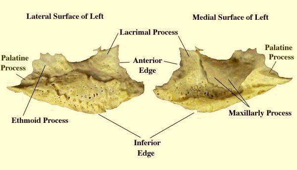
5 Inferior nasal concha Each inferior nasal concha consists of a curved plate of bone attached to the lateral wall of the nasal cavity. Each consists of inferior and superior borders, medial and lateral surfaces, and anterior and posterior ends. The superior border serves to attach the bone to the lateral wall of the nose, articulating with four different bones. The anterior third of the superior border articulates with the conchal crest of the maxilla and the posterior third with the conchal crest of the perpendicular plate of the palatine bone. The middle third of the upper border shows three small processes: lacrimal, maxillary and ethmoidal processes. The concha s longer inferior border lies free within the nasal cavity. It is thickened and often curves inwards. The inferior nasal concha exhibits the following landmarks: Anterior end. Posterior end. Lateral surface. Medial surface. Ethmoidal process. Lacrimal process. Maxillary process.
6
7 Lacrimal bone The two bones are small, thin and rectangular. They each lie in the anterior part of the medial wall of the orbit. Anteriorly, the lacrimal bone articulates with the frontal process of the maxilla. Posteriorly, the bone meets the orbital plate of the ethmoid. Superiorly, it joins the frontal bone and inferiorly, the maxilla. The lacrimal bone exhibits the following landmarks: Nasal surface. Orbital surface. Posterior lacrimal crests. Lacrimal grooves. Fossa for lacrimal sac. Lacrimal hamulus. Descending process. Attachments The lacrimal part of the orbicularis oculi muscle arises from the lateral surface of the lacrimal bone.
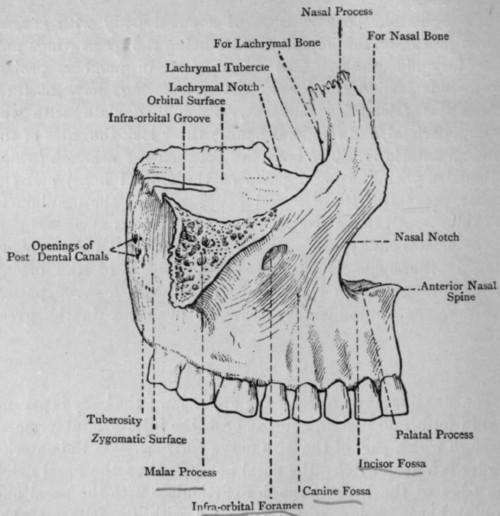
8 Maxilla The maxillary bones occupy the central part of the face and support the teeth of the upper jaw. They contribute to much of the skeleton of the upper face including the nasal aperture, the bridge of the nose, the floor of the orbital cavities and the bones of the cheeks. Each bone consists of a body and four processes: the frontal, zygomatic, alveolar and palatine processes. The body forms the main bulk of the bone and possesses anterior, orbital, nasal and infratemporal (posterior) surfaces. The maxillary bones show many articulations. On the face, the maxillary bones articulate with each other, the nasal bones, the nasal cartilages and the frontal bone. Laterally, they articulate with the zygomatic bones. Each maxillary bone also joins with the vomer, the septal cartilage, the lacrimal bone, the ethmoid bone and the inferior nasal concha to contribute to the skeleton of the nasal fossa and the orbit. Attachments The maxilla provides attachment for buccinator, depressor septi, incisivus labii superioris, levator anguli oris, levator labii superioris, orbicularis oculi, and nasalis.
9 The maxillary bone exhibits the following features: Body Infra-orbital foramen. Infra-orbital groove. Canine fossa. Nasal notch. Anterior nasal spine. Alveolar canals. Greater palatine groove. Inferior meatus. Maxillary hiatus. Maxillary sinus. Frontal process Anterior lacrimal crest. Agger nasi. Ethmoidal crest. Fossa for lacrimal sac. Lacrimal groove. Conchal crest. Middle meatus. Zygomatic process Alveolar process Tuberosity. Canine eminence. Palatine process Floor of nasal cavity. Incisive canal. Incisive fossa. Nasal crest. Alveolar ridges. Depressions for palatine gland.
10
11 Nasal bone The two nasal bones form the upper part of the bridge of the nose. Each nasal bone is quadrilateral, being longer than it is wide. The nasal bone exhibits the following features: Superior border. Inferior border. Medial border. Lateral border. Internal surface. External surface. Grooves for anterior ethmoidal nerve. Vascular foramina. The superior border articulates with the nasal part of the frontal bone. The inferior border forms the superior boundary of the anterior nasal aperture. The lateral border meets the frontal process of the maxilla and the medial border meets its fellow in the midline. Attachments The procerus muscle is attached to the external surface of the nasal bone and the lateral nasal cartilage to the inferior border.
12 Palatine bone The palatine bone is found at the back part of the nasal cavity between the maxilla and the pterygoid process of the sphenoid bone. It contributes to the floor and lateral wall of the nasal cavity, the roof of the mouth, and the floor of the orbit; it contributes to the formation of the pterygopalatine and pterygoid fossae; and one fissure, the inferior orbital fissure. The palatine bone is L-shaped, and consists of a horizontal plate and a vertical perpendicular plate and four processes: the pyramidal, orbital, maxillary and sphenoidal processes. The horizontal plate is a quadrilateral plate of bone that forms the posterior quarter of the hard palate when articulated with its fellow; it has a nasal surface and four borders: anterior, posterior, medial and lateral. The perpendicular plate is rectangular, the vertical dimension being approximately twice that of the antero-posterior dimension. It has two surfaces, the maxillary and nasal, and four borders: anterior, posterior, superior and inferior.
13 The palatine bone exhibits the following features: Horizontal plate Palatine surface. Nasal surface. Anterior border. Posterior border. Medial border. Lateral border. Palatine crest. Posterior nasal spine. Pyramidal process. Lesser palatine foramina. Perpendicular plate Maxillary surface. Nasal surface. Anterior border. Posterior border. Superior border. Inferior border. Pterygopalatine canal. Palatovaginal canal. Conchal crest. Ethmoid crest. Greater palatine foramina. Greater palatine groove. Maxillary process. Sphenoidal process. Orbital process. Sphenopalatine notch.
14 Vomer The vomer is a single thin plate of bone, shaped like a ploughshare and forms the postero-inferior portion of the nasal septum (slide). It possesses two lateral surfaces and four borders, anterior, inferior, superior and posterior. All borders accept the posterior border articulate with the adjacent bones. The posterior border is free and does not articulate with any other structures. It is slightly concave and slopes antero-inferiorly to form a prominent midline ridge between the two posterior nasal apertures.
15 The vomer exhibits the following landmarks: Ala. Site of articulation with septal cartilage. Site of articulation with ethmoid bone. Site of articulation with maxilla and palatine bone. Site of articulation with sphenoid bone. Groove for nasopalatine nerve and vessels. Groove for ethmoidal nerve. The anterior border is the longest border on the bone and slopes antero-inferiorly. It articulates with the nasal septal cartilage and with the perpendicular plate of the ethmoid bone. The inferior border lies anterior to the superior border and articulates with the median maxillary and palatine nasal crests. Finally, the superior border articulates with the vaginal process and the body of the sphenoid bone.
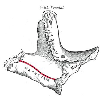
16 Zygomatic bone The two zygomatic bones form the skeleton of the cheeks. Each bone has: three surfaces; lateral (antero-lateral), temporal (postero-medial) and orbital, five borders; orbital (antero-superior), maxillary (antero-inferior), temporal (postero- superior), postero-inferior and postero-medial and two processes, the frontal and temporal. The lateral surface is smooth and slightly convex. The entire surface of the zygomatic bone viewed medially is called the temporal surface. It can be subdivided into two regions. Anteriorly, it shows a roughened area for articulation with the zygomatic process of the maxilla. Posteriorly, the temporal surface includes the lower surface of the orbital plate and the temporal surface of the temporal process. This posterior region is smooth and forms the anterior boundary of the temporal fossa.
17 The zygomatic bone exhibits the following landmarks: Body. Temporal process. Frontal process. Orbital surface (plate). Orbital border. Maxillary border. Temporal border. Postero-inferior border. Postero-medial border. Marginal tubercle. Zygomatico-orbital foramen. Zygomaticofacial foramen. Zygomaticotemporal foramen. Attachments The zygomatic bone provides attachment for the: Levator labii superioris. Masseter. Temporalis. Temporal fascia. Zygomaticus major. Zygomaticus minor.
18
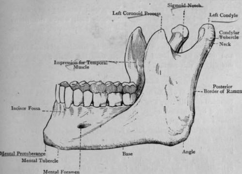
19 Mandible The mandible consists of a horizontal, horseshoe-shaped shaped body and two vertical rami. The body of the mandible supports the mandibular teeth within the alveolar processes. The rami of the mandible articulate with the temporal bones at the temporomandibular joints.
20 The mandible exhibits the following prominent landmarks: Body Alveolar ridge (process). Menti symphysis. Mental protuberance. Mental tubercle. Incisive fossa. Digastric fossa. Inferior mental spine (genial tubercle). Superior mental spine (genial tubercle). Mylohyoid line. Sublingular fossa. Submandibular fossa. Mental foramen. Oblique line. Rami Coronoid process. Condylar process. Head of condylar process. Neck of condylar process. Pterygoid fovea. Mandibular notch. Anterior border. Posterior border. Lingula. Mylohyoid groove. Obtuse angle.
21 Attachments It gives attachments to anterior bellies of the digastric muscle, geniohyoid (slide), genioglossus, mylohyoid (slide), masseter, buccinator, medial and lateral pterygoid, mentalis, temporalis, incisivus labii inferioris, depressor anguli oris, platysma muscles and part of the orbicularis oris. It also gives attachment to the sphenomandibular and stylomandibular ligaments and the pterygomandibular raphe.
22
Bones of the skull & face
 Bones of the skull & face Cranium= brain case or helmet Copyright The McGraw-Hill Companies, Inc. Permission required for reproduction or display. The cranium is composed of eight bones : frontal Occipital
Skull basic structures. Neurocranium
 Assoc. Prof. Květuše Lovásová, M.V.D., PhD. Skull basic structures Skull consists of two groups of bones: neurocranium (bones forming the brain box) splanchnocranium (bones forming the facial skeleton)
Anatomic Relations Summary. Done by: Sohayyla Yasin Dababseh
 Anatomic Relations Summary Done by: Sohayyla Yasin Dababseh Anatomic Relations Lecture 1 Part-1 - The medial wall of the nose is the septum. - The vestibule lies directly inside the nostrils (Nares). -
Infratemporal fossa: Tikrit University college of Dentistry Dr.Ban I.S. head & neck Anatomy 2 nd y.
 Infratemporal fossa: This is a space lying beneath the base of the skull between the lateral wall of the pharynx and the ramus of the mandible. It is also referred to as the parapharyngeal or lateral pharyngeal
Chapter 7 Part A The Skeleton
 Chapter 7 Part A The Skeleton Why This Matters Understanding the anatomy of the skeleton enables you to anticipate problems such as pelvic dimensions that may affect labor and delivery The Skeleton The
The Skull and Temporomandibular joint II Prof. Abdulameer Al-Nuaimi. E. mail:
 The Skull and Temporomandibular joint II Prof. Abdulameer Al-Nuaimi E-mail: a.al-nuaimi@sheffield.ac.uk E. mail: abdulameerh@yahoo.com Temporal fossa The temporal fossa is a depression on the temporal
Temporal region. temporal & infratemporal fossae. Zhou Hong Ying Dept. of Anatomy
 Temporal region temporal & infratemporal fossae Zhou Hong Ying Dept. of Anatomy Temporal region is divided by zygomatic arch into temporal & infratemporal fossae. Temporal Fossa Infratemporal fossa Temporal
Upper arch. 1Prosthodontics. Dr.Bassam Ali Al-Turaihi. Basic anatomy & & landmark of denture & mouth
 1Prosthodontics Lecture 2 Dr.Bassam Ali Al-Turaihi Basic anatomy & & landmark of denture & mouth Upper arch Palatine process of maxilla: it form the anterior three quarter of the hard palate. Horizontal
SKULL / CRANIUM BONES OF THE NEUROCRANIUM (7) Occipital bone (1) Sphenoid bone (1) Temporal bone (2) Frontal bone (1) Parietal bone (2)
 Occipital bone (1) Sphenoid bone (1) Temporal bone (2) Frontal bone (1) Parietal bone (2)") Important! 1. Memorizing these pages only does not guarantee the succesfull passing of the midterm test or the semifinal exam. 2. The handout has not been supervised, and I can not guarantee, that these
Important! 1. Memorizing these pages only does not guarantee the succesfull passing of the midterm test or the semifinal exam. 2. The handout has not been supervised, and I can not guarantee, that these
The cribriform plate. ethmoid bone. Ethmoid bone consists from: 1) A horizontal cribriform plate. 2) A perpendicular plate. 3) Two lateral labyrinths.
 A horizontal cribriform plate. 2) A perpendicular plate. 3) Two lateral labyrinths.") ethmoid bone Ethmoid bone consists from: 1) A horizontal cribriform plate. 2) A perpendicular plate. 3) Two lateral labyrinths. The cribriform plate 1) Connect the two labyrinths to the perpendicular plate.
ethmoid bone Ethmoid bone consists from: 1) A horizontal cribriform plate. 2) A perpendicular plate. 3) Two lateral labyrinths. The cribriform plate 1) Connect the two labyrinths to the perpendicular plate.
PTERYGOPALATINE FOSSA
 PTERYGOPALATINE FOSSA Outline Anatomical Structure and Boundaries Foramina and Communications with other spaces and cavities Contents Pterygopalatine Ganglion Especial emphasis on certain arteries and
University of Palestine. Midterm Exam 2013/2014 Total Grade:
 [ Course No: DNTS2208 Course Title: Head and Neck Anatomy Date: 17/11/1024 No. of Questions: (52) Time: 2hours Using Calculator (No) University of Palestine Midterm Exam 2013/2014 Total Grade: Instructor
Bisection of Head & Nasal Cavity 頭部對切以及鼻腔. 解剖學科馮琮涵副教授 分機
 Bisection of Head & Nasal Cavity 頭部對切以及鼻腔 解剖學科馮琮涵副教授 分機 3250 E-mail: thfong@tmu.edu.tw Outline: The structure of nose The concha and meatus in nasal cavity The openings of paranasal sinuses Canals, foramens
Skull-2. Norma Basalis Interna Norma Basalis Externa. Dr. Heba Kalbouneh Associate Professor of Anatomy and Histology
 Skull-2 Norma Basalis Interna Norma Basalis Externa Dr. Heba Kalbouneh Associate Professor of Anatomy and Histology Norma basalis interna Base of the skull- superior view The interior of the base of the
Dr.Ban I.S. head & neck anatomy 2 nd y جامعة تكريت كلية طب االسنان مادة التشريح املرحلة الثانية أ.م.د. بان امساعيل صديق 6102/6102
 جامعة تكريت كلية طب االسنان مادة التشريح املرحلة الثانية أ.م.د. بان امساعيل صديق 6102/6102 Pterygopalatine fossa: The pterygopalatine fossa is a cone-shaped depression, It is located between the maxilla,
Maxilla, ORBIT and infratemporal fossa. Neophytos C Demetriades MD, DDS, MSc Associate professor European University of Cyprus School of Medicine
 Maxilla, ORBIT and infratemporal fossa Neophytos C Demetriades MD, DDS, MSc Associate professor European University of Cyprus School of Medicine MAXILLA Superior, middle, and inferior meatus Frontal sinus
Anatomy and Physiology. Bones, Sutures, Teeth, Processes and Foramina of the Human Skull
 Anatomy and Physiology Chapter 6 DRO Bones, Sutures, Teeth, Processes and Foramina of the Human Skull Name: Period: Bones of the Human Skull Bones of the Cranium: Frontal bone: forms the forehead and the
Biology 218 Human Anatomy. Adapted from Martini Human Anatomy 7th ed. Chapter 6 The Skeletal System: Axial Division
 Adapted from Martini Human Anatomy 7th ed. Chapter 6 The Skeletal System: Axial Division Introduction The axial skeleton: Composed of bones along the central axis of the body Divided into three regions:
University of Palestine. Midterm Exam 2013/2014 Total Grade:
 Course No: DNTS2208 Course Title: Head and Neck Anatomy Date: 09/11/2013 No. of Questions: (50) Time: 1hour Using Calculator (No) University of Palestine Midterm Exam 2013/2014 Total Grade: Instructor
Skeletal System: Skull.
 Skeletal System: Skull www.fisiokinesiterapia.biz Bones of the Skull SPLANCHNOCRANIUM Nasal (2) Maxilla (2) Lacrimal (2) Zygomatic (2) Palatine (2) Inferior concha (2) Vomer Mandible NEUROCRANIUM Frontal
Oral cavity landmarks
 By: Dr. Ahmed Rabah Oral cavity landmarks The knowledge of oral anatomy and physiology will help the operator and provides enough landmarks to act as positive guide during denture construction. This subject
Introduction to Local Anesthesia and Review of Anatomy
 5-Sep Introduction and Anatomy Review 12-Sep Neurophysiology and Pain 19-Sep Physiology and Pharmacology part 1 26-Sep Physiology and Pharmacology part 2 Introduction to Local Anesthesia and Review of
Nasal region. cartilages: septal cartilage (l); lateral nasal cartilage (2); greater alar cartilages (2); lesser alar cartilages (?
; lateral nasal cartilage (2); greater alar cartilages (2); lesser alar cartilages (?") Nasal region skull bones: nasal and frontal processes of maxilla cartilages: septal cartilage (l); lateral nasal cartilage (2); greater alar cartilages (2); lesser alar cartilages (?) 1 Nasal cavity Roof
Nasal region skull bones: nasal and frontal processes of maxilla cartilages: septal cartilage (l); lateral nasal cartilage (2); greater alar cartilages (2); lesser alar cartilages (?) 1 Nasal cavity Roof
Omran Saeed. Luma Taweel. Mohammad Almohtaseb. 1 P a g e
 2 Omran Saeed Luma Taweel Mohammad Almohtaseb 1 P a g e I didn t include all the photos in this sheet in order to keep it as small as possible so if you need more clarification please refer to slides In
Nose & Mouth OUTLINE. Nose. - Nasal Cavity & Its Walls. - Paranasal Sinuses. - Neurovascular Structures. Mouth. - Oral Cavity & Its Contents
 Dept. of Human Anatomy, Si Chuan University Zhou hongying eaglezhyxzy@163.com Nose & Mouth OUTLINE Nose - Nasal Cavity & Its Walls - Paranasal Sinuses - Neurovascular Structures Mouth - Oral Cavity & Its
Chapter 7. Skeletal System
 Chapter 7 Skeletal System 1 Skull A. The skull is made up of 22 bones: 8 cranial bones, 13 facial bones, and the mandible. B. The Cranium encloses and protects the brain, provides attachments for muscles,
Cranium Facial bones. Sternum Rib
 Figure 7.1 The human skeleton. Skull Thoracic cage (ribs and sternum) Cranium Facial bones Sternum Rib Bones of pectoral girdle Vertebral column Sacrum Vertebra Bones of pelvic girdle (a) Anterior view
Skeletal System -Axial System. Chapter 7 Part A
 Skeletal System -Axial System Chapter 7 Part A Skeleton Learn: Names of the s. Identify specific landmarks that allow: Bones to fit into each other, Organs to fit into the cavities, Muscles to attach,
Trigeminal Nerve Worksheets, Distributions Page 1
 Trigeminal Nerve Worksheet #1 Distribution by Nerve Dr. Darren Hoffmann Dental Gross Anatomy, Spring 2013 We have drawn out each of the branches of CN V in lecture and you have an idea now for their basic
3-Deep fascia: is absent (except over the parotid gland & buccopharngeal fascia covering the buccinator muscle)
") The Face 1-Skin of the Face The skin of the face is: Elastic Vascular (bleed profusely however heal rapidly) Rich in sweat and sebaceous glands (can cause acne in adults) It is connected to the underlying
The Face 1-Skin of the Face The skin of the face is: Elastic Vascular (bleed profusely however heal rapidly) Rich in sweat and sebaceous glands (can cause acne in adults) It is connected to the underlying
AXIAL SKELETON SKULL
 AXIAL SKELETON SKULL CRANIAL BONES (8 total flat bones w/ 2 paired) 1. Frontal forms forehead & upper portion of eyesocket (orbital) 2. Parietal paired bones; form superior & lateral walls of cranium 3.
MAXILLA, ORBIT & PTERYGOPALATINE FOSSA. Neophytos C Demetriades MD, DDS, MSc Associate professor European University of Cyprus School of Medicine
 MAXILLA, ORBIT & PTERYGOPALATINE FOSSA Neophytos C Demetriades MD, DDS, MSc Associate professor European University of Cyprus School of Medicine Maxilla MAXILLA Superior, middle, and inferior meatus Frontal
Dr. Sami Zaqout, IUG Medical School
 The skull The skull is composed of several separate bones united at immobile joints called sutures. Exceptions? Frontal bone Occipital bone Vault Cranium Sphenoid bone Zygomatic bones Base Ethmoid bone
Mohammad Hisham Al-Mohtaseb. Lina Mansour. Reyad Jabiri. 0 P a g e
 2 Mohammad Hisham Al-Mohtaseb Lina Mansour Reyad Jabiri 0 P a g e This is only correction for the last year sheet according to our record. If you already studied this sheet just read the yellow notes which
Dr.Noor Hashem Mohammad Lecture (5)
") Dr.Noor Hashem Mohammad Lecture (5) 2016-2017 If the mandible is discarded, the anterior part of this aspect of the skull is seen to be formed by the hard palate. The palatal processes of the maxillae
Dr.Noor Hashem Mohammad Lecture (5) 2016-2017 If the mandible is discarded, the anterior part of this aspect of the skull is seen to be formed by the hard palate. The palatal processes of the maxillae
Structure Location Function
 Frontal Bone Cranium forms the forehead and roof of the orbits Occipital Bone Cranium forms posterior and inferior portions of the cranium Temporal Bone Cranium inferior to the parietal bone forms the
Anatomy images for MSS practical exam- 2019
 Anatomy images for MSS practical exam- 2019 Ilium Ischium Pubis Acetabulaum Iliac crest Iliac tubercle ASIS (muscle and ligament attached) AIIS (muscle attached) PSIS PIIS Ischial spine Ischial tuberosity
Temporal fossa Infratemporal fossa Pterygopalatine fossa Terminal branches of external carotid artery Pterygoid venous plexus
 Outline of content Temporal fossa Infratemporal fossa Pterygopalatine fossa Terminal branches of external carotid artery Pterygoid venous plexus Boundary Content Communication Mandibular division of trigeminal
Chapter 7: Skeletal System: Gross Anatomy
 Chapter 7: Skeletal System: Gross Anatomy I. General Considerations A. How many bones in an average adult skeleton? B. Anatomic features of bones are based on II. Axial Skeleton A. Skull 1. Functionally
Parotid Gland. Parotid Gland. Largest of 3 paired salivary glands (submandibular; sublingual) Ramus of Mandible. Medial pterygoid.
 Ramus of Mandible. Medial pterygoid.") Parotid region Parotid Gland Largest of 3 paired salivary glands (submandibular; sublingual) Ramus of Mandible Medial pterygoid Cross section of mandible Masseter D S SCM Parotid Gland Mastoid Process
Parotid region Parotid Gland Largest of 3 paired salivary glands (submandibular; sublingual) Ramus of Mandible Medial pterygoid Cross section of mandible Masseter D S SCM Parotid Gland Mastoid Process
Parotid Gland, Temporomandibular Joint and Infratemporal Fossa
 M1 - Anatomy Parotid Gland, Temporomandibular Joint and Infratemporal Fossa Jeff Dupree Sanger 9-057 jldupree@vcu.edu Parotid gland: wraps around the mandible positioned between the mandible and the sphenoid
Head and Face Anatomy
 Head and Face Anatomy Epicranial region The Scalp The soft tissue that covers the vault of skull. Extends from supraorbital margin to superior nuchal line. Layers of the scalp S C A L P = skin = connective
Lec [8]: Mandibular nerve:
![Lec [8]: Mandibular nerve:](/thumbs/94/121295776.jpg "Lec [8]: Mandibular nerve:") Lec [8]: Mandibular nerve: The mandibular branch from the trigeminal ganglion lies in the middle cranial fossa lateral to the cavernous sinus. With the motor root of the trigeminal nerve [motor roots lies
Lec [8]: Mandibular nerve: The mandibular branch from the trigeminal ganglion lies in the middle cranial fossa lateral to the cavernous sinus. With the motor root of the trigeminal nerve [motor roots lies
Anatomy #1; Respiratory Nose and the Nasal Cavity December 1st, 2013
 Note #1: the doctor skipped some slides in the lecture. Those slides are not included in this sheet and so you will have to review the slides to study them. The reason they were not included is because
SKULL AS A WHOLE + ANTERIOR CRANIAL FOSSA
 SKULL AS A WHOLE + ANTERIOR CRANIAL FOSSA LEARNING OBJECTIVES At the end of this lecture, the student should be able to know: Parts of skeleton (axial and appendicular) Parts of skull Sutures of skull
APPENDICULAR SKELETON 126 AXIAL SKELETON SKELETAL SYSTEM. Cranium. Skull. Face. Skull and associated bones. Auditory ossicles. Associated bones.
 SKELETAL SYSTEM 206 AXIAL SKELETON 80 APPENDICULAR SKELETON 26 Skull Skull and associated s 29 Cranium Face Auditory ossicles 8 4 6 Associated s Hyoid Thoracic cage 25 Sternum Ribs 24 Vertebrae 24 column
The orbit-1. Dr. Heba Kalbouneh Assistant Professor of Anatomy and Histology
 The orbit-1 Dr. Heba Kalbouneh Assistant Professor of Anatomy and Histology Orbital plate of frontal bone Orbital plate of ethmoid bone Lesser wing of sphenoid Greater wing of sphenoid Lacrimal bone Orbital
Dr.Sepideh Falah-kooshki
 Dr.Sepideh Falah-kooshki MAXILLA Premaxillary/median palatal suture (radiolucent). Incisive fossa and foramen (radiolucent). Nasal passages (radiolucent). Nasal septum (radiopaque). Anterior nasal spine
The sebaceous glands (glands of Zeis) open directly into the eyelash follicles, ciliary glands (glands of Moll) are modified sweat glands that open
 open directly into the eyelash follicles, ciliary glands (glands of Moll) are modified sweat glands that open") The Orbital Region The orbits are a pair of bony cavities that contain the eyeballs; their associated muscles, nerves, vessels, and fat; and most of the lacrimal apparatus upper eyelid is larger and more
The Orbital Region The orbits are a pair of bony cavities that contain the eyeballs; their associated muscles, nerves, vessels, and fat; and most of the lacrimal apparatus upper eyelid is larger and more
THE SKELETAL SYSTEM. Focus on the Skull
 THE SKELETAL SYSTEM Focus on the Skull Review Anatomical Terms Anterior/Posterior Dorsal/Ventral Medial/Lateral Superior/Inferior Bone Markings - Review Projections for attachment of muscles, ligaments
Bony orbit Roof The orbital plate of the frontal bone Lateral wall: the zygomatic bone and the greater wing of the sphenoid
 Bony orbit Roof: Formed by: The orbital plate of the frontal bone, which separates the orbital cavity from the anterior cranial fossa and the frontal lobe of the cerebral hemisphere Lateral wall: Formed
Neck of Condylar. Process. Anterior Border of Ramus. Mandibular. Foramen. Posterior Border of Ramus Incisive Fossa.
 Learning Outcomes The Mandible Surface Anatomy Muscle Attachments The (FOM) Muscles of the FOM The Tongue Muscles of the Tongue The Submandibular Region Submandibular Gland Sublingual Gland Lingual The
The Axial Skeleton. C h a p t e r. PowerPoint Lecture Slides prepared by Jason LaPres Lone Star College - North Harris
 C h a p t e r 7 The Axial Skeleton PowerPoint Lecture Slides prepared by Jason LaPres Lone Star College - North Harris Copyright 2009 Pearson Education, Inc., publishing as Pearson Benjamin Cummings An
Anatomy of the Trigeminal Nerve
 19 Anatomy of the Trigeminal Nerve.1 Introduction 0. The Central Part of the Trigeminal Nerve 1..1 Origin 1.. Trigeminal Nuclei.3 The Peripheral Part of the Trigeminal Nerve 4.3.1 Ophthalmic Nerve 4.3.
YOU MUST BRING YOUR OWN GLOVES FOR THIS ACTIVITY.
 ACTIVITY 3: AXIAL SKELETON AND LONG BONE DISSECTION Objectives: 1) How to get ready: Read Chapter 7, McKinley et al., Human Anatomy, 5e. All text references are for this textbook. Learning the meanings
3. The Jaw and Related Structures
 Overview and objectives of this dissection 3. The Jaw and Related Structures The goal of this dissection is to observe the muscles of jaw raising. You will also have the opportunity to observe several
Trigeminal Nerve (V)
") Trigeminal Nerve (V) Lecture Objectives Discuss briefly how the face is developed. Follow up the course of trigeminal nerve from its point of central connections, exit and down to its target areas. Describe
Trigeminal Nerve (V) Lecture Objectives Discuss briefly how the face is developed. Follow up the course of trigeminal nerve from its point of central connections, exit and down to its target areas. Describe
Dr. Sami Zaqout Faculty of Medicine IUG
 Auricle External Ear External auditory meatus The Ear Middle Ear (Tympanic Cavity) Auditory ossicles Internal Ear (Labyrinth) Bony labyrinth Membranous labyrinth External Ear Auricle External auditory
Crafton Hills College Human Anatomy & Physiology Axial Skeleton
 A. Major Divisions Crafton Hills College Human Anatomy & Physiology Axial keleton 1. Axial: Part of skeleton lies along long axis of body 2. Appendicular: Bones & features of the appendages B. AXIAL KELETON
ACTIVITY 3: AXIAL SKELETON AND LONG BONE DISSECTION COW BONE DISSECTION
 ACTIVITY 3: AXIAL SKELETON AND LONG BONE DISSECTION Objectives: 1) How to get ready: Read Chapter 7, McKinley et al., Human Anatomy, 4e. All text references are for this textbook. Learning the meanings
HEAD & NECK BY NUMBERS THIRD EDITION Copyright 2013, Anatomy Numbers (Phoenix, AZ) All rights reserved
 All rights reserved") HEAD & NECK BY NUMBERS THIRD EDITION Copyright 2013, Anatomy Numbers (Phoenix, AZ) All rights reserved No part of this publication may be reproduced or transmitted in any form or by any means, electronic
HEAD & NECK BY NUMBERS THIRD EDITION Copyright 2013, Anatomy Numbers (Phoenix, AZ) All rights reserved No part of this publication may be reproduced or transmitted in any form or by any means, electronic
Face. Definition: The area between the two ears and from the chin to the eye brows. The muscles of the face
 Face Definition: The area between the two ears and from the chin to the eye brows. The muscles of the face The muscle of facial expression (include the muscle of the face and the scalp). All are derived
Trigeminal Nerve Anatomy. Dr. Mohamed Rahil Ali
 Trigeminal Nerve Anatomy Dr. Mohamed Rahil Ali Trigeminal nerve Largest cranial nerve Mixed nerve Small motor root and large sensory root Motor root Nucleus of motor root present in the pons and medulla
Skull-2. Norma Basalis Interna. Dr. Heba Kalbouneh Assistant Professor of Anatomy and Histology
 Skull-2 Norma Basalis Interna Dr. Heba Kalbouneh Assistant Professor of Anatomy and Histology Norma basalis interna Base of the skull- superior view The interior of the base of the skull is divided into
Human Anatomy and Physiology - Problem Drill 07: The Skeletal System Axial Skeleton
 Human Anatomy and Physiology - Problem Drill 07: The Skeletal System Axial Skeleton Question No. 1 of 10 Which of the following statements about the axial skeleton is correct? Question #01 A. The axial
Tikrit University collage of dentistry Dr.Ban I.S. head & neck anatomy 2 nd y. Lec [5] / Temporal fossa :
![Tikrit University collage of dentistry Dr.Ban I.S. head & neck anatomy 2 nd y. Lec [5] / Temporal fossa :](/thumbs/88/115294566.jpg "Tikrit University collage of dentistry Dr.Ban I.S. head & neck anatomy 2 nd y. Lec [5] / Temporal fossa :") Lec [5] / Temporal fossa : Borders of the Temporal Fossa: Superior: Superior temporal line. Inferior: gap between zygomatic arch and infratemporal crest of sphenoid bone. Anterior: Frontal process of the
Lec [5] / Temporal fossa : Borders of the Temporal Fossa: Superior: Superior temporal line. Inferior: gap between zygomatic arch and infratemporal crest of sphenoid bone. Anterior: Frontal process of the
External Acoustic Meatus. Mastoid Process. Zygomatic Process. Temporal Bone
 Bone lab review 1. Frontal Bone 2. Supra-Orbital Foramen 3. Orbit (Orbital Cavity) 4. Superior Orbital Fissure 5. Inferior Orbital Fissure 6. Zygomatic Bone 7. Infra-Orbital Foramen 8. Maxilla 9. Mandible
BOGOMOLETS NATIONAL MEDICAL UNIVERSITY. Department of human anatomy GUIDELINES
 BOGOMOLETS NATIONAL MEDICAL UNIVERSITY Department of human anatomy GUIDELINES Academic discipline HUMAN ANATOMY Module 1 Content module 2 The theme of the Eye-socket, bone nasal lesson cavity. bony palate
Chapter 7: Head & Neck
 Chapter 7: Head & Neck Osteology I. Overview A. Skull The cranium is composed of irregularly shaped bones that are fused together at unique joints called sutures The skull provides durable protection from
Tracing the Cranial Nerves Osteologically
 CN I II III IV V 1 Supra-orbital ethmoidal nn. Ext. nasal V 2 Tracing the Cranial Nerves Osteologically Nucleus of Origin Olfactory tracts of frontal lobe of cerebrum Optic tracts from optic chiasma and
Muscles of the Face, Head, and Neck
 Muscles of the Face, Head, and Neck 1 How Muscles Are Named Many muscles named using such features as Location Function Shape Direction of fibers Number of heads or divisions Points of attachment Size
University of Palestine. Final Exam 1 st Semester 2014/2015 Total Grade: 60
 Question One: MCQ: 1- The coronal suture joins the a) frontal and parietal bones. b) left and right parietal bones. c) parietal and occipital bones. d) parietal, squamous temporal and greater wing of the
Skeletal system. Prof. Abdulameer Al-Nuaimi. E. mail:
 Skeletal system Prof. Abdulameer Al-Nuaimi E-mail: a.al-nuaimi@sheffield.ac.uk E. mail: abdulameerh@yahoo.com Functions of Bone and The Skeletal System Support: The skeleton serves as the structural framework
Temporomandibular Joint. Dr Noman ullah wazir
 Temporomandibular Joint Dr Noman ullah wazir Type of Joint TMJ is a Synovial joint between : The condylar head of the mandible. The mandibular fossa of squamous part of temporal bone. The joint cavity
Figure (2-6): Labial frenum and labial notch.
: Labial frenum and labial notch.") The anatomy of the edentulous ridge in the maxilla and mandible is very important for the design of a complete denture. The consistency of the mucosa and architecture of the underlying bone is different
The anatomy of the edentulous ridge in the maxilla and mandible is very important for the design of a complete denture. The consistency of the mucosa and architecture of the underlying bone is different
*in general the blood supply of the nose comes from branches of the internal and external carotid arteries.
 In the previous lecture we talked about the anatomy of the nasal cavity, today we will talk about its blood supply, venous drainage, innervations, and finally about the paranasal sinuses. When we describe
OPEN ACCESS ATLAS OF OTOLARYNGOLOGY, HEAD & NECK OPERATIVE SURGERY
 OPEN ACCESS ATLAS OF OTOLARYNGOLOGY, HEAD & NECK OPERATIVE SURGERY INFERIOR MAXILLECTOMY Tumours of the hard palate and superior alveolus may be resected by inferior maxillectomy (Figure 1). A Le Fort
Trigeminal nerve. Slide in bold and please go back to see the pictures, if I skipped any part of record that because it wasn t clear to me
 Trigeminal nerve Slide in bold and please go back to see the pictures, if I skipped any part of record that because it wasn t clear to me Hala nsour 2/26/2018 P a g e 1 this lecture contain two topics
1. In regards to the bones of the face, which of the following is NOT TRUE?
 Anatomy and Physiology Fall Exam II: Form B Name: 1. In regards to the bones of the face, which of the following is NOT TRUE? A. The vomer bone articulates with the dorsal surface of the palatine process
Face and Scalp 解剖學科鄭授德
 Face and Scalp 解剖學科鄭授德 本教材之圖片取自於 1 Gray s Anatomy for Students, 3rd ed, 2015, by Drake, Vogl, and Mitchell 2 Clinically Oriented Anatomy, 7th ed, 2014, by Moore, Dalley, and Agur 3 Clinically Oriented
function - sensory & postganglionic sympathetic [communication from the internal carotid plexus in the cavernous sinus] innervation of the mucosa of
![function - sensory & postganglionic sympathetic [communication from the internal carotid plexus in the cavernous sinus] innervation of the mucosa of](/thumbs/74/71276096.jpg "function - sensory & postganglionic sympathetic [communication from the internal carotid plexus in the cavernous sinus] innervation of the mucosa of") Nerves I. Cranial nerves A. Olfactory (CN I) 1. Olfactory bulb 2. Olfactory tract B. Optic n. (CNII) function - carries visual sensory information from the neural retina to the diencephalon & midbrain
Nerves I. Cranial nerves A. Olfactory (CN I) 1. Olfactory bulb 2. Olfactory tract B. Optic n. (CNII) function - carries visual sensory information from the neural retina to the diencephalon & midbrain
View of a Skull, 1489 by Leonardo Da Vinci. Kaan Yücel M.D., Ph.D Tuesday
 View of a Skull, 1489 by Leonardo Da Vinci Kaan Yücel M.D., Ph.D. 26.11.2013 Tuesday 1.SKULL skeleton of the head cranium 22 bones excluding ossicles of the ear 1.SKULL Mandible Lower jaw bone Neurocranium
Biology 2401 The Skeletal System
 Biology 2401 The Skeletal System Purpose: The lab will describe the microscopic and gross anatomy of bone, identify bones of the body, and identify important bone markings. I. Overview of the Skeleton
CHAPTER 7, PART II (BONES)
") Anatomy Name: CHAPTER 7, PART II (BONES) Entry #: INSTRUCTIONS: 1) READ Chapter 7, pg. 140-161. 2) Using the outline, make a note card for each underlined bone name or phrase. 3) On each note card, put
Anatomy Name: CHAPTER 7, PART II (BONES) Entry #: INSTRUCTIONS: 1) READ Chapter 7, pg. 140-161. 2) Using the outline, make a note card for each underlined bone name or phrase. 3) On each note card, put
Introduction to Occlusion and Mechanics of Mandibular Movement
 Introduction to Occlusion and Mechanics of Mandibular Movement Dr. Pauline Hayes Garrett Department of Endodontics, Prosthodontics, and Operative Dentistry University of Maryland, Baltimore Assigned reading
Chapter 8A. The Skeletal System: The Axial Skeleton. The Skeletal System: The Axial Skeleton. Types of Bones. Types of Bones
 Chapter 8A The Skeletal System: The Axial Skeleton The Skeletal System: The Axial Skeleton 206 named bones Axial Skeleton 80 bones lie along longitudinal axis skull, hyoid, vertebrae, ribs, sternum, ear
بسم هللا الرحمن الرحيم السالم عليكم ورحمة هللا وبركاته
 بسم هللا الرحمن الرحيم السالم عليكم ورحمة هللا وبركاته Lecture one Introduction to General Anatomy By Anatomist Dr. Hatem A. Hatem Department of Anatomy, Histology& Embryology College of Dentistry-Ibn
Lips and labial mucosa
 Lips and labial mucosa External portion of the lips: the vermilion border and the skin Vermilion border : the exposed red portion of the lip, covered by mucous membrane, no mucous glands Boundary: the
The Skull DANIL HAMMOUDI.MD
 The Skull DANIL HAMMOUDI.MD summary of bones/structures in Chapter 15 of the manual need tp be print as soon as possible http://www.mnsu.edu/emuseum/biology/humananatomy/skeletal/skul l/frontal/frontal.html
ANATOMY & PHYSIOLOGY I Laboratory Version B Name Section. REVIEW SHEET Exercise 10 Axial Skeleton
 ANATOMY & PHYSIOLOGY I Laboratory Version B Name Section REVIEW SHEET Exercise 10 Axial Skeleton 1 POINT EACH. THE SKULL MULTIPLE CHOICE 1. The major components of the axial skeleton include the 7. The
Riverside Community College Anatomy & Physiology 2B SPRING 2012 EXAM #1-ABC (Nervous System)
") Riverside Community College Anatomy & Physiology 2B SPRING 2012 EXAM #1-ABC (Nervous System) Name: 1) This vertebra is an example of a(n). 1) A) thoracic B) axis C) atlas D) lumbar E) sacral 1 2) W hich
Riverside Community College Anatomy & Physiology 2B SPRING 2012 EXAM #1-ABC (Nervous System) Name: 1) This vertebra is an example of a(n). 1) A) thoracic B) axis C) atlas D) lumbar E) sacral 1 2) W hich
TEST YOURSELF- Chapter 7
 TEST YOURSELF- Chapter 7 Cranial Bones 1. Give the name of the bone for each of the following markings. Some of the markings are found on more than one bone. List all that apply. Cranium a. Frontal squama:
Chapter 35: Anatomy. Daniel O. Graney, Shan R. Baker. Nasal Pyramid. Bony pyramid
 Chapter 35: Anatomy Daniel O. Graney, Shan R. Baker Nasal Pyramid The nose is a pyramidal structure with its apex projecting anteriorly and its base attached to the facial skeleton. The superior part of
Prosthodontics Dr.Yassen H.
 Prosthodontics Dr.Yassen H. Lecture -2- Anatomy & Physiology Related to Prosthodontics (Myology) Muscles are divided or classified into: 1. Muscles of facial expression. 2. Suprahyoid muscles. 3. Infrahyoid
Axial skeleton bones and markings
 Axial skeleton bones and markings Skull Cranial bones Frontal x 1 Supraorbital foramen Occipital x 1 Foramen magnum Occipital condyles Superior nuchal line Inferior nuchal line Anterior cranial fossa External
The Pharynx. Dr. Nabil Khouri MD. MSc, Ph.D
 The Pharynx Dr. Nabil Khouri MD. MSc, Ph.D Introduction The pharynx is the Musculo-fascial halfcylinder that links the oral and nasal cavities in the head to the larynx and esophagus in the neck Common
BONE CHALLENGE DANIL HAMMOUDI.MD
 BONE CHALLENGE DANIL HAMMOUDI.MD Bone Basic functions? A. support B. protection C. movement assistance in D. RBC formation-hemopoiesis E. mineral homeostasis +importance of calcium F. energy supply -yellow
Skull. Sphenoid and Ethmoid bones
 Skull. Sphenoid and Ethmoid bones PhD., Dr. David Lendvai Department of Anatomy, Histology and Embriology Semmelweis University, Faculty of Medicine 2018. Skeletal system Structure of the skull Border