Alterations in Synaptic Strength Preceding Axon Withdrawal
|
|
|
- Vernon Gibson
- 6 years ago
- Views:
Transcription
1 Alterations in Synaptic Strength Preceding Axon Withdrawal H. Colman, J. Nabekura, J.W. Lichtman presented by Ana Fiallos
.")
2 Synaptic Transmission at the Neuromuscular Junction Motor neurons with cell bodies in the spinal cord have long axons that branch near muscles. Typically, an axon makes a single point of synaptic contact with a skeletal muscle fiber, i.e. the neuromuscular junction (NMJ). The process of fusion of synaptic vesicles and release of ACh occurs at differentiated regions of the presynaptic membrane called active zones. The end-plate is synonymous with NMJ. An end-plate potential (EPP) is an example of an excitatory postsynaptic potential caused by the transient opening of AChRs.
3 Neat Fact About ACh A classic experiment performed by Loewi in 1921 is cited as the first definitive evidence for chemical neurotransmission. He repeatedly stimulated the vagus nerve of a frog heart, which caused a slowing of the heartbeat, and collected the artificial saline that emerged from the ventricle. When he later perfused the same heart with the fluid previously collected, the fluid alone caused the heart to slow down. Later the active compound in the fluid was discovered to be acetylcholine, originally called Vagusstoff.
4 Objective of Experiment The experimenters examined the functional and structural synaptic changes that occur when muscle fibers transition from polyneuronal to singly innervated cells during postnatal development (P1 P9). In adult mammals a one-to-one relationship exists between motor nerve terminals and muscle fibers. However, this is not true in the embryo and in the postnatal mouse. At intermediate stages of development several axons converge on each muscle fiber, and soon after birth all inputs but one are eliminated. It is hypothesize that synapse elimination must be part of a competitive process. What governs this competition? The purpose of this transient polyneuronal innervation remains a mystery. - one possibility might be to ensure that each muscle fiber is innervated. - another is that synapse elimination provides a way by which activity can change the strength of particular connections.
5 Methods Intracellular recordings from newborn and adult muscle fibers temporarily innervated by two axons were done. One suction electrode was applied (for stimulation) to each of the two nearby nerve branches which converged at the same junction. Shown in Figure 1a. Muscle fibers innervated by two axons were chosen for the study. EPPs were recorded from a common area on the muscle were the two axons converged. Recordings were done from the trapezius muscle and sternomastoid muscle in the neonatal mouse.
6 Quantal Content as a Measurement of Neurotransmitter Release Quantal content is the discrete number of synaptic vesicles that fuse at the presynaptic terminal. It is thought that EPPs are the summation of many unitary events, each having a magnitude of 0.4mV (discovered by Del Castillo and Katz). Quantal content is measured under conditions in which there is a high concentration of magnesium and/or a low concentration of calcium. The increase in magnesium and/or the decrease of calcium greatly reduces the probability that ACh will be released when the motor neuron is stimulated. A high magnesium concentration partially blocks presynaptic calcium channels which decrease calcium entry into the presynaptic terminal and thus the amplitude of EPPs. Experiments done by Del Castillo and Katz show that the amplitudes of the peak responses occur in discrete multiples of approximately 0.4mV: 0, 0.4,0.8, and 1.2mV.
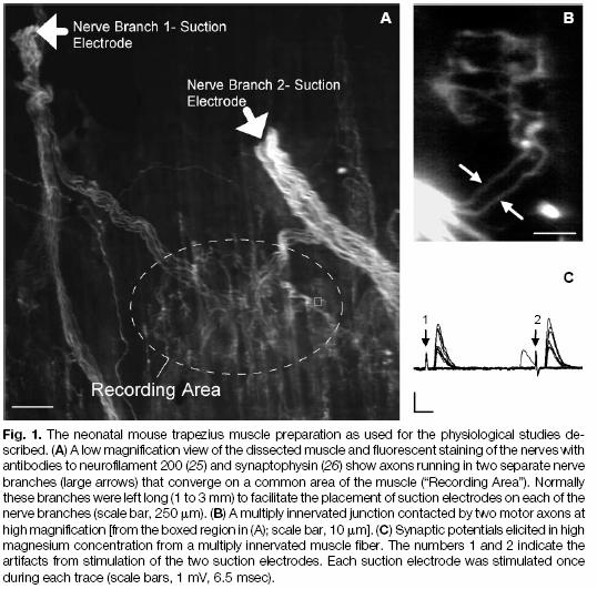
7 Figure 1
, but over time there is an increase in disparity in quantal contents of inputs co-innervating a")
8 Quantal Content Ratios as indicators of synaptic strength Synaptic strength is the ratio of the quantal content of one input vs. the other input Fig 2A shows how initially synaptic strengths were similar (quantal content ratio <2), but over time there is an increase in disparity in quantal contents of inputs co-innervating a muscle fiber. Fig 2B ( black bars indicate q.c.r of < 2, white bars q.c.r. of 2 to 4, gray bars q.c.r. of >4) demonstrates that over time there is a shift in the percentage of multiply innervated junctions with low quantal content ratios towards quantal content ratios of more than 4.

9 Quantal content disparity predicts single innervation Fig.2C shows the comparison of the synaptic strengths of multiply innervated fibers with the synaptic strength of an input of a singly innervated fiber on the same muscle. For each pair of axonal innervations converging at a NMJ there is a weaker and a stronger input. For the case were the quantal content ratio with respect to each other is <2, when the stronger s quantal content is taken in proportion to the quantal content of the singly innervated you get black bar with <2. In conclusion, the stronger input from the pair with a quantal content ratio >4 have a synaptic strength similar to that of a singly innervated NMJ. Therefore, fibers with the widest disparities of quantal content ratios are the direct predecessors to singly innervated muscle fibers. Fig.2D Increasing disparity in quantal content of the inputs co-innervating a muscle fiber is due to both an absolute increase in the quantal content of the stronger input and the decrease of quantal content of the weaker input.

10 Changes in Quantal Efficacy Under the conditions of high magnesium concentrations, depolarization induced by individual quanta (quantal efficacy) were observed. Fig.3A G shows that one input gives rise to a significantly smaller quantal responses than the other input. A - response to nerve stimulation B - amplitude of synaptic response in color. C traces ranked by amp. largest EPP amp. on top. D ranked traces in C represented in height and color. E evoked quantal responses arranged in order of amp. Quantal content of axon 1 is 0.23 and quantal content of axon 2 is F- axon 1 shows a gradual decrement in amp. without an obvious cut-off between smallest evoked events and failures. G axon 2 shows a distinct cut-off between smallest evoked potentials and failures.

11 H axon 1 has a quantal content of 0.31 axon 2 has a quantal content of I shows a gradual decline for axon 1; the largest EPPs from axon 1 are comparable to the weakest EPPs from axon2. J show a distinct cut-off between smallest potentials and failures for axon 2 K shows that the larger MEPP falls within the distribution of single quantal responses from axon 2 and that the smaller MEPP falls within the distribution of EPPs from axon 1. These MEPPs are spontaneous. MEPP miniature end-plate potential
 of both the weak and strong inputs.")
12 Quantal efficacy measured by amplitude of MEPPs Spontaneous MEPPs were assayed by adjusting the strength of suction electrode to stimulate one input repetitively (10 Hz) to cause an increase in MEPP frequency. Fig.4, A-C, show a muscle fiber from a 9-day old mouse with one strong and one weak input. A response (EPPs) of both the weak and strong inputs. B EPP of the larger input; C EPP of the weaker input. D When both inputs were stimulated together repetitively, large (open arrow) and small (filled arrow) MEPPs were seen after each EPP. E Repetitive stimulation of large EPP alone was followed by large MEPPs. F Repetitive stimulation of small EPP alone was followed by small MEPP. G the distribution of MEPP amplitudes after stimulation of both inputs. H distribution of MEPP after stimulation of large input I distribution of MEPPs after stimulation of weaker input had a lower mean than (H).
13 How is quantal efficacy during synapse elimination explained? Small MEPPs are also associated with weaker inputs in multiply innervated cells. To determine whether changes in quantal efficacy could be explained by a reduction in the amount of acetylcholine molecules per vesicle, repetitive stimulation under the presence of hemicholinium-3 (depletes synaptic vesicles of acetylcholine) were done. Stimulation in the presence of hemicholinium-3 and high magnesium decreased the amplitude of evoked quanta to a size similar to that observed for the small quantal events in multiply innervated muscles fibers, and the median rise times of the small events were as fast or faster than before. Since the small events observed during synapse elimination are not mimicked by using hemicholinium 3 they turned to decreases in the density of AChRs in the areas of the postsynaptic membrane. Given that a low density of AChRs could account for small quantal responses, area with low density of receptor density were identified in NMJs and overlying nerve terminals were studied for signs of synaptic vesicle at these sites.

14 Synaptic vesicles proteins were found overlying some areas of decreased receptor density (above). This supports the idea that small quantal responses observed are explained by a reduction in postsynaptic sensitivity. Synaptophysin is an integral membrane protein in synaptic vesicles.

15 Once an input is four times stronger than the other, axon withdrawal of the weaker input would appear imminent
16 Conclusions The rapid nature of the changes in synaptic strength may be related to the activity dependence of synaptic competition. The ability of an axon to induce elimination of its competitor is related to its synaptic efficacy. The skewing observed in the experiments in quantal content and efficacy would be expected to cause further changes in synaptic strength by inducing the elimination of synaptic sites and progressively tipping the competitive balance in favor of one axon over another.
Quantal Analysis Problems
 Quantal Analysis Problems 1. Imagine you had performed an experiment on a muscle preparation from a Drosophila larva. In this experiment, intracellular recordings were made from an identified muscle fibre,
Activity Dependent Changes At the Developing Neuromuscular Junction
 Activity Dependent Changes At the Developing Neuromuscular Junction (slides 16, 17 and 18 have been slightly modified for clarity) MCP Lecture 2-3 9.013/7.68 04 Neuromuscular Junction Development 1. Muscle
CHAPTER 44: Neurons and Nervous Systems
 CHAPTER 44: Neurons and Nervous Systems 1. What are the three different types of neurons and what are their functions? a. b. c. 2. Label and list the function of each part of the neuron. 3. How does the
Cellular Bioelectricity
 ELEC ENG 3BB3: Cellular Bioelectricity Notes for Lecture 22 Friday, February 28, 2014 10. THE NEUROMUSCULAR JUNCTION We will look at: Structure of the neuromuscular junction Evidence for the quantal nature
Neuromuscular Junction Testing ELBA Y. GERENA MALDONADO, MD ACTING ASSISTANT PROFESSOR UNIVERSITY OF WASHINGTON MEDICAL CENTER
 Neuromuscular Junction Testing ELBA Y. GERENA MALDONADO, MD ACTING ASSISTANT PROFESSOR UNIVERSITY OF WASHINGTON MEDICAL CENTER Objectives Neurophysiology Electrodiagnostic Evaluation Clinical Application
NEURONS COMMUNICATE WITH OTHER CELLS AT SYNAPSES 34.3
 NEURONS COMMUNICATE WITH OTHER CELLS AT SYNAPSES 34.3 NEURONS COMMUNICATE WITH OTHER CELLS AT SYNAPSES Neurons communicate with other neurons or target cells at synapses. Chemical synapse: a very narrow
Synaptic Transmission
 Synaptic Transmission Postsynaptic Mechanisms Synapses electrical and chemical Part I Neurotransmitters categories and life cycle Neurotransmitters examples and postsynaptic effects Pathology Part II Neurotransmitter
BIPN100 F15 Human Physiology 1 Lecture 3. Synaptic Transmission p. 1
 BIPN100 F15 Human Physiology 1 Lecture 3. Synaptic Transmission p. 1 Terms you should know: synapse, neuromuscular junction (NMJ), pre-synaptic, post-synaptic, synaptic cleft, acetylcholine (ACh), acetylcholine
Neuroscience 201A (2016) - Problems in Synaptic Physiology
 - Problems in Synaptic Physiology") Question 1: The record below in A shows an EPSC recorded from a cerebellar granule cell following stimulation (at the gap in the record) of a mossy fiber input. These responses are, then, evoked by stimulation.
Question 1: The record below in A shows an EPSC recorded from a cerebellar granule cell following stimulation (at the gap in the record) of a mossy fiber input. These responses are, then, evoked by stimulation.
ANSC (FSTC) 607 Physiology and Biochemistry of Muscle as a Food MOTOR INNERVATION AND MUSCLE CONTRACTION
 607 Physiology and Biochemistry of Muscle as a Food MOTOR INNERVATION AND MUSCLE CONTRACTION") ANSC (FSTC) 607 Physiology and Biochemistry of Muscle as a Food MOTOR INNERVATION AND MUSCLE CONTRACTION I. Motor innervation of muscle A. Motor neuron 1. Branched (can innervate many myofibers) à terminal
ANSC (FSTC) 607 Physiology and Biochemistry of Muscle as a Food MOTOR INNERVATION AND MUSCLE CONTRACTION I. Motor innervation of muscle A. Motor neuron 1. Branched (can innervate many myofibers) à terminal
Neuromuscular Transmission Diomedes E. Logothetis, Ph.D. (Dr. DeSimone s lecture notes revised) Learning Objectives:
 Learning Objectives:") Neuromuscular Transmission Diomedes E. Logothetis, Ph.D. (Dr. DeSimone s lecture notes revised) Learning Objectives: 1. Know the subunit composition of nicotinic ACh channels, general topology of the α
Neuromuscular Transmission Diomedes E. Logothetis, Ph.D. (Dr. DeSimone s lecture notes revised) Learning Objectives: 1. Know the subunit composition of nicotinic ACh channels, general topology of the α
Introduction to Neurobiology
 Biology 240 General Zoology Introduction to Neurobiology Nervous System functions: communication of information via nerve signals integration and processing of information control of physiological and
Homeostatic regulation of synaptic strength and the safety factor for neuromuscular transmission
 The Life Cycle of Neuromuscular Synapses Homeostatic regulation of synaptic strength and the safety factor for neuromuscular transmission 1. Synaptic transmission, safety factor and sizestrength relationships
Effects of adrenaline on nerve terminals in the superior cervical ganglion of the rabbit
 Br. J. Pharmac. (1971), 41, 331-338. Effects of adrenaline on nerve terminals in the superior cervical ganglion of the rabbit D. D. CHRIST AND S. NISHI Neurophysiology Laboratory, Department of Pharmacology,
Assessing neuromuscular transmission in mice with Huntington s disease
 Assessing neuromuscular transmission in mice with Huntington s disease Gabriela Garza-Vazquez Research Mentor: Dr. Andrew Voss Abstract: Huntington s disease (HD) is a degenerative genetic illness that
photometry on the extruded cytoplasm.
 Answers To Midterm 2011 Question 1. a) Isoproterenol. Used to dissect presynaptic and postsynaptic components of sympathetic modulation of neuromuscular junction (Orbelli effect). Specifically activates
Bernard Katz ( )
") Bernard Katz (1911-2003) German-born biophysicist and neurophysiologist. Studied medicine at the University of Leipzig, then fled to Britain in 1935. Worked at University College London (UCL) under Archibald
Bernard Katz (1911-2003) German-born biophysicist and neurophysiologist. Studied medicine at the University of Leipzig, then fled to Britain in 1935. Worked at University College London (UCL) under Archibald
BIONB/BME/ECE 4910 Neuronal Simulation Assignments 1, Spring 2013
 BIONB/BME/ECE 4910 Neuronal Simulation Assignments 1, Spring 2013 Tutorial Assignment Page Due Date Week 1/Assignment 1: Introduction to NIA 1 January 28 The Membrane Tutorial 9 Week 2/Assignment 2: Passive
Lecture 22: A little Neurobiology
 BIO 5099: Molecular Biology for Computer Scientists (et al) Lecture 22: A little Neurobiology http://compbio.uchsc.edu/hunter/bio5099 Larry.Hunter@uchsc.edu Nervous system development Part of the ectoderm
Chapter 8 11/1/2012. Synaptic Components are Ancient. Syncytium or Synapses? Synapse Formation and Function. Early Calcium Spikes
 Chapter 8 Synaptic Components are Ancient Synapse Formation and Function Fig 8.1 Syncytium or Synapses? Electrical Development Synapses Improve in Function with Time Fig 8.2 Fig 8.3 Early Calcium Spikes
B C. i) At what region(s) on the drawing above would you expect a high density of voltage-gated sodium channels?
 At what region(s) on the drawing above would you expect a high density of voltage-gated sodium channels?") MIT Department of Biology 7.013: Introductory Biology - Spring 2005 Instructors: Professor Hazel Sive, Professor Tyler Jacks, Dr. Claudette Gardel 7.013 SECTION NEUROBIOLOGY 2 Part A ligand-gated sodium
MIT Department of Biology 7.013: Introductory Biology - Spring 2005 Instructors: Professor Hazel Sive, Professor Tyler Jacks, Dr. Claudette Gardel 7.013 SECTION NEUROBIOLOGY 2 Part A ligand-gated sodium
Anatomy of a Neuron. Copyright 2000 by BSCS and Videodiscovery, Inc. Permission granted for classroom use. Master 2.1
 Anatomy of a Neuron Master 2.1 Neurons Interact With Other Neurons Through Synapses Master 2.2 How Do Neurons Communicate? 1 2 3 4 5 6 Master 2.3 Neurons Communicate by Neurotransmission Neurons communicate
MCB MIDTERM EXAM #1 MONDAY MARCH 3, 2008 ANSWER KEY
 MCB 160 - MIDTERM EXAM #1 MONDAY MARCH 3, 2008 ANSWER KEY Name ID# Instructions: -Only tests written in pen will be regarded -Please submit a written request indicating where and why you deserve more points
Anatomy Review. Graphics are used with permission of: Pearson Education Inc., publishing as Benjamin Cummings (
 Anatomy Review Graphics are used with permission of: Pearson Education Inc., publishing as Benjamin Cummings (http://www.aw-bc.com) Page 1. Introduction Neurons communicate with other cells at junctions
Concept 48.1 Neuron organization and structure reflect function in information transfer
 Name Chapter 48: Neurons, Synapses, and Signaling Period Chapter 48: Neurons, Synapses, and Signaling Concept 48.1 Neuron organization and structure reflect function in information transfer 1. What is
Chapter 45: Synapses Transmission of Nerve Impulses Between Neurons. Chad Smurthwaite & Jordan Shellmire
 Chapter 45: Synapses Transmission of Nerve Impulses Between Neurons Chad Smurthwaite & Jordan Shellmire The Chemical Synapse The most common type of synapse used for signal transmission in the central
Medicine, University of Lund, Sweden
 336 J. Phy8iol. (1961), 156, pp. 336-343 With 6 text-ftgures Printed in Great Britain AN ELECTROPHYSIOLOGIC STUDY OF THE NEURO- MUSCULAR JUNCTION IN MYASTHENIA GRAVIS BY 0. DAHLBACK, D. ELMQVIST, T. R.
What effect would an AChE inhibitor have at the neuromuscular junction?
 CASE 4 A 32-year-old woman presents to her primary care physician s office with difficulty chewing food. She states that when she eats certain foods that require a significant amount of chewing (meat),
Portions from Chapter 6 CHAPTER 7. The Nervous System: Neurons and Synapses. Chapter 7 Outline. and Supporting Cells
 CHAPTER 7 The Nervous System: Neurons and Synapses Chapter 7 Outline Neurons and Supporting Cells Activity in Axons The Synapse Acetylcholine as a Neurotransmitter Monoamines as Neurotransmitters Other
Synaptic transmission
 Outline Synaptic transmission Sompol Tapechum M.D., Ph.D. Department of Physiology Faculty of Medicine Siriraj Hospital, Bangkok, Thailand. sisth@mahidol.ac.th 2 Structure of synapse Modes of synaptic
Synapse Formation. Steven McLoon Department of Neuroscience University of Minnesota
 Synapse Formation Steven McLoon Department of Neuroscience University of Minnesota 1 Course News Midterm Exam Monday, Nov 13 9:30-11:30am Bring a #2 pencil!! 2 Course News Lecture schedule: Mon (Oct 31)
Fundamentals of the Nervous System and Nervous Tissue: Part C
 PowerPoint Lecture Slides prepared by Janice Meeking, Mount Royal College C H A P T E R 11 Fundamentals of the Nervous System and Nervous Tissue: Part C Warm Up What is a neurotransmitter? What is the
Chapter 11 Introduction to the Nervous System and Nervous Tissue Chapter Outline
 Chapter 11 Introduction to the Nervous System and Nervous Tissue Chapter Outline Module 11.1 Overview of the Nervous System (Figures 11.1-11.3) A. The nervous system controls our perception and experience
Synthesis. Storage. Physiology and Pathophysiology of Neuromuscular Transmission. Release. Action. Inactivation. Myasthenia Gravis Before
 Synthesis Physiology and Pathophysiology of Neuromuscular Transmission Storage Release Action Inactivation Myasthenia gravis and LEMS are autoimmune diseases Myasthenia Gravis Before LEMS: Ca channel antibodies
Nervous System. Master controlling and communicating system of the body. Secrete chemicals called neurotransmitters
 Nervous System Master controlling and communicating system of the body Interacts with the endocrine system to control and coordinate the body s responses to changes in its environment, as well as growth,
Function of the Nervous System
 Nervous System Function of the Nervous System Receive sensory information, interpret it, and send out appropriate commands to form a response Composed of neurons (functional unit of the nervous system)
Neurons, Synapses and Signaling. Chapter 48
 Neurons, Synapses and Signaling Chapter 48 Warm Up Exercise What types of cells can receive a nerve signal? Nervous Organization Neurons- nerve cells. Brain- organized into clusters of neurons, called
Biol 219 Lec 12 Fall 2016
 Cell-to-Cell: Neurons Communicate at Synapses Electrical synapses pass electrical signals through gap junctions Signal can be bi-directional Synchronizes the activity of a network of cells Primarily in
Chapter 11: Functional Organization of Nervous Tissue
 Chapter 11: Functional Organization of Nervous Tissue I. Functions of the Nervous System A. List and describe the five major nervous system functions: 1. 2. 3. 4. 5. II. Divisions of the Nervous System
5-Nervous system II: Physiology of Neurons
 5-Nervous system II: Physiology of Neurons AXON ION GRADIENTS ACTION POTENTIAL (axon conduction) GRADED POTENTIAL (cell-cell communication at synapse) SYNAPSE STRUCTURE & FUNCTION NEURAL INTEGRATION CNS
Synaptic Transmission
 Synaptic Transmission Graphics are used with permission of: Pearson Education Inc., publishing as Benjamin Cummings (http://www.aw-bc.com) Page 1. Introduction Synaptic transmission involves the release
STRUCTURAL ELEMENTS OF THE NERVOUS SYSTEM
 STRUCTURAL ELEMENTS OF THE NERVOUS SYSTEM STRUCTURE AND MAINTENANCE OF NEURONS (a) (b) Dendrites Cell body Initial segment collateral terminals (a) Diagrammatic representation of a neuron. The break in
The Nervous System. Nervous System Functions 1. gather sensory input 2. integration- process and interpret sensory input 3. cause motor output
 The Nervous System Nervous System Functions 1. gather sensory input 2. integration- process and interpret sensory input 3. cause motor output The Nervous System 2 Parts of the Nervous System 1. central
Chapter 9 Refinement of Synaptic Connections
 Chapter 9 Refinement of Synaptic Connections Afferent Projection Error during Development During development there is a constant rearrangement of synaptic connections, new synapses are formed and old synapses
Neurons, Synapses, and Signaling
 Neurons, Synapses, and Signaling The Neuron is the functional unit of the nervous system. Neurons are composed of a cell body, which contains the nucleus and organelles; Dendrites which are extensions
Version A. AP* Biology: Nervous System. Questions 1 and 2. Name: Period
 Name: Period Version A AP* Biology: Nervous System Directions: Each of the questions or incomplete statements below is followed by four suggested answers or completions. Select the one that is best in
10.1: Introduction. Cell types in neural tissue: Neurons Neuroglial cells (also known as neuroglia, glia, and glial cells) Dendrites.
 Dendrites.") 10.1: Introduction Copyright The McGraw-Hill Companies, Inc. Permission required for reproduction or display. Cell types in neural tissue: Neurons Neuroglial cells (also known as neuroglia, glia, and glial
10.1: Introduction Copyright The McGraw-Hill Companies, Inc. Permission required for reproduction or display. Cell types in neural tissue: Neurons Neuroglial cells (also known as neuroglia, glia, and glial
All questions below pertain to mandatory material: all slides, and mandatory homework (if any).
.") ECOL 182 Spring 2008 Dr. Ferriere s lectures Lecture 6: Nervous system and brain Quiz Book reference: LIFE-The Science of Biology, 8 th Edition. http://bcs.whfreeman.com/thelifewire8e/ All questions below
ECOL 182 Spring 2008 Dr. Ferriere s lectures Lecture 6: Nervous system and brain Quiz Book reference: LIFE-The Science of Biology, 8 th Edition. http://bcs.whfreeman.com/thelifewire8e/ All questions below
Branches of the Nervous System
 The Nervous System Branches of the Nervous System There are 2 main branches of the nervous system Central Nervous System Brain Spinal Cord Peripheral Nervous System All nerves leading to rest of body Anatomy
Synapses and Neurotransmitters
 Synapses and Neurotransmitters Action Potentials We have been talking about action potentials and how they allow an electrical impulse to travel from the dendrites to the end plates of a neuron. These
Chapter 9. Nervous System
 Chapter 9 Nervous System Central Nervous System (CNS) vs. Peripheral Nervous System(PNS) CNS Brain Spinal cord PNS Peripheral nerves connecting CNS to the body Cranial nerves Spinal nerves Neurons transmit
Thursday, January 22, Nerve impulse
 Nerve impulse Transmembrane Potential caused by ions moving through cell membrane at different rates Two main ions of concern Na + - Sodium K + - potassium Cell membrane not freely permeable therefore
Synaptic Transmission
 5 Synaptic Transmission Overview THE HUMAN BRAIN CONTAINS AT LEAST 1 billion neurons, each with the ability to influence many other cells. Clearly, sophisticated and highly efficient mechanisms are needed
Communication within a Neuron
 Neuronal Communication, Ph.D. Communication within a Neuron Measuring Electrical Potentials of Axons The Membrane Potential The Action Potential Conduction of the Action Potential 1 The withdrawal reflex
Spatial Distribution of Calcium Entry Evoked by Single Action Potentials within the Presynaptic Active Zone
 Spatial Distribution of Calcium Entry Evoked by Single Action Potentials within the Presynaptic Active Zone Elliot S. Wachman,, Robert E. Poage,, Joel R. Stiles, Daniel L. Farkas,, and Stephen D. Meriney
SUPPLEMENTARY INFORMATION
 Supplementary Figure 1. Normal AMPAR-mediated fepsp input-output curve in CA3-Psen cdko mice. Input-output curves, which are plotted initial slopes of the evoked fepsp as function of the amplitude of the
MOLECULAR AND CELLULAR NEUROSCIENCE
 MOLECULAR AND CELLULAR NEUROSCIENCE BMP-218 November 4, 2014 DIVISIONS OF THE NERVOUS SYSTEM The nervous system is composed of two primary divisions: 1. CNS - Central Nervous System (Brain + Spinal Cord)
Nervous System. 2. Receives information from the environment from CNS to organs and glands. 1. Relays messages, processes info, analyzes data
 Nervous System 1. Relays messages, processes info, analyzes data 2. Receives information from the environment from CNS to organs and glands 3. Transmits impulses from CNS to muscles and glands 4. Transmits
AP Biology Unit 6. The Nervous System
 AP Biology Unit 6 The Nervous System Branches of the Nervous System There are 2 main branches of the nervous system Central Nervous System Brain Spinal Cord Peripheral Nervous System All nerves leading
number Done by Corrected by Doctor
 number 13 Done by Tamara Wahbeh Corrected by Doctor Omar Shaheen In this sheet the following concepts will be covered: 1. Divisions of the nervous system 2. Anatomy of the ANS. 3. ANS innervations. 4.
Membrane Potentials. (And Neuromuscular Junctions)
") Membrane Potentials (And Neuromuscular Junctions) Skeletal Muscles Irritability & contractility Motor neurons & motor units Muscle cells have two important and unique properties: They are irritable and
Membrane Potentials (And Neuromuscular Junctions) Skeletal Muscles Irritability & contractility Motor neurons & motor units Muscle cells have two important and unique properties: They are irritable and
SYNAPTIC TRANSMISSION 1
 SYNAPTIC TRANSMISSION 1 I. OVERVIEW A. In order to pass and process information and mediate responses cells communicate with other cells. These notes examine the two means whereby excitable cells can rapidly
Chapter 4 Neuronal Physiology
 Chapter 4 Neuronal Physiology V edit. Pg. 99-131 VI edit. Pg. 85-113 VII edit. Pg. 87-113 Input Zone Dendrites and Cell body Nucleus Trigger Zone Axon hillock Conducting Zone Axon (may be from 1mm to more
SYNAPTIC COMMUNICATION
 BASICS OF NEUROBIOLOGY SYNAPTIC COMMUNICATION ZSOLT LIPOSITS 1 NERVE ENDINGS II. Interneuronal communication 2 INTERNEURONAL COMMUNICATION I. ELECTRONIC SYNAPSE GAP JUNCTION II. CHEMICAL SYNAPSE SYNAPSES
PMT. Explain the importance of reflex actions (3) Page 1 of 19
 Page 1 of 19") Q1. When a finger accidentally touches a hot object, a reflex action occurs. The biceps muscle contracts, causing the arm to be flexed and the finger is pulled away. The diagram shows the arrangement of
Q1. When a finger accidentally touches a hot object, a reflex action occurs. The biceps muscle contracts, causing the arm to be flexed and the finger is pulled away. The diagram shows the arrangement of
Chapter 11: Nervous System and Nervous Tissue
 Chapter 11: Nervous System and Nervous Tissue I. Functions and divisions of the nervous system A. Sensory input: monitor changes in internal and external environment B. Integrations: make decisions about
Cellular Neurobiology BIPN140
 Cellular Neurobiology BIPN140 1st Midterm Exam Ready for Pickup By the elevator on the 3 rd Floor of Pacific Hall (waiver) Exam Depot Window at the north entrance to Pacific Hall (no waiver) Mon-Fri, 10:00
NEUROMUSCULAR BLOCKING AGENTS
 NEUROMUSCULAR BLOCKING AGENTS Edward JN Ishac, Ph.D. Associate Professor, Pharmacology and Toxicology Smith 742, 828-2127, Email: eishac@vcu.edu Learning Objectives: 1. Understand the physiology of the
Chapter 3 subtitles Action potentials
 CELLULAR NEUROPHYSIOLOGY CONSTANCE HAMMOND Chapter 3 subtitles Action potentials Introduction (3:15) This third chapter explains the calcium current triggered by the arrival of the action potential in
DOI: /jphysiol The Physiological Society Rapid Report
 (2002), 545.2, pp. 337 343 DOI: 10.1113/jphysiol.2002.032516 The Physiological Society 2002 www.jphysiol.org Rapid Report Regulation by Rab3A of an endogenous modulator of neurotransmitter release at mouse
1) Drop off in the Bi 150 box outside Baxter 331 or to the head TA (jcolas).
 Drop off in the Bi 150 box outside Baxter 331 or to the head TA (jcolas).") Bi/CNS/NB 150 Problem Set 3 Due: Tuesday, Oct. 27, at 4:30 pm Instructions: 1) Drop off in the Bi 150 box outside Baxter 331 or e-mail to the head TA (jcolas). 2) Submit with this cover page. 3) Use a
Bi/CNS/NB 150 Problem Set 3 Due: Tuesday, Oct. 27, at 4:30 pm Instructions: 1) Drop off in the Bi 150 box outside Baxter 331 or e-mail to the head TA (jcolas). 2) Submit with this cover page. 3) Use a
Outline. Neuron Structure. Week 4 - Nervous System. The Nervous System: Neurons and Synapses
 Outline Week 4 - The Nervous System: Neurons and Synapses Neurons Neuron structures Types of neurons Electrical activity of neurons Depolarization, repolarization, hyperpolarization Synapses Release of
1) Drop off in the Bi 150 box outside Baxter 331 or to the head TA (jcolas).
 Drop off in the Bi 150 box outside Baxter 331 or to the head TA (jcolas).") Bi/CNS/NB 150 Problem Set 3 Due: Tuesday, Oct. 27, at 4:30 pm Instructions: 1) Drop off in the Bi 150 box outside Baxter 331 or e-mail to the head TA (jcolas). 2) Submit with this cover page. 3) Use a
Bi/CNS/NB 150 Problem Set 3 Due: Tuesday, Oct. 27, at 4:30 pm Instructions: 1) Drop off in the Bi 150 box outside Baxter 331 or e-mail to the head TA (jcolas). 2) Submit with this cover page. 3) Use a
Chapter 2: Cellular Mechanisms and Cognition
 Chapter 2: Cellular Mechanisms and Cognition MULTIPLE CHOICE 1. Two principles about neurons were defined by Ramón y Cajal. The principle of connectional specificity states that, whereas the principle
EE 791 Lecture 2 Jan 19, 2015
 EE 791 Lecture 2 Jan 19, 2015 Action Potential Conduction And Neural Organization EE 791-Lecture 2 1 Core-conductor model: In the core-conductor model we approximate an axon or a segment of a dendrite
Organismic Biology Bio 207. Lecture 6. Muscle and movement; sliding filaments; E-C coupling; length-tension relationships; biomechanics. Prof.
 Organismic Biology Bio 207 Lecture 6 Muscle and movement; sliding filaments; E-C coupling; length-tension relationships; biomechanics Prof. Simchon Today s Agenda Skeletal muscle Neuro Muscular Junction
BASICS OF NEUROBIOLOGY NERVE ENDINGS ZSOLT LIPOSITS
 BASICS OF NEUROBIOLOGY NERVE ENDINGS ZSOLT LIPOSITS 1 11. előadás. Prof. Liposits Zsolt NERVE ENDINGS I. Effectors and receptors 2 NERVE ENDINGS NEURONS COMMUNICATE WITH NON-NEURONAL ELEMENTS VIA SPECIALIZED
Study Guide Answer Key Nervous System
 Biology 12 Human Biology Textbook: BC Biology 12 Study Guide Answer Key Nervous System 1. Draw a neuron, label 3 parts and give the function of those parts. Dendrite: carry signals to the cell body Cell
Functions of Nervous System Neuron Structure
 Chapter 10 Nervous System I Divisions of the Nervous System Cell Types of Neural Tissue neurons neuroglial cells Central Nervous System brain spinal cord Peripheral Nervous System nerves cranial nerves
The Nervous System. Dr. ZHANG Xiong Dept. of Physiology ZJU School of Medicine.
 The Nervous System Dr. ZHANG Xiong Dept. of Physiology ZJU School of Medicine Http://10.10.10.151 Part 1. Summary of the nervous system The Nervous System Central Nervous System Brain + Spinal Cord Peripheral
Section: Chapter 5: Multiple Choice. 1. The structure of synapses is best viewed with a(n):
:") Section: Chapter 5: Multiple Choice 1. The structure of synapses is best viewed with a(n): p.155 electron microscope. light microscope. confocal microscope. nissle-stained microscopic procedure. 2. Electron
Section: Chapter 5: Multiple Choice 1. The structure of synapses is best viewed with a(n): p.155 electron microscope. light microscope. confocal microscope. nissle-stained microscopic procedure. 2. Electron
Action potential. Definition: an all-or-none change in voltage that propagates itself down the axon
 Action potential Definition: an all-or-none change in voltage that propagates itself down the axon Action potential Definition: an all-or-none change in voltage that propagates itself down the axon Naturally
The action potential travels down both branches because each branch is a typical axon with voltage dependent Na + and K+ channels.
 BIO 360 - MIDTERM FALL 2018 This is an open book, open notes exam. PLEASE WRITE YOUR NAME ON EACH SHEET. Read each question carefully and answer as well as you can. Point values are shown at the beginning
Ameen Alsaras. Ameen Alsaras. Mohd.Khatatbeh
 9 Ameen Alsaras Ameen Alsaras Mohd.Khatatbeh Nerve Cells (Neurons) *Remember: The neural cell consists of: 1-Cell body 2-Dendrites 3-Axon which ends as axon terminals. The conduction of impulse through
Chapter 10: Muscles. Vocabulary: aponeurosis, fatigue
 Chapter 10: Muscles 37. Describe the structural components of skeletal muscle tissue from the molecular to the organ level. 38. Describe the structure, function, and importance of sarcomeres. 39. Identify
NERVOUS SYSTEM 1 CHAPTER 10 BIO 211: ANATOMY & PHYSIOLOGY I
 BIO 211: ANATOMY & PHYSIOLOGY I 1 Ch 10 A Ch 10 B This set CHAPTER 10 NERVOUS SYSTEM 1 BASIC STRUCTURE and FUNCTION Dr. Lawrence G. Altman www.lawrencegaltman.com Some illustrations are courtesy of McGraw-Hill.
浙江大学医学院基础医学整合课程 各论 III. The Nervous System. Dr. ZHANG Xiong Dept. of Physiology ZJU School of Medicine
 The Nervous System Dr. ZHANG Xiong Dept. of Physiology ZJU School of Medicine xiongzhang@zju.edu.cn http://10.202.77.12/ 1 Part 1. Summary of the nervous system 2 The Nervous System Central Nervous System
Elizabeth Biopsychology (PSY 302) The Synapses 08/29/2017. The Synapses
 The Synapses 08/29/2017. The Synapses") Elizabeth Biopsychology (PSY 302) The Synapses 08/29/2017 The Synapses Conduction of a Depolarization o In dendrites: passive propagation : There is attenuation of signal transmission -Further away they
Elizabeth Biopsychology (PSY 302) The Synapses 08/29/2017 The Synapses Conduction of a Depolarization o In dendrites: passive propagation : There is attenuation of signal transmission -Further away they
Neurons: Structure and communication
 Neurons: Structure and communication http://faculty.washington.edu/chudler/gall1.html Common Components of a Neuron Dendrites Input, receives neurotransmitters Soma Processing, decision Axon Transmits
PSK4U THE NEUROMUSCULAR SYSTEM
 PSK4U THE NEUROMUSCULAR SYSTEM REVIEW Review of muscle so we can see how the neuromuscular system works This is not on today's note Skeletal Muscle Cell: Cellular System A) Excitation System Electrical
Chapter 3 Neurotransmitter release
 NEUROPHYSIOLOGIE CELLULAIRE CONSTANCE HAMMOND Chapter 3 Neurotransmitter release In chapter 3, we proose 3 videos: Observation Calcium Channel, Ca 2+ Unitary and Total Currents Ca 2+ and Neurotransmitter
Cardiac muscle is different from other types of muscle in that cardiac muscle
 6 E X E R C I S E Cardiovascular Physiology O B J E C T I V E S 1. To define autorhythmicity, sinoatrial node, pacemaker cells, and vagus nerves 2. To understand the effects of the sympathetic and parasympathetic
Neural Tissue. Chapter 12 Part B
 Neural Tissue Chapter 12 Part B CNS Tumors - Neurons stop dividing at age 4 but glial cells retain the capacity to divide. - Primary CNS tumors in adults- division of abnormal neuroglia rather than from
DO NOW: ANSWER ON PG 73
 DO NOW: ANSWER ON PG 73 1. Name 1 neurotransmitter that we have learned about. 2. Draw a basic graph of a neuron action potential. Label resting potential, threshold, depolarization, and repolarization
35-2 The Nervous System Slide 1 of 38
 1 of 38 35-2 The Nervous System The nervous system controls and coordinates functions throughout the body and responds to internal and external stimuli. 2 of 38 Neurons Neurons The messages carried by
Axon Nerve impulse. Axoplasm Receptor. Axomembrane Stimuli. Schwann cell Effector. Myelin Cell body
 Nervous System Review 1. Explain a reflex arc. 2. Know the structure, function and location of a sensory neuron, interneuron, and motor neuron 3. What is (a) Neuron Axon Nerve impulse Axoplasm Receptor
Neurons. Pyramidal neurons in mouse cerebral cortex expressing green fluorescent protein. The red staining indicates GABAergic interneurons.
 Neurons Pyramidal neurons in mouse cerebral cortex expressing green fluorescent protein. The red staining indicates GABAergic interneurons. MBL, Woods Hole R Cheung MSc Bioelectronics: PGEE11106 1 Neuron
transmission, it has similarly been proposed (see Fatt & Katz, 1952b; fashion, corresponding to the dropping out of whole miniature units
 553 J. Physiol. (I954) I24, 553-559 THE EFFECT OF MAGNESIUM ON THE ACTIVITY OF MOTOR NERVE ENDINGS By J. DEL CASTILLO AND B. KATZ From the Department of Biophysics, University College, London (Received
Baraa Ayed. Mohammad khatatbeh. 1 P a g e
 4 Baraa Ayed أسامة الخض Mohammad khatatbeh 1 P a g e Today we want to talk about these concepts: Excitation-Contraction coupling Smooth muscles (Generally speaking) Excitation-Contraction coupling Excitation-Contraction