IV. Cerebrovascular diseases
|
|
|
- Arabella Hodges
- 6 years ago
- Views:
Transcription
1 IV. Cerebrovascular diseases
2 - Cerebrovascular disease denotes brain disorders caused by pathologic processes involving the blood vessels. - The three main pathogenic mechanisms are: 1. Thrombotic occlusion of vessels 2. Embolic occlusion of vessels 3. Vascular rupture. - Thrombosis and embolism cause ischemic injury or infarction of specific regions of the brain, depending on the vessel involved.
3 - Hemorrhage accompanies rupture of vessels, leading to direct tissue damage as well as secondary ischemic injury Stroke : - Is the clinical designation applied to a. Abrupt onset of focal or global neurological symptoms b. caused by ischemia or hemorrhage c. and these symptoms must continue for more than 24 hours
4 d. and there should be permanent damage to the brain. Transient ischemic attack(tia): a- Applied that the neurological symptoms resolve within 24 hours b. No irreversible tissue damage c. The cause is small emboli from the carotids or vertebrobasilar circulation that resolve before causing irreversible injury Example Amarex fugax
5 - From the standpoint of the pathophysiology and pathologic anatomy, it is convenient to consider cerebrovascular disease as two processes a. Hypoxia, ischemia and infarction b. Hemorrhage
6 A. Hypoxia, ischemia and infarction - The brain may be deprived of oxygen by several mechanisms a. Functional hypoxia in a setting of a low partial pressure of oxygen or impaired oxygen-carrying capacity b. Global Ischemia; either transient or permanent due to tissue hypoperfusion, which can be caused by, hypotension or shock c. Vascular obstruction.
7 I. Global ischemia - Widespread ischemic injury can occur in the setting of severe systemic hypotension, when systolic pressure fall below 50 mm Hg such as in cardiac arrest, and shock because of failure of autoregulation - The clinical outcome varies with the severity and duration of the insult
8 A. Mild transient Global Ischemia - There may be only a transient postischemic confusional state, with eventual complete recovery. - Neurons are more susceptible to mild ischemic injury than glial cells - Neurons are the most susceptible followed by oligodendrocytes, astocytes then endothelial cells
9 - The most susceptible neurons to mild transient global ischemia are: 1. The pyramidal cells of the CA1 region of the hippocampus 2. Middle cortical lamina of the neocortex, layers 3, 5, and 6 (called laminar necrosis) 3. Purkinje cells of the cerebellum.
10 Note: - Neuronal loss in transient global ischemia is due to excitotoxicity - The susceptible neurons have many receptors to the excitatory neurotransmitter glutamate - So in transient global ischemia, the astrocytes release glutamate that binds to its neuronal receptors NMDA (N-methyl D-aspartate) leading to increase intracellular calcium and activation of enzymes that leads to death of these neurons
11 B. Severe global ischemia - Widespread neuronal death occurs irrespective of regional vulnerability.(pan necrosis) a. Persistent vegetative state ( awake but not aware) - In severe global cerebral ischemia, individuals who survive in this state often remain severely impaired neurologically. b. Brain death - Other patients meet the clinical criteria for "brain death," including:
12 1. Evidence of diffuse cortical injury (isoelectric, or "flat," electroencephalogram 2. And brain stem damage, including absent reflexes and respiratory drive.. Border zone ("watershed") infarcts - Are wedge-shaped areas of infarction that occur in those regions of the brain and spinal cord that lie at the most distal fields of arterial perfusion. "- In the cerebral hemispheres, the border zone between the anterior and the middle cerebral artery distributions is at greatest risk(double watershed area).
13 - Damage to this region produces a band of necrosis over the cerebral convexity a few centimeters lateral to the inter-hemispheric fissure. - Border zone infarcts are usually seen after hypotensive episodes..
14 2. Focal Cerebral Ischemia - Cerebral arterial occlusion leads to focal ischemia and-if sustained-to infarction of CNS tissue in the distribution of the compromised vessel. - The size, location, and shape of the infarct and the extent of tissue damage that results are determined by modifying variables, most importantly the adequacy of collateral flow - The major source of collateral flow is the circle of Willis.
15 - Partial collateralization is also provided over the surface of the brain through cortical-leptomeningeal anastomoses. NOTE - In contrast, there is little if any collateral flow for thalamus, basal ganglia, and deep white matter which are supplied by deep penetrating vessels.
16 - Occlusive vascular disease of severity sufficient to lead to cerebral infarction may be due to 1. In situ thrombosis - The majority of thrombotic occlusions causing cerebral infarctions are due to atherosclerosis - The most common sites of primary thrombosis are a. The carotid bifurcation, b. The origin of the middle cerebral artery, c. And at either end of the basilar artery
17 - The venous side of the circulation may also undergo thrombosis and cause significant cerebral ischemia - The striking example is the thrombosis of the superior sagittal sinus which can occur with infections or hypercoagulability state
18 2. Embolization from a distant source. - Overall, embolic infarctions are more common than thrombosis. - Sources of emboli: a. Cardiac mural thrombi are a frequent source b. Thromboemboli also arise in arteries, most often from atheromatous plaques within the carotid arteries. c. Paradoxical emboli, particularly in children with cardiac anomalies; d. Emboli of other materials (tumor, fat, or air).
19 NOTES - The territory of distribution of the middle cerebral artery-the direct extension of the internal carotid artery-is most frequently affected by embolic infarction; - Emboli tend to lodge where vessels branch or in areas of pre-existing luminal stenosis - Infarcts can be divided into two broad groups hemorrhagic and non-hemorrhagic based on their macroscopic and corresponding radiologic appearance
20 Microscopically, 1. After the first 12 hours: a. Red neurons and both cytotoxic and vasogenic edema predominate b. Swelling of endothelial and glial cells, mainly astrocytes d. Disintegration and myelinated fibers. 2. Up to 48 hours, there is some neutrophilic emigration
21 weeks a. Mononuclear phagocytic cells predominate and macrophages containing myelin breakdown products or blood may persist in the lesion for months to years. b. As the process of phagocytosis and liquefaction proceeds, ---there will be gemistocytic gliosis 4. After several months : - Fibrillary astrocytosis
22 Notes: a. In the cerebral cortex the cavity is delimited from the meninges and subarachnoid space by a gliotic layer of tissue, derived from the molecular layer of cortex. b- The pia and arachnoid are not affected and do not contribute to the healing process.
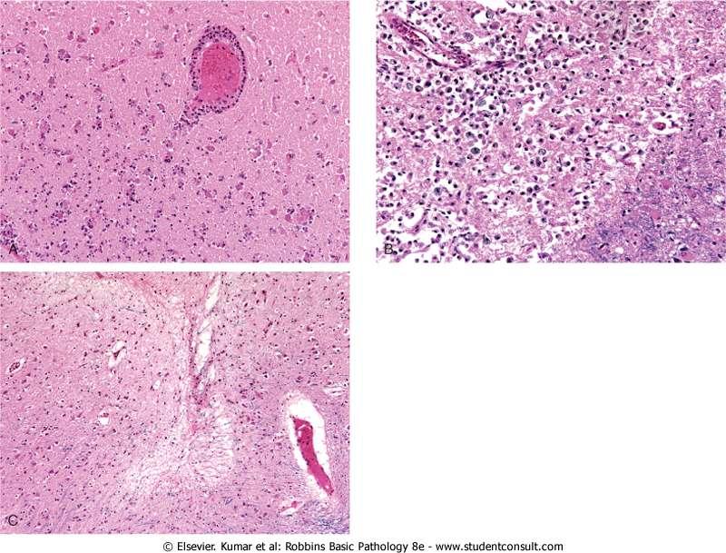
23 Cerebral infarction
24 B. Intracranial Hemorrhage - - Hemorrhage within the skull can occur in a variety of locations, and each location is associated with a set of underlying causes. 1. Hemorrhages within the brain itself can occur: a. Secondary to hypertension (most common) b. Cerebral amyloid angiopathy c. Arterio-venous malformation, d. A cavernous malformation, or e.. An intra-parenchymal tumor especially oligodendroglioma, glioblastoma or metastaic renal cell carcinoma and melanoma.
25 2. Subarachnoid hemorrhages: - Are most commonly seen with aneurysms but occur also with other vascular malformations. 3. Hemorrhages associated with the dura (in either subdural or epidural spaces) usually due to trauma.
26 1. Primary Brain Parenchymal Hemorrhage - Spontaneous (non-traumatic) intra-parenchymal hemorrhages occur most commonly in mid to late adult life, with a peak incidence at about 60 years of age - Most are caused by rupture of a small intraparenchymal vessel A. Hypertension : - Is the leading underlying cause, and brain hemorrhage accounts for roughly 15% of deaths among persons with chronic hypertension - It has a peak incidence at about 60 years
27 - Intracerebral hemorrhage can be clinically devastating when it affects large portions of the brain or extends into ventricular system or, it can affect small regions and be clinically silent. - Massive Hypertensive intraparenchymal hemorrhages occur in: a. Basal ganglia,--most common location b. Thalamus c. Pons d. and Cerebellum
28 Mechanisms of massive hemorrhage in Hypertension: 1. Hyaline arteriolar sclerosis - Affects the deep penetrating arteries and arterioles that supply the basal ganglia and the brain stem - Affected arteriolar walls are weakened and are more vulnerable to rupture 2. Chronic hypertension results in formation of minute aneurysms (Charcot-Bouchard microaneurysms ) - Form in vessels less than 300 μm in diameter.
29 Other CNS disorders caused by hypertension: 1. Lacunes or lacunar infarcts : - Small cavitary infarcts, just a few millimeters in size, and the main locations are: a. In basal ganglia and thalamus b. The internal capsule, and the pons. - Are caused by occlusion of a single penetrating vessel
30 2. Slit hemorrhages due to rupture of small penetrating vessels and with time, these hemorrhages resorb leaving slit-like cavities surrounded by black discoloration 3.. Acute hypertensive encephalopathy : - Most often is associated with sudden sustained rises in diastolic blood pressure to greater than 130 mm Hg and characterized :
31 a. By increased intracranial pressure and b Global cerebral dysfunction, manifesting as headaches, confusion, vomiting, convulsions, and sometimes coma. - Rapid therapeutic intervention to reduce the intracranial pressure is essential.
32 2. Cerebral Amyloid Angiopathy (CAA) : - Is the second most common cause of spontaneous parenchymal hemorrhage - Is a disease in which amyloidogenic peptides, deposit in the walls of meningeal and cortical vessels - Amyloid deposition (Aβ amyloid) weakens vessel walls and increases the risk of hemorrhages - The hemorrhage differs in distribution from those associated with hypertension
Cerebrovascular diseases-2
 Cerebrovascular diseases-2 Primary angiitis of CNS - Other causes of infarction i. Hypercoagulable states ii. Drug-abuse such as amphetamine, heroin and cocain Note - The venous side of the circulation
CEREBROVASCULAR DISEASES. By: Shifaa AlQa qa
 CEREBROVASCULAR DISEASES By: Shifaa AlQa qa Cerebrovascular diseases Brain disorders caused by pathologic processes involving blood vessels 3 pathogenic mechanisms (1) thrombotic occlusion, (2) embolic
CEREBROVASCULAR DISEASES. By: Shifaa AlQa qa
 CEREBROVASCULAR DISEASES By: Shifaa AlQa qa Cerebrovascular diseases Brain disorders caused by pathologic processes involving blood vessels 3 pathogenic mechanisms (1) thrombotic occlusion, (2) embolic
CNS pathology Third year medical students. Dr Heyam Awad 2018 Lecture 7: Non traumatic brain haemorrhage
 CNS pathology Third year medical students Dr Heyam Awad 2018 Lecture 7: Non traumatic brain haemorrhage ILOS To list the causes of intracranial haemorrhage. To understand the pathogenesis of each cause.
HYPERTENSIVE ENCEPHALOPATHY
 HYPERTENSIVE ENCEPHALOPATHY Reversible posterior leukoencephalopathy syndrome Cause Renal disease Pheochromocytoma Disseminated vasculitis Eclampsia Acute toxemia Medications & illicit drugs (cocaine)
CNS pathology Third year medical students. Dr Heyam Awad 2018 Lecture 5: disturbed fluid balance and increased intracranial pressure
 CNS pathology Third year medical students Dr Heyam Awad 2018 Lecture 5: disturbed fluid balance and increased intracranial pressure ILOs Understand causes and symptoms of increased intracranial pressure.
Cerebral Vascular Diseases. Nabila Hamdi MD, PhD
 Cerebral Vascular Diseases Nabila Hamdi MD, PhD Outline I. Stroke statistics II. Cerebral circulation III. Clinical symptoms of stroke IV. Pathogenesis of cerebral infarcts (Stroke) 1. Ischemic - Thrombotic
V. CENTRAL NERVOUS SYSTEM TRAUMA
 V. CENTRAL NERVOUS SYSTEM TRAUMA I. Concussion - Is a clinical syndrome of altered consiousness secondary to head injury - Brought by a change in the momentum of the head when a moving head suddenly arrested
Marc Norman, Ph.D. - Do Not Use without Permission 1. Cerebrovascular Accidents. Marc Norman, Ph.D. Department of Psychiatry
 Cerebrovascular Accidents Marc Norman, Ph.D. Department of Psychiatry Neuropsychiatry and Behavioral Medicine Neuropsychology Clinical Training Seminar 1 5 http://www.nlm.nih.gov/medlineplus/ency/images/ency/fullsize/18009.jpg
2. Subarachnoid Hemorrhage
 Causes: 2. Subarachnoid Hemorrhage A. Saccular (berry) aneurysm - Is the most frequent cause of clinically significant subarachnoid hemorrhage is rupture of a saccular (berry) aneurysm. B. Vascular malformation
Neuropathology lecture series. III. Neuropathology of Cerebrovascular Disease. Physiology of cerebral blood flow
 Neuropathology lecture series III. Neuropathology of Cerebrovascular Disease Physiology of cerebral blood flow Brain makes up only 2% of body weight Percentage of cardiac output: 15-20% Percentage of O
PTA 106 Unit 1 Lecture 3
 PTA 106 Unit 1 Lecture 3 The Basics Arteries: Carry blood away from the heart toward tissues. They typically have thicker vessels walls to handle increased pressure. Contain internal and external elastic
Cerebrovascular Disease
 Neuropathology lecture series Cerebrovascular Disease Physiology of cerebral blood flow Brain makes up only 2% of body weight Percentage of cardiac output: 15-20% Percentage of O 2 consumption (resting):
Blood Supply. Allen Chung, class of 2013
 Blood Supply Allen Chung, class of 2013 Objectives Understand the importance of the cerebral circulation. Understand stroke and the types of vascular problems that cause it. Understand ischemic penumbra
Overview of Stroke: Etiologies, Demographics, Syndromes, and Outcomes. Alex Abou-Chebl, MD, FSVIN Medical Director, Stroke Baptist Health Louisville
 Overview of Stroke: Etiologies, Demographics, Syndromes, and Outcomes Alex Abou-Chebl, MD, FSVIN Medical Director, Stroke Baptist Health Louisville Disclosure Statement of Financial Interest Within the
ISCHEMIC STROKE IMAGING
 ISCHEMIC STROKE IMAGING ผศ.พญ พญ.จ ร ร ตน ธรรมโรจน ภาคว ชาร งส ว ทยา คณะแพทยศาสตร มหาว ทยาล ยขอนแก น A case of acute hemiplegia Which side is the abnormality, right or left? Early Right MCA infarction
11/2/2016. Stroke. Carl F. McComas, M.D. November 3, Disclosures. None (of any kind)
") Stroke Carl F. McComas, M.D. November 3, 2016 None (of any kind) Disclosures 1 HYPERTENSION Stroke The seat of apoplexy seems to be within the same portion of the of the brain.... Both affects, the imagination,
Stroke Carl F. McComas, M.D. November 3, 2016 None (of any kind) Disclosures 1 HYPERTENSION Stroke The seat of apoplexy seems to be within the same portion of the of the brain.... Both affects, the imagination,
Essentials of Clinical MR, 2 nd edition. 14. Ischemia and Infarction II
 14. Ischemia and Infarction II Lacunar infarcts are small deep parenchymal lesions involving the basal ganglia, internal capsule, thalamus, and brainstem. The vascular supply of these areas includes the
CENTRAL NERVOUS SYSTEM TRAUMA and Subarachnoid Hemorrhage. By: Shifaa AlQa qa
 CENTRAL NERVOUS SYSTEM TRAUMA and Subarachnoid Hemorrhage By: Shifaa AlQa qa Subarachnoid Hemorrhage Causes: Rupture of a saccular (berry) aneurysm Vascular malformation Trauma Hematologic disturbances
Principles Arteries & Veins of the CNS LO14
 Principles Arteries & Veins of the CNS LO14 14. Identify (on cadaver specimens, models and diagrams) and name the principal arteries and veins of the CNS: Why is it important to understand blood supply
Cerebrovascular Disease
 Neuropathology lecture series Cerebrovascular Disease Kurenai Tanji, M.D., Ph.D. December 11, 2007 Physiology of cerebral blood flow Brain makes up only 2% of body weight Percentage of cardiac output:
CNS VASCULAR DISEASE. Reid R. Heffner, M.D. Department of Pathology/Anatomy UB Jacobs School of Medicine January 15, 2019
 CNS VASCULAR DISEASE Reid R. Heffner, M.D. Department of Pathology/Anatomy UB Jacobs School of Medicine January 15, 2019 I HAVE NO CONFLICTS OF INTEREST OR ANY DISCLOSURES TO DECLARE. I HAVE NO FINANCIAL
NEURO IMAGING 2. Dr. Said Huwaijah Chairman of radiology Dep, Damascus Univercity
 NEURO IMAGING 2 Dr. Said Huwaijah Chairman of radiology Dep, Damascus Univercity I. EPIDURAL HEMATOMA (EDH) LOCATION Seventy to seventy-five percent occur in temporoparietal region. CAUSE Most likely caused
What Are We Going to Do? Fourth Year Meds Clinical Neuroanatomy. Hydrocephalus and Effects of Interruption of CSF Flow. Tube Blockage Doctrine
 Fourth Year Meds Clinical Neuroanatomy Ventricles, CSF, Brain Swelling etc. David A. Ramsay, Neuropathologist, LHSC What Are We Going to Do? Hydrocephalus and some effects of the interruption of CSF flow
Cerebro-vascular stroke
 Cerebro-vascular stroke CT Terminology Hypodense lesion = lesion of lower density than the normal brain tissue Hyperdense lesion = lesion of higher density than normal brain tissue Isodense lesion = lesion
Vascular Cognitive Impairment-- NEUROPATHOLOGIC ISSUES. VCI vs. IVD/DEMENTIA with VASCULAR DISEASE (IVD) advanced pathology
 advanced pathology") Vascular Cognitive Impairment-- NEUROPATHOLOGIC ISSUES VCI vs. IVD/DEMENTIA with VASCULAR DISEASE (IVD) advanced pathology HANDLING the BRAIN at AUTOPSY: What to FIX vs. what to FREEZE? --no need to be
Vascular Cognitive Impairment-- NEUROPATHOLOGIC ISSUES VCI vs. IVD/DEMENTIA with VASCULAR DISEASE (IVD) advanced pathology HANDLING the BRAIN at AUTOPSY: What to FIX vs. what to FREEZE? --no need to be
Hemodynamic Disorders, Thrombosis, and Shock. Richard A. McPherson, M.D.
 Hemodynamic Disorders, Thrombosis, and Shock Richard A. McPherson, M.D. Edema The accumulation of abnormal amounts of fluid in intercellular spaces of body cavities. Inflammation and release of mediators
[(PHY-3a) Initials of MD reviewing films] [(PHY-3b) Initials of 2 nd opinion MD]
![[(PHY-3a) Initials of MD reviewing films] [(PHY-3b) Initials of 2 nd opinion MD]](/thumbs/89/98619893.jpg "[(PHY-3a) Initials of MD reviewing films] [(PHY-3b) Initials of 2 nd opinion MD]") 2015 PHYSICIAN SIGN-OFF (1) STUDY NO (PHY-1) CASE, PER PHYSICIAN REVIEW 1=yes 2=no [strictly meets case definition] (PHY-1a) CASE, IN PHYSICIAN S OPINION 1=yes 2=no (PHY-2) (PHY-3) [based on all available
2015 PHYSICIAN SIGN-OFF (1) STUDY NO (PHY-1) CASE, PER PHYSICIAN REVIEW 1=yes 2=no [strictly meets case definition] (PHY-1a) CASE, IN PHYSICIAN S OPINION 1=yes 2=no (PHY-2) (PHY-3) [based on all available
Classical CNS Disease Patterns
 Classical CNS Disease Patterns Inflammatory Traumatic In response to the trauma of having his head bashed in GM would have experienced some of these features. NOT TWO LITTLE PEENY WEENY I CM LACERATIONS.
Cerebrovascular Disorders. Blood, Brain, and Energy. Blood Supply to the Brain 2/14/11
 Cerebrovascular Disorders Blood, Brain, and Energy 20% of body s oxygen usage No oxygen/glucose reserves Hypoxia - reduced oxygen Anoxia - Absence of oxygen supply Cell death can occur in as little as
CNS pathology Third year medical students,2019. Dr Heyam Awad Lecture 2: Disturbed fluid balance and increased intracranial pressure
 CNS pathology Third year medical students,2019 Dr Heyam Awad Lecture 2: Disturbed fluid balance and increased intracranial pressure ILOs Understand causes and symptoms of increased intracranial pressure.
Traumatic brain injuries are caused by external mechanical forces such as: - Falls - Transport-related accidents - Assault
 PP2231 Brain injury Cerebrum consists of frontal, parietal, occipital and temporal lobes Diencephalon consists of thalamus, hypothalamus Cerbellum Brain stem consists of midbrain, pons, medulla Central
Neuroradiology: Imaging and Stroke
 Neuroradiology: Imaging and Stroke Stroke 2017 William Gallmann January 28, 2017 Stroke Arterial ischemia/infarct accounts for ~85% Cerebral venous occlusions - 0.5-1% Spontaneous intracranial hemorrhage
Characteristic features of CNS pathology. By: Shifaa AlQa qa
 Characteristic features of CNS pathology By: Shifaa AlQa qa Normal brain: - The neocortex (gray matter): six layers: outer plexiform, outer granular, outer pyramidal, inner granular, inner pyramidal, polymorphous
Acute stroke. Ischaemic stroke. Characteristics. Temporal classification. Clinical features. Interpretation of Emergency Head CT
 Ischaemic stroke Characteristics Stroke is the third most common cause of death in the UK, and the leading cause of disability. 80% of strokes are ischaemic Large vessel occlusive atheromatous disease
Ischemic heart disease
 Ischemic heart disease Introduction In > 90% of cases: the cause is: reduced coronary blood flow secondary to: obstructive atherosclerotic vascular disease so most of the time it is called: coronary artery
An Introduc+on to Stroke
 An Introduc+on to Stroke Elizabeth Huntoon MS, MD Assistant Professor Department of Physical Medicine and Rehabilita>on Vanderbilt University School of Medicine Defini+on Sudden focal neurologic deficit
An Introduction to Imaging the Brain. Dr Amy Davis
 An Introduction to Imaging the Brain Dr Amy Davis Common reasons for imaging: Clinical scenarios: - Trauma (NICE guidelines) - Stroke - Tumours - Seizure - Neurological degeneration memory, motor dysfunction,
M555 Medical Neuroscience Blood Flow in CNS Meninges Blood Brain Barrier CSF
 M555 Medical Neuroscience Blood Flow in CNS Meninges Blood Brain Barrier CSF Arterial Blood Flow to CNS approximately % of what goes wrong within the skull that produces neurological deficits is vascular
Head Trauma Inservice (October)
") John Tramell - Head Trauma Inservice, October 2005.doc Page 1 Head Trauma Inservice (October) Head trauma is the leading cause of death in trauma patients. Having a basic understanding of the anatomy and
John Tramell - Head Trauma Inservice, October 2005.doc Page 1 Head Trauma Inservice (October) Head trauma is the leading cause of death in trauma patients. Having a basic understanding of the anatomy and
OBJECTIVES. At the end of the lecture, students should be able to: List the cerebral arteries.
 DR JAMILA EL MEDANY OBJECTIVES At the end of the lecture, students should be able to: List the cerebral arteries. Describe the cerebral arterial supply regarding the origin, distribution and branches.
Enhancement of Cranial US: Utility of Supplementary Acoustic Windows and Doppler Harriet J. Paltiel, MD
 Enhancement of Cranial US: Utility of Supplementary Acoustic Windows and Doppler Harriet J. Paltiel, MD Boston Children s Hospital Harvard Medical School None Disclosures Conventional US Anterior fontanelle
UPSTATE Comprehensive Stroke Center. Neurosurgical Interventions Satish Krishnamurthy MD, MCh
 UPSTATE Comprehensive Stroke Center Neurosurgical Interventions Satish Krishnamurthy MD, MCh Regional cerebral blood flow is important Some essential facts Neurons are obligatory glucose users Under anerobic
NACC Vascular Consortium. NACC Vascular Consortium. NACC Vascular Consortium
 NACC Vascular Consortium NACC Vascular Consortium Participating centers: Oregon Health and Science University ADC Rush University ADC Mount Sinai School of Medicine ADC Boston University ADC In consultation
STROKE - IMAGING. Dr RAJASEKHAR REDDY 2nd Yr P.G. RADIODIAGNOSIS KIMS,Narkatpalli.
 STROKE - IMAGING Dr RAJASEKHAR REDDY 2nd Yr P.G. RADIODIAGNOSIS KIMS,Narkatpalli. STROKE Describes a clinical event that consists of sudden onset of neurological symptoms Types Infarction - occlusion of
Meninges and Ventricles
 Meninges and Ventricles Irene Yu, class of 2019 LEARNING OBJECTIVES Describe the meningeal layers, the dural infolds, and the spaces they create. Name the contents of the subarachnoid space. Describe the
The dura is sensitive to stretching, which produces the sensation of headache.
 Dural Nerve Supply Branches of the trigeminal, vagus, and first three cervical nerves and branches from the sympathetic system pass to the dura. Numerous sensory endings are in the dura. The dura is sensitive
The NIHSS score is 4 (considering 2 pts for the ataxia involving upper and lower limbs.
 Neuroscience case 5 1. Speech comprehension, ability to speak, and word use were normal in Mr. Washburn, indicating that aphasia (cortical language problem) was not involved. However, he did have a problem
CT INTERPRETATION COURSE
 CT INTERPRETATION COURSE Refresher Course ASTRACAT October 2012 Stroke is a Clinical Diagnosis A clinical syndrome characterised by rapidly developing clinical symptoms and/or signs of focal loss of cerebral
Traumatic Brain Injury TBI Presented by Bill Masten
 1 2 Cerebrum two hemispheres and four lobes. Cerebellum (little brain) coordinates the back and forth ballet of motion. It judges the timing of every movement precisely. Brainstem coordinates the bodies
Stroke/TIA. Tom Bedwell
 Stroke/TIA Tom Bedwell tab1g11@soton.ac.uk The Plan Definitions Anatomy Recap Aetiology Pathology Syndromes Brocas / Wernickes Investigations Management Prevention & Prognosis TIAs Key Definitions Transient
Head CT Scan Interpretation: A Five-Step Approach to Seeing Inside the Head Lawrence B. Stack, MD
 Head CT Scan Interpretation: A Five-Step Approach to Seeing Inside the Head Lawrence B. Stack, MD Five Step Approach 1. Adequate study 2. Bone windows 3. Ventricles 4. Quadrigeminal cistern 5. Parenchyma
Vascular Disorders. Nervous System Disorders (Part B-1) Module 8 -Chapter 14. Cerebrovascular disease S/S 1/9/2013
 Module 8 -Chapter 14. Cerebrovascular disease S/S 1/9/2013") Nervous System Disorders (Part B-1) Module 8 -Chapter 14 Overview ACUTE NEUROLOGIC DISORDERS Vascular Disorders Infections/Inflammation/Toxins Metabolic, Endocrinologic, Nutritional, Toxic Neoplastic Traumatic
Nervous System Disorders (Part B-1) Module 8 -Chapter 14 Overview ACUTE NEUROLOGIC DISORDERS Vascular Disorders Infections/Inflammation/Toxins Metabolic, Endocrinologic, Nutritional, Toxic Neoplastic Traumatic
WHITE PAPER: A GUIDE TO UNDERSTANDING SUBARACHNOID HEMORRHAGE
 WHITE PAPER: A GUIDE TO UNDERSTANDING SUBARACHNOID HEMORRHAGE Subarachnoid Hemorrhage is a serious, life-threatening type of hemorrhagic stroke caused by bleeding into the space surrounding the brain,
WHI Form Report of Cardiovascular Outcome Ver (For items 1-11, each question specifies mark one or mark all that apply.
 WHI Form - Report of Cardiovascular Outcome Ver. 6. COMMENTS To be completed by Physician Adjudicator Date Completed: - - (M/D/Y) Adjudicator Code: OMB# 095-044 Exp: 4/06 -Affix label here- Clinical Center/ID:
MCHENRY WESTERN LAKE COUNTY EMS SYSTEM Paramedic, EMT-B and PHRN Optional Continuing Education 2019 #7 Strokes
 MCHENRY WESTERN LAKE COUNTY EMS SYSTEM Paramedic, EMT-B and PHRN Optional Continuing Education 2019 #7 Strokes Stroke is the third leading cause of death and the leading cause of adult disability in the
Nicolas Bianchi M.D. May 15th, 2012
 Nicolas Bianchi M.D. May 15th, 2012 New concepts in TIA Differential Diagnosis Stroke Syndromes To learn the new definitions and concepts on TIA as a condition of high risk for stroke. To recognize the
Brain Injuries. Presented By Dr. Said Said Elshama
 Brain Injuries Presented By Dr. Said Said Elshama Types of head injuries 1- Scalp injuries 2- Skull injuries 3- Intra Cranial injuries ( Brain ) Anatomical structure of meninges Intra- Cranial Injuries
Module 3. The Blood Supply of the Brain
 Module 3. The Blood Supply of the Brain Relating Vascular and Functional Anatomy Objectives for Module 3 Knowledge! Describe or sketch the course of the major arteries and their branches that comprise
CMS Limitations Guide - Radiology Services
 CMS Limitations Guide - Radiology Services Starting October 1, 2015, CMS will update their existing medical necessity limitations on tests and procedures to correspond to ICD-10 codes. This limitations
Medical Review Guidelines Magnetic Resonance Angiography
 Medical Review Guidelines Magnetic Resonance Angiography Medical Guideline Number: MRG2001-05 Effective Date: 2/13/01 Revised Date: 2/14/2006 OHCA Reference OAC 317:30-5-24. Radiology. (f) Magnetic Resonance
Stroke 101. Maine Cardiovascular Health Summit. Eileen Hawkins, RN, MSN, CNRN Pen Bay Stroke Program Coordinator November 7, 2013
 Stroke 101 Maine Cardiovascular Health Summit Eileen Hawkins, RN, MSN, CNRN Pen Bay Stroke Program Coordinator November 7, 2013 Stroke Statistics Definition of stroke Risk factors Warning signs Treatment
The central nervous system
 Sectc.qxd 29/06/99 09:42 Page 81 Section C The central nervous system CNS haemorrhage Subarachnoid haemorrhage Cerebral infarction Brain atrophy Ring enhancing lesions MRI of the pituitary Multiple sclerosis
Brain Attack. Strategies in the Management of Acute Ischemic Stroke: Neuroscience Clerkship. Case Medical Center
 Brain Attack Strategies in the Management of Acute Ischemic Stroke: Neuroscience Clerkship Stroke is a common and devastating disorder Third leading antecedent of death in American men, and second among
Emergency Department Stroke Registry Indicator Specifications 2018 Report Year (07/01/2017 to 06/30/2018 Discharge Dates)
") 2018 Report Year (07/01/2017 to 06/30/2018 Discharge Dates) Summary of Changes I62.9 added to hemorrhagic stroke ICD-10-CM diagnosis code list (table 3) Measure Description Methodology Rationale Measurement
2018 Report Year (07/01/2017 to 06/30/2018 Discharge Dates) Summary of Changes I62.9 added to hemorrhagic stroke ICD-10-CM diagnosis code list (table 3) Measure Description Methodology Rationale Measurement
TOXIC AND NUTRITIONAL DISORDER MODULE
 TOXIC AND NUTRITIONAL DISORDER MODULE Objectives: For each of the following entities the student should be able to: 1. Describe the etiology/pathogenesis and/or pathophysiology, gross and microscopic morphology
THE ESSENTIAL BRAIN INJURY GUIDE
 THE ESSENTIAL BRAIN INJURY GUIDE Neuroanatomy & Neuroplasticity Section 2 Contributors Erin D. Bigler, PhD Michael R. Hoane, PhD Stephanie Kolakowsky-Hayner, PhD, CBIST, FACRM Dorothy A. Kozlowski, PhD
Supplemental Digital Content: Definitions Based on the International Classification of Diseases, Ninth Revision, Clinical Modification
 Supplemental Digital Content: Definitions Based on the International Classification of Diseases, Ninth Revision, Clinical Modification (ICD-9-CM) Diagnose and Procedures Codes 1. ICD-9-CM definition of
INDIANA HEALTH COVERAGE PROGRAMS
 INDIANA HEALTH COVERAGE PROGRAMS PROVIDER CODE TABLES Note: Due to possible changes in Indiana Health Coverage Programs (IHCP) policy or national coding updates, inclusion of a code on the code tables
Vascular Malformations of the Brain. William A. Cox, M.D. Forensic Pathologist/Neuropathologist. September 8, 2014
 Vascular Malformations of the Brain William A. Cox, M.D. Forensic Pathologist/Neuropathologist September 8, 2014 Vascular malformations of the brain are classified into four principal groups: arteriovenous
CEREBRO VASCULAR ACCIDENTS
 CEREBRO VASCULAR S MICHAEL OPONG-KUSI, DO MBA MORTON CLINIC, TULSA, OK, USA 8/9/2012 1 Cerebrovascular Accident Third Leading cause of deaths (USA) 750,000 strokes in USA per year. 150,000 deaths in USA
The tale of global hypoxic ischaemic injury
 The tale of global hypoxic ischaemic injury Poster No.: C-0400 Congress: ECR 2016 Type: Educational Exhibit Authors: L. M. Zammit, R. Grech ; Paola/MT, Dublin 9/IE Keywords: CNS, CT, MR, Education, Computer
HEART AND SOUL STUDY OUTCOME EVENT - MORBIDITY REVIEW FORM
 REVIEW DATE REVIEWER'S ID HEART AND SOUL STUDY OUTCOME EVENT - MORBIDITY REVIEW FORM : DISCHARGE DATE: RECORDS FROM: Hospitalization ER Please check all that may apply: Myocardial Infarction Pages 2, 3,
ATHEROSCLEROSIS. Secondary changes are found in other coats of the vessel wall.
 ATHEROSCLEROSIS Atherosclerosis Atherosclerosis is a disease process affecting the intima of the aorta and large and medium arteries, taking the form of focal thickening or plaques of fibrous tissue and
DOWNLOAD PDF OTHER CEREBROVASCULAR SYNDROMES
 Chapter 1 : ICD 10 Code for Other vascular syndromes of brain in cerebrovascular diseases G Other symptoms of cerebrovascular disease include migraines, seizures, epilepsy, or cognitive decline. However,
Cerebral small vessel disease
 Cerebral small vessel disease What is it? What are the clinical syndromes? How do we diagnose it? What is the pathophysiology? New insights from genetics? Possible therapies? Small Vessel disease Changes
Medical Neuroscience Tutorial Notes
 Medical Neuroscience Tutorial Notes Blood Supply to the Brain MAP TO NEUROSCIENCE CORE CONCEPTS 1 NCC1. The brain is the body's most complex organ. LEARNING OBJECTIVES After study of the assigned learning
Module 1. Introduction to Stroke and Stroke Prevention
 Module 1. Introduction to Stroke and Stroke Prevention Overview Objectives for Module 1 Knowledge! Compare and contrast ischemic stroke and transient ischemic attack (TIA)! Distinguish ischemic from hemorrhagic
For Emergency Doctors. Dr Suzanne Smallbane November 2011
 For Emergency Doctors Dr Suzanne Smallbane November 2011 A: Orbit B: Sphenoid Sinus C: Temporal Lobe D: EAC E: Mastoid air cells F: Cerebellar hemisphere A: Frontal lobe B: Frontal bone C: Dorsum sellae
Case 9511 Hypertensive microangiopathy
 Case 9511 Hypertensive microangiopathy Schepers S, Barthels C Section: Neuroradiology Published: 2011, Nov. 3 Patient: 67 year(s), male Authors' Institution Department of Radiology, Jessa ziekenhuis campus
ATHEROSCLEROSIS زيد ثامر جابر. Zaid. Th. Jaber
 ATHEROSCLEROSIS زيد ثامر جابر Zaid. Th. Jaber Objectives 1- Review the normal histological features of blood vessels walls. 2-define the atherosclerosis. 3- display the risk factors of atherosclerosis.
The CNS Part II pg
 The CNS Part II pg. 455-474 Protection of the Brain Objectives Describe how the meninges, cerebrospinal fluid, and the blood brain barrier protect the CNS. Explain how Cerebrospinal fluid is formed, and
Heat-Related Deaths in Hot Cities: Estimates of Human Tolerance to High Temperature Thresholds
 Supplementary Information Heat-Related Deaths in Hot Cities: Estimates of Human Tolerance to High Temperature Thresholds Selection of ICD-10 Codes Indicating Conditions and Diseases that Might Be Directly
Stroke: Every Minute Counts! Primary Stroke Center, Ingalls Memorial Hospital
 Stroke: Every Minute Counts! Primary Stroke Center, Ingalls Memorial Hospital Objectives Describe the A & P of the nervous system Outline pathophysiological changes in the nervous system that may alter
HEAD AND NECK IMAGING. James Chen (MS IV)
") HEAD AND NECK IMAGING James Chen (MS IV) Anatomy Course Johns Hopkins School of Medicine Sept. 27, 2011 OBJECTIVES Introduce cross sectional imaging of head and neck Computed tomography (CT) Review head
HEAD AND NECK IMAGING James Chen (MS IV) Anatomy Course Johns Hopkins School of Medicine Sept. 27, 2011 OBJECTIVES Introduce cross sectional imaging of head and neck Computed tomography (CT) Review head
Histology of the CNS
 Histology of the CNS Lecture Objectives Describe the histology of the cerebral cortex layers. Describe the histological features of the cerebellum; layers and cells of cerebellar cortex. Describe the elements
Blood Supply of the CNS
 Blood Supply of the CNS Lecture Objectives Describe the four arteries supplying the CNS. Follow up each artery to its destination. Describe the circle of Willis and its branches. Discuss the principle
LOSS OF CONSCIOUSNESS & ASSESSMENT. Sheba Medical Center Acute Medicine Department MATTHEW WRIGHT
 LOSS OF CONSCIOUSNESS & ASSESSMENT Sheba Medical Center Acute Medicine Department MATTHEW WRIGHT OUTLINE Causes Head Injury Clinical Features Complications Rapid Assessment Glasgow Coma Scale Classification
SWISS SOCIETY OF NEONATOLOGY. Neonatal cerebral infarction
 SWISS SOCIETY OF NEONATOLOGY Neonatal cerebral infarction May 2002 2 Mann C, Neonatal and Pediatric Intensive Care Unit, Landeskrankenhaus und Akademisches Lehrkrankenhaus Feldkirch, Austria Swiss Society
Dating Neurological Injury
 Dating Neurological Injury wwwwwwwww Jeff L. Creasy Dating Neurological Injury A Forensic Guide for Radiologists, Other Expert Medical Witnesses, and Attorneys Jeff L. Creasy Associate Professor of Neuroradiology
Principles of Anatomy and Physiology
 Principles of Anatomy and Physiology 14 th Edition CHAPTER 14 The Brain and Cranial Nerves Introduction The purpose of the chapter is to: 1. Understand how the brain is organized, protected, and supplied
secondary effects and sequelae of head trauma.
 Neuroimaging of vascular/secondary secondary effects and sequelae of head trauma. Andrès Server Alonso Department of Neuroradiology Division of Radiology Ullevål University Hospital Oslo, Norway. Guidelines
Mild Traumatic Brain Injury
 Mild Traumatic Brain Injury Concussions This presentation is for information purposes only, not for any commercial purpose, and may not be sold or redistributed. David Wesley, M.D. Outline Epidemiology
HEMODYNAMIC DISORDERS
 HEMODYNAMIC DISORDERS Normal fluid homeostasis requires vessel wall integrity as well as maintenance of intravascular pressure and osmolarity within certain physiologic ranges. Increases in vascular volume
Blood Supply. Steven McLoon Department of Neuroscience University of Minnesota
 Blood Supply Steven McLoon Department of Neuroscience University of Minnesota 1 Course News Review Sessions with Dr. Riedl Tuesdays 4-5pm in MCB 3-146 (the main lab room) The first exam is coming soon!
Circulatory Disturbances 5: Thrombosis, Embolism, Infarction, Shock
 Circulatory Disturbances 5: Thrombosis, Embolism, Infarction, Shock Shannon Martinson, Feb 2016 http://people.upei.ca/smartinson/ VPM 152 General Pathology Thrombosis, Embolism, Infarction, Shock Learning
Disorders of the Nervous System. Disorders of the Neurological System. General Endpoints of CNS Disease. General Endpoints of CNS Disease
 HD in Nursing-Pathophysiology Disorders of the Nervous System What are some disorders of the nervous system? Disorders of the Neurological System Dr. C.H. Lai The nervous system is vulnerable to various
Myocardial Infarction
 Myocardial Infarction MI = heart attack Defined as necrosis of heart muscle resulting from ischemia. A very significant cause of death worldwide. of these deaths, 33% -50% die before they can reach the