Bony orbit Roof The orbital plate of the frontal bone Lateral wall: the zygomatic bone and the greater wing of the sphenoid
|
|
|
- Ross Murphy
- 6 years ago
- Views:
Transcription


1 Bony orbit Roof: Formed by: The orbital plate of the frontal bone, which separates the orbital cavity from the anterior cranial fossa and the frontal lobe of the cerebral hemisphere Lateral wall: Formed by : the zygomatic bone and the greater wing of the sphenoid

 The body of")

2 Medial wall: Formed from before backward by: The frontal process of the maxilla The lacrimal bone The orbital plate of the ethmoid (which separates the orbital cavity from the ethmoid sinuses) The body of the sphenoid Floor :Formed by: the orbital plate of the maxilla, which separates the orbital cavity from the maxillary sinus
: It transmits the supraorbital nerve and blood")
")
3 Openings Into the Orbital Cavity 1-Supraorbital notch (Foramen): It transmits the supraorbital nerve and blood vessels 2-Infraorbital groove and canal: Situated they transmit the infraorbital nerve (a continuation of the maxillary nerve) and blood vessels.


4 3-Inferior orbital fissure: Located posteriorly between the maxilla and the greater wing of the sphenoid it communicates with the pterygopalatine fossa. It transmits 1-the maxillary nerve and its zygomatic branch 2-the inferior ophthalmic vein and sympathetic nerves. 4-Nasolacrimal canal: Located anteriorly on the medial wall; it communicates with the inferior meatus of the nose It transmits the nasolacrimal duct.

 the abducent nerve, the nasociliary nerve the")

5 5-Superior orbital fissure: Located posteriorly between the greater and lesser wings of the sphenoid it communicates with the middle cranial fossa. It transmits the lacrimal nerve the frontal nerve the trochlear nerve the oculomotor nerve (upper and lower divisions) the abducent nerve, the nasociliary nerve the superior ophthalmic vein. 6-Optic canal: Located posteriorly in the lesser wing of the sphenoid it communicates with the middle cranial fossa. It transmits the optic nerve and the ophthalmic artery

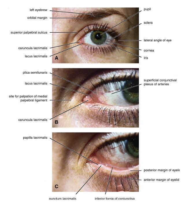
6 The Orbital Region The orbits are a pair of bony cavities that contain the eyeballs Eyelids The eyelids (they act like the curtains) protect the eye from injury and excessive light by their closure The upper eyelid is larger and more mobile than the lower because of its attachment to the levator palpebrae superioris The upper and lower eyelids meet each other at the medial and lateral angles. The palpebral fissure is the elliptical opening between the eyelids The palpebral fissure is the entrance into the conjunctival sac
 open directly into the eyelash follicles.")
 Contains the palpebral part of")
7 structure of the eye lids 1-skin:thin and can be easily become oedematous (with fluid or blood) Contains The sebaceous glands (glands of Zeis) open directly into the eyelash follicles. The ciliary glands (glands of Moll) are modified sweat glands that open separately between adjacent lashes 2- Superficial fascia: ( remember we said earlier No fat) Contains the palpebral part of orbicularis occuli


8 3- Palpebral fascia The framework of the eyelids is formed by a fibrous sheet, the orbital septum The orbital septum is attached to the periosteum at the orbital margins. The orbital septum is thickened at the margins of the lids to form the superior and inferior TARSAL PLATES. The lateral ends of the tarsal plates are attached by a band, the lateral palpebral ligament, the orbital margin. The medial ends of the plates are attached by a band, the medial palpebral ligament, to the lacrimal bone The tarsal glands are embedded in the posterior surface of the tarsal plates


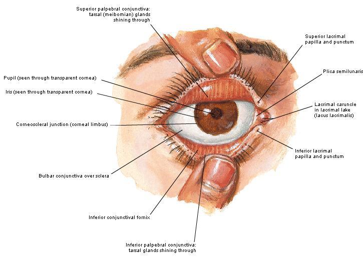
9 4-The conjunctiva is a thin mucous membrane that lines the eyelids It is reflected at the superior and inferior fornices onto the anterior surface of the eyeball The upper lateral part of the superior fornix is pierced by the ducts of the lacrimal gland The conjunctiva thus forms a potential space, the conjunctival sac, which is open at the palpebral fissure.
10


11 Movements of the Eyelids The position of the eyelids at rest depends on the tone of : 1-The orbicularis oculi 2-The levator palpebrae superioris muscles and the position of the eyeball. The eyelids are closed by : 1-The contraction of the orbicularis oculi and 2-The relaxation of the levator palpebrae superioris muscles The eye is opened by: THE LEVATOR PALPEBRAE SUPERIORIS Raising the upper lid the superior tarsal muscle which is part of the levator palpebrae superioris, helps maintain eyelid elevation and are innervated by postganglionic sympathetic fibers from the superior cervical ganglion Loss of oculomotor nerve [III] function results in complete ptosis or drooping of the superior eyelid, whereas loss of sympathetic innervation to the superior tarsal muscle results in partial ptosis


12 Horner's syndrome Horner's syndrome is caused by a lesion in the sympathetic trunk in the neck that results in sympathetic dysfunction. It is characterized by three typical features: 1-Pupillary constriction due to paralysis of the dilator pupillae muscle; 2-Partial ptosis (drooping of the upper eyelid) due to paralysis of the superior tarsal muscle of the levator palpebrae superioris; 3-Absence of sweating on the ipsilateral side of the face and the neck due to absence of innervation of the sweat glands.


13 Lacrimal Gland The lacrimal gland consists of: 1-a large orbital part 2- a small palpebral part which are continuous with each other around the lateral edge of the aponeurosis of the levator palpebrae superioris. It is situated above the eyeball in the anterior and upper part of the orbit posterior to the orbital septum The gland opens into the lateral part of the superior fornix of the conjunctiva by 12 ducts.
14


 long and emerges from the lower end of the lacrimal sac The duct descends downward,")
15 Lacrimal Ducts The tears circulate across the cornea and accumulate in the lacus lacrimalis. From here the tears enter the canaliculi lacrimales through the puncta lacrimalis. The canaliculi lacrimales pass medially and open into the lacrimal sac which lies in the lacrimal groove behind the medial palpebral ligament and is the upper blind end of the nasolacrimal duct. The nasolacrimal duct is about 0.5 in. (1.3 cm) long and emerges from the lower end of the lacrimal sac The duct descends downward, backward, and laterally in a bony canal and opens into the inferior meatus of the nose.


16 5-They then pass into its zygomatic branch and the zygomaticotemp oral nerve 6-They reach the lacrimal gland within the lacrimal nerve 2-The preganglionic fibers reach the pterygopalatine ganglion (sphenopalatine ganglion) via the nervus intermedius and its great petrosal branch 4-joins the maxillary nerve. 1-The parasympathetic secretomotor nerve supply is derived from the lacrimal nucleus of the facial nerve 3-via the nerve of the pterygoid canal. The postganglionic fibers leave the ganglion


17 5-finally the lacrimal nerve 4- via the zygomatic nerve, the zygomaticotempor al nerve 4-the maxillary nerve the maxillary nerve 2-travels in the deep petrosal nerve 3-then in the nerve of the pterygoid canal, 1-The sympathetic postganglionic nerve supply is from the internal carotid plexus
18


19 Nerves of the Orbit Optic Nerve The optic nerve enters the orbit from the middle cranial fossa by passing through the optic canal It is accompanied by the ophthalmic artery, which lies on its lower lateral side. The nerve is surrounded by sheaths of pia mater, arachnoid mater, and dura mater It runs forward and laterally within the cone of the recti muscles and pierces the sclera at a point medial to the posterior pole of the eyeball Remember that the meninges fuse with the sclera so that the subarachnoid space with its contained cerebrospinal fluid extends forward from the middle cranial fossa, around the optic nerve, and through the optic canal, as far as the eyeball. A rise in pressure of the cerebrospinal fluid within the cranial cavity therefore is transmitted to the back of the eyeball.


20 Lacrimal Nerve arises from the ophthalmic division of the trigeminal nerve It enters the orbit through the upper part of the superior orbital fissure passes forward along the upper border of the lateral rectus muscle It is joined by a branch of the zygomaticotemporal nerve, which later leaves it to enter the lacrimal gland


21 Frontal Nerve The frontal nerve arises from the ophthalmic division of the trigeminal nerve It enters the orbit through the upper part of the superior orbital fissure and passes forward on the upper surface of the levator palpebrae superioris beneath the roof of the orbit It divides into the supratrochlear and supraorbital nerves that wind around the upper margin of the orbital cavity to supply the skin of the forehead; the supraorbital nerve also supplies the mucous membrane of the frontal air sinus.
22 Trochlear Nerve The trochlear nerve enters the orbit through the upper part of the superior orbital fissure It runs forward and supplies the superior oblique muscle


23 Oculomotor Nerve The superior ramus of the oculomotor nerve enters the orbit through the lower part of the superior orbital fissure It supplies the superior rectus muscle then pierces it, and supplies the levator palpebrae superioris muscle The inferior ramus of the oculomotor nerve enters the orbit in a similar manner and supplies the inferior rectus, the medial rectus, and the inferior oblique muscles. The nerve to the inferior oblique gives off a branch that passes to the ciliary ganglion and carries parasympathetic fibers to the sphincter pupillae and the ciliary muscle SO4 LR6


24 Nasociliary Nerve The nasociliary nerve arises from the ophthalmic division of the trigeminal nerve. It enters the orbit through the lower part of the superior orbital fissure It crosses above the optic nerve, runs forward along the upper margin of the medial rectus muscle, and ends by dividing into the anterior ethmoidal and infratrochlear nerves Branches of the Nasociliary Nerve 1-The communicating branch to the ciliary ganglion is a sensory nerve. The sensory fibers from the eyeball pass to the ciliary ganglion via the short ciliary nerves without interruption, and then join the nasociliary nerve by means of the communicating branch. 2-The long ciliary nerves, two or three in number, arise from the nasociliary nerve as it crosses the optic nerve They contain sympathetic fibers for the dilator pupillae muscle. The nerves pass forward with the short ciliary nerves and pierce the sclera of the eyeball. They continue forward between the sclera and the choroid to reach the iris.

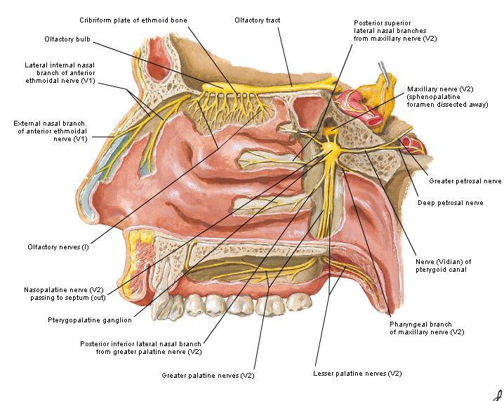
25 3-The posterior ethmoidal nerve supplies the ethmoidal and sphenoidal air sinuses 4-The infratrochlear nerve supplies the skin of the medial part of the upper eyelid and the adjacent part of the nose 5-The anterior ethmoidal nerve passes through the anterior ethmoidal foramen After supplying an area of mucous membrane in the nasal cavity, it appears on the face as the external nasal nerve at the lower border of the nasal bone, and supplies the skin of the nose down as far as the tip


26 The Sixth Cranial nerve ABDUCENT NERVE The abducent nerve enters the orbit through the lower part of the superior orbital fissure It supplies the lateral rectus muscle


27 Ciliary Ganglion Is a parasympathetic ganglion About the size of a pinhead and situated in the posterior part of the orbit. It receives its preganglionic parasympathetic fibers from the oculomotor nerve via the nerve to the inferior oblique muscle The postganglionic fibers leave the ganglion in the short ciliary nerves, which enter the back of the eyeball and supply the sphincter pupillae and the ciliary muscle. It receives its sympathetic fibers from the internal carotid sympathetic plexus in the orbit and run through the ganglion without interruption.


 The ciliary arteries The lacrimal artery to the lacrimal gland The")
28 Ophthalmic Artery is a branch of the internal carotid artery It enters the orbit through the optic canal with the optic nerve It runs forward and crosses the optic nerve to reach the medial wall of the orbit. It gives off numerous branches, which accompany the nerves in the orbital cavity Branches of the Ophthalmic Artery The central artery of the retina is a small branch that pierces the meningeal sheaths of the optic nerve to gain entrance to the nerve It runs in the substance of the optic nerve and enters the eyeball at the center of the optic disc. Here, it divides into branches, which may be studied in a patient through an ophthalmoscope The muscular branches (of the ophthalmic artery) The ciliary arteries The lacrimal artery to the lacrimal gland The supratrochlear and supraorbital arteries are distributed to the skin of the forehead

29 Ophthalmic Veins The superior ophthalmic vein communicates in front with the facial vein The inferior ophthalmic vein communicates through the inferior orbital fissure with the pterygoid venous plexus. Both veins pass backward through the superior orbital fissure and drain into the cavernous sinus.
The sebaceous glands (glands of Zeis) open directly into the eyelash follicles, ciliary glands (glands of Moll) are modified sweat glands that open
 open directly into the eyelash follicles, ciliary glands (glands of Moll) are modified sweat glands that open") The Orbital Region The orbits are a pair of bony cavities that contain the eyeballs; their associated muscles, nerves, vessels, and fat; and most of the lacrimal apparatus upper eyelid is larger and more
The Orbital Region The orbits are a pair of bony cavities that contain the eyeballs; their associated muscles, nerves, vessels, and fat; and most of the lacrimal apparatus upper eyelid is larger and more
The orbit-2. Dr. Heba Kalbouneh Assistant Professor of Anatomy and Histology
 The orbit-2 Dr. Heba Kalbouneh Assistant Professor of Anatomy and Histology Eyelids The eyelids (act like the curtains) protect the eye from injury and excessive light by their closure The upper eyelid
1 Eyelids. Lacrimal Apparatus. Orbital Region. 3 The Orbit. The Eye
 1 1 Eyelids Orbital Region 2 Lacrimal Apparatus 3 The Orbit 4 The Eye 2 Eyelids The eyelids protect the eye from injury and excessive light by their closure. The upper eyelid is larger and more mobile
Bony orbit. Lateral wall: Formed by : the zygomatic bone and the greater wing of the sphenoid
 Bony orbit Roof: Formed by: The orbital plate of the frontal bone, which separates the orbital cavity from the anterior cranial fossa and the frontal lobe of the cerebral hemisphere Lateral wall: Formed
The orbit-1. Dr. Heba Kalbouneh Assistant Professor of Anatomy and Histology
 The orbit-1 Dr. Heba Kalbouneh Assistant Professor of Anatomy and Histology Orbital plate of frontal bone Orbital plate of ethmoid bone Lesser wing of sphenoid Greater wing of sphenoid Lacrimal bone Orbital
Maxilla, ORBIT and infratemporal fossa. Neophytos C Demetriades MD, DDS, MSc Associate professor European University of Cyprus School of Medicine
 Maxilla, ORBIT and infratemporal fossa Neophytos C Demetriades MD, DDS, MSc Associate professor European University of Cyprus School of Medicine MAXILLA Superior, middle, and inferior meatus Frontal sinus
MAXILLA, ORBIT & PTERYGOPALATINE FOSSA. Neophytos C Demetriades MD, DDS, MSc Associate professor European University of Cyprus School of Medicine
 MAXILLA, ORBIT & PTERYGOPALATINE FOSSA Neophytos C Demetriades MD, DDS, MSc Associate professor European University of Cyprus School of Medicine Maxilla MAXILLA Superior, middle, and inferior meatus Frontal
213: HUMAN FUNCTIONAL ANATOMY: PRACTICAL CLASS 12 Cranial cavity, eye and orbit
 213: HUMAN FUNCTIONAL ANATOMY: PRACTICAL CLASS 12 Cranial cavity, eye and orbit OSTEOLOGY Identify the bones which comprise the walls of the orbit: maxilla, zygomatic, ethmoid, lachrymal, frontal, and
Unit VIII Problem 8 Anatomy: Orbit and Eyeball
 Unit VIII Problem 8 Anatomy: Orbit and Eyeball - The bony orbit: it is protecting our eyeball and resembling a pyramid: With a base directed: anterolaterally. And an apex directed: posteromedially. Notes:
GNK485 The eye and related structures. Prof MC Bosman 2012
 GNK485 The eye and related structures Prof MC Bosman 2012 Surface anatomy Bony orbit Eyeball and Lacrimal apparatus Extra-ocular muscles Movements of the eye Innervation Arterial supply and venous drainage
Dr.Ban I.S. head & neck anatomy 2 nd y جامعة تكريت كلية طب االسنان مادة التشريح املرحلة الثانية أ.م.د. بان امساعيل صديق 6102/6102
 جامعة تكريت كلية طب االسنان مادة التشريح املرحلة الثانية أ.م.د. بان امساعيل صديق 6102/6102 Pterygopalatine fossa: The pterygopalatine fossa is a cone-shaped depression, It is located between the maxilla,
Omran Saeed. Luma Taweel. Mohammad Almohtaseb. 1 P a g e
 2 Omran Saeed Luma Taweel Mohammad Almohtaseb 1 P a g e I didn t include all the photos in this sheet in order to keep it as small as possible so if you need more clarification please refer to slides In
3-Deep fascia: is absent (except over the parotid gland & buccopharngeal fascia covering the buccinator muscle)
") The Face 1-Skin of the Face The skin of the face is: Elastic Vascular (bleed profusely however heal rapidly) Rich in sweat and sebaceous glands (can cause acne in adults) It is connected to the underlying
The Face 1-Skin of the Face The skin of the face is: Elastic Vascular (bleed profusely however heal rapidly) Rich in sweat and sebaceous glands (can cause acne in adults) It is connected to the underlying
Face. Definition: The area between the two ears and from the chin to the eye brows. The muscles of the face
 Face Definition: The area between the two ears and from the chin to the eye brows. The muscles of the face The muscle of facial expression (include the muscle of the face and the scalp). All are derived
PTERYGOPALATINE FOSSA
 PTERYGOPALATINE FOSSA Outline Anatomical Structure and Boundaries Foramina and Communications with other spaces and cavities Contents Pterygopalatine Ganglion Especial emphasis on certain arteries and
Mohammad Hisham Al-Mohtaseb. Lina Mansour. Reyad Jabiri. 0 P a g e
 2 Mohammad Hisham Al-Mohtaseb Lina Mansour Reyad Jabiri 0 P a g e This is only correction for the last year sheet according to our record. If you already studied this sheet just read the yellow notes which
mistake ;slides in bold but you still have to go back to our slides to see the figure, tables and some scheme
 Khozama jehad : I am doing my best and I am sorry for any unintended mistake ;slides in bold but you still have to go back to our slides to see the figure, tables and some scheme The Orbit, Orbital Contents
Bony orbit. Sup. Med. Inf. Lat. frontal bone. frontal process of maxilla. zygomatic process of maxilla zygomatic bone
 Orbit 解剖學科鄭授德 本教材之圖片取自於 1. Gray s Anatomy for Students, 3rd ed., 2015, by Drake, Vogl, and Mitchell 2. Clinically Oriented Anatomy, 7th ed., 2014, by Moore, Dalley, and Agur 3. Anatomy, an Essential Textbook,
Infratemporal fossa: Tikrit University college of Dentistry Dr.Ban I.S. head & neck Anatomy 2 nd y.
 Infratemporal fossa: This is a space lying beneath the base of the skull between the lateral wall of the pharynx and the ramus of the mandible. It is also referred to as the parapharyngeal or lateral pharyngeal
Lecture 10 Orbit and control of eye movements
 Lecture 10 Orbit and control of eye movements Overview of structures in the orbit (Moore pp 899, Netter Plate 1) The orbit contains the eye, from which the optic nerve exits into the cranial cavity optic
Ocular Anatomy for the Paraoptometric
 Ocular Anatomy for the Paraoptometric Minnesota Optometric Association Paraoptometric CE Friday September 30, 2016 Lindsay A. Sicks, OD, FAAO Assistant Professor, Illinois College of Optometry lsicks@ico.edu
Chapter(2):the lid page (1) THE LID
:the lid page (1) THE LID") Chapter(2):the lid page (1) THE LID Anatomy of the lid: * Check movie anatomy of the lid model The eyelids are two movable muco-cutaneous folds which protect the eye on closure. The are joined temporary
Chapter(2):the lid page (1) THE LID Anatomy of the lid: * Check movie anatomy of the lid model The eyelids are two movable muco-cutaneous folds which protect the eye on closure. The are joined temporary
REVIEW OF HEAD AND NECK CRANIAL NERVES AND EVERYTHING ELSE
 REVIEW OF HEAD AND NECK CRANIAL NERVES AND EVERYTHING ELSE OLFACTORY NERVE CN I ANTERIOR CRANIAL FOSSA CRISTA GALLI OF ETHMOID OLFACTORY FORAMINA IN CRIBIFORM PLATE OF ETHMOID BONE CN I OLFACTORY NERVE
Anatomic Relations Summary. Done by: Sohayyla Yasin Dababseh
 Anatomic Relations Summary Done by: Sohayyla Yasin Dababseh Anatomic Relations Lecture 1 Part-1 - The medial wall of the nose is the septum. - The vestibule lies directly inside the nostrils (Nares). -
The Orbit. The Orbit OCULAR ANATOMY AND DISSECTION 9/25/2014. The eye is a 23 mm organ...how difficult can this be? Openings in the orbit
 The eye is a 23 mm organ...how difficult can this be? OCULAR ANATOMY AND DISSECTION JEFFREY M. GAMBLE, OD COLUMBIA EYE CONSULTANTS OPTOMETRY & UNIVERSITY OF MISSOURI DEPARTMENT OF OPHTHALMOLOGY CLINICAL
Trigeminal Nerve Worksheets, Distributions Page 1
 Trigeminal Nerve Worksheet #1 Distribution by Nerve Dr. Darren Hoffmann Dental Gross Anatomy, Spring 2013 We have drawn out each of the branches of CN V in lecture and you have an idea now for their basic
The Special Senses: Part A
 PowerPoint Lecture Slides prepared by Janice Meeking, Mount Royal College CHAPTER 15 The Special Senses: Part A Warm Up What is the function of the eyeball? List any structures of the eyeball that you
Dr. Sami Zaqout, IUG Medical School
 The skull The skull is composed of several separate bones united at immobile joints called sutures. Exceptions? Frontal bone Occipital bone Vault Cranium Sphenoid bone Zygomatic bones Base Ethmoid bone
Trigeminal Nerve (V)
") Trigeminal Nerve (V) Lecture Objectives Discuss briefly how the face is developed. Follow up the course of trigeminal nerve from its point of central connections, exit and down to its target areas. Describe
Trigeminal Nerve (V) Lecture Objectives Discuss briefly how the face is developed. Follow up the course of trigeminal nerve from its point of central connections, exit and down to its target areas. Describe
Lec [8]: Mandibular nerve:
![Lec [8]: Mandibular nerve:](/thumbs/94/121295776.jpg "Lec [8]: Mandibular nerve:") Lec [8]: Mandibular nerve: The mandibular branch from the trigeminal ganglion lies in the middle cranial fossa lateral to the cavernous sinus. With the motor root of the trigeminal nerve [motor roots lies
Lec [8]: Mandibular nerve: The mandibular branch from the trigeminal ganglion lies in the middle cranial fossa lateral to the cavernous sinus. With the motor root of the trigeminal nerve [motor roots lies
Tikrit University collage of dentistry Dr.Ban I.S. head & neck anatomy 2 nd y. Lec [5] / Temporal fossa :
![Tikrit University collage of dentistry Dr.Ban I.S. head & neck anatomy 2 nd y. Lec [5] / Temporal fossa :](/thumbs/88/115294566.jpg "Tikrit University collage of dentistry Dr.Ban I.S. head & neck anatomy 2 nd y. Lec [5] / Temporal fossa :") Lec [5] / Temporal fossa : Borders of the Temporal Fossa: Superior: Superior temporal line. Inferior: gap between zygomatic arch and infratemporal crest of sphenoid bone. Anterior: Frontal process of the
Lec [5] / Temporal fossa : Borders of the Temporal Fossa: Superior: Superior temporal line. Inferior: gap between zygomatic arch and infratemporal crest of sphenoid bone. Anterior: Frontal process of the
HEAD AND NECK ANATOMY PRACTICE QUESTIONS
 HEAD AND NECK ANATOMY PRACTICE QUESTIONS 1. A patient complains that he has lost sensation on his face and that the skin of his face feels numb. The physician tests tactile acuity by touching the forehead
Dr.Ban I.S. head & neck anatomy 2 nd y. جامعة تكريت كلية طب االسنان املرحلة الثانية أ.م.د. بان امساعيل صديق 6102/6102
 جامعة تكريت كلية طب االسنان التشريح مادة املرحلة الثانية أ.م.د. بان امساعيل صديق 6102/6102 Parotid region The part of the face in front of the ear and below the zygomatic arch is the parotid region. The
Temporal region. temporal & infratemporal fossae. Zhou Hong Ying Dept. of Anatomy
 Temporal region temporal & infratemporal fossae Zhou Hong Ying Dept. of Anatomy Temporal region is divided by zygomatic arch into temporal & infratemporal fossae. Temporal Fossa Infratemporal fossa Temporal
Tikrit University College of Dentistry Dr.Ban I.S. head & neck anatomy 2 nd y.
 Lec [3]/The scalp The scalp extends from the supraorbital margins anteriorly to the nuchal lines at the back of the skull and down to the temporal lines at the sides. The forehead, from eyebrows to hairline,
Head and Face Anatomy
 Head and Face Anatomy Epicranial region The Scalp The soft tissue that covers the vault of skull. Extends from supraorbital margin to superior nuchal line. Layers of the scalp S C A L P = skin = connective
Superior View of the Skull (Norma Verticalis) Anteriorly the frontal bone articulates with the two parietal bones AT THE CORONAL SUTURE
 Anteriorly the frontal bone articulates with the two parietal bones AT THE CORONAL SUTURE") Superior View of the Skull (Norma Verticalis) Anteriorly the frontal bone articulates with the two parietal bones AT THE CORONAL SUTURE 1 The two parietal bones articulate in the midline AT THE SAGITTAL
Superior View of the Skull (Norma Verticalis) Anteriorly the frontal bone articulates with the two parietal bones AT THE CORONAL SUTURE 1 The two parietal bones articulate in the midline AT THE SAGITTAL
Sense of Vision. Chapter 8. The Eye and Vision. The Eye Orbit. Eyebrows, Eyelids, Eyelashes. Accessory Organs 5/3/2016.
 Sense of Vision Chapter 8 Special Senses The Eye and Vision 70 percent of all sensory receptors are in the eyes Each eye has over 1 million nerve fibers Protection for the eye Most of the eye is enclosed
Major Anatomic Components of the Orbit
 Major Anatomic Components of the Orbit 1. Osseous Framework 2. Globe 3. Optic nerve and sheath 4. Extraocular muscles Bony Orbit Seven Bones Frontal bone Zygomatic bone Maxillary bone Ethmoid bone Sphenoid
Temporal fossa Infratemporal fossa Pterygopalatine fossa Terminal branches of external carotid artery Pterygoid venous plexus
 Outline of content Temporal fossa Infratemporal fossa Pterygopalatine fossa Terminal branches of external carotid artery Pterygoid venous plexus Boundary Content Communication Mandibular division of trigeminal
human anatomy 2016 lecture fifteen Dr meethak ali ahmed neurosurgeon
 Cranial Nerves Organization of the Cranial Nerves The cranial nerves are named as follows: I. Olfactory II. Optic III. Oculomotor IV. Trochlear V. Trigeminal VI. Abducent VII. Facial VIII. Vestibulocochlear
Cranial Cavity REFERENCES: OBJECTIVES OSTEOLOGY. Stephen A. Gudas, PT, PhD
 Stephen A. Gudas, PT, PhD Cranial Cavity REFERENCES: Moore and Agur, Essential Clinical Anatomy (ECA), 3rd ed., pp. 496 498; 500 507; 512 514 Grant s Atlas 12 th ed., Figs 7.6; 7.19 7.30. Grant s Dissector
THE SPECIAL SENSES. Introduction Vision
 THE SPECIAL SENSES Introduction Vision RECEPTORS Structures designed to respond to stimuli Variable complexity RECEPTORS: GENERAL PROPERTIES Transducers Receptor Potential Generator Potential RECEPTORS
Parotid Gland, Temporomandibular Joint and Infratemporal Fossa
 M1 - Anatomy Parotid Gland, Temporomandibular Joint and Infratemporal Fossa Jeff Dupree Sanger 9-057 jldupree@vcu.edu Parotid gland: wraps around the mandible positioned between the mandible and the sphenoid
Anatomy for ophthalmic anaesthesia
 British Journal of Anaesthesia 1995; 75: 80 87 REVIEW ARTICLES Anatomy for ophthalmic anaesthesia R. W. JOHNSON Study of the anatomy of the orbit, its contents and surrounding structures allows the anaesthetist
Trigeminal nerve. Slide in bold and please go back to see the pictures, if I skipped any part of record that because it wasn t clear to me
 Trigeminal nerve Slide in bold and please go back to see the pictures, if I skipped any part of record that because it wasn t clear to me Hala nsour 2/26/2018 P a g e 1 this lecture contain two topics
*in general the blood supply of the nose comes from branches of the internal and external carotid arteries.
 In the previous lecture we talked about the anatomy of the nasal cavity, today we will talk about its blood supply, venous drainage, innervations, and finally about the paranasal sinuses. When we describe
Bisection of Head & Nasal Cavity 頭部對切以及鼻腔. 解剖學科馮琮涵副教授 分機
 Bisection of Head & Nasal Cavity 頭部對切以及鼻腔 解剖學科馮琮涵副教授 分機 3250 E-mail: thfong@tmu.edu.tw Outline: The structure of nose The concha and meatus in nasal cavity The openings of paranasal sinuses Canals, foramens
Introduction to Local Anesthesia and Review of Anatomy
 5-Sep Introduction and Anatomy Review 12-Sep Neurophysiology and Pain 19-Sep Physiology and Pharmacology part 1 26-Sep Physiology and Pharmacology part 2 Introduction to Local Anesthesia and Review of
4/22/16. Eye. External Anatomy of Eye. Accessory Structures. Bio 40B Dr. Kandula
 Eye Bio 40B Dr. Kandula External Anatomy of Eye Accessory Structures l Eyebrows l Levator Palpebrae Superioris - opens eye l Eyelashes l Ciliary glands modified sweat glands l Small sebaceous glands l
External Occipital Protuberance
 Osteology Exterior Skull Frontal Bone Glabella Superciliary Arch Supraorbital Notch/Foramen Nasion (junction w/ Nasal bone) Frontal/Metopic Suture (usually absent in adult, b/w ossification centers of
Veins of the Face and the Neck
 Veins of the Face and the Neck Facial Vein The facial vein is formed at the medial angle of the eye by the union of the supraorbital and supratrochlear veins. connected through the ophthalmic veins with
Unit 18: Cranial Cavity and Contents
 Unit 18: Cranial Cavity and Contents Dissection Instructions: The calvaria is to be removed without damage to the dura mater which is attached to the inner surface of the calvaria. Cut through the outer
The SCALP. Prof. Dr. Muhammad Imran Qureshi
 The SCALP By Prof. Dr. Muhammad Imran Qureshi The SCALP includes FIVE layers external to the Calvaria. These are: S: Skin & Superficial Fascia C: Connective Tissue A: Aponeurosis (Epicranial) L: Loose
Eye Movements. Geometry of the Orbit. Extraocular Muscles
 Eye Movements Geometry of the Orbit The eye (oculus) is located in the anterior aspect of the orbit: the equator of the eye (defined by a coronal plane passing through its middle) lies at the margin of
THIEME. Scalp and Superficial Temporal Region
 CHAPTER 2 Scalp and Superficial Temporal Region Scalp Learning Objectives At the end of the dissection of the scalp, you should be able to identify, understand and correlate the clinical aspects: Layers
TRANSVERSE SECTION PLANE Scalp 2. Cranium. 13. Superior sagittal sinus
 TRANSVERSE SECTION PLANE 1 1. Scalp 2. Cranium 3. Superior sagittal sinus 4. Dura mater 5. Falx cerebri 6. Frontal lobes of the cerebrum 7. Middle meningeal artery 8. Cortex, grey matter 9. Cerebral vessels
Dr.Ban I.S. head & neck anatomy 2 nd y. جامعة تكريت كلية طب االسنان املرحلة الثانية
 جامعة تكريت كلية طب االسنان التشريح مادة املرحلة الثانية أ.م.د. بان امساعيل صديق 6102-6102 1 The Palate The palate forms the roof of the mouth and the floor of the nasal cavity. It is divided into two
Nasal region. cartilages: septal cartilage (l); lateral nasal cartilage (2); greater alar cartilages (2); lesser alar cartilages (?
; lateral nasal cartilage (2); greater alar cartilages (2); lesser alar cartilages (?") Nasal region skull bones: nasal and frontal processes of maxilla cartilages: septal cartilage (l); lateral nasal cartilage (2); greater alar cartilages (2); lesser alar cartilages (?) 1 Nasal cavity Roof
Nasal region skull bones: nasal and frontal processes of maxilla cartilages: septal cartilage (l); lateral nasal cartilage (2); greater alar cartilages (2); lesser alar cartilages (?) 1 Nasal cavity Roof
Trigeminal Nerve Anatomy. Dr. Mohamed Rahil Ali
 Trigeminal Nerve Anatomy Dr. Mohamed Rahil Ali Trigeminal nerve Largest cranial nerve Mixed nerve Small motor root and large sensory root Motor root Nucleus of motor root present in the pons and medulla
The dura is sensitive to stretching, which produces the sensation of headache.
 Dural Nerve Supply Branches of the trigeminal, vagus, and first three cervical nerves and branches from the sympathetic system pass to the dura. Numerous sensory endings are in the dura. The dura is sensitive
Bones Ethmoid bone Inferior nasal concha Lacrimal bone Maxilla Nasal bone Palatine bone Vomer Zygomatic bone Mandible
 splanchnocranium - Consists of part of skull that is derived from branchial arches - The facial bones are the bones of the anterior and lower human skull Bones Ethmoid bone Inferior nasal concha Lacrimal
CN I Olfactory. CN II Optic. CN III Oculomotor. Special Sensory Efferent fibers to Olfactory Bulb. Cribiform Plate of Ethmoid
 CN I Olfactory Efferent fibers to Olfactory Bulb Olfactory Tract Olfactory Bulb Cribiform Plate of Ethmoid Anosmia Loss of sense of smell Uncinate Fits olfactory hallucinations To Olfactory Epithelium
Brain ميهاربا لض اف دمح ا د The Meninges 1- Dura Mater of the Brain endosteal layer does not extend meningeal layer falx cerebri tentorium cerebelli
 .احمد د فاضل ابراهيم Lecture 15 Brain The Meninges Three protective membranes or meninges surround the brain in the skull: the dura mater, the arachnoid mater, and the pia mater 1- Dura Mater of the Brain
Skull-2. Norma Basalis Interna Norma Basalis Externa. Dr. Heba Kalbouneh Associate Professor of Anatomy and Histology
 Skull-2 Norma Basalis Interna Norma Basalis Externa Dr. Heba Kalbouneh Associate Professor of Anatomy and Histology Norma basalis interna Base of the skull- superior view The interior of the base of the
Cranial nerves.
 Cranial nerves eaglezhyxzy@163.com Key Points of Learning Name Components Passing through Peripheral distribution Central connection Function Cranial nerves Ⅰ olfactory Ⅱ optic Ⅲ occulomotor Ⅳ trochlear
The Skull and Temporomandibular joint II Prof. Abdulameer Al-Nuaimi. E. mail:
 The Skull and Temporomandibular joint II Prof. Abdulameer Al-Nuaimi E-mail: a.al-nuaimi@sheffield.ac.uk E. mail: abdulameerh@yahoo.com Temporal fossa The temporal fossa is a depression on the temporal
Skull-2. Norma Basalis Interna. Dr. Heba Kalbouneh Assistant Professor of Anatomy and Histology
 Skull-2 Norma Basalis Interna Dr. Heba Kalbouneh Assistant Professor of Anatomy and Histology Norma basalis interna Base of the skull- superior view The interior of the base of the skull is divided into
Nose & Mouth OUTLINE. Nose. - Nasal Cavity & Its Walls. - Paranasal Sinuses. - Neurovascular Structures. Mouth. - Oral Cavity & Its Contents
 Dept. of Human Anatomy, Si Chuan University Zhou hongying eaglezhyxzy@163.com Nose & Mouth OUTLINE Nose - Nasal Cavity & Its Walls - Paranasal Sinuses - Neurovascular Structures Mouth - Oral Cavity & Its
Anatomy and Physiology. Bones, Sutures, Teeth, Processes and Foramina of the Human Skull
 Anatomy and Physiology Chapter 6 DRO Bones, Sutures, Teeth, Processes and Foramina of the Human Skull Name: Period: Bones of the Human Skull Bones of the Cranium: Frontal bone: forms the forehead and the
function - sensory & postganglionic sympathetic [communication from the internal carotid plexus in the cavernous sinus] innervation of the mucosa of
![function - sensory & postganglionic sympathetic [communication from the internal carotid plexus in the cavernous sinus] innervation of the mucosa of](/thumbs/74/71276096.jpg "function - sensory & postganglionic sympathetic [communication from the internal carotid plexus in the cavernous sinus] innervation of the mucosa of") Nerves I. Cranial nerves A. Olfactory (CN I) 1. Olfactory bulb 2. Olfactory tract B. Optic n. (CNII) function - carries visual sensory information from the neural retina to the diencephalon & midbrain
Nerves I. Cranial nerves A. Olfactory (CN I) 1. Olfactory bulb 2. Olfactory tract B. Optic n. (CNII) function - carries visual sensory information from the neural retina to the diencephalon & midbrain
 This article was originally published in the Encyclopedia of the Eye, published by Elsevier, and the attached copy is provided by Elsevier for the author's benefit and for the benefit of the author's institution,
Dr.Ban I.S. head & neck anatomy 2 nd y. جامعة تكريت كلية طب االسنان مادة التشريح املرحلة الثانية أ.م.د. بان امساعيل صديق 6102/6102
 جامعة تكريت كلية طب االسنان مادة التشريح املرحلة الثانية أ.م.د. بان امساعيل صديق 6102/6102 The scalp The scalp extends from the supraorbital margins anteriorly to the nuchal lines at the back of the skull
Chapter 7: Head & Neck
 Chapter 7: Head & Neck Osteology I. Overview A. Skull The cranium is composed of irregularly shaped bones that are fused together at unique joints called sutures The skull provides durable protection from
Introduction to Head and Neck Anatomy
 Introduction to Head and Neck Anatomy Nervous Tissue Controls and integrates all body activities within limits that maintain life Three basic functions 1. sensing changes with sensory receptors 2. interpreting
Cranial Nerve VII - Facial Nerve. The facial nerve has 3 main components with distinct functions
 Cranial Nerve VII - Facial Nerve The facial nerve has 3 main components with distinct functions Somatic motor efferent Supplies the muscles of facial expression; posterior belly of digastric muscle; stylohyoid,
Parotid Gland. Parotid Gland. Largest of 3 paired salivary glands (submandibular; sublingual) Ramus of Mandible. Medial pterygoid.
 Ramus of Mandible. Medial pterygoid.") Parotid region Parotid Gland Largest of 3 paired salivary glands (submandibular; sublingual) Ramus of Mandible Medial pterygoid Cross section of mandible Masseter D S SCM Parotid Gland Mastoid Process
Parotid region Parotid Gland Largest of 3 paired salivary glands (submandibular; sublingual) Ramus of Mandible Medial pterygoid Cross section of mandible Masseter D S SCM Parotid Gland Mastoid Process
The Ear The ear consists of : 1-THE EXTERNAL EAR 2-THE MIDDLE EAR, OR TYMPANIC CAVITY 3-THE INTERNAL EAR, OR LABYRINTH 1-THE EXTERNAL EAR.
 The Ear The ear consists of : 1-THE EXTERNAL EAR 2-THE MIDDLE EAR, OR TYMPANIC CAVITY 3-THE INTERNAL EAR, OR LABYRINTH 1-THE EXTERNAL EAR Made of A-AURICLE B-EXTERNAL AUDITORY MEATUS A-AURICLE It consists
C h a p t e r PowerPoint Lecture Slides prepared by Jason LaPres North Harris College Houston, Texas
 C h a p t e r 15 The Nervous System: The Brain and Cranial Nerves PowerPoint Lecture Slides prepared by Jason LaPres North Harris College Houston, Texas Copyright 2009 Pearson Education, Inc., publishing
INSERTION* SURGICAL ANATOMY OF THE LEVATOR PALPEBRAE. impossible to dissect and separate these layers. That the levator aponeurosis
 Brit. J. Ophthal. (1962) 46, 503. SURGICAL ANATOMY OF THE LEVATOR PALPEBRAE INSERTION* BY EDWARD EPSTEIN Johannesburg, Union of South Africa THE text-book description of the anatomy of the upper eyelid
Biology 323 Human Anatomy for Biology Majors Week 10; Lecture 1; Tuesday Dr. Stuart S. Sumida. Cranial Nerves and Soft Tissues of the Skull
 Biology 323 Human Anatomy for Biology Majors Week 10; Lecture 1; Tuesday Dr. Stuart S. Sumida Cranial Nerves and Soft Tissues of the Skull FOREBRAIN MIDBRAIN HINDBRAIN Forebrain: Cerebrum Perception,
Bones of the skull & face
 Bones of the skull & face Cranium= brain case or helmet Copyright The McGraw-Hill Companies, Inc. Permission required for reproduction or display. The cranium is composed of eight bones : frontal Occipital
Tracing the Cranial Nerves Osteologically
 CN I II III IV V 1 Supra-orbital ethmoidal nn. Ext. nasal V 2 Tracing the Cranial Nerves Osteologically Nucleus of Origin Olfactory tracts of frontal lobe of cerebrum Optic tracts from optic chiasma and
Dr. Sami Zaqout Faculty of Medicine IUG
 The Nose External Nose Nasal Cavity External Nose Blood and Nerve Supplies of the External Nose Blood Supply of the External Nose The skin of the external nose Branches of the ophthalmic and the maxillary
The Eye. The Orbit. The EYE What a Trip!!! - The Anterior Segment 5/12/2015. Jill J Luebbert, CPOT, ABOC
 The EYE What a Trip!!! - The Anterior Segment Jill J Luebbert, CPOT, ABOC The Eye The Orbit Bony socket containing the eye and most of its accessory organs consisting of 7 bones 1 The Seven Bones of the
Anatomy of the orbit. Lay-out. Imaging technique. 3 x 3. brief overview of the basic anatomy of the orbit and its structures
 Anatomy of the orbit Prof. Pia C Sundgren MD, PhD Department of Diagnostic Radiology, Clinical Sciences, Lund University, Sweden Lay-out brief overview of the basic anatomy of the orbit and its structures
Anatomy images for MSS practical exam- 2019
 Anatomy images for MSS practical exam- 2019 Ilium Ischium Pubis Acetabulaum Iliac crest Iliac tubercle ASIS (muscle and ligament attached) AIIS (muscle attached) PSIS PIIS Ischial spine Ischial tuberosity
VISUAL REFLEXES. B. The oculomotor nucleus, Edinger-Westphal nucleus, and oculomotor nerve at level of the superior colliculus.
 Neuroanatomy Suzanne Stensaas February 24, 2011, 10:00-12:00 p.m. Reading: Waxman Ch. 15 HyperBrain: Ch 7 with quizzes and or Lab 7 videotape http://www-medlib.med.utah.edu/kw/hyperbrain/anim/reflex.html
Anatomy #1; Respiratory Nose and the Nasal Cavity December 1st, 2013
 Note #1: the doctor skipped some slides in the lecture. Those slides are not included in this sheet and so you will have to review the slides to study them. The reason they were not included is because
Learning Outcomes. The Carotid 20/02/2013. Scalp, Face, Parotid. Layers of the Scalp. The Parotid Gland. The Scalp. The Carotid The Facial Artery
 Learning Outcomes The Scalp Layers of the Scalp Bleeding from the Scalp The Carotid The Facial Artery Major Muscles of the Face and Jaw(s) Muscles of Mastication Muscles of Facial Expression The Parotid
The Seventh Cranial Nerve The Facial By Prof. Dr. Muhammad Imran Qureshi
 The Seventh Cranial Nerve The Facial By Prof. Dr. Muhammad Imran Qureshi Functional Components: SVE: Fibers originate from nucleus of facial nerve, and supply facial muscles GVE: Fibers derived from superior
This lab activity is aligned with Visible Body s Human Anatomy Atlas app.
 1 This lab activity is aligned with Visible Body s Human Anatomy Atlas app. Learn more at visiblebody.com/professors We've split our Cranial Nerves lab activity into two parts. Part 1 is pre-lab exercises
For the following questions, indicate the letter that corresponds to the SINGLE MOST APPROPRIATE ANSWER
 GROSS ANATOMY EXAMINATION May 15, 2000 For the following questions, indicate the letter that corresponds to the SINGLE MOST APPROPRIATE ANSWER 1. Pain associated with an infection limited to the middle
Chapter 7 Part A The Skeleton
 Chapter 7 Part A The Skeleton Why This Matters Understanding the anatomy of the skeleton enables you to anticipate problems such as pelvic dimensions that may affect labor and delivery The Skeleton The
Anatomy of the Trigeminal Nerve
 19 Anatomy of the Trigeminal Nerve.1 Introduction 0. The Central Part of the Trigeminal Nerve 1..1 Origin 1.. Trigeminal Nuclei.3 The Peripheral Part of the Trigeminal Nerve 4.3.1 Ophthalmic Nerve 4.3.
SKULL AS A WHOLE + ANTERIOR CRANIAL FOSSA
 SKULL AS A WHOLE + ANTERIOR CRANIAL FOSSA LEARNING OBJECTIVES At the end of this lecture, the student should be able to know: Parts of skeleton (axial and appendicular) Parts of skull Sutures of skull