Nature Neuroscience: doi: /nn Supplementary Figure 1. Distribution of starter cells for RV-mediated retrograde tracing.
|
|
|
- Diane Little
- 5 years ago
- Views:
Transcription
, barrel field of somatosensory cortex (SSbfd) and secondary motor cortex (MOs).")
1 Supplementary Figure 1 Distribution of starter cells for RV-mediated retrograde tracing. Parcellation of cortical areas is based on Allen Mouse Brain Atlas and drawn to scale. Thick white curves, outlines of VIS, SS, AUD, ACA and MO. Thin gray curves, outlines of primary visual cortex (VISp), barrel field of somatosensory cortex (SSbfd) and secondary motor cortex (MOs). Color scale, normalized starter cell density. Scale bar, 1 mm.

2 Supplementary Figure 2 Laminar distribution of starter cells for RV-mediated retrograde tracing from VIS, SS and AUD.
 Fluorescence images of LGd, VP and MG (yellow box in coronal diagram), showing RV-labeled input neurons (red) to VIS, SS and AUD, respectively (scale bar, 100 µm). Red, tdtomato; blue, DAPI.")
3 Supplementary Figure 3 Whole-brain distributions of inputs to VIS, SS and AUD. (a) Viral vectors and injection procedure for RV-mediated trans-synaptic retrograde tracing. (b) Fluorescence images of LGd, VP and MG (yellow box in coronal diagram), showing RV-labeled input neurons (red) to VIS, SS and AUD, respectively (scale bar, 100 µm). Red, tdtomato; blue, DAPI. (c-e) RV-labeled neurons detected in all samples of each group (VIS, blue; SS, red; AUD, green; scale bar, 1 mm). White masks indicate injection sites excluded from data analysis. (f) Percentages of retrogradely labeled input neurons in selected cortical and subcortical brain regions (VIS, n = 3 mice; SS, n = 3; AUD, n = 3). Included are cortical areas with > 2% labeling and thalamic structures with > 1% labeling. Each circle represents one mouse. Error bar, ± s.e.m.

4 Supplementary Figure 4 Frontal cortical inputs to VIS, SS and AUD.
 Fluorescence images of LGd/LGv, VP and MG (yellow box in coronal diagram), showing the axons (red) from VIS, SS and AUD, respectively (scale bar, 200 µm). Red, mcherry; blue, DAPI.")
 Percentages of labeled axons in cortical and subcortical brain structures (VIS, n = 3 mice; SS, n = 3; AUD, n = 3).")
5 Supplementary Figure 5 Whole-brain distributions of axonal projections from VIS, SS and AUD. (a) Viral vector and injection procedure for tracing axonal projections. (b) Fluorescence images of LGd/LGv, VP and MG (yellow box in coronal diagram), showing the axons (red) from VIS, SS and AUD, respectively (scale bar, 200 µm). Red, mcherry; blue, DAPI. (c-e) Axons detected in all samples of each group (VIS, blue; SS, red; AUD, green; scale bar, 1 mm). White masks indicate injection sites excluded from data analysis. (f) Percentages of labeled axons in cortical and subcortical brain structures (VIS, n = 3 mice; SS, n = 3; AUD, n = 3). Included are cortical areas with > 1% labeling and thalamic structures with > 0.8% labeling. Each circle represents one mouse. Error bar, ±s.e.m.
 Fluorescence images of VIS, SS and AUD (red box in coronal diagram), showing the axons from ACA or MO (scale bar, 200 µm). Red, mcherry; blue, DAPI.")
6 Supplementary Figure 6 Example experiments showing reciprocal connections between the frontal cortices and the sensory cortices. (a) Fluorescence images of VIS, SS and AUD (red box in coronal diagram), showing the axons from ACA or MO (scale bar, 200 µm). Red, mcherry; blue, DAPI. (b) Right, fluorescence images of VIS, SS and AUD (red box in coronal diagram), showing RV-labeled input neurons (red) to ACA or MO (scale bar, 200 µm). Inset, enlarged view of the region in white box (scale bar, 10 µm). Red, tdtomato; blue, DAPI.

 in VIS, PTLp")
7 Supplementary Figure 7 ACA innervates Pvalb +, Sst + and Vip + interneurons and glutamatergic neurons (Camk2α + ) in VIS, PTLp and RSP.
8 (a) Viral vectors and injection procedure for RV-mediated transsynaptic retrograde tracing. (b) Upper panel, viral injection sites in VIS (red box in coronal diagram) of Pvalb-Cre, Sst-Cre and Vip-Cre mice (scale bar, 200 µm). Inset, enlarged view of the region in white box showing starter cells (yellow; scale bar, 20 µm). Lower panel, fluorescence images of ACA and MO (red box in coronal diagram) showing RV-labeled input neurons (green) to each subtype of interneurons in VIS (scale bar, 200 µm). Green, EGFP; red, mcherry; blue, DAPI. (c) Similar to (b), for ACA inputs to PV+, SOM+, VIP+ interneurons and glutamatergic neurons in PTLp. (d) Similar to (b), for ACA inputs to Pvalb+, Sst+, Vip+ interneurons and glutamatergic neurons in RSP. (e) Percentages of input neurons in ACA retrogradely labeled from VIS, PTLp and RSP in Camk2α-Cre (yellow), Pvalb-Cre (blue), Sst-Cre (red) and Vip-Cre mice (green). n = 3 mice in each group. Each circle represents one mouse. Error bar, ± s.e.m.


9 Supplementary Figure 8 ACA interneurons (Pval +, Sst + and Vip + ) are innervated by VIS, PTLp and RSP.
10 (a) Left, viral injection sites in ACA (red box in coronal diagram) of Pvalb-Cre, Sst-Cre and Vip-Cre mice (scale bar, 200 µm). Inset, enlarged view of the region in white box showing starter cells (yellow; scale bar, 20 µm). Right, fluorescence images of VIS, PTLp and RSP (red box in coronal diagram) showing RV-labeled input neurons (green) to each subtype of interneurons in ACA (scale bar, 200 µm). Green, EGFP; red, mcherry; blue, DAPI. (b) Percentages of retrogradely labeled input neurons in selected cortical and subcortical brain regions (n = 3 mice in each group). Each circle represents one mouse. Included are the same structures as in Fig. 2h.
 Left, bright field image of ACA and MO showing RV-labeled neurons from SS (scale bar, 200 µm). Inset, enlarged view of the region in red box (scale bar, 20 µm).")
11 Supplementary Figure 9 Whole-brain distributions of axonal projections from MO SS neurons. (a) Viral vectors and injection procedure for tracing the axonal projections from MO SS neurons. (b) Left, bright field image of ACA and MO showing RV-labeled neurons from SS (scale bar, 200 µm). Inset, enlarged view of the region in red box (scale bar, 20 µm). Immunostaining for tdtomato was performed to convert the fluorescence signal (tdtomato expressed by RV) into nickel-enhanced DAB signal; Middle, image of detected axon signals in SS (scale bar, 200 µm); right, bright field image of the region in green box, showing RV-labeled axons of MO SS neurons (scale bar, 100 µm). (c) Axons detected in all MO SS samples (scale bar, 1 mm). White masks indicate injection sites excluded from data analysis. (d) Percentages of labeled axons in selected cortical and subcortical brain structures (n = 3 mice). Each circle represents one mouse. Error bar, ±s.e.m. Data for MO axons are the same as in Figure 2D, shown here to facilitate comparison.
 Viral vectors and injection procedure for labeling the presynaptic sites of ACA VIS and ACA SC neurons.")
 from ACA VIS neurons.")
12 Supplementary Figure 10 ACA VIS and ACA SC neurons form synapses in the identified cortical and subcortical areas. (a) Viral vectors and injection procedure for labeling the presynaptic sites of ACA VIS and ACA SC neurons. (b) Upper panel, fluorescence images of VIS, PTLp, RSP and STR (red box in coronal diagram) showing axons (green) and putative presynaptic sites (red) from ACA VIS neurons. Lower panel, similar to upper panel, showing axons (green) and putative presynaptic sites (red) from ACA SC neurons in RSP, PrL/ILA, LP/LD, STR and SC. Green, mgfp; red, mruby. Scale bar, 20 µm.
 Left, fluorescence images of AAV-EF1α-mCherry-IRES-WGA-Cre injection site (red) in VIS (upper panel) and SC (lower panel). Right, WGA-Cre-induced EYFP expression (green) in ACA.")
13 Supplementary Figure 11 WGA-Cre-induced EYFP expression in ACA VIS and ACA SC neurons. (a) Viral vectors and injection procedure for retrograde labeling of ACA VIS and ACA SC neurons with EYFP. (b) Left, fluorescence images of AAV-EF1α-mCherry-IRES-WGA-Cre injection site (red) in VIS (upper panel) and SC (lower panel). Right, WGA-Cre-induced EYFP expression (green) in ACA. Note that mcherry fluorescence is located in neuronal somatas at the injection site (left), but not in the ACA (right). White arrowheads, neurons expressing EYFP. Blue, DAPI.
 Viral vectors and retrobead injection procedure for retrograde labeling of ACA VIS and ACA SC neurons with EYFP (mediated by WGA-Cre) or red retrobeads.")
14 Supplementary Figure 12 WGA-Cre-labeled ACA VIS and ACA SC neurons (EYFP + ) largely overlapped with Retrobead-labeled ACA VIS and ACA SC neurons, respectively. (a) Viral vectors and retrobead injection procedure for retrograde labeling of ACA VIS and ACA SC neurons with EYFP (mediated by WGA-Cre) or red retrobeads. (b) Left, fluorescence images of AAV-EF1α-mCherry-IRES-WGA-Cre and red retrobeads injection site (red) in VIS (upper panel) and SC (lower panel). Middle, retrobeads (red) and WGA-Cre-induced EYFP expression (green) in ACA. Right, enlarged view of the region in white box. White arrowheads, neurons containing both EYFP and retrobeads. Blue, DAPI. Note that most EYFP+ neurons in ACA were also labeled by red retrobeads (VIS injection, 83 ± 4% (s.e.m.), n = 3 mice; SC injection, 80 ± 4%, n = 3 mice).
 Viral vectors and retrobead injection procedure for retrograde labeling of ACA VIS and ACA SC neurons with EYFP (mediated by WGA-Cre) or red retrobeads.")
15 Supplementary Figure 13 WGA-Cre-labeled ACA VIS and ACA SC neurons (EYFP + ) showed no overlap with Retrobead-labeled ACA SC and ACA VIS neurons, respectively. (a) Viral vectors and retrobead injection procedure for retrograde labeling of ACA VIS and ACA SC neurons with EYFP (mediated by WGA-Cre) or red retrobeads. (b) Upper panel: Left, fluorescence image of AAV-EF1α-mCherry-IRES-WGA-Cre injection site (red) in VIS. Middle, red retrobead injection site in SC. Right, retrobeads (red) and WGA-Cre-induced EYFP expression (green) in ACA. Enlarged view is from the region in white box. Lower panel, similar to upper panel, but the AAV was injected in SC and red retrobeads were injected in VIS. Green arrowheads, neurons expressing EYFP. Red arrowheads, neurons containing red retrobeads. Blue, DAPI. Note that the EYFP+ neurons in ACA were not labeled by red retrobeads (0%, n = 3 mice in each group).

16 Supplementary Figure 14 Control experiments for WGA-Cre-mediated trans-synaptic tracing in ACA. (a) Viral vectors and injection procedure for negative control. AAV expressing WGA-Cre was injected into VIS or SC of wild-type mice and AAV with Cre-dependent expression of TVA receptors was injected into the ACA, followed by injection of RV expressing EGFP into ACA. Note that AAV with Cre-dependent expression of rabies glycoprotein was omitted in this control experiment. (b) Left, fluorescence images of AAV-EF1α-mCherry-IRES-WGA-Cre injection site (red) in VIS (upper panel) and SC (lower panel). Middle, injection site of other AAV and RV in ACA containing AAV/RV infected neurons (green). Right, without rabies glycoprotein, no RV-labeled neurons were found in frontal cortical regions such as PL/ILA. Note that due to the leaky TVA expression (Cre-independent) 19,21, the RV labeled neurons were not restricted in the layer 5 of ACA in the group with WGA-Cre expression in SC. Since leaky expression of rabies glycoprotein is unlikely sufficient to reconstitute infectious RV particles, this does not compromise long-distance tracing. (c) Schematic of the whole-brain distribution of RV-labeled neurons without rabies glycoprotein. RV-labeled neurons were located in the regions within 700 mm anterior and posterior to the center of injection site in ACA (Bregma +0.3 mm), indicated by green shading.
17
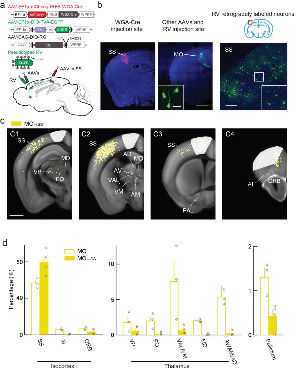
18 Supplementary Figure 15 Whole-brain distributions of inputs to MO SS neurons. (a) Viral vectors and injection procedure for RV-mediated transsynaptic retrograde tracing from MO SS neurons. (b) Left, fluorescence images of AAV-EF1α-mCherry-IRES-WGA-Cre injection site in SS (scale bar, 1 mm). Middle, injection site of other AAVs and RV in MO (scale bar, 1 mm). Inset, enlarged view of the region in white box showing AAV/RV infected neurons (green, scale bar, 20 µm). Right, retrogradely labeled neurons (green) in SS (scale bar, 200 µm). Inset, enlarged view of the region in white box (scale bar, 20 µm). Green, EGFP; red, mcherry; blue, DAPI. (c) RV-labeled neurons detected in all MO SS samples. White masks indicated the injection sites excluded from data analysis (scale bar, 1 mm). (d) Percentages of retrogradely labeled neurons in selected cortical and subcortical brain structures (MO SS, n = 4 mice). Each circle represents one mouse. Error bar, ±s.e.m. Data for MO inputs are the same as in Figure 3D.
Nature Neuroscience doi: /nn Supplementary Figure 1. Characterization of viral injections.
 Supplementary Figure 1 Characterization of viral injections. (a) Dorsal view of a mouse brain (dashed white outline) after receiving a large, unilateral thalamic injection (~100 nl); demonstrating that
Supplementary Materials for
 advances.sciencemag.org/cgi/content/full/1/10/e1500775/dc1 Supplementary Materials for Structural-functional connectivity deficits of neocortical circuits in the Fmr1 /y mouse model of autism Matthias
SUPPLEMENTARY INFORMATION
 SUPPLEMENTARY INFORMATION doi:10.1038/nature11306 Supplementary Figures Supplementary Figure 1. Basic characterization of GFP+ RGLs in the dentate gyrus of adult nestin-gfp mice. a, Sample confocal images
Supplementary figure 1: LII/III GIN-cells show morphological characteristics of MC
 1 2 1 3 Supplementary figure 1: LII/III GIN-cells show morphological characteristics of MC 4 5 6 7 (a) Reconstructions of LII/III GIN-cells with somato-dendritic compartments in orange and axonal arborizations
Nature Neuroscience: doi: /nn Supplementary Figure 1. ACx plasticity is required for fear conditioning.
 Supplementary Figure 1 ACx plasticity is required for fear conditioning. (a) Freezing time of conditioned and control mice before CS presentation and during CS presentation in a new context. Student s
Nature Neuroscience: doi: /nn Supplementary Figure 1. Diverse anorexigenic signals induce c-fos expression in CEl PKC-δ + neurons
 Supplementary Figure 1 Diverse anorexigenic signals induce c-fos expression in CEl PKC-δ + neurons a-c. Quantification of CEl c-fos expression in mice intraperitoneal injected with anorexigenic drugs (a),
Unique functional properties of somatostatin-expressing GABAergic neurons in mouse barrel cortex
 Supplementary Information Unique functional properties of somatostatin-expressing GABAergic neurons in mouse barrel cortex Luc Gentet, Yves Kremer, Hiroki Taniguchi, Josh Huang, Jochen Staiger and Carl
Nature Neuroscience: doi: /nn Supplementary Figure 1. Trial structure for go/no-go behavior
 Supplementary Figure 1 Trial structure for go/no-go behavior a, Overall timeline of experiments. Day 1: A1 mapping, injection of AAV1-SYN-GCAMP6s, cranial window and headpost implantation. Water restriction
Nature Neuroscience: doi: /nn Supplementary Figure 1. MADM labeling of thalamic clones.
 Supplementary Figure 1 MADM labeling of thalamic clones. (a) Confocal images of an E12 Nestin-CreERT2;Ai9-tdTomato brain treated with TM at E10 and stained for BLBP (green), a radial glial progenitor-specific
File name: Supplementary Information Description: Supplementary Figures, Supplementary Table and Supplementary References
 File name: Supplementary Information Description: Supplementary Figures, Supplementary Table and Supplementary References File name: Supplementary Data 1 Description: Summary datasheets showing the spatial
Nature Neuroscience: doi: /nn Supplementary Figure 1
 Supplementary Figure 1 Subcellular segregation of VGluT2-IR and TH-IR within the same VGluT2-TH axon (wild type rats). (a-e) Serial sections of a dual VGluT2-TH labeled axon. This axon (blue outline) has
Wenqin Hu, Cuiping Tian, Tun Li, Mingpo Yang, Han Hou & Yousheng Shu
 Distinct contributions of Na v 1.6 and Na v 1.2 in action potential initiation and backpropagation Wenqin Hu, Cuiping Tian, Tun Li, Mingpo Yang, Han Hou & Yousheng Shu Supplementary figure and legend Supplementary
Tuning properties of individual circuit components and stimulus-specificity of experience-driven changes.
 Supplementary Figure 1 Tuning properties of individual circuit components and stimulus-specificity of experience-driven changes. (a) Left, circuit schematic with the imaged component (L2/3 excitatory neurons)
Nature Neuroscience: doi: /nn Supplementary Figure 1. Lick response during the delayed Go versus No-Go task.
 Supplementary Figure 1 Lick response during the delayed Go versus No-Go task. Trial-averaged lick rate was averaged across all mice used for pyramidal cell imaging (n = 9). Different colors denote different
Supplementary Information
 Supplementary Information Title Degeneration and impaired regeneration of gray matter oligodendrocytes in amyotrophic lateral sclerosis Authors Shin H. Kang, Ying Li, Masahiro Fukaya, Ileana Lorenzini,
Nature Neuroscience: doi: /nn Supplementary Figure 1. Splenic atrophy and leucopenia caused by T3 SCI.
 Supplementary Figure 1 Splenic atrophy and leucopenia caused by T3 SCI. (a) Gross anatomy of representative spleens from control and T3 SCI mice at 28 days post-injury. (b and c) Hematoxylin and eosin
Nature Neuroscience: doi: /nn.4642
 Supplementary Figure 1 Recording sites and example waveform clustering, as well as electrophysiological recordings of auditory CS and shock processing following overtraining. (a) Recording sites in LC
Supplemental Information. A Labeled-Line Neural Circuit. for Pheromone-Mediated Sexual Behaviors in Mice
 Neuron, Volume Supplemental Information A Labeled-Line Neural Circuit for Pheromone-Mediated Sexual Behaviors in Mice Kentaro K. Ishii, Takuya Osakada, Hiromi Mori, Nobuhiko Miyasaka, Yoshihiro Yoshihara,
Nature Neuroscience: doi: /nn Supplementary Figure 1. Large-scale calcium imaging in vivo.
 Supplementary Figure 1 Large-scale calcium imaging in vivo. (a) Schematic illustration of the in vivo camera imaging set-up for large-scale calcium imaging. (b) High-magnification two-photon image from
Nature Neuroscience: doi: /nn Supplementary Figure 1. Confirmation that optogenetic inhibition of dopaminergic neurons affects choice
 Supplementary Figure 1 Confirmation that optogenetic inhibition of dopaminergic neurons affects choice (a) Sample behavioral trace as in Figure 1d, but with NpHR stimulation trials depicted as green blocks
SUPPLEMENTARY INFORMATION
 doi:10.1038/nature22324 Effects of photoinhibition on licking Photoinhibition of ALM or thalamus caused only small changes in lick early rates, no response rates, and licking latency. ALM photoinhibition
Supplemental Information. Dorsal Raphe Dual Serotonin-Glutamate Neurons. Drive Reward by Establishing Excitatory Synapses
 Cell Reports, Volume 26 Supplemental Information Dorsal Raphe Dual Serotonin-Glutamate Neurons Drive Reward by Establishing Excitatory Synapses on VTA Mesoaccumbens Dopamine Neurons Hui-Ling Wang, Shiliang
Nature Neuroscience: doi: /nn.4335
 Supplementary Figure 1 Cholinergic neurons projecting to the VTA are concentrated in the caudal mesopontine region. (a) Schematic showing the sites of retrograde tracer injections in the VTA: cholera toxin
Structural basis for the role of inhibition in facilitating adult brain plasticity
 Structural basis for the role of inhibition in facilitating adult brain plasticity Jerry L. Chen, Walter C. Lin, Jae Won Cha, Peter T. So, Yoshiyuki Kubota & Elly Nedivi SUPPLEMENTARY FIGURES 1-6 a b M
Supplementary Figure 1 Information on transgenic mouse models and their recording and optogenetic equipment. (a) 108 (b-c) (d) (e) (f) (g)
 108 (b-c) (d) (e) (f) (g)") Supplementary Figure 1 Information on transgenic mouse models and their recording and optogenetic equipment. (a) In four mice, cre-dependent expression of the hyperpolarizing opsin Arch in pyramidal cells
Supplementary Figure 1 Information on transgenic mouse models and their recording and optogenetic equipment. (a) In four mice, cre-dependent expression of the hyperpolarizing opsin Arch in pyramidal cells
A Sensorimotor Circuit in Mouse Cortex for Visual Flow Predictions
 Article A Sensorimotor Circuit in Mouse Cortex for Visual Flow Predictions Highlights d Mouse A4b/M sends a dense topographically organized input to V1 d d d Motor-related signals from A4b/M drive motor
Zhu et al, page 1. Supplementary Figures
 Zhu et al, page 1 Supplementary Figures Supplementary Figure 1: Visual behavior and avoidance behavioral response in EPM trials. (a) Measures of visual behavior that performed the light avoidance behavior
Supplementary Materials for
 www.sciencetranslationalmedicine.org/cgi/content/full/4/117/117ra8/dc1 Supplementary Materials for Notch4 Normalization Reduces Blood Vessel Size in Arteriovenous Malformations Patrick A. Murphy, Tyson
Supplementary Figure 1
 8w Pia II/III IV V VI PV EYFP EYFP PV EYFP PV d PV EYFP Supplementary Figure a Spike probability x - PV-Cre d Spike probability x - RS RS b e Spike probability Spike probability.6......8..... FS FS c f
Supplementary Figure 1. SDS-FRL localization of CB 1 in the distal CA3 area of the rat hippocampus. (a-d) Axon terminals (t) in stratum pyramidale
 Axon terminals (t) in stratum pyramidale") Supplementary Figure 1. SDS-FRL localization of CB 1 in the distal CA3 area of the rat hippocampus. (a-d) Axon terminals (t) in stratum pyramidale (b) show stronger immunolabeling for CB 1 than those in
Supplementary Figure 1. SDS-FRL localization of CB 1 in the distal CA3 area of the rat hippocampus. (a-d) Axon terminals (t) in stratum pyramidale (b) show stronger immunolabeling for CB 1 than those in
Nature Neuroscience: doi: /nn Supplementary Figure 1
 Supplementary Figure 1 Drd1a-Cre driven ChR2 expression in the SCN. (a) Low-magnification image of a representative Drd1a-ChR2 coronal brain section (n = 2) showing endogenous tdtomato fluorescence (magenta).
SUPPLEMENTARY INFORMATION
 SUPPLEMENTARY INFORMATION doi:10.1038/nature12107 Supplementary Figure 1. CLARITY preserves GFP and TdTomato signals. (a) 3D rendering of a 1mm-thick Thy1-EGFP M line mouse brain block processed by CLARITY
Supplementary Figure 1. ACE robotic platform. A. Overview of the rig setup showing major hardware components of ACE (Automatic single Cell
 2 Supplementary Figure 1. ACE robotic platform. A. Overview of the rig setup showing major hardware components of ACE (Automatic single Cell Experimenter) including the MultiClamp 700B, Digidata 1440A,
Supplementary Figure 1
 Supplementary Figure 1 Arcuate ChIEF-tdTomato neurons expressed TH These micrographs show that TH-Cre-ChIEF-tdTomato (magenta), expressed by AAV in a TH-Cre mouse, were immunostained with TH (green) in
Supplementary Figure 1. Microglia do not show signs of classical immune activation following MD a-b. Images showing immunoreactivity for MHCII (a)
") 1 Supplementary Figure 1. Microglia do not show signs of classical immune activation following MD a-b. Images showing immunoreactivity for MHCII (a) and CD45 (b) in fixed sections of binocular visual cortex
1 Supplementary Figure 1. Microglia do not show signs of classical immune activation following MD a-b. Images showing immunoreactivity for MHCII (a) and CD45 (b) in fixed sections of binocular visual cortex
Nature Neuroscience: doi: /nn Supplementary Figure 1
 Supplementary Figure 1 Distribution of GlyT2::eGFP fibers in the mouse thalamus at three different coronal levels. Note the innervation centered in the rostral (CL, PC) and caudal (PF) nuclear groups of
An acetylcholine-activated microcircuit drives temporal dynamics of cortical activity
 An acetylcholine-activated microcircuit drives temporal dynamics of cortical activity Naiyan Chen, Hiroki Sugihara, & Mriganka Sur Nature America, nc. All rights reserved. Cholinergic modulation of cortex
Nature Biotechnology: doi: /nbt Supplementary Figure 1. Analysis of hair bundle morphology in Ush1c c.216g>a mice at P18 by SEM.
 Supplementary Figure 1 Analysis of hair bundle morphology in Ush1c c.216g>a mice at P18 by SEM. (a-c) Heterozygous c.216ga mice displayed normal hair bundle morphology at P18. (d-i) Disorganized hair bundles
Nature Neuroscience: doi: /nn Supplementary Figure 1
 Supplementary Figure 1 Atlas representations of the midcingulate (MCC) region targeted in this study compared against the anterior cingulate (ACC) region commonly reported. Coronal sections are shown on
Comparison of open chromatin regions between dentate granule cells and other tissues and neural cell types.
 Supplementary Figure 1 Comparison of open chromatin regions between dentate granule cells and other tissues and neural cell types. (a) Pearson correlation heatmap among open chromatin profiles of different
Supplementary Information
 1 Supplementary Information A role for primary cilia in glutamatergic synaptic integration of adult-orn neurons Natsuko Kumamoto 1,4,5, Yan Gu 1,4, Jia Wang 1,4, Stephen Janoschka 1,2, Ken-Ichi Takemaru
SUPPLEMENTARY INFORMATION
 doi:1.138/nature1139 a d Whisker angle (deg) Whisking repeatability Control Muscimol.4.3.2.1 -.1 8 4-4 1 2 3 4 Performance (d') Pole 8 4-4 1 2 3 4 5 Time (s) b Mean protraction angle (deg) e Hit rate (p
Nature Medicine: doi: /nm.4322
 1 2 3 4 5 6 7 8 9 10 11 Supplementary Figure 1. Predicted RNA structure of 3 UTR and sequence alignment of deleted nucleotides. (a) Predicted RNA secondary structure of ZIKV 3 UTR. The stem-loop structure
Supplementary Figure 1
 Supplementary Figure 1 The average sigmoid parametric curves of capillary dilation time courses and average time to 50% peak capillary diameter dilation computed from individual capillary responses averaged
Summary of behavioral performances for mice in imaging experiments.
 Supplementary Figure 1 Summary of behavioral performances for mice in imaging experiments. (a) Task performance for mice during M2 imaging experiments. Open triangles, individual experiments. Filled triangles,
293T cells were transfected with indicated expression vectors and the whole-cell extracts were subjected
 SUPPLEMENTARY INFORMATION Supplementary Figure 1. Formation of a complex between Slo1 and CRL4A CRBN E3 ligase. (a) HEK 293T cells were transfected with indicated expression vectors and the whole-cell
Nature Neuroscience: doi: /nn Supplementary Figure 1. Iliopsoas and quadratus lumborum motor neurons in the L2 spinal segment.
 Supplementary Figure 1 Iliopsoas and quadratus lumborum motor neurons in the L2 spinal segment. (A) IL and QL motor neurons were labeled after CTb-488 (green) muscle injections at birth. At P4, the L2
SUPPLEMENTARY INFORMATION
 Supplementary Figure 1. Normal AMPAR-mediated fepsp input-output curve in CA3-Psen cdko mice. Input-output curves, which are plotted initial slopes of the evoked fepsp as function of the amplitude of the
Nature Neuroscience: doi: /nn Supplementary Figure 1. Visualization of AT1a-positive cells using AT1a lacz/+ mouse.
 Supplementary Figure 1 Visualization of AT1a-positive cells using AT1a lacz/+ mouse. (a f) Immunohistochemical detection of β-gal in the mouse brain. Coronal sections at the respective anteroposterior
A toolbox of Cre-dependent optogenetic transgenic mice for light-induced activation and silencing
 A toolox of Cre-dependent optogenetic transgenic mice for light-induced activation and silencing Linda Madisen 1, Tianyi Mao 2,7, Henner Koch 3, Jia-min Zhuo, Antal Berenyi 5, Shigeyoshi Fujisawa 5, Yun-Wei
Parallel, Redundant Circuit Organization for Homeostatic Control of Feeding Behavior
 Parallel, Redundant Circuit Organization for Homeostatic Control of Feeding Behavior J. Nicholas Betley, 1,2 Zhen Fang Huang Cao, 1,2 Kimberly D. Ritola, 1 and Scott M. Sternson 1, * 1 Janelia Farm Research
Supplementary Figure 1: Validation of labeling specificity of immature OSNs and presynaptic terminals. (A) (B) (C) (D) (E)
 (B) (C) (D) (E)") Supplementary Figure 1: Validation of labeling specificity of immature OSNs and presynaptic terminals. (A) Confocal images of septal olfactory epithelium of an adult Gγ8-sypGFP-tdTom mouse showing colocalization
Supplementary Figure 1: Validation of labeling specificity of immature OSNs and presynaptic terminals. (A) Confocal images of septal olfactory epithelium of an adult Gγ8-sypGFP-tdTom mouse showing colocalization
a 0,8 Figure S1 8 h 12 h y = 0,036x + 0,2115 y = 0,0366x + 0,206 Labeling index Labeling index ctrl shrna Time (h) Time (h) ctrl shrna S G2 M G1
 Time (h) ctrl shrna S G2 M G1") (GFP+ BrdU+)/GFP+ Labeling index Labeling index Figure S a, b, y =,x +, y =,x +,,,,,,,, Time (h) - - Time (h) c d S G M G h M G S G M G S G h Time of BrdU injection after electroporation (h) M G S G M
(GFP+ BrdU+)/GFP+ Labeling index Labeling index Figure S a, b, y =,x +, y =,x +,,,,,,,, Time (h) - - Time (h) c d S G M G h M G S G M G S G h Time of BrdU injection after electroporation (h) M G S G M
Lack of GPR88 enhances medium spiny neuron activity and alters. motor- and cue- dependent behaviors
 Lack of GPR88 enhances medium spiny neuron activity and alters motor- and cue- dependent behaviors Albert Quintana, Elisenda Sanz, Wengang Wang, Granville P. Storey, Ali D. Güler Matthew J. Wanat, Bryan
doi: /nature09554
 SUPPLEMENTARY INFORMATION doi:10.1038/nature09554 Supplementary Figure 1: Optical Tracing with New Photoactivatable GFP Variants Reveals Enhanced Labeling of Neuronal Processes We qualitatively compare
In the cortex, many neurons selectively respond
 PRESYNAPTIC NETWORKS Single-cell initiated monosynaptic tracing reveals layer-specific cortical network modules Adrian Wertz, * Stuart Trenholm, * Keisuke Yonehara, Daniel Hillier, Zoltan Raics, Marcus
You submitted this quiz on Sun 19 May :32 PM IST (UTC +0530). You got a score of out of
. You got a score of out of") Feedback Ex6 You submitted this quiz on Sun 19 May 2013 9:32 PM IST (UTC +0530). You got a score of 10.00 out of 10.00. Question 1 What is common to Parkinson, Alzheimer and Autism? Electrical (deep brain)
Feedback Ex6 You submitted this quiz on Sun 19 May 2013 9:32 PM IST (UTC +0530). You got a score of 10.00 out of 10.00. Question 1 What is common to Parkinson, Alzheimer and Autism? Electrical (deep brain)
Target-specific M1 inputs to infragranular S1 pyramidal neurons
 J Neurophysiol 116: 1261 1274, 216. First published June 22, 216; doi:1.1152/jn.132.215. Target-specific M1 inputs to infragranular S1 pyramidal neurons Amanda K. Kinnischtzke, Erika E. Fanselow, and Daniel
Supplementary Figure 1
 Supplementary Figure 1 Supplementary Figure 1 SNARE Probes for FRET/2pFLIM Analysis Used in the Present Study. mturquoise (mtq) and Venus (Ven) are in blue and yellow, respectively. The soluble N-ethylmaleimide-sensitive
Nucleus Accumbens Subnuclei Regulate Motivated Behavior via Direct Inhibition and Disinhibition of VTA Dopamine Subpopulations
 Article Nucleus Accumbens Subnuclei Regulate Motivated Behavior via Direct Inhibition and Disinhibition of VTA Dopamine Subpopulations Highlights d Mesolimbic DA subpopulations are embedded within different
Reward prediction based on stimulus categorization in. primate lateral prefrontal cortex
 Reward prediction based on stimulus categorization in primate lateral prefrontal cortex Xiaochuan Pan, Kosuke Sawa, Ichiro Tsuda, Minoro Tsukada, Masamichi Sakagami Supplementary Information This PDF file
SUPPLEMENTARY INFORMATION
 doi: 10.1038/nature06310 SUPPLEMENTARY INFORMATION www.nature.com/nature 1 www.nature.com/nature 2 www.nature.com/nature 3 Supplementary Figure S1 Spontaneous duration of wake, SWS and REM sleep (expressed
mm Distance (mm)
") b a Magnet Illumination Coverslips MPs Objective 2575 µm 1875 µm 1575 µm 1075 µm 875 µm 545 µm 20µm 2 3 0.5 0.3mm 1 1000 100 10 1 0.1 1000 100 10 1 0.1 Field Induction (Gauss) 1.5 0 5 10 15 20 Distance
b a Magnet Illumination Coverslips MPs Objective 2575 µm 1875 µm 1575 µm 1075 µm 875 µm 545 µm 20µm 2 3 0.5 0.3mm 1 1000 100 10 1 0.1 1000 100 10 1 0.1 Field Induction (Gauss) 1.5 0 5 10 15 20 Distance
Nature Methods: doi: /nmeth Supplementary Figure 1. Activity in turtle dorsal cortex is sparse.
 Supplementary Figure 1 Activity in turtle dorsal cortex is sparse. a. Probability distribution of firing rates across the population (notice log scale) in our data. The range of firing rates is wide but
Serotonergic Control of the Developing Cerebellum M. Oostland
 Serotonergic Control of the Developing Cerebellum M. Oostland Summary Brain development is a precise and crucial process, dependent on many factors. The neurotransmitter serotonin is one of the factors
Supplementary Figure 1: Signaling centers contain few proliferating cells, express p21, and
 Supplementary Figure 1: Signaling centers contain few proliferating cells, express p21, and exclude YAP from the nucleus. (a) Schematic diagram of an E10.5 mouse embryo. (b,c) Sections at B and C in (a)
Supplementary Figure 1. Properties of various IZUMO1 monoclonal antibodies and behavior of SPACA6. (a) (b) (c) (d) (e) (f) (g) .
 (b) (c) (d) (e) (f) (g) .") Supplementary Figure 1. Properties of various IZUMO1 monoclonal antibodies and behavior of SPACA6. (a) The inhibitory effects of new antibodies (Mab17 and Mab18). They were investigated in in vitro fertilization
Supplementary Figure 1. Properties of various IZUMO1 monoclonal antibodies and behavior of SPACA6. (a) The inhibitory effects of new antibodies (Mab17 and Mab18). They were investigated in in vitro fertilization
Supplementary Figure 1
 Supplementary Figure 1 Localization of virus injections. (a) Schematic showing the approximate center of AAV-DIO-ChR2-YFP injection sites in the NAc of Dyn-cre mice (n=8 mice, 16 injections; caudate/putamen,
Nature Neuroscience: doi: /nn Supplementary Figure 1. Behavioral training.
 Supplementary Figure 1 Behavioral training. a, Mazes used for behavioral training. Asterisks indicate reward location. Only some example mazes are shown (for example, right choice and not left choice maze
Movement Initiation Signals in Mouse Whisker Motor Cortex
 Article Movement Initiation Signals in Mouse Whisker Motor Cortex Highlights d Optogenetic excitation (inactivation) of wm1 evokes (inhibits) whisking d d d Layer-specific neuronal activity in wm1 encodes
Conditional Deletion of All Neurexins Defines Diversity of Essential Synaptic Organizer Functions for Neurexins
 Article Conditional Deletion of All Neurexins Defines Diversity of Essential Synaptic Organizer Functions for Neurexins Highlights d Pan-neurexin KO causes diverse synaptic phenotypes in a synapse-specific
Authors: K. L. Arendt, M. Royo, M. Fernández-Monreal, S. Knafo, C. N. Petrok, J.
 SUPPLEMENTARY INFORMATION Title: PIP 3 controls synaptic function by maintaining AMPA receptor clustering at the postsynaptic membrane Authors: K. L. Arendt, M. Royo, M. Fernández-Monreal, S. Knafo, C.
Supplementary Figure 1
 Supplementary Figure 1 Genetic labeling of microglia Male and female 2-3 month-old CreERT2;R26-tdTomato mice or CreERT2;R26-tdTomato;Iba1-eGFP transgenic mice were treated with 1x, 2x (48 h apart), or
BMI risk SNPs associate with increased CADM1 and CADM2 expression in the cerebellum of human subjects.
 Supplementary Figure 1 BMI risk SNPs associate with increased CADM1 and CADM2 expression in the cerebellum of human subjects. Boxplots show the 25% and 75% quantiles of normalized mrna expression levels
Parallel Driving and Modulatory Pathways Link the Prefrontal Cortex and Thalamus
 Boston University OpenBU Health Sciences http://open.bu.edu SAR: Health Sciences: Scholarly Papers 2007-9-5 Parallel Driving and Modulatory Pathways Link the Prefrontal Cortex and Thalamus Zikopoulos,
1. Microfluidic device characteristics and cell compartmentalization
 Supplementary figures: 1. Microfluidic device characteristics and cell compartmentalization Microfluidic channels were molded in PDMS (Fig. S1 A and B) over silicon masters, which were structured with
SUPPLEMENTARY INFORMATION
 SUPPLEMENTARY INFORMATION Supplementary Figure 1. The expression of ephrin-b2 H2BGFP persists in the post-hearingonset organ of Corti and is specifically restricted to supporting cells. Sox2 immunolabeling
Supplementary Figure 1. Nature Neuroscience: doi: /nn.4547
 Supplementary Figure 1 Characterization of the Microfetti mouse model. (a) Gating strategy for 8-color flow analysis of peripheral Ly-6C + monocytes from Microfetti mice 5-7 days after TAM treatment. Living
Thalamo-Cortical Relationships Ultrastructure of Thalamic Synaptic Glomerulus
 Central Visual Pathways V1/2 NEUR 3001 dvanced Visual Neuroscience The Lateral Geniculate Nucleus () is more than a relay station LP SC Professor Tom Salt UCL Institute of Ophthalmology Retina t.salt@ucl.ac.uk
Prss56, a novel marker of adult neurogenesis in the mouse brain. - Supplemental Figures 1 to 5- Brain Structure and Function
 Prss56, a novel marker of adult neurogenesis in the mouse brain - Supplemental Figures 1 to 5- Brain Structure and Function Alexandre Jourdon 1,2, Aurélie Gresset 1, Nathalie Spassky 1, Patrick Charnay
SUPPLEMENTARY INFORMATION
 SUPPLEMENTARY INFORMATION Human cerebral cortex development from pluripotent stem cells to functional excitatory synapses Yichen Shi 1,2, Peter Kirwan 1,2, James Smith 1,2, Hugh P.C. Robinson 3 and Frederick
Basal Ganglia. Introduction. Basal Ganglia at a Glance. Role of the BG
 Basal Ganglia Shepherd (2004) Chapter 9 Charles J. Wilson Instructor: Yoonsuck Choe; CPSC 644 Cortical Networks Introduction A set of nuclei in the forebrain and midbrain area in mammals, birds, and reptiles.
SUPPLEMENTARY FIG. S2. Representative counting fields used in quantification of the in vitro neural differentiation of pattern of dnscs.
 Supplementary Data SUPPLEMENTARY FIG. S1. Representative counting fields used in quantification of the in vitro neural differentiation of pattern of anpcs. A panel of lineage-specific markers were used
Supplemental Information. Evoked Axonal Oxytocin Release. in the Central Amygdala. Attenuates Fear Response
 Neuron, Volume 73 Supplemental Information Evoked Axonal Oxytocin Release in the Central Amygdala Attenuates Fear Response H. Sophie Knobloch, Alexandre Charlet, Lena C. Hoffmann, Marina Eliava, Sergey
Supplementary Materials for VAMP4 directs synaptic vesicles to a pool that selectively maintains asynchronous neurotransmission
 Supplementary Materials for VAMP4 directs synaptic vesicles to a pool that selectively maintains asynchronous neurotransmission Jesica Raingo, Mikhail Khvotchev, Pei Liu, Frederic Darios, Ying C. Li, Denise
Nature Neuroscience: doi: /nn Supplementary Figure 1
 Supplementary Figure 1 Relative expression of K IR2.1 transcript to enos was reduced 29-fold in capillaries from knockout animals. Relative expression of K IR2.1 transcript to enos was reduced 29-fold
SUPPLEMENTARY INFORMATION
 Supplementary Figure 1. Behavioural effects of ketamine in non-stressed and stressed mice. Naive C57BL/6 adult male mice (n=10/group) were given a single dose of saline vehicle or ketamine (3.0 mg/kg,
SUPPLEMENTARY INFORMATION
 Figure S1. Loss of Ena/VASP proteins inhibits filopodia and neuritogenesis. (a) Bar graph of filopodia number per stage 1 control and mmvvee (Mena/ VASP/EVL-null) neurons at 40hrs in culture. Loss of all
Nature Neuroscience: doi: /nn Supplementary Figure 1
 Supplementary Figure 1 Quantification of myelin fragments in the aging brain (a) Electron microscopy on corpus callosum is shown for a 18-month-old wild type mice. Myelin fragments (arrows) were detected
NERVOUS SYSTEM & SENSES TEACHER COPY
 NERVOUS SYSTEM & SENSES TEACHER COPY FUNCTIONS OF THE NERVOUS SYSTEM What are the three functions of the Nervous System? 1. Receives information about what is happening inside and outside of your body
Supplemental Information. A Visual-Cue-Dependent Memory Circuit. for Place Navigation
 Neuron, Volume 99 Supplemental Information A Visual-Cue-Dependent Memory Circuit for Place Navigation Han Qin, Ling Fu, Bo Hu, Xiang Liao, Jian Lu, Wenjing He, Shanshan Liang, Kuan Zhang, Ruijie Li, Jiwei
Visual system invades the endbrain: pathways to striatum and cortex (continued) Why this happened in evolution
 Why this happened in evolution") Visual system invades the endbrain: pathways to striatum and cortex (continued) Why this happened in evolution What were the adaptive advantages? Visual information reaching the striatum directly: Advantages
Visual system invades the endbrain: pathways to striatum and cortex (continued) Why this happened in evolution What were the adaptive advantages? Visual information reaching the striatum directly: Advantages
Neocortex. Cortical Structures in the Brain. Neocortex Facts. Laminar Organization. Bark-like (cortical) structures: Shepherd (2004) Chapter 12
 structures: Shepherd (2004) Chapter 12") Neocortex Shepherd (2004) Chapter 12 Rodney Douglas, Henry Markram, and Kevan Martin Instructor: Yoonsuck Choe; CPSC 644 Cortical Networks Cortical Structures in the Brain Bark-like (cortical) structures:
Neocortex Shepherd (2004) Chapter 12 Rodney Douglas, Henry Markram, and Kevan Martin Instructor: Yoonsuck Choe; CPSC 644 Cortical Networks Cortical Structures in the Brain Bark-like (cortical) structures:
a b c periosteum parietal bone bone marrow dura periosteum suture mesenchyme osteogenic front suture mesenchyme 1
 coronary suture sagittal suture DOI: 10.1038/ncb3139 a b c e parietal bone suture mesenchyme parietal bone bone marrow ura ura ura f parietal bone ura suture mesenchyme bone g ura osteogenic front suture
A Cortical Circuit for Gain Control by Behavioral State
 A Cortical Circuit for Gain Control by Behavioral State Yu Fu, 1, * Jason M. Tucciarone, 2,3 J. Sebastian Espinosa, 1 Nengyin Sheng, 4 Daniel P. Darcy, 1 Roger A. Nicoll, 4 Z. Josh Huang, 2 and Michael
SOMATOSENSORY SYSTEMS
 SOMATOSENSORY SYSTEMS Schematic diagram illustrating the neural pathways that convey somatosensory information to the cortex and, subsequently, to the motor system. Double arrows show reciprocal connections.
Hormonal gain control of a medial preoptic area social reward circuit
 CORRECTION NOTICE Nat. Neurosci. 20, 449 458 (2017) Hormonal gain control of a medial preoptic area social reward circuit Jenna A McHenry, James M Otis, Mark A Rossi, J Elliott Robinson, Oksana Kosyk,
label the basement membrane). Different fixation methods of EB-perfused P8 mice to optimize the combination
. Different fixation methods of EB-perfused P8 mice to optimize the combination") Supplementary Figure 1 Optimization of the tissue fixation protocol to combine EB perfusion and IB4 endothelial tip cell staining in the postnatal mouse brain. a-l Labeling of EB-perfused P8 mice with
Supplementary Figure 1 Optimization of the tissue fixation protocol to combine EB perfusion and IB4 endothelial tip cell staining in the postnatal mouse brain. a-l Labeling of EB-perfused P8 mice with
Broad Integration of Expression Maps and Co-Expression Networks Compassing Novel Gene Functions in the Brain
 Supplementary Information Broad Integration of Expression Maps and Co-Expression Networks Compassing Novel Gene Functions in the Brain Yuko Okamura-Oho a, b, *, Kazuro Shimokawa c, Masaomi Nishimura b,
(a) Significant biological processes (upper panel) and disease biomarkers (lower panel)
 Significant biological processes (upper panel) and disease biomarkers (lower panel)") Supplementary Figure 1. Functional enrichment analyses of secretomic proteins. (a) Significant biological processes (upper panel) and disease biomarkers (lower panel) 2 involved by hrab37-mediated secretory
Supplementary Figure 1. Functional enrichment analyses of secretomic proteins. (a) Significant biological processes (upper panel) and disease biomarkers (lower panel) 2 involved by hrab37-mediated secretory