Cerebral Vascular Diseases. Nabila Hamdi MD, PhD
|
|
|
- Sandra Bruce
- 5 years ago
- Views:
Transcription

1 Cerebral Vascular Diseases Nabila Hamdi MD, PhD
2 Outline I. Stroke statistics II. Cerebral circulation III. Clinical symptoms of stroke IV. Pathogenesis of cerebral infarcts (Stroke) 1. Ischemic - Thrombotic - Embolic - Lacunar/small vessel disease 2. Hemorrhagic - Primary brain parenchymal hemorrhage - Subarachnoid hemorrhage & saccular aneurysm V. Traumatic brain vascular injury - Epidural hematoma - Subdural hematoma 2
3 ILOs 1. To distinguish ischemic stroke from hemorrhagic stroke in terms of etiology and pathology 2. To know the most common causes of ischemic infarcts 3. Explain small vessel disease, mention 2 conditions that cause it and know its effects on the brain 4. Know 4 different causes of intracerebral hemorrhage 5. Understand the role of hypertension in the pathogenesis of hemorrhagic strokes 6. Explain the subarachnoid hemorrhage resulting from the rupture of saccular aneurysms 7. Distinguish intracerebral hemorrhage from traumatic brain hemorrhages 8. Differentiate between epidural and subdural hematoma 3
4 Stroke Statistics About 795,000 Americans each year suffer a new or recurrent stroke. That means, on average, a stroke occurs every 40 seconds. No. 3 cause of death after cardiovascular diseases and cancer About 1 of every 18 deaths. On average, every 4 minutes someone dies of stroke. Of all strokes, 87% are ischemic, 10% are intracerebral hemorrhages, and 3% are subarachnoid hemorrhages The estimated direct and indirect cost of stroke for 2009 is $68.9 billion (inpatient care, rehabilitation and follow-up care necessary for lasting deficits) 4

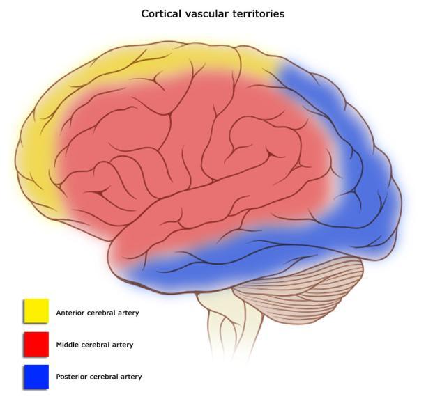
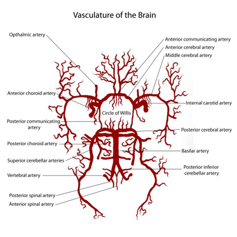
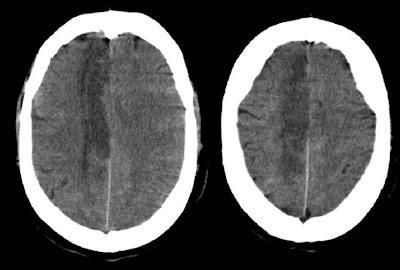
5 Cerebral Circulation Neuro4Students, Cerebrovascular attack 5

6 Cerebral Circulation Circle of Willis: source of collateral flow major little if any collateral flow for the deep penetrating vessels (thalamus, basal ganglia, and deep white matter) 6


7 Stroke Ischemic (Clots) 87% Hemorrhagic (Bleeds) 13% Stroke Ischemic injury/infarction of specific regions of the brain, depending on the vessel involved. Hemorrhage leads to direct tissue damage as well as secondary ischemic injury 3% subarachnoid hemorrhage 10% intracerebral hemorrhage 7
 1/3 of patients with TIA develop clinically significant infarcts within 5 years")
8 Stroke Focal neurological deficit Longer than 24 hours Less than 24 hours (minutes) Permanent tissue damage No permanent tissue damage Stroke Transient ischemic attacks (TIAs) 1/3 of patients with TIA develop clinically significant infarcts within 5 years 8


9 Signs & Symptoms of Stroke 9
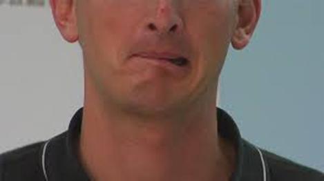
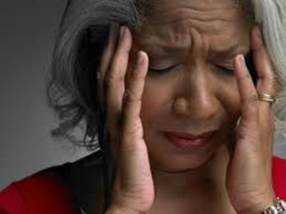
10 Signs & Symptoms of Stroke Hemiplegia: Sudden paralysis of a leg, arm or one side of the face Hemiparesis: Sudden numbness or weakness of arm, leg or face Consciousness: +/- loss of consciousness Aphasia: loss/impairment of the power to use or comprehend words. Dysarthria: affects the mechanics of speech. Amaurosis fugas sudden loss of vision in one or both eyes: gray or black shade coming down over their eye Hemianopsia: loss of half of the visual field. Ataxia: Sudden trouble walking, dizziness, loss of balance or coordination, leading to difficulty in walking normally Vertigo (spinning form of feeling dizzy) Sudden severe headache with no known cause Memory, emotions, orientation, Incontinence 10


11 Warning Symptoms of Stroke Does one side of the face droop or is it numb? Ask the person to smile. Is the person's smile uneven? Is one arm weak or numb? Ask the person to raise both arms. Does one arm drift downward? Is speech slurred? Is the person unable to speak or hard to understand? Ask the person to repeat a simple sentence. Is the sentence repeated correctly? even if the symptoms go away, call emergency and get the person to the hospital immediately. Check the time so you'll know when the first symptoms appeared 11
12 Ischemic Stroke Definition: Ischemic stroke or cerebral infarct is a focal brain necrosis due to complete and prolonged ischemia that affects all tissue elements, neurons, glia and vessels. Risk factors: Modifiable Hypertension Age >55 Diabetes Atrial fibrillation Smoking Hyperlipidemia Carotid stenosis Male gender Black race Non modifiable Family history of stroke Personal history of stroke Sickle Cell Disease Lack of physical activity Major Causes: 1. Atherosclerosis 2. Embolisms 3. Small vessel disease 4. Vascular spasm (following hemorrhagic stroke) Other: Vasculitis, hypercoagulability, dissection of a vessel wall, sickel cell disease


13 Ischemic Stroke 1. Atherosclerosis (most common cause) Kindly refer to CVS lecture Neuro4Students Cerebrovascular attack 13

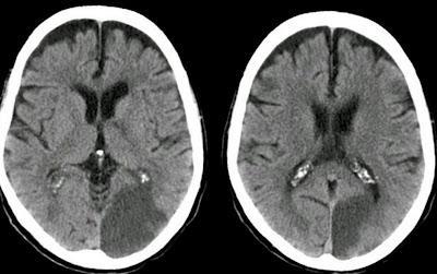
14 Left MCA Right ACA Left PCA Neuroradiology Unit, S P Institute of Neurosciences,Solapur,Maharashtra, INDIA Radiology department of the Rijnland Hospital in Leiderdorp, the Netherlands Lacunar infarct 14

15 1. Atherosclerosis Risk factors! Ischemic Stroke Atheromatous plaques can cause narrowing or occlusion of the vascular lumen by themselves or after rupture and thrombosis Bifurcation points of large arteries & major cervical and intracranial arteries (blood flow!) Atherothrombotic infarcts evolve within hours or days The most severe atherosclerotic lesions are typically encountered within large vessels However, Some patients have an asymptomatic occlusion of a cervical internal carotid artery?! Cerebrovascular anatomy and common sites of atherosclerosis 15

16 2. Embolism Ischemic Stroke Most emboli are fragments of blood clot that originate in the heart or major vessels MI, atrial fibrillation and other arythmias, endocarditis Rarer causes are fat, air and tumor emboli Embolic infarcts have an abrupt onset Assumed if: - source of embolism is present - multiple infarcts in the brain - infarcts in other organs - absence of atherosclerosis - absence of other vascular disease 16 UCSF Department of Surgery, The University of California, San Francisco
 Atherosclerosis of small arteries Small")
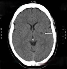
17 3. Lacunar Infarcts Ischemic Stroke Occlusion of deep penetrating branches of major cerebral arteries Deeper parts of the brain (basal ganglia, thalamus, deep white matter) and brain stem. The infarcts are generally from 2-20 mm in diameter Typically, no impairments in cognition, memory, speech, or level of consciousness (cortex not affected) Atherosclerosis of small arteries Small vessel disease; hyaline arteriosclerosis in hypertension and diabetes, but occurs in old age w/o these predisposing conditions. Lacunar stroke 17
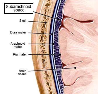
18 Intracranial Hemorrhages Subarachnoid Hemorrhage (Saccular Aneurysms) Non- Traumatic Primary Brain Parenchymal Hemorrhage Epidural Hematoma Subdural Hematoma Traumatic 18

19 Primary Brain Parenchymal Hemorrhage Spontaneous, nontraumatic intraparenchymal hemorrhages Rupture of small penetrating arteries (basal ganglia & thalamus) Accounts for 15% of deaths among patients with chronic hypertension Hypertension is the most underlying cause Small vessel disease: arteriolar walls affected by hyaline change are weaker than normal vessels and are therefore more vulnerable to rupture. Other causes: aging, smoking, oral contraceptives, drug abuse, excessive alcohol intake 19
 Risk of herniation of cerebellum and brain stem compression deep coma,")
20 Primary Brain Parenchymal Hemorrhage Brain is asymmetrically distorted effect + associated edema) (mass Hematoma may dissect into the ventricles Onset is always abrupt with evidence of increased intracranial pressure: severe headache, vomiting, seizures, rapid loss of consciousness, papilledema (swelling of optic disc) Risk of herniation of cerebellum and brain stem compression deep coma, irregular respirations, dilated nonresponsive pupils and spasticity Massive hypertensive hemorrhage rupturing into a lateral ventricle 20

21 Normal cerebellar tonsils Cerebellar tonsils are pushed through the foramen magnum into the spinal canal Cerebellar Herniation 21 Mayfield Clinic, University of Cincinnati Department of Neurosurgery
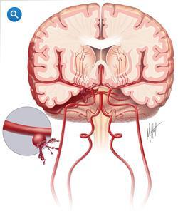
22 Subarachnoid Hemorrhage 22
23 Subarachnoid Hemorrhage Rupture of saccular aneurysms is the most common cause of nontraumatic SAH Saccular (berry) aneurysms are present in 1% of the general population Arise most commonly at arterial bifurcations in the territories of ICA To a less extent in posterior (vertebrobasilar) circulation Enlarge with time and are at greatest risk for rupture once they reach 6-10 mm in diameter Relative frequency of common sites of saccular (berry) aneurysms in the circle of Willis 23
24 Subarachnoid Hemorrhage SAH resulting from rupture of saccular aneurysm is less common than primary cerebral hemorrhage, with women being more affected than males with most cases before age of 50 Abrupt onset with severe headache, often described as the "worst headache of my life, vomiting and loss of consciousness (increased ICP) Meningeal signs are usually present (neck rigidity and pain, back pain, and bilateral leg pain). Seizures during the acute phase of SAH occur in 10-25% of patients. They result from the sudden rise in ICP or direct cortical irritation by blood. Blood in CSF(lumbar punction) 50% dye within several days of onset of symptoms Might be acutely complicated by cerebral infarcts (arterial spasm), acute hydrocephalus (increased accumulation of CSF in ventricles) and herniation. 24

25 Traumatic Vascular Injury 25

26 Traumatic Vascular Injury C. Large organizing subdural hematoma attached to the dura B. Epidural hematoma covering a portion of the dura 26
 Neurosurgical emergency requiring prompt")
27 Epidural Hematoma Neurosurgical emergency Rupture of a meningeal artery Middle meningeal artery, fracture of temporal bone Compression of subjacent dura Risks: herniation, brain stem compression and death Lucid interval is typical: time in which the conditions of the patients improve after a head trauma before deteriorating (loss of consciousness) Neurosurgical emergency requiring prompt drainage Rescuers say that he maintained consciousness when they first reached him, but his health quickly deteriorated 27
 Size vary from small to massive hemorrhage with mass effect Sudden and progressive worsening of symptoms No lucid interval!")
28 Subdural Hematoma Tearing of bridging veins that extend from brain surface to dural sinuses Rapid change in head velocity: head blows, violent shaking in infants In elderly, even after minor trauma: brain atrophy, veins are stretched out (more space for movement) Size vary from small to massive hemorrhage with mass effect Sudden and progressive worsening of symptoms No lucid interval!! 28
29 References ROBBINS Basic Pathology 8 th Edition Cerebrovascular diseases, Clinical aspects. Dr. Michael P. Merchut FERNE: Foundation for Education and Research in Neurological Emergencies, Stroke Pathophysiology, Sid Shah, MD 29

30 30
Overview of Stroke: Etiologies, Demographics, Syndromes, and Outcomes. Alex Abou-Chebl, MD, FSVIN Medical Director, Stroke Baptist Health Louisville
 Overview of Stroke: Etiologies, Demographics, Syndromes, and Outcomes Alex Abou-Chebl, MD, FSVIN Medical Director, Stroke Baptist Health Louisville Disclosure Statement of Financial Interest Within the
Vascular Disorders. Nervous System Disorders (Part B-1) Module 8 -Chapter 14. Cerebrovascular disease S/S 1/9/2013
 Module 8 -Chapter 14. Cerebrovascular disease S/S 1/9/2013") Nervous System Disorders (Part B-1) Module 8 -Chapter 14 Overview ACUTE NEUROLOGIC DISORDERS Vascular Disorders Infections/Inflammation/Toxins Metabolic, Endocrinologic, Nutritional, Toxic Neoplastic Traumatic
Nervous System Disorders (Part B-1) Module 8 -Chapter 14 Overview ACUTE NEUROLOGIC DISORDERS Vascular Disorders Infections/Inflammation/Toxins Metabolic, Endocrinologic, Nutritional, Toxic Neoplastic Traumatic
Cerebrovascular Disorders. Blood, Brain, and Energy. Blood Supply to the Brain 2/14/11
 Cerebrovascular Disorders Blood, Brain, and Energy 20% of body s oxygen usage No oxygen/glucose reserves Hypoxia - reduced oxygen Anoxia - Absence of oxygen supply Cell death can occur in as little as
/ / / / / / Hospital Abstraction: Stroke/TIA. Participant ID: Hospital Code: Multi-Ethnic Study of Atherosclerosis
 Multi-Ethnic Study of Atherosclerosis Participant ID: Hospital Code: Hospital Abstraction: Stroke/TIA History and Hospital Record 1. Was the participant hospitalized as an immediate consequence of this
E X P L A I N I N G STROKE
 EXPLAINING STROKE Introduction Explaining Stroke is a practical step-by-step booklet that explains how a stroke happens, different types of stroke and how to prevent a stroke. Many people think a stroke
Classical CNS Disease Patterns
 Classical CNS Disease Patterns Inflammatory Traumatic In response to the trauma of having his head bashed in GM would have experienced some of these features. NOT TWO LITTLE PEENY WEENY I CM LACERATIONS.
CEREBROVASCULAR DISEASES. By: Shifaa AlQa qa
 CEREBROVASCULAR DISEASES By: Shifaa AlQa qa Cerebrovascular diseases Brain disorders caused by pathologic processes involving blood vessels 3 pathogenic mechanisms (1) thrombotic occlusion, (2) embolic
Nicolas Bianchi M.D. May 15th, 2012
 Nicolas Bianchi M.D. May 15th, 2012 New concepts in TIA Differential Diagnosis Stroke Syndromes To learn the new definitions and concepts on TIA as a condition of high risk for stroke. To recognize the
NEURO IMAGING 2. Dr. Said Huwaijah Chairman of radiology Dep, Damascus Univercity
 NEURO IMAGING 2 Dr. Said Huwaijah Chairman of radiology Dep, Damascus Univercity I. EPIDURAL HEMATOMA (EDH) LOCATION Seventy to seventy-five percent occur in temporoparietal region. CAUSE Most likely caused
Acute stroke. Ischaemic stroke. Characteristics. Temporal classification. Clinical features. Interpretation of Emergency Head CT
 Ischaemic stroke Characteristics Stroke is the third most common cause of death in the UK, and the leading cause of disability. 80% of strokes are ischaemic Large vessel occlusive atheromatous disease
Marc Norman, Ph.D. - Do Not Use without Permission 1. Cerebrovascular Accidents. Marc Norman, Ph.D. Department of Psychiatry
 Cerebrovascular Accidents Marc Norman, Ph.D. Department of Psychiatry Neuropsychiatry and Behavioral Medicine Neuropsychology Clinical Training Seminar 1 5 http://www.nlm.nih.gov/medlineplus/ency/images/ency/fullsize/18009.jpg
Cerebrovascular diseases-2
 Cerebrovascular diseases-2 Primary angiitis of CNS - Other causes of infarction i. Hypercoagulable states ii. Drug-abuse such as amphetamine, heroin and cocain Note - The venous side of the circulation
Stroke School for Internists Part 1
 Stroke School for Internists Part 1 November 4, 2017 Dr. Albert Jin Dr. Gurpreet Jaswal Disclosures I receive a stipend for my role as Medical Director of the Stroke Network of SEO I have no commercial
IV. Cerebrovascular diseases
 IV. Cerebrovascular diseases - Cerebrovascular disease denotes brain disorders caused by pathologic processes involving the blood vessels. - The three main pathogenic mechanisms are: 1. Thrombotic occlusion
CEREBRO VASCULAR ACCIDENTS
 CEREBRO VASCULAR S MICHAEL OPONG-KUSI, DO MBA MORTON CLINIC, TULSA, OK, USA 8/9/2012 1 Cerebrovascular Accident Third Leading cause of deaths (USA) 750,000 strokes in USA per year. 150,000 deaths in USA
Stroke 101. Maine Cardiovascular Health Summit. Eileen Hawkins, RN, MSN, CNRN Pen Bay Stroke Program Coordinator November 7, 2013
 Stroke 101 Maine Cardiovascular Health Summit Eileen Hawkins, RN, MSN, CNRN Pen Bay Stroke Program Coordinator November 7, 2013 Stroke Statistics Definition of stroke Risk factors Warning signs Treatment
V. CENTRAL NERVOUS SYSTEM TRAUMA
 V. CENTRAL NERVOUS SYSTEM TRAUMA I. Concussion - Is a clinical syndrome of altered consiousness secondary to head injury - Brought by a change in the momentum of the head when a moving head suddenly arrested
CENTRAL NERVOUS SYSTEM TRAUMA and Subarachnoid Hemorrhage. By: Shifaa AlQa qa
 CENTRAL NERVOUS SYSTEM TRAUMA and Subarachnoid Hemorrhage By: Shifaa AlQa qa Subarachnoid Hemorrhage Causes: Rupture of a saccular (berry) aneurysm Vascular malformation Trauma Hematologic disturbances
Stroke - Intracranial hemorrhage. Dr. Amitesh Aggarwal Associate Professor Department of Medicine
 Stroke - Intracranial hemorrhage Dr. Amitesh Aggarwal Associate Professor Department of Medicine Etiology and pathogenesis ICH accounts for ~10% of all strokes 30 day mortality - 35 45% Incidence rates
Neuropathology lecture series. III. Neuropathology of Cerebrovascular Disease. Physiology of cerebral blood flow
 Neuropathology lecture series III. Neuropathology of Cerebrovascular Disease Physiology of cerebral blood flow Brain makes up only 2% of body weight Percentage of cardiac output: 15-20% Percentage of O
Cerebrovascular Disease
 Neuropathology lecture series Cerebrovascular Disease Physiology of cerebral blood flow Brain makes up only 2% of body weight Percentage of cardiac output: 15-20% Percentage of O 2 consumption (resting):
PTA 106 Unit 1 Lecture 3
 PTA 106 Unit 1 Lecture 3 The Basics Arteries: Carry blood away from the heart toward tissues. They typically have thicker vessels walls to handle increased pressure. Contain internal and external elastic
What Are We Going to Do? Fourth Year Meds Clinical Neuroanatomy. Hydrocephalus and Effects of Interruption of CSF Flow. Tube Blockage Doctrine
 Fourth Year Meds Clinical Neuroanatomy Ventricles, CSF, Brain Swelling etc. David A. Ramsay, Neuropathologist, LHSC What Are We Going to Do? Hydrocephalus and some effects of the interruption of CSF flow
Alan Barber. Professor of Clinical Neurology University of Auckland
 Alan Barber Professor of Clinical Neurology University of Auckland Presented with L numbness & slurred speech 2 episodes; 10 mins & 2 hrs Hypertension Type II DM Examination P 80/min reg, BP 160/95, normal
[(PHY-3a) Initials of MD reviewing films] [(PHY-3b) Initials of 2 nd opinion MD]
![[(PHY-3a) Initials of MD reviewing films] [(PHY-3b) Initials of 2 nd opinion MD]](/thumbs/89/98619893.jpg "[(PHY-3a) Initials of MD reviewing films] [(PHY-3b) Initials of 2 nd opinion MD]") 2015 PHYSICIAN SIGN-OFF (1) STUDY NO (PHY-1) CASE, PER PHYSICIAN REVIEW 1=yes 2=no [strictly meets case definition] (PHY-1a) CASE, IN PHYSICIAN S OPINION 1=yes 2=no (PHY-2) (PHY-3) [based on all available
2015 PHYSICIAN SIGN-OFF (1) STUDY NO (PHY-1) CASE, PER PHYSICIAN REVIEW 1=yes 2=no [strictly meets case definition] (PHY-1a) CASE, IN PHYSICIAN S OPINION 1=yes 2=no (PHY-2) (PHY-3) [based on all available
CMS Limitations Guide - Radiology Services
 CMS Limitations Guide - Radiology Services Starting October 1, 2015, CMS will update their existing medical necessity limitations on tests and procedures to correspond to ICD-10 codes. This limitations
11/2/2016. Stroke. Carl F. McComas, M.D. November 3, Disclosures. None (of any kind)
") Stroke Carl F. McComas, M.D. November 3, 2016 None (of any kind) Disclosures 1 HYPERTENSION Stroke The seat of apoplexy seems to be within the same portion of the of the brain.... Both affects, the imagination,
Stroke Carl F. McComas, M.D. November 3, 2016 None (of any kind) Disclosures 1 HYPERTENSION Stroke The seat of apoplexy seems to be within the same portion of the of the brain.... Both affects, the imagination,
Chapter 15 Neurological Emergencies Stroke (1 of 2) Stroke (2 of 2) Seizures Altered Mental Status (AMS)
 Stroke (2 of 2) Seizures Altered Mental Status (AMS)") 1 2 3 4 5 Chapter 15 Neurological Emergencies Stroke (1 of 2) Stroke is the leading cause of death in the United States. After heart disease and cancer It is common in geriatric patients. More than women
1 2 3 4 5 Chapter 15 Neurological Emergencies Stroke (1 of 2) Stroke is the leading cause of death in the United States. After heart disease and cancer It is common in geriatric patients. More than women
Disorders of the Nervous System. Disorders of the Neurological System. General Endpoints of CNS Disease. General Endpoints of CNS Disease
 HD in Nursing-Pathophysiology Disorders of the Nervous System What are some disorders of the nervous system? Disorders of the Neurological System Dr. C.H. Lai The nervous system is vulnerable to various
IDPH EMS Region Five. Stroke Education
 IDPH EMS Region Five Stroke Education Time is Brain!!!!! Time is Brain!!!! Stroke refers to any spontaneous damage to the brain caused by an abnormality of the blood supply by means of a clot or bleed.
CNS VASCULAR DISEASE. Reid R. Heffner, M.D. Department of Pathology/Anatomy UB Jacobs School of Medicine January 15, 2019
 CNS VASCULAR DISEASE Reid R. Heffner, M.D. Department of Pathology/Anatomy UB Jacobs School of Medicine January 15, 2019 I HAVE NO CONFLICTS OF INTEREST OR ANY DISCLOSURES TO DECLARE. I HAVE NO FINANCIAL
Chapter 15 Neurological Emergencies Stroke (1 of 2) Stroke (2 of 2) Seizures Altered Mental Status (AMS) Brain Structure and Function
 Stroke (2 of 2) Seizures Altered Mental Status (AMS) Brain Structure and Function") 1 Chapter 15 Neurological Emergencies 2 Stroke (1 of 2) Stroke is the leading cause of death in the United States. After heart disease and cancer It is common in geriatric patients. More than women have
1 Chapter 15 Neurological Emergencies 2 Stroke (1 of 2) Stroke is the leading cause of death in the United States. After heart disease and cancer It is common in geriatric patients. More than women have
Strokes , The Patient Education Institute, Inc. hp Last reviewed: 11/11/2017 1
 Strokes Introduction A stroke or a brain attack is a very serious condition that can result in death and significant disability. This disease is ranked as the third leading cause of death in the United
ISCHEMIC STROKE IMAGING
 ISCHEMIC STROKE IMAGING ผศ.พญ พญ.จ ร ร ตน ธรรมโรจน ภาคว ชาร งส ว ทยา คณะแพทยศาสตร มหาว ทยาล ยขอนแก น A case of acute hemiplegia Which side is the abnormality, right or left? Early Right MCA infarction
CNS pathology Third year medical students. Dr Heyam Awad 2018 Lecture 7: Non traumatic brain haemorrhage
 CNS pathology Third year medical students Dr Heyam Awad 2018 Lecture 7: Non traumatic brain haemorrhage ILOS To list the causes of intracranial haemorrhage. To understand the pathogenesis of each cause.
Understanding Stroke
 MINTO PREVENTION & REHABILITATION CENTRE CENTRE DE PREVENTION ET DE READAPTATION MINTO Understanding Stroke About This Kit Stroke is the fourth leading cause of death in Canada after heart disease and
Stroke: Every Minute Counts! Primary Stroke Center, Ingalls Memorial Hospital
 Stroke: Every Minute Counts! Primary Stroke Center, Ingalls Memorial Hospital Objectives Describe the A & P of the nervous system Outline pathophysiological changes in the nervous system that may alter
Spasm of the extracranial internal carotid artery resulting from blunt trauma demonstrated by angiography
 Spasm of the extracranial internal carotid artery resulting from blunt trauma demonstrated by angiography Case report ELISHA S. GURDJIAN, M.D., BLAISE AUDET, M.D., RENATO W. SIBAYAN, M.D., AND LLYWELLYN
Blood Supply. Allen Chung, class of 2013
 Blood Supply Allen Chung, class of 2013 Objectives Understand the importance of the cerebral circulation. Understand stroke and the types of vascular problems that cause it. Understand ischemic penumbra
Neurosurgical Management of Stroke
 Overview Hemorrhagic Stroke Ischemic Stroke Aneurysmal Subarachnoid hemorrhage Neurosurgical Management of Stroke Jesse Liu, MD Instructor, Neurological Surgery Initial management In hospital management
Stroke/TIA. Tom Bedwell
 Stroke/TIA Tom Bedwell tab1g11@soton.ac.uk The Plan Definitions Anatomy Recap Aetiology Pathology Syndromes Brocas / Wernickes Investigations Management Prevention & Prognosis TIAs Key Definitions Transient
2. Subarachnoid Hemorrhage
 Causes: 2. Subarachnoid Hemorrhage A. Saccular (berry) aneurysm - Is the most frequent cause of clinically significant subarachnoid hemorrhage is rupture of a saccular (berry) aneurysm. B. Vascular malformation
Pre-Hospital Stroke Care: Bringing It To The Street. by Bob Atkins, NREMT-Paramedic AEMD EMS Director Bedford Regional Medical Center
 Pre-Hospital Stroke Care: Bringing It To The Street by Bob Atkins, NREMT-Paramedic AEMD EMS Director Bedford Regional Medical Center Overview/Objectives Explain the reasons or rational behind the importance
MCHENRY WESTERN LAKE COUNTY EMS SYSTEM Paramedic, EMT-B and PHRN Optional Continuing Education 2019 #7 Strokes
 MCHENRY WESTERN LAKE COUNTY EMS SYSTEM Paramedic, EMT-B and PHRN Optional Continuing Education 2019 #7 Strokes Stroke is the third leading cause of death and the leading cause of adult disability in the
The NIHSS score is 4 (considering 2 pts for the ataxia involving upper and lower limbs.
 Neuroscience case 5 1. Speech comprehension, ability to speak, and word use were normal in Mr. Washburn, indicating that aphasia (cortical language problem) was not involved. However, he did have a problem
CNS pathology Third year medical students. Dr Heyam Awad 2018 Lecture 5: disturbed fluid balance and increased intracranial pressure
 CNS pathology Third year medical students Dr Heyam Awad 2018 Lecture 5: disturbed fluid balance and increased intracranial pressure ILOs Understand causes and symptoms of increased intracranial pressure.
Stroke Prevention. For more information about stroke, call University Hospital s Heart Line at 706/ or toll free at 866/
 Stroke Prevention Drug Use: The use of illicit drugs, including cocaine and crack cocaine, can cause stroke. Cocaine may act on other risk factors, such as hypertension, heart disease and vascular disease,
Cerebrovascular Disease
 Neuropathology lecture series Cerebrovascular Disease Kurenai Tanji, M.D., Ph.D. December 11, 2007 Physiology of cerebral blood flow Brain makes up only 2% of body weight Percentage of cardiac output:
Neuroanatomy of a Stroke. Joni Clark, MD Professor of Neurology Barrow Neurologic Institute
 Neuroanatomy of a Stroke Joni Clark, MD Professor of Neurology Barrow Neurologic Institute No disclosures Stroke case presentations Review signs and symptoms Review pertinent exam findings Identify the
NEURORADIOLOGY DIL part 3
 NEURORADIOLOGY DIL part 3 Bleeds and hemorrhages K. Agyem MD, G. Hall MD, D. Palathinkal MD, Alexandre Menard March/April 2015 OVERVIEW Introduction to Neuroimaging - DIL part 1 Basic Brain Anatomy - DIL
Essentials of Clinical MR, 2 nd edition. 14. Ischemia and Infarction II
 14. Ischemia and Infarction II Lacunar infarcts are small deep parenchymal lesions involving the basal ganglia, internal capsule, thalamus, and brainstem. The vascular supply of these areas includes the
Stroke: clinical presentations, symptoms and signs
 Stroke: clinical presentations, symptoms and signs Professor Peter Sandercock University of Edinburgh EAN teaching course Burkina Faso 8 th November 2017 Clinical diagnosis is important to Ensure stroke
Various Stroke and Symptoms and Causes
 Various Stroke and Symptoms and Causes ShinJung Park Daegu Dongho high School, Korea G12 [Abstract] Stroke is one of the most common leading causes of death. In America, it is the 5 th leading cause of
Tutorials. By Dr Sharon Truter
 Tutorials By Dr Sharon Truter To the Tutorials By Dr Sharon Truter What to expect from the Tutorials What to expect from these tutorials Outlines, structure, guided reading, explanations, mnemonics Begin
Meninges and Ventricles
 Meninges and Ventricles Irene Yu, class of 2019 LEARNING OBJECTIVES Describe the meningeal layers, the dural infolds, and the spaces they create. Name the contents of the subarachnoid space. Describe the
INCREASED INTRACRANIAL PRESSURE
 INCREASED INTRACRANIAL PRESSURE Sheba Medical Center, Acute Medicine Department Irene Frantzis P-Year student SGUL 2013 Normal Values Normal intracranial volume: 1700 ml Volume of brain: 1200-1400 ml CSF:
If you have any of these symptoms or see them in someone else, call 911! Treatment can be more effective if given quickly. Every minute counts!
 Sudden numbness or weakness of face, arm or leg, especially on one side of the body Sudden confusion, trouble speaking or understanding Sudden trouble seeing in one or both eyes Sudden trouble walking,
Case Conference: Neuroradiology. Case 1: Tumor Case 1: 22yo F w/ HA and prior Seizures
 Case Conference: Neuroradiology Case 1: 22yo F w/ HA and prior Seizures David E. Rex, MD, PhD Stanford University Hospital Department of Radiology Case 1: Tumor Most likely gangiloglioma, oligodendroglioma,
Key Clinical Concepts
 Cerebrovascular Review and General Vascular Syndromes, Including Those That Impact Dizziness Key Clinical Concepts Basic Review of Cerebrovascular Circulation Circulation to the brain is divided into anterior
CEREBROVASCULAR DISEASES. By: Shifaa AlQa qa
 CEREBROVASCULAR DISEASES By: Shifaa AlQa qa Cerebrovascular diseases Brain disorders caused by pathologic processes involving blood vessels 3 pathogenic mechanisms (1) thrombotic occlusion, (2) embolic
M555 Medical Neuroscience Blood Flow in CNS Meninges Blood Brain Barrier CSF
 M555 Medical Neuroscience Blood Flow in CNS Meninges Blood Brain Barrier CSF Arterial Blood Flow to CNS approximately % of what goes wrong within the skull that produces neurological deficits is vascular
An Introduc+on to Stroke
 An Introduc+on to Stroke Elizabeth Huntoon MS, MD Assistant Professor Department of Physical Medicine and Rehabilita>on Vanderbilt University School of Medicine Defini+on Sudden focal neurologic deficit
CBT/OTEP 442 Stroke. Seattle-King County EMS. Print version of EMS Online Course
 Seattle-King County EMS Seattle-King County Emergency Medical Services Division Public Health - Seattle/King County 401 5th Avenue, Suite 1200 Seattle, WA 98104 (206) 296-4693 January 2009 (rev. 4/1/09)
WHITE PAPER: A GUIDE TO UNDERSTANDING SUBARACHNOID HEMORRHAGE
 WHITE PAPER: A GUIDE TO UNDERSTANDING SUBARACHNOID HEMORRHAGE Subarachnoid Hemorrhage is a serious, life-threatening type of hemorrhagic stroke caused by bleeding into the space surrounding the brain,
Cerebrovascular Disease
 Cerebrovascular Disease I. INTRODUCTION Cerebrovascular disease (CVD) includes all disorders in which an area of the brain is transiently or permanently affected by ischemia or bleeding and one or more
Head CT Scan Interpretation: A Five-Step Approach to Seeing Inside the Head Lawrence B. Stack, MD
 Head CT Scan Interpretation: A Five-Step Approach to Seeing Inside the Head Lawrence B. Stack, MD Five Step Approach 1. Adequate study 2. Bone windows 3. Ventricles 4. Quadrigeminal cistern 5. Parenchyma
OBJECTIVES. At the end of the lecture, students should be able to: List the cerebral arteries.
 DR JAMILA EL MEDANY OBJECTIVES At the end of the lecture, students should be able to: List the cerebral arteries. Describe the cerebral arterial supply regarding the origin, distribution and branches.
Alan Barber. Professor of Clinical Neurology University of Auckland
 Alan Barber Professor of Clinical Neurology University of Auckland Presented with L numbness & slurred speech 2 episodes; 10 mins & 2 hrs Hypertension Type II DM Examination pulse 80/min reg, BP 160/95
Stroke & the Emergency Department. Dr. Barry Moynihan, March 2 nd, 2012
 Stroke & the Emergency Department Dr. Barry Moynihan, March 2 nd, 2012 Outline Primer Stroke anatomy & clinical syndromes Diagnosing stroke Anterior / Posterior Thrombolysis Haemorrhage The London model
8th Annual NKY TBI Conference 3/28/2014
 Closed Head Injury: Headache to Herniation A N T H O N Y T. K R A M E R U N I V E R S I T Y O F C I N C I N N A T I B L U E A S H E M S T E C H N O L O G Y P R O G R A M Objectives Describe the pathological
Non-Traumatic Neuro Emergencies
 Department of Radiology University of California San Diego Non-Traumatic Neuro Emergencies John R. Hesselink, M.D. Nontraumatic Neuroemergencies 1. Acute focal neurological deficit 2. Worst headache of
Chapter 15. Neurologic Emergencies
 Chapter 15 Neurologic Emergencies Introduction (1 of 4) Stroke is the third leading cause of death in the United States. After heart disease and cancer It is common in geriatric patients. More men than
It s Not All One Sided. James Smith, MD (Smitty)
") It s Not All One Sided James Smith, MD (Smitty) Disclosures Chair, NE State EMS Board Medical Director, Emergency Services, GPH Medical Director, GPH LifeNet, NPFD, Maxwell, Tryon, Chase County, NP Rec
It s Not All One Sided James Smith, MD (Smitty) Disclosures Chair, NE State EMS Board Medical Director, Emergency Services, GPH Medical Director, GPH LifeNet, NPFD, Maxwell, Tryon, Chase County, NP Rec
Brain Attacks and Acute Stroke Management
 Brain Attacks and Acute Stroke Management WWW.RN.ORG Reviewed January, 2017, Expires January, 2019 Provider Information and Specifics available on our Website Unauthorized Distribution Prohibited 2017
LOSS OF CONSCIOUSNESS & ASSESSMENT. Sheba Medical Center Acute Medicine Department MATTHEW WRIGHT
 LOSS OF CONSCIOUSNESS & ASSESSMENT Sheba Medical Center Acute Medicine Department MATTHEW WRIGHT OUTLINE Causes Head Injury Clinical Features Complications Rapid Assessment Glasgow Coma Scale Classification
North Oaks Trauma Symposium Friday, November 3, 2017
 Traumatic Intracranial Hemorrhage Aaron C. Sigler, DO, MS Neurosurgery Tulane Neurosciences None Disclosures Overview Anatomy Epidural hematoma Subdural hematoma Cerebral contusions Outline Traumatic ICH
ANASTAMOSIS FOR BRAIN STEM ISCHEMIA/Khodadad et al.
 ANASTAMOSIS FOR BRAIN STEM ISCHEMIA/Khodadad et al. visualization of the posterior inferior cerebellar artery. The patient, now 11 months post-operative, has shown further neurological improvement since
DISORDERS OF THE NERVOUS SYSTEM
 DISORDERS OF THE NERVOUS SYSTEM Bell Work What s your reaction time? Go to this website and check it out: https://www.justpark.com/creative/reaction-timetest/ Read the following brief article and summarize
Cerebro-vascular stroke
 Cerebro-vascular stroke CT Terminology Hypodense lesion = lesion of lower density than the normal brain tissue Hyperdense lesion = lesion of higher density than normal brain tissue Isodense lesion = lesion
CVA. Alison Atwater PA-C
 CVA Alison Atwater PA-C Types of CVAs Ischemic strokes 80% of strokes 2/3 are thrombotic 1/3 are embolic emboli from the heart or arteries feeding the brain such as carotids, vertebral and basilar etc
OVER- REACT. HOW MANY OF THE 10 STROKE SYMPTOMS DO YOU KNOW? Learn them inside > If you suspect STROKE, CALL 911 immediately
 OVER- REACT We were out with friends when my husband suddenly had trouble walking. I suspected stroke, so I CALLED 911 IMMEDIATELY. It helped save his life. If you suspect STROKE, CALL 911 immediately
secondary effects and sequelae of head trauma.
 Neuroimaging of vascular/secondary secondary effects and sequelae of head trauma. Andrès Server Alonso Department of Neuroradiology Division of Radiology Ullevål University Hospital Oslo, Norway. Guidelines
Most hypertensive: headache, vomiting, seizures, changes in mental status, fever, changes EKG
 Wk 2. Management of Clients with Stroke 1. Stroke neurologic changes by interruption in blood supply to brain 1) Etiology Ischemia: thrombosis or embolism thrombotic strokes > embolic strokes (1) Thrombosis
Brain Injuries. Presented By Dr. Said Said Elshama
 Brain Injuries Presented By Dr. Said Said Elshama Types of head injuries 1- Scalp injuries 2- Skull injuries 3- Intra Cranial injuries ( Brain ) Anatomical structure of meninges Intra- Cranial Injuries
The central nervous system
 Sectc.qxd 29/06/99 09:42 Page 81 Section C The central nervous system CNS haemorrhage Subarachnoid haemorrhage Cerebral infarction Brain atrophy Ring enhancing lesions MRI of the pituitary Multiple sclerosis
Principles Arteries & Veins of the CNS LO14
 Principles Arteries & Veins of the CNS LO14 14. Identify (on cadaver specimens, models and diagrams) and name the principal arteries and veins of the CNS: Why is it important to understand blood supply
 CLINICAL FEATURES THAT SUPPORT ATHEROSCLEROTIC STROKE 1. cerebral cortical impairment (aphasia, neglect, restricted motor involvement, etc.) or brain stem or cerebellar dysfunction 2. lacunar clinical
Medical Review Guidelines Magnetic Resonance Angiography
 Medical Review Guidelines Magnetic Resonance Angiography Medical Guideline Number: MRG2001-05 Effective Date: 2/13/01 Revised Date: 2/14/2006 OHCA Reference OAC 317:30-5-24. Radiology. (f) Magnetic Resonance
WHAT IS A STROKE? What causes a stroke? What disabilities can result from a stroke?
 Know Stroke Stroke is the third leading cause of death in the United States and a leading cause of serious, long-term disability in adults. About 600,000 new strokes are reported in the U.S. each year.
THE FRAMINGHAM STUDY Protocol for data set vr_soe_2009_m_0522 CRITERIA FOR EVENTS. 1. Cardiovascular Disease
 THE FRAMINGHAM STUDY Protocol for data set vr_soe_2009_m_0522 CRITERIA FOR EVENTS 1. Cardiovascular Disease Cardiovascular disease is considered to have developed if there was a definite manifestation
HEAD AND NECK IMAGING. James Chen (MS IV)
") HEAD AND NECK IMAGING James Chen (MS IV) Anatomy Course Johns Hopkins School of Medicine Sept. 27, 2011 OBJECTIVES Introduce cross sectional imaging of head and neck Computed tomography (CT) Review head
HEAD AND NECK IMAGING James Chen (MS IV) Anatomy Course Johns Hopkins School of Medicine Sept. 27, 2011 OBJECTIVES Introduce cross sectional imaging of head and neck Computed tomography (CT) Review head
HPI Signs and Symptoms Considerations
 SECTION: Adult General Medical Emergencies PROTOCOL TITLE: Medical Stroke/TIA REVISED: 07/2017 Protocol OVERVIEW: Stroke is a major cause of disability and a leading cause of death in the U.S. There are
Assessing the Stroke Patient. Arlene Boudreaux, MSN, RN, CCRN, CNRN
 Assessing the Stroke Patient Arlene Boudreaux, MSN, RN, CCRN, CNRN Cincinnati Pre-Hospital Stroke Scale May be done by EMS o One of many o F facial droop on one side o A arm drift (hold a pizza box, close
Alan Barber. Professor of Clinical Neurology University of Auckland
 Alan Barber Professor of Clinical Neurology University of Auckland Presented with Non-fluent dysphasia R facial weakness Background Ischaemic heart disease Hypertension Hyperlipidemia L MCA branch
Traumatic Brain Injury TBI Presented by Bill Masten
 1 2 Cerebrum two hemispheres and four lobes. Cerebellum (little brain) coordinates the back and forth ballet of motion. It judges the timing of every movement precisely. Brainstem coordinates the bodies
The dura is sensitive to stretching, which produces the sensation of headache.
 Dural Nerve Supply Branches of the trigeminal, vagus, and first three cervical nerves and branches from the sympathetic system pass to the dura. Numerous sensory endings are in the dura. The dura is sensitive
Identifying Cerebrovascular Disorders. Wengui Yu, MD, PhD Department of Neurology, University of California, Irvine
 Identifying Cerebrovascular Disorders Wengui Yu, MD, PhD Department of Neurology, University of California, Irvine Objectives Review different types of cerebrovascular disorders. Briefly discuss etiology,
Disclosures. CREST Trial: Summary. Lecture Outline 4/16/2015. Cervical Atherosclerotic Disease
 Disclosures Your Patient Has Carotid Bulb Stenosis and a Tandem Intracranial Stenosis: How Do SAMMPRIS and Other Evidence Inform Your Treatment? UCSF Vascular Symposium 2015 Steven W. Hetts, MD Associate