Paranasaalsiinuste kuvamine. Jüri Hirmo Tartu 2010
|
|
|
- Herbert Allen
- 5 years ago
- Views:
Transcription
1 Paranasaalsiinuste kuvamine Jüri Hirmo Tartu 2010

2 Michelle A. Michel, MD Professor of Radiology and Otolaryngology Medical College of Wisconsin, Milwaukee RSNA IVP Estonia October 2009
3 PNS kuvamise meetodid Rö-ülesvõtted (suured muutused) KT (luuline anatoomia) MRT (haiguse levik) UH maksillaarsiinustest
4 UH maksillaarsiinustest Aitab differentseerida Tu, limaskesta paksenemist ja sekreeti. Röntgenkiirguse puudumine. Reaalajas. Odav. Operaatorsõltuvus.
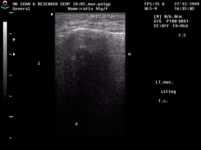
5 UH maksillaarsiinusest: norm
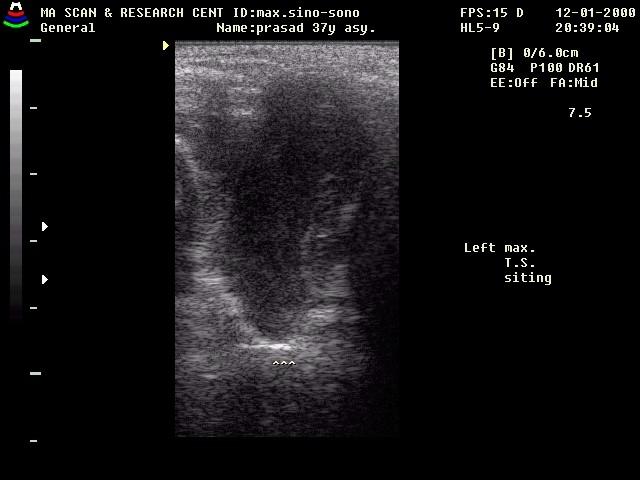
6 UH maksillaarsiinusest: sinusiit
: näha hästi frontaal- ja etmoidaalsiinused.")
7 Kuvamine: Röntgen Oktsipitomentaalne ülesvõte (Waters, nina-lõuaots): näeb kõiki ninakõrvalkoopaid, eriti hästi maksillaarsiinuseid. Oktsipitofrontaalne ülesvõte (Caldwell, nina-otsmik): näha hästi frontaal- ja etmoidaalsiinused. Külgülesvõte või Submentovertikaalne ülesvõte annavad vahel lisainformatsiooni.

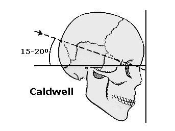
8 Waters ja Caldwell
9 KT Koronaarses tasapinnas Patsient kõhuli, kael väljasirutatud 1-3 mm järjestikku kihid + MPR Luu algoritm Aksiaalses tasapinnas Patsient selili 0,625 1,25 mm + MPR Luu algoritm
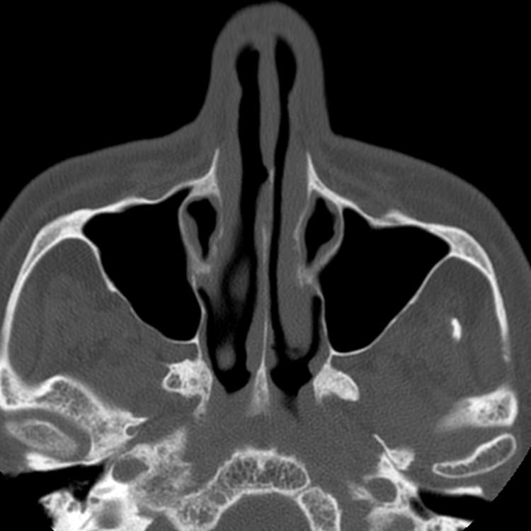
10 KT
11 Kuvamine: KT KT uuring koronaarses tasapinnas vaade sama mis KNK arstil paranasaale uurides. Ainuüksi KT põhjal ei diagnoosita paranasaalsiinuste haigusi, vajalik ka anamnees, kliiniline- ja endoskoopiline leid.
12 Kuvamine: KT KT uuringu pildil on info hetkeseisust: Kui patsiendil on hiljuti (4-6 nädalat) olnud sinusiit siis on muutused näha. Kui patsiendil on korduvad sinusiidid, aga pole olnud haigusnähte pikemat aega, siis võib leid olla normis Ägeda ülemiste hingamisteede infektsiooni puhul soovitatav KT edasi lükata.
13 Kuvamine: KT Ainus mis ei muutu nii kiirelt on luulised struktuurid ja veresoonte-närvide paiknemine siinuste suhtes. Hea radioloog : Teab, mis infot NKK arst tahab KT-st Vaatab, et uuring oleks optimaalselt tehtud Annab vastuses kliiniliselt olulist infot Tase saavutatav ainult regulaarse tagasiside ja haigusjuhtude ühise arutamisega.
14 Kuvamine: MRT Seeninfektsioon Sinusiidi tüsistused Kasvajad Haiguse levik väljaspoole PNS-d
15 MRT Coronal T1 4mm / 1mm 4:22 Coronal STIR 5:21 Axial T1 5:10 Axial T2 FSE FS 3:08 Axial T1 post Gad fat sat 4:02 Coronal T1 post Gad fat sat 6:10 No contrast for anosmia screening
16 Siinuste areng Ninaõõne väljasopistused Alguse piirkondadest saavad ostiumid Arengut mõjutavad: Limaskesta kasv Luude areng Teiste struktuuride regressioon Õhurõhu gradiendid

17 Siinuste areng Maksillaar Tekkivad esimesena (vastsündinul kitsad pilud), valmis 15 a Etmoid Teke sünnil, valmis a Sfenoid Sünnil minimaalsed, valmis 10 a Frontaal Sünnil puuduvad, teke 2-3 a, orbita lae tasemel 8 a
18 Funktsioon Taju, hingamine, kaitse, refleks Maitse haistmine (gustatory olfaction) Hingamine Õhu juhtimine Soojendamine Niisutamine Puhastamine
 Verevarustus ühel poolel intensiivistub ninakarbikud")
19 Nasaalne tsükkel Esineb 80-90% inimestel 30 minutit paar tundi (2-6) Verevarustus ühel poolel intensiivistub ninakarbikud paksenevad


20 Mukotsiliaarne kliirens Messerklinger 1950 ja 1960-nendatel Mukotsiliaarse kliirensi uuringud Endoskoopiline sekreedi transpordi jälgimine Lima liigub geneetiliselt määratud teed ostiumide suunas Sekreedi transport 1 cm/min

21 Sekreedi transport Kliiniliselt olulised siinused: Frontaal Eesmised etmoidid Maksillaar Kommunikatsioon keskmise ninakäiguga eelkambrite kaudu Frontaalne retsess Ethmoid infundibulum Operatsioonid suunatud eelkambritele

22 Normanatoomia Ninaõõs Lamina cribiforme Karbikud ja käigud Kõva suulagi Septum nasi
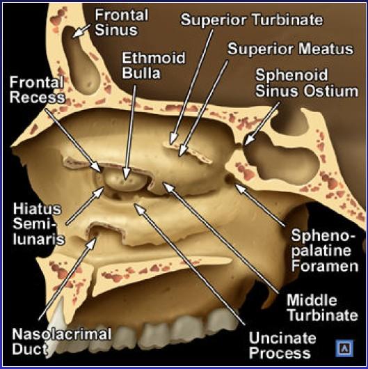
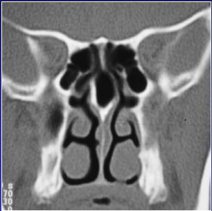
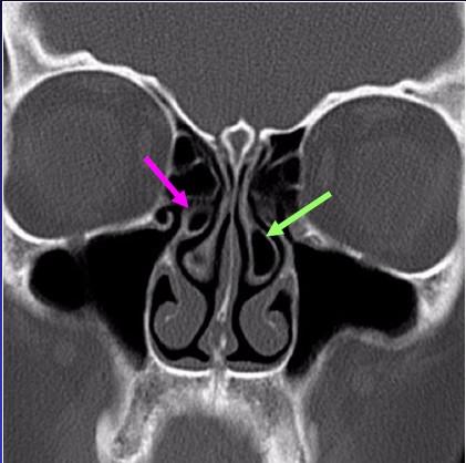
23 Normaalanatoomia

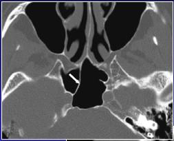
24 Foramen sphenopalatinum Ninaõõne lateraalses seinas taga-üleval Läbivad närvid ja veresooned Kaetud limaskesta poolt Otseühendus fossa pterygopalatina ja ninaõõne vahel
25 Foramen sphenopalatinum
26 Foramen sphenopalatinum

27 Frontaalsiinus Drenaazh frontaalretsessi Ductus nasofrontalis frontal infundibulum Frontaalsest ostiumist madalamal Proksimaalsel dreneerumine varieerub olenevalt processus uncinatusest Distaalsel dreneerub keskmise ninakäigu esiossa
28 Frontaalretsess
29 Frontaalretsess
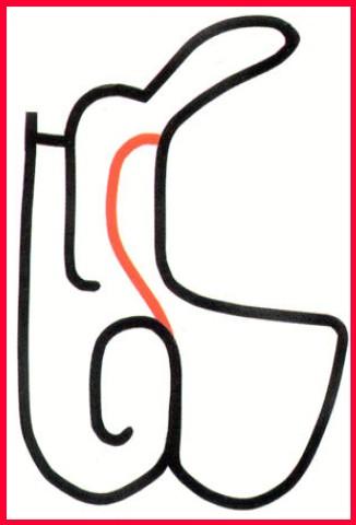
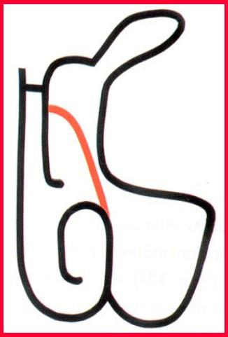
30 Frontaalsiinus ja uncinatus
31 Frontaalretsess ia uncinatus

32 Processus uncinatus

33 Drenaazh Maksillaarsiinus: hiatus semilunaris keskmine ninakäik Etmoidid: ees keskmine ninakäik, taga sfenoetmoidaalretsess Sfenoid: sfenoetmoidaalne retsess Ductus nasolacrimalis: alumine ninakäik
34 Ductus nasolacrimalis

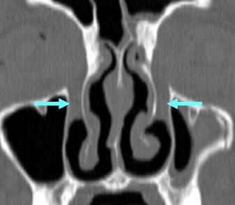
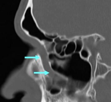
35 Keskmine ninakäik: Drenaazh Frontaalsiinusest frontaalretsessi kaudu Etmoidbulla ja etmoidrakustik anterioorsel Maksillaarsiinus infundibulumi kaudu
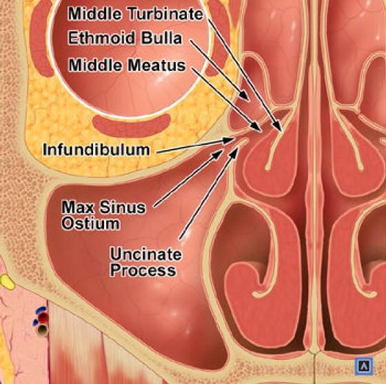
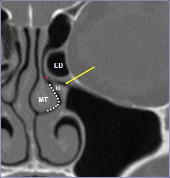
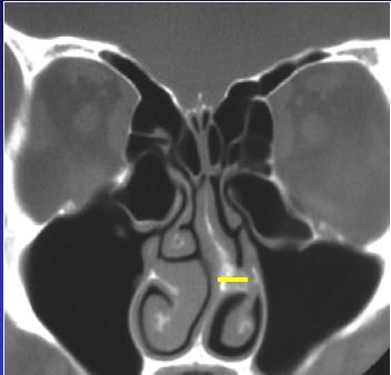
36 Ostiomeataalkompleks
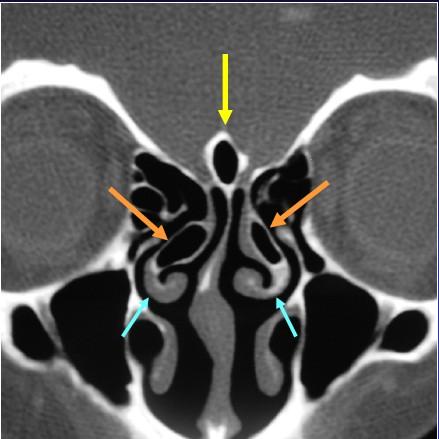
37 Anatoomilised variatsioonid Paranasaalide ja ninakarbikute konfiguratsioon, sama individuaalne nagu sõrmejäljed Võib soodustada põletiku või obstruktsiooni teket Suurendab FESS-i tüsistuste tõenäosust

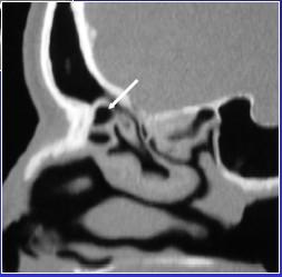
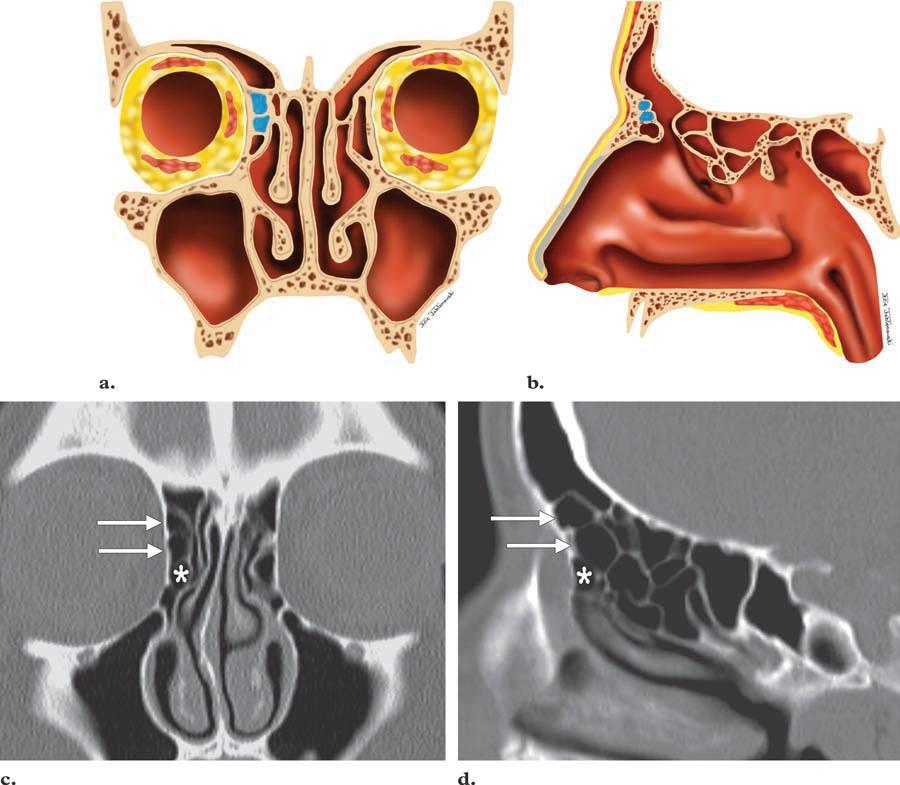
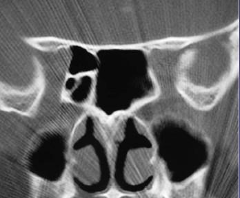
38 Agger nasi rakk ( % inimestest)
 Septumi")
39 Ninavahesein Deviatsioon Eksostoosid (Spurs) Septumi retsessid
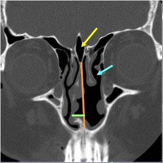
40 Ninavahesein
41 Tüüp I frontaalrakk
42 Tüüp II frontaalrakk
43 Tüüp III frontaalrakk
44 Tüüp IV froontaalrakk
45 Processus uncinatus variatsioonid Pneumatiseeritud (ahendab infundibulumi või keskmist ninakäiku) Deviatsioonis või atüüpilise kinnitusega (lamina papyraceaga, koljupõhimikuga või keskmise karbikuga) Atelektaatiline (lateraliseerunud orbita põhja suunas, ahendab infundiibulumi)

")
 Pneumatiseerunud")
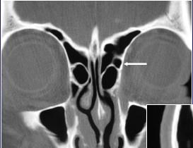
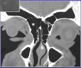
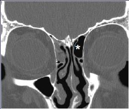
46 Keskmine karbik Hüpoplastiline (tüüpiline ninavaheseina deviatsiooniga) Paradoksaalne (konkaavne ninavaheseina suunas) Pneumatiseerunud (concha bullosa)
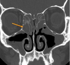
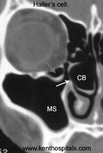
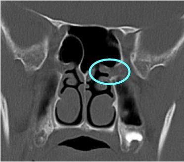
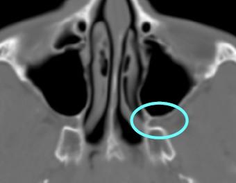
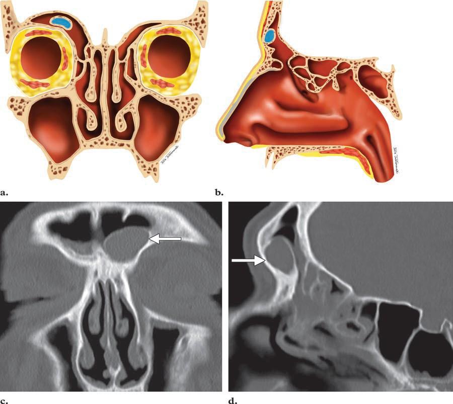
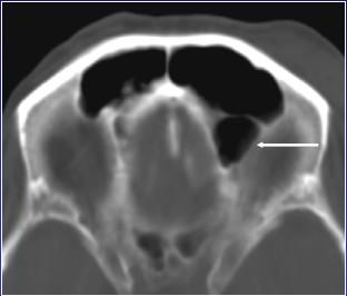
47 Infraorbital etmoidrakk (Halleri rakk)

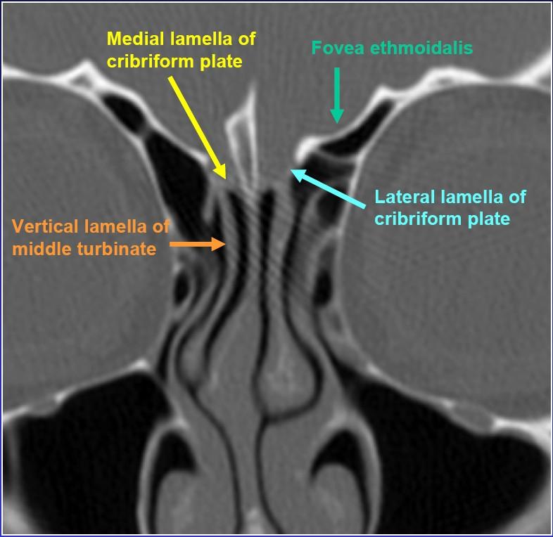
48 Variatsioonid Dehistsentne lamina papyracea Madal fovea ethmoidalis Hüpoplastiline labürint
49 Supraorbitaalne etmoidrakk
50 Sfenoidaalsiinus
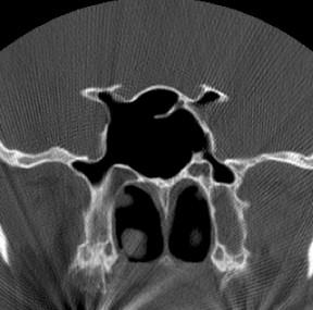
51 Sfenoidaalsiinus
52 Variatsioonid
53 Variatsioonid
54 Kasutatud allikad Professor Michelle A. Micheli loengud Failed Endoscopic Sinus Surgery: Spectrum of CT Findings in the Frontal Recess RadioGraphics 2009; 29:
Paranasaalsiinuste normivariandid ja nende tähtsus. Artur Aramaa III a radioloogia resident 2017
 Paranasaalsiinuste normivariandid ja nende tähtsus Artur Aramaa III a radioloogia resident 2017 Millest juttu tuleb Lühidalt olulisest anatoomiast Normivariantide esinemissagedus Normivariantide seos haigustega?
Boundaries Septum Turbinates & Meati Lamellae Drainage Pathways Variants
 The Fastest 20 Minutes in Michelle A. Michel, MD Professor of Radiology and Otolaryngology Medical College of Wisconsin, Milwaukee Overview Nasal cavity Anterior skull base Ostiomeatal complex Frontal
Computed tomography road map of the paranasal sinuses for treatment planning
 Computed tomography road map of the paranasal sinuses for treatment planning Poster No.: C-2607 Congress: ECR 2013 Type: Educational Exhibit Authors: N. Schembri, A. S. Gatt, D. Ellul, J. Brunton; Dundee/UK
Ninahingamise takistus. Eliise Annus
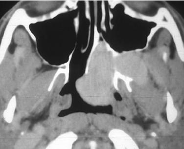
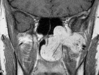
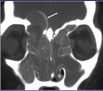
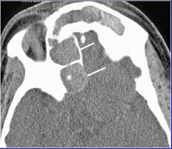
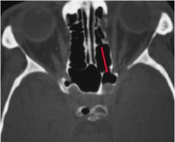
 Ninahingamise takistus Eliise Annus N, 8 21.08.2014 Ninahingamistakistus pikemat aega Alates 11.08 oli angiin, sai ospamoxi 8 päeva Ema märkas kasvajat uvula taga Laps norskab tihti Nasaalne hääl Rö KT
Chapter Five. 1 of 8 11/3/2008 2:52 PM.
 1 of 8 11/3/2008 2:52 PM Email : myousefmian@hotmail.com Chapter Five FRONT COVER Introduction Acknowledgement CHAPTERS Chapter One Chapter Two Chapter Three Chapter Four Chapter Five Chapter Six Chapter
Skull Base Danger Zones in FESS
 Skull Base Danger Zones in FESS Poster No.: C-2278 Congress: ECR 2014 Type: Educational Exhibit Authors: L. Renza Lozada, R. Carreño Gonzalez, G. Quintana Sanchez, 1 2 1 1 1 2 R. E. Figueroa ; Malaga/ES,
Communication issue - What should the radiologist report before functional endoscopic sinus surgery
 Communication issue - What should the radiologist report before functional endoscopic sinus surgery Poster No.: C-0509 Congress: ECR 2015 Type: Educational Exhibit Authors: A. M. Dobra 1, C. A. Badiu 1,
Radiological anatomy of frontal sinus By drtbalu
 2009 Radiological anatomy of frontal sinus By drtbalu Anatomy of frontal sinus is highly variable. Precise understanding of these variables will help a surgeon to avoid unnecessary complications during
CT OF THE PARANASAL SINUSES : NORMAL ANATOMY, VARIANTS AND PATHOLOGY
 Journal of Optoelectronics and Biomedical Materials Vol.2 Issue 4, October-December 2010, p. 281 289 CT OF THE PARANASAL SINUSES : NORMAL ANATOMY, VARIANTS AND PATHOLOGY AMIT N D DWIVEDI *, KAPIL KUMAR
FESS imaging - the role of MDCT
 FESS imaging - the role of MDCT Poster No.: C-0179 Congress: ECR 2013 Type: Educational Exhibit Authors: J. Plascak, K. Makaruha, B. Klasic, L. Kavur, V. Vidjak; Zagreb/HR Keywords: Image verification,
JMSCR Vol 04 Issue 05 Page May 2016
 www.jmscr.igmpublication.org Impact Factor 5.244 Index Copernicus Value: 5.88 ISSN (e)-2347-176x ISSN (p) 2455-0450 DOI: http://dx.doi.org/10.18535/jmscr/v4i5.25 Radiologic Variations of Nose and Paranasal
Three-Dimensional Volumetric Display of the Nasal Ostiomeatal Channels and Paranasal Sinuses
 Downloaded from www.ajronline.org by 37.44.202.192 on 12/22/17 from IP address 37.44.202.192. Copyright RRS. For personal use only; all rights reserved Three-Dimensional Volumetric Display of the Nasal
BOGOMOLETS NATIONAL MEDICAL UNIVERSITY. Department of human anatomy GUIDELINES
 BOGOMOLETS NATIONAL MEDICAL UNIVERSITY Department of human anatomy GUIDELINES Academic discipline HUMAN ANATOMY Module 1 Content module 2 The theme of the Eye-socket, bone nasal lesson cavity. bony palate
International Journal of Biological & Medical Research
 Int J Biol Med Res.2015;6(1):4775-4781 Contents lists available at BioMedSciDirect Publications International Journal of Biological & Medical Research Journal homepage: www.biomedscidirect.com BioMedSciDirect
Transnasal Endoscopic Sinonasal Surgery
 Reda kamel, Cadaveric dissection 1 Transnasal Endoscopic Sinonasal Surgery Cadaver Dissection Guide For Endoscopic Sinus Surgery Cairo University Egypt Reda Kamel Professor of Rhinology Cairo University
Computed tomographic evaluation of anatomical variations of paranasal sinus region
 International Journal of Research in Medical Sciences Gupta S et al. Int J Res Med Sci. 2016 Jul;4(7):2909-2913 www.msjonline.org pissn 2320-6071 eissn 2320-6012 Research Article DOI: http://dx.doi.org/10.18203/2320-6012.ijrms20161975
SINUS ANATOMY AND FUNCTION
 EMBRYOLOGY AND DEVELOPMENT SINUS ANATOMY AND FUNCTION -4 th week gestation: -frontonasal process identified, arises over developing forebrain -ectodermal -contributes to nasal capsule -9 th and 10 th week
CT anatomy of paranasal sinuses.
 CT anatomy of paranasal sinuses. Poster No.: C-2117 Congress: ECR 2017 Type: Educational Exhibit Authors: O. Dib, H. Chahinez, B. Asma, C. abdelouahab, M. Ourrad El, 1 2 1 1 1 2 1 3 3 B. Nacereddine ;
Südamepuudulikkus: iseloomulikud muutused Rö-pildil ning KT-uuringul. Tatjana Vask
 Südamepuudulikkus: iseloomulikud muutused Rö-pildil ning KT-uuringul Tatjana Vask Piltdiagnostika kardioloogias 2012 Täna kavas: Rindkere Rö- ja KT-uuringud südamepuudulikkusega patsientidel Südamepuudulikkusega
A Targeted Endoscopic Approach to Chronic Isolated Frontal Sinusitis
 Otolaryngology Head and Neck Surgery (2006) 134, 28-32 ORIGINAL RESEARCH A Targeted Endoscopic Approach to Chronic Isolated Frontal Sinusitis Roee Landsberg, MD, Yoram Segev, MD, Michael Friedman, MD,
The Frontal Sinus Drainage Pathway and Related Structures
 Pictorial Essay The Frontal Sinus Drainage Pathway and Related Structures David L. Daniels, Mahmood F. Mafee, Michelle M. Smith, Timothy L. Smith, Thomas P. Naidich, W. Douglas Brown, William E. Bolger,
PROBLEM RECOMMENDATION
 PREVENTION (MINIMIZING) IN ENDOSCOPIC Steven D. Schaefer, MD Professor and Chair Department of Otolaryngology PREVENTION AND Intraoperative Hemorrhage Loss of Orientation Inability to Identify/Preserve
STUDY OF SINONASAL VARIATIONS BY CT SCAN AND NASAL ENDOSCOPY IN CHRONIC SINUSITIS: A PROSPECTIVE CLINICAL STUDY
 STUDY OF SINONASAL VARIATIONS BY CT SCAN AND NASAL ENDOSCOPY IN CHRONIC SINUSITIS: A PROSPECTIVE CLINICAL STUDY A. V. S. Hanumantha Rao 1, B. Vijay Kumar 2, J. Suresh Babu 3 1Associate Professor, Department
Reasons for Failure and Surgical Revisions. Stil Kountakis, MD, PhD Professor and Chief, Division of Rhinology
 Reasons for Failure and Surgical Revisions Stil Kountakis, MD, PhD Professor and Chief, Division of Rhinology Medical College of Georgia of Georgia Regents University Department of Otolaryngology / Head
We are IntechOpen, the world s leading publisher of Open Access books Built by scientists, for scientists. International authors and editors
 We are IntechOpen, the world s leading publisher of Open Access books Built by scientists, for scientists 3,350 108,000 1.7 M Open access books available International authors and editors Downloads Our
The Egyptian Journal of Hospital Medicine (July 2017) Vol.68 (3), Page
 Vol.68 (3), Page") The Egyptian Journal of Hospital Medicine (July 2017) Vol.68 (3), Page 1390-1394 Anatomical Variations of Nasal Structures in Chronic Rhinosinusitis as Detected by Computed Tomography Scan Omar Adnan Hasan,
The Egyptian Journal of Hospital Medicine (July 2017) Vol.68 (3), Page 1390-1394 Anatomical Variations of Nasal Structures in Chronic Rhinosinusitis as Detected by Computed Tomography Scan Omar Adnan Hasan,
Pathological consequences of anatomical variations in the sino-nasal region: how can radiologists help clinicians?
 Pathological consequences of anatomical variations in the sino-nasal region: how can radiologists help clinicians? Poster No.: C-0735 Congress: ECR 2016 Type: Educational Exhibit Authors: M. E. Laino,
Difficult airway management- our experience
 Difficult airway management- our experience J. Starkopf, A. Sell, A. Sõrmus, J. Samarütel Clinic of Anaesthesiology and Intensive Care Tartu University Clinics Estonia Clinic of Anaesthesiology and Intensive
Anatomical variants of the uncinate process CT scan imaging study
 Romanian Journal of Rhinology, Vol. 2, No. 7, July - September 2012 original Study Anatomical variants of the uncinate process CT scan imaging study Vasilica Baldea 1, Mihail Dan Cobzeanu 2, Florina Mihalcea
Anatomical Analysis of the Frontal Recess Cells in Endoscopic Sinus Surgery An Indian Perspective
 ORIGINAL ARTICLE Anatomical Analysis of the Frontal Recess Cells in Endoscopic Sinus Surgery An Indian Perspective 1 Dhingra Shruti, 2 Agarwal AK, 3 Passey JC, 4 Kaul JM 1 Resident, Department of Otolaryngology
JMSCR Vol 05 Issue 09 Page September 2017
 www.jmscr.igmpublication.org Impact Factor 5.84 Index Copernicus Value: 71.58 ISSN (e)-2347-176x ISSN (p) 2455-0450 DOI: https://dx.doi.org/10.18535/jmscr/v5i9.52 Relationship of Agger Nasi Cell and Uncinate
Imaging of the Paranasal Sinuses
 14. Sommerschule Imaging of the Paranasal Sinuses Bettlach 24.08.2018 Christoph Schlegel Conventional Radiology NNH-Status: okzipito-frontal: frontal sinus, anterior ethmoid okzipito-nasal : maxillary
Chapter Six. 1 of 6 11/3/2008 2:21 PM.
 1 of 6 11/3/2008 2:21 PM Email : myousefmian@hotmail.com Chapter Six FRONT COVER Introduction Acknowledgement CHAPTERS Chapter One Chapter Two Chapter Three Chapter Four Chapter Five Chapter Six Chapter
Morphological Changes of the Ethmoid and Maxillary Cavities after Endoscopic Sinus Surgery, A Quantitative Digital Analysis.
 Morphological Changes of the Ethmoid and Maxillary Cavities after Endoscopic Sinus Surgery, A Quantitative Digital Analysis Thesis Submitted for fulfillment of M.D. degree in Otorhinolaryngology By: Hisham
Anatomical Variations in Osteomeatal Complex among Patients undergoing Functional Endoscopic Sinus Surgery
 V Narendrakumar, V Subramanian Original article 10.5005/jp-journals-10013-1259 Anatomical Variations in Osteomeatal Complex among Patients undergoing Functional Endoscopic Sinus Surgery 1 V Narendrakumar,
IJCMR 606. ORIGINAL RESEARCH Correlation of Computed Tomography And Nasal Endoscopic Findings In Chronic Rhinosinusitis A Clinical Study
 IJCMR 606 ORIGINAL RESEARCH Correlation of Computed Tomography And Nasal Endoscopic Findings In Chronic Rhinosinusitis A Clinical Study Umeek Jeelani 1, Ulfat Ara Wani 2, Shabir Khanday 3, Shahi Jahan
A CORRELATION STUDY OF PARANASAL SINUSES BETWEEN OPERATIVE ENDOSCOPIC FINDINGS IN FESS AND PREOPERATIVE CT SCAN
 A CORRELATION STUDY OF PARANASAL SINUSES BETWEEN OPERATIVE ENDOSCOPIC FINDINGS IN FESS AND PREOPERATIVE CT SCAN Polisetti Ravi Babu 1, Bhennur Durga Prasad 2, Lanke Sowmya 3, K.S.B.S. Krishna Sasanka 4
Katya A. Shpilberg 1 Simon C. Daniel 1 Amish H. Doshi 1 William Lawson 2 Peter M. Som 1. Neuroradiology/Head and Neck Imaging Original Research
 Neuroradiology/Head and Neck Imaging Original Research Shpilberg et al. CT of Paranasal Sinuses and Nasal Cavity Neuroradiology/Head and Neck Imaging Original Research Katya A. Shpilberg 1 Simon C. Daniel
Professor Dr.Muhammad Ajmal Dr.Tehmina Nazir. HOLY FAMILY HOSPITAL Rawalpindi
 Professor Dr.Muhammad Ajmal Dr.Tehmina Nazir HOLY FAMILY HOSPITAL Rawalpindi SCHEME OF PRESENTATION PLAIN X-RAYS CT SCAN MRI CONCLUSION IMAGING MODALITIES PLAIN X-RAYS CT SCAN MRI OCCIPITOMENTAL/WATER
ORIGINAL ARTICLE RELATIONSHIP OF CONCHA BULLOSA WITH OSTEOMEATAL UNIT BLOCKAGE. TOMOGRAPHIC STUDY IN 200 PATIENTS.
 RELATIONSHIP OF CONCHA BULLOSA WITH OSTEOMEATAL UNIT BLOCKAGE. TOMOGRAPHIC STUDY IN 200 PATIENTS. Shrikrishna B H 1, Jyothi A C 2, Sanjay G 3, Sandeep Samson G 4. 1. Associate Professor, Department of
A Computer-Assisted Anatomical Study of the Nasofrontal Region
 The Laryngoscope Lippincott Williams & Wilkins, Inc., Philadelphia 2001 The American Laryngological, Rhinological and Otological Society, Inc. A Computer-Assisted Anatomical Study of the Nasofrontal Region
Prevalence of Anatomical Variations of the Sinonasal Region and their Relationship with Chronic Rhinosinusitis
 Prevalence of Anatomical Variations of the Sinonasal Region and their Relationship with Chronic Rhinosinusitis Karki S, 1 Pokharel M, 2 Suwal S, 1 Poudel R 1 ABSTRACT Background 1 Department of Radiology
Imaging Anatomy in Revision Sinus Surgery
 Chapter 1 Imaging Anatomy in Revision Sinus Surgery Ramon E. Figueroa 1 Core Messages An intimate knowledge of sinus anatomy and a clear understanding of the baseline postsurgical anatomy are required
Diagnostic Performance of Multidetector Computed Tomography (MDCT) in Diagnosis of Sinus Variations
 in Diagnosis of Sinus Variations") Signature: Pol J Radiol, 2017; 82: 713-725 DOI: 10.12659/PJR.903684 ORIGINL RTICLE Received: 2017.02.08 ccepted: 2017.02.23 Published: 2017.11.17 uthors Contribution: Study Design Data Collection C Statistical
Signature: Pol J Radiol, 2017; 82: 713-725 DOI: 10.12659/PJR.903684 ORIGINL RTICLE Received: 2017.02.08 ccepted: 2017.02.23 Published: 2017.11.17 uthors Contribution: Study Design Data Collection C Statistical
Liberaalne vähiravikorraldus keskhaiglad versus regionaalhaiglad
 Liberaalne vähiravikorraldus keskhaiglad versus regionaalhaiglad Andrus Arak, MD, PhD onkoloog, üldkirurg Pärnus 06.05.2016 Liberaalne - salliv, vabameelne Optimaalne - parim, sobivaim, ökonoomseim Konservatiivne
A Study of Anatomical Variations in Patients with Chronic Rhinosinusitis.
 DOI: 10.2127/aimdr.201..2.EN1 Original Article ISSN (O):239-222; ISSN (P):239-21 A Study of Anatomical Variations in Patients with Chronic Rhinosinusitis. Smruti Swain 1 1 Associate Professor, Department
Skull and Axial Skeleton
 Published on Second Faculty of Medicine, Charles University (http://www.lf2.cuni.cz ) Skull and Axial Skeleton Description of the test The examination of the skull skeleton is in oral format. It consists
A radiological study of anatomical variations in ostiomeatal complex in patients with chronic rhinosinusitis
 International Journal of Otorhinolaryngology and Head and Neck Surgery Rajneesh et al. Int J Otorhinolaryngol Head Neck Surg. 2017 Jul;3(3):528-533 http://www.ijorl.com pissn 2454-5929 eissn 2454-5937
3D Accuitomo Clinical Case Evidence
 3D Accuitomo Clinical Case Evidence The Advantages of DVT for Ear-, Nose- & Throat-Diagnostic Thinking ahead. Focused on life. Editorial Dear Colleagues, Index of Contents I am very happy to present you
The Study of the Agger Nasi Cell
 The Study of the Agger Nasi Cell The agger nasi (from agger meaning "mound or heap") is a small ridge on the lateral side of the nasal cavity. It is located midway at the anterior edge of the middle nasal
Haigusjuht. Helen Kikerpill II a arst-resident radioloogia
 Haigusjuht Helen Kikerpill II a arst-resident radioloogia 21.04.2011 Kliinilised andmed: valu paremas puusas [Manaster et al] 21.04.2011 MRT uuring lülisamba nimmeosast Olulist spinaalkanali ahenemist
International Journal of Health Sciences and Research ISSN:
 International Journal of Health Sciences and Research www.ijhsr.org ISSN: 2249-9571 Original Research Article Anatomical Study of the Middle Meatus with Emphasis to the Maxillary Ostium and Their Clinical
Bones Ethmoid bone Inferior nasal concha Lacrimal bone Maxilla Nasal bone Palatine bone Vomer Zygomatic bone Mandible
 splanchnocranium - Consists of part of skull that is derived from branchial arches - The facial bones are the bones of the anterior and lower human skull Bones Ethmoid bone Inferior nasal concha Lacrimal
F.E.S.S. FUNCTIONAL ENDOSCOPIC SINUS SURGERY. Mohammed Yousaf MIAN, MBBS, FRCS,FICS
 F.E.S.S. FUNCTIONAL ENDOSCOPIC SINUS SURGERY Mohammed Yousaf MIAN, MBBS, FRCS,FICS Consultant Otolaryngology, Head and Neck Surgery Furness General Hospital, Dalton Lane, Barrow in Furness, Cumbria LA14
Incidence of accessory ostia in patients with chronic maxillary sinusitis
 International Journal of Otorhinolaryngology and Head and Neck Surgery Ghosh P et al. Int J Otorhinolaryngol Head Neck Surg. 2018 Mar;4(2):443-447 http://www.ijorl.com pissn 2454-5929 eissn 2454-5937 Original
Spheno-Ethmoidectomy
 Diagnostic and Therapeutic Endoscopy, Vol. 5, pp. 1-8 Reprints available directly from the publisher Photocopying permitted by license only (C) 1998 OPA (Overseas Publishers Association) N.V. Published
Anatomical Variants in Frontal Recess Region and their Impact on Frontal Sinus Surgery in Chronic Sinusitis
 American Journal of Health Research 2015; 3(3): 140-145 Published online April 23, 2015 (http://www.sciencepublishinggroup.com/j/ajhr) doi: 10.11648/j.ajhr.20150303.15 ISSN: 2330-8788 (Print); ISSN: 2330-8796
Surgical Anatomy 2 of the Paranasal Sinuses
 Chapter 2 Surgical Anatomy 2 of the Paranasal Sinuses Zoukaa B. Sargi, Roy R. Casiano Core Messages There are learned anatomical landmarks that can help surgeons perform safe endoscopic sinus surgery.
S INONASAL imaging has progressed methodically as each new generation
 David M. Yousem, MD Imaging of Sinonasal Inflammatory Disease Changes in imaging sinonasal inflammatory disease have paralleled changes in the treatment of chronic sinusitis. As functional endoscopic sinus
Endoscopic Sinus Surgery. A Practical Approach
 Endoscopic Sinus Surgery A Practical Approach Springer London Berlin Heidelberg New York Barcelona Budapest Hong Kong Milan Paris Santa Clara Singapore Tokyo S.K. Kaluskar With a Contribution by Professor
AComputedTomographyAidedClinicalReportonAnatomicalVariationsoftheParanasalSinuses
 Global Journal of Medical Research: K Interdisciplinary Volume 16 Issue 6 Version 1.0 Type: Double Blind Peer Reviewed International Research Journal Publisher: Global Journals Inc. (USA) Online ISSN:
The Relation between Anatomical Variations of Osteomeatal Complex & Nasal Structures and Chronic Sinusitis by Computed Tomography
 International Journal of Medical Imaging 2015; 3(2): 16-20 Published online March 6, 2015 (http://www.sciencepublishinggroup.com/j/ijmi) doi: 10.11648/j.ijmi.20150302.12 ISSN: 2330-8303 (Print); ISSN:
ORIGINAL ARTICLE ANATOMICAL VARIATIONS OF THE OSTEOMEATAL COMPLEX TOMOGRAPHIC FINDINGS IN 100 PATIENTS
 ANATOMICAL VARIATIONS OF THE OSTEOMEATAL COMPLEX TOMOGRAPHIC FINDINGS IN 100 PATIENTS Jyothi A. C 1, Shrikrishna B. H 2, Sanjay G 3, Sandeep Samson G 4. 1. Associate Professor, Department of ENT and head
Evaluation of Anatomical Variations in Ostiomeatal Unit by Computed Tomography
 Original Article Print ISSN: 2321-6379 Online ISSN: 2321-595X DOI: 10.17354/ijss/2018/83 Evaluation of Anatomical Variations in Ostiomeatal Unit by Computed Tomography Sushilkumar Kale 1, K Preetha 2 1
A COMPUTERIZED TOMOGRAPHIC STUDY OF UNCINATE PROCESS OF ETHMOID BONE
 Original Article A COMPUTERIZED TOMOGRAPHIC STUDY OF UNCINATE PROCESS OF ETHMOID BONE N. Vinay Kumar * 1, E. Kamala 2, T. S. Guga Priya 3, S. D. NalinaKumari 4. *1,2 Assistant professor, Department of
Towards the Understanding of Sinonasal Anatomical Variations A Cadaveric Study
 ORIGINAL ARTICLE Towards the Understanding of Sinonasal Anatomical Variations A Cadaveric Study Towards the Understanding of Sinonasal Anatomical Variations A Cadaveric Study 1 Daisy Sahni, 2 Rupa Mehta,
The advent of high-resolution computerized tomography
 An anatomic classification of the ethmoidal bulla REUBEN C. SETLIFF, III, MD, PETER J. CATALANO, MD, FACS, LISA A. CATALANO, MPH, and CHAD FRANCIS, BA, Sioux Falls, South Dakota, and Burlington, Massachusetts
Original Article Effect of lamina papyracea ingression on orbito-ocular complications after functional endoscopic sinus surgery
 Int J Clin Exp Med 2016;9(6):10317-10321 www.ijcem.com /ISSN:1940-5901/IJCEM0025371 Original Article Effect of lamina papyracea ingression on orbito-ocular complications after functional endoscopic sinus
Variations in Paranasal Sinus Anatomy: Implications for the Pathophysiology of Chronic Rhinosinusitis and Safety of Endoscopic Sinus Surgery
 ARTICLE Variations in Paranasal Sinus Anatomy: Implications for the Pathophysiology of Chronic Rhinosinusitis and Safety of Endoscopic Sinus Surgery S.A.R. Nouraei, MBBChir, MRCS, A.R. Elisay, MD, A. DiMarco,
Radiological significance of the sinonasal anatomical variants in recurrent acute rhinosinusitis patients
 International Journal of Research in Medical Sciences Sahu N et al. Int J Res Med Sci. 2017 Jun;5(6):2379-2384 www.msjonline.org pissn 2320-6071 eissn 2320-6012 Original Research Article DOI: http://dx.doi.org/10.18203/2320-6012.ijrms20172121
Sinonasal Imaging. Mamdouh Mahfouz MD Professor of Radiology Cairo University. ssregypt.com
 Sinonasal Imaging Mamdouh Mahfouz MD Professor of Radiology Cairo University ssregypt.com Scanning Techniques Routine Study CORONAL Coronal 3-5mm sections from the posterior wall of the sphenoid sinus
Nasal Anatomy and Evaluation
 See discussions, stats, and author profiles for this publication at: https://www.researchgate.net/publication/283830743 Nasal Anatomy and Evaluation Article October 2015 DOI: 10.1007/978-3-319-10332-7_2
Welcome to the Functional Endoscopic Sinus Surgery
 Welcome to Endoscopic Sinus Surgery http://endoscopicsinussurgery.co.uk/index.html 1 of 1 11/3/2008 3:20 PM Email : myousefmian@hotmail.com FRONT COVER Welcome to the Functional Endoscopic Sinus Surgery
The International Frontal Sinus Anatomy Classification (IFAC) and Classification of the Extent of Endoscopic Frontal...
 and Classification of the Extent of Endoscopic Frontal...") See discussions, stats, and author profiles for this publication at: https://www.researchgate.net/publication/298902054 The International Frontal Sinus Anatomy Classification (IFAC) and Classification
See discussions, stats, and author profiles for this publication at: https://www.researchgate.net/publication/298902054 The International Frontal Sinus Anatomy Classification (IFAC) and Classification
Conventional Sinus Surgery Vs Fess
 IOSR Journal of Dental and Medical Sciences (IOSR-JDMS) e-issn: 2279-0853, p-issn: 2279-0861.Volume 16, Issue 7 Ver. III (July. 2017), PP 44-51 www.iosrjournals.org Conventional Sinus Surgery Vs Fess *
FRONTAL SINUPLASTY P R E P A R E D A N D P R E S E N T E D B Y : D R. Y A H Y A F A G E E H R 4 16/ 12/ 2013
 FRONTAL SINUPLASTY P R E P A R E D A N D P R E S E N T E D B Y : D R. Y A H Y A F A G E E H R 4 16/ 12/ 2013 ANATOMY: FRONTAL SINUS Not present at birth Starts developing at 4 years Radiographically visualized
OPEN ACCESS ATLAS OF OTOLARYNGOLOGY, HEAD & NECK OPERATIVE SURGERY
 OPEN ACCESS ATLAS OF OTOLARYNGOLOGY, HEAD & NECK OPERATIVE SURGERY INFERIOR MAXILLECTOMY Tumours of the hard palate and superior alveolus may be resected by inferior maxillectomy (Figure 1). A Le Fort
The Incidence of Concha Bullosa and Its Association with Chronic Rhinosinusitis Deviated Nasal Septum and Osteomeatal Complex Obstruction
 1 Bahrain Medical Bulletin, Vol. 33, No. 4, December 2011 The Incidence of Concha Bullosa and Its Association with Chronic Rhinosinusitis Deviated Nasal Septum and Osteomeatal Complex Obstruction Fatma
The Relationship of Concha Bullosa with Nasal Septal Deviation and Paranasal Sinus Disease
 International Journal of Advances in Health Sciences (IJHS) ISSN 2349-7033 Vol2, Issue6, 2015, pp762-770 http://www.ijhsonline.com Research Article The Relationship of Concha Bullosa with Nasal Septal
MATERIALS AND METHODS
 sinus surgery as described by Messerklinger, wherein infundibulotomy forms an integral step by removal of the uncinate process [Stammberger and Posawetz, 1990; Kennedy et al, 1985; Rice, 1989]. Even though
University of Palestine. Midterm Exam 2013/2014 Total Grade:
 Course No: DNTS2208 Course Title: Head and Neck Anatomy Date: 09/11/2013 No. of Questions: (50) Time: 1hour Using Calculator (No) University of Palestine Midterm Exam 2013/2014 Total Grade: Instructor
Major inflammatory patterns of chronic sinonasal diseases and their. accompanied anatomical variations; CT scan review
 Major inflammatory patterns of chronic sinonasal diseases and their accompanied anatomical variations; CT scan review Dr. Qays Ahmed Hassan AL-Timimy ABSTRACT Background: Because of wide use of Functional
DISSERTATION ON A PROSPECTIVE STUDY OF ANATOMICAL VARIATION OF OSTEOMEATAL COMPLEX IN CHRONIC SINUSITIS PATIENTS
 DISSERTATION ON A PROSPECTIVE STUDY OF ANATOMICAL VARIATION OF OSTEOMEATAL COMPLEX IN CHRONIC SINUSITIS PATIENTS Submitted in partial fulfillment of the requirements for M.S. DEGREE BRANCH -IV OTORHINOLARYNGOLOGY
ROLE OF ANATOMICAL OBSTRUCTION IN THE PATHOGENESIS OF CHRONIC SINUSITIS
 From the SelectedWorks of Balasubramanian Thiagarajan July 1, 2012 ROLE OF ANATOMICAL OBSTRUCTION IN THE PATHOGENESIS OF CHRONIC SINUSITIS Balasubramanian Thiagarajan Available at: https://works.bepress.com/drtbalu/51/
Cables 152, 153 Camera systems , video stack/cameras , 153 Cantholysis, inferior
 296 A Abscess periorbital 45 goal of surgery 44 subperiosteal 18, 20 Accessory ostium 108 109, 114 116 anterior to uncinate process 114, 115 in posterior fontanelle 116 Adenoameloblastoma 43 Adenocarcinoma
Computerised tomographic profile of ethmoid roof on basis of keros classification among ethnic Kashmiri s
 International Journal of Otorhinolaryngology and Head and Neck Surgery Salroo IN et al. Int J Otorhinolaryngol Head Neck Surg. 2016 Jan;2(1):1-5 http://www.ijorl.com pissn 2454-5929 eissn 2454-5937 Research
Chronic Frontal Rhinosinusitis: Diagnosis and Management
 Chapter Chronic Frontal Rhinosinusitis: Diagnosis and Management Core Messages Despite significant advances in surgical techniques, technology, and knowledge of pathophysiology, management of chronic frontal
HAIGUSJUHT 23 AASTANE MEES ALAKÕHUVALUGA. Jevgeni Kulikov 3. aasta resident
 HAIGUSJUHT 23 AASTANE MEES ALAKÕHUVALUGA Jevgeni Kulikov 3. aasta resident Anamnees 10.10.2017 öösel kõhuvalu, korduvalt oksendanud, oksemassid tavapärased, kõht käib läbi. Kõhuvalu kirjeldab rohkem kui
Frontal sinus disease continues to be one of the great
 Unilateral transnasal endoscopic approach to frontal sinuses: Draf IIc Mohammed K. Al Komser, M.D., M.A.S. and Andrew N. Goldberg, M.D., M.S.C.E. ABSTRACT For chronic sinusitis surgery, the Draf III approach
COMPARISON OF CONE BEAM COMPUTED TOMOGRAPHY AND COMPUTED TOMOGRAPHY EXAMINATIONS of PARANASAL SINUSES: PRELIMINARY STUDY
 CLINICAL DENTISTRY AND RESEARCH 2013; 37(1): 10-18 COMPARISON OF CONE BEAM COMPUTED TOMOGRAPHY AND COMPUTED TOMOGRAPHY EXAMINATIONS of PARANASAL SINUSES: PRELIMINARY STUDY Filiz Namdar Pekiner, DDS, PhD
Bisection of Head & Nasal Cavity 頭部對切以及鼻腔. 解剖學科馮琮涵副教授 分機
 Bisection of Head & Nasal Cavity 頭部對切以及鼻腔 解剖學科馮琮涵副教授 分機 3250 E-mail: thfong@tmu.edu.tw Outline: The structure of nose The concha and meatus in nasal cavity The openings of paranasal sinuses Canals, foramens
Incidence of sinonasal anatomical variations associated with chronic sinusitis by CT scan in Karaikal, South India
 International Journal of Otorhinolaryngology and Head and Neck Surgery Gouripur K et al. Int J Otorhinolaryngol Head Neck Surg. 2017 Jul;3(3):576-580 http://www.ijorl.com pissn 2454-5929 eissn 2454-5937
EVALUATION OF PATIENT'S OUTCOME AFTER ENDOSCOPIC SINUS SURGERY
 Basrah Journal Of Surgery Bas J Surg, September, 16, 2010 EVALUATION OF PATIENT'S OUTCOME AFTER ENDOSCOPIC SINUS SURGERY Hiwa A Abdulkareem MB.ChB, FICMS, CABS(ENT); Teaching Hospital; University of Sulaimania.
Mari-Liis Ilmoja SA Tallinna Lastehaigla
 Mari-Liis Ilmoja SA Tallinna Lastehaigla 22.05.2015 24,165 anaesthesias 724 adverse events; 31:1000 53% respiratory Risk factors: infancy, higher ASA status, ENT surgery Perioperative anaesthetic morbidity
A comprehensive study on complications of endoscopic sinus surgery
 International Journal of Otorhinolaryngology and Head and Neck Surgery Shyras JAD et al. Int J Otorhinolaryngol Head Neck Surg. 2017 Jul;3(3):472-477 http://www.ijorl.com pissn 2454-5929 eissn 2454-5937
The View through the Nose: ENT considerations for Pituitary/Skull Base Surgery
 The View through the Nose: ENT considerations for Pituitary/Skull Base Surgery Edsel Kim, M.D. Otolaryngology-Head and Neck Surgery The Oregon Clinic Providence Brain and Spine Institute Pituitary, Thyroid
Anatomy #1; Respiratory Nose and the Nasal Cavity December 1st, 2013
 Note #1: the doctor skipped some slides in the lecture. Those slides are not included in this sheet and so you will have to review the slides to study them. The reason they were not included is because
Pneumatization of Mastoid Air Cells, Temporal Bone, Ethmoid and Sphenoid Sinuses. Any Correlation?
 DOI 10.1007/s12070-014-0745-z ORIGINAL ARTICLE Pneumatization of Mastoid Air Cells, Temporal Bone, Ethmoid and Sphenoid Sinuses. Any Correlation? Khalid Hindi Sarmad Alazzawi Rajagopalan Raman Narayanan
Nasal region. cartilages: septal cartilage (l); lateral nasal cartilage (2); greater alar cartilages (2); lesser alar cartilages (?
; lateral nasal cartilage (2); greater alar cartilages (2); lesser alar cartilages (?") Nasal region skull bones: nasal and frontal processes of maxilla cartilages: septal cartilage (l); lateral nasal cartilage (2); greater alar cartilages (2); lesser alar cartilages (?) 1 Nasal cavity Roof
Nasal region skull bones: nasal and frontal processes of maxilla cartilages: septal cartilage (l); lateral nasal cartilage (2); greater alar cartilages (2); lesser alar cartilages (?) 1 Nasal cavity Roof