Fetal Medicine. Case Presentations. Dr Ermos Nicolaou Fetal Medicine Unit Chris Hani Baragwanath Hospital. October 2003
|
|
|
- Camron Lester
- 5 years ago
- Views:
Transcription
1 Case Presentations Dr Ermos Nicolaou Fetal Medicine Unit Chris Hani Baragwanath Hospital October 2003
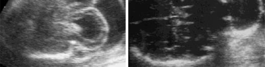
2 Case 1 Ms A M 22year old P0 G1 Referred from Sebokeng Hospital at 36w for polyhydramnios On Ultrasound: Mild/Moderate ventriculomegaly Large cystic mass arisisng from the posterior aspect of the fetal skull; 120x 170mm in diameter No other abnormalities seen Dx: Large Meningocele Plan: In view of the advanced gestational age no inetrvention justifiable Elective C/S at 38w and post natal assessment
3
4
5
6
7
8 Case 1 Elective C/S performed; no complications Antenatal findings confirmed
9
10
11
12 Case 1 Baby admitted to neuro-surgical ward Neurological assessment normal at this point CT scan: Occipital meningocele No brain tissue involved Moderate dilatation of the ventricles
13 Case 1 Surgery performed on day 9 post delivery Uneventful procedure Baby discharged on day 4 For follow up in 1 week
14
15
16
17
18 Case 2 Mrs N N 32 year old P0 G1 Had normal NT at 12 weeks Presents at 20 w 2d with a central supra-ocular mass
19
20
21 Case 2 On scan: Anterior/ frontal encephalocele, lemon sign Nasal bone not visible Central ossification failure Rest of face appears normal No other abnormalities seen
22 Case 2 Counselling: 1. TOP 2. Conservative management with close monitoring? Patient absconded
23 These include NEURAL TUBE DEFECTS 1. Anencephaly 2. Spina bifida 3. Encephalocoele In anencephaly there is absence of the cranial vault (acrania) with secondary degeneration of the brain Encephaloceles are cranial defects, usually occipital, with herniated fluid-filled or brain-filled cysts
24 NEURAL TUBE DEFECTS In spina bifida the neural arch, usually in the lumbosacral region, is incomplete with secondary damage to the exposed nerves
25 NEURAL TUBE DEFECTS Prevalence Subject to large geographical and temporal variations In the UK the prevalence is about 5 per 1,000 births Anencephaly and spina bifida, with an approximately equal prevalence, account for 95% of the cases and encephalocele for the remaining 5%
26 NEURAL TUBE DEFECTS Etiology Chromosomal abnormalities, single mutant genes, and maternal diabetes mellitus or ingestion of teratogens, such as antiepileptic drugs, are implicated in about 10% of the cases precise etiology for the majority of these defects is unknown When a parent or previous sibling has had a neural tube defect, the risk of recurrence is 5-10%. Periconceptual supplementation of the maternal diet with folate reduces by about half the risk of developing these defects
27 NEURAL TUBE DEFECTS Anencephaly Diagnosis of anencephaly during the second trimester of pregnancy is based on the demonstration of absent cranial vault and cerebral hemispheres Associated spinal lesions are found in up to 50% of cases In the first trimester the diagnosis can be made after 11 weeks, when ossification of the skull normally occurs
28 NEURAL TUBE DEFECTS Anencephaly
29 NEURAL TUBE DEFECTS Spina bifida Diagnosis of spina bifida requires the systematic examination of each neural arch from the cervical to the sacral region both transversely and longitudinally In the transverse scan the normal neural arch appears as a closed circle with an intact skin covering, whereas in spina bifida the arch is "U" shaped and there is an associated bulging meningocoele (thin-walled cyst) or myelomeningocoele

30 NEURAL TUBE DEFECTS Spina bifida
31 NEURAL TUBE DEFECTS Spina bifida The diagnosis of spina bifida has been greatly enhanced by the recognition of associated abnormalities in the skull and brain Secondary to the Arnold-Chiari malformation and include 1. frontal bone scalloping (lemon sign), 2. and obliteration of the cisterna magna 3. with either an "absent" cerebellum or abnormal anterior curvature of the cerebellar hemispheres (banana sign).


32 NEURAL TUBE DEFECTS Spina bifida

33 NEURAL TUBE DEFECTS Spina bifida
34 NEURAL TUBE DEFECTS Spina bifida A variable degree of ventricular enlargement is present in virtually all cases of open spina bifida at birth, but in only about 70% of cases in the midtrimester
35 NEURAL TUBE DEFECTS Encephalocele Encephaloceles are recognised as cranial defects with herniated fluid-filled or brain-filled cysts most commonly found in an occipital location (75% of the cases) but alternative sites include the frontoethmoidal and parietal regions
36 ENCEPHALOCELE - DEFINITION A neural tube defect affecting the skull resulting in the herniation of the meninges and portions of the brain through a bony midline defect in the skull.

37 NEURAL TUBE DEFECTS Encephalocele
38 ENCEPHALOCELE - EPIDEMIOLOGY incidence: 1/10th as common as spinal neural tube defects risk factors: multifactorial inheritance pattern
39 ENCEPHALOCELE -associated anomalies 1. Syndromes Dandy-Walker Syndrome Klippel-Feil Syndrome Meckel-Gruber Syndrome rare autosomal recessive disorder occipital encephalocele associated with microcephaly, holoprosencephaly, cleft lip or palate, polydactyly, abnormal genitalia, polycystic kidneys 2. Malformations Arnold-Chiari malformation, porencephaly, agenesis of the corpus callosum, myelodysplasia, optic nerve dysplasia, cleft palate
40 ENCEPHALOCELE -PATHOGENESIS Background two major forms of dysraphism affecting the skull: 1. Cranial Meningocele consists of a CSF-filled meningeal sac only skull equivalent of a spinal meningocele 2. Cranial Encephalocele 1. portions of the brain found in the herniated meningeal sac include cerebral cortex, cerebellum, brainstem, and/or ventricles 2. neural tissue within encephalocele is often abnormal 3. the amount of compromised and deformed neural tissue determines the extent of cerebral dysfunction 4. brain tissue not extending into the encephalocele may be structurally and functionally abnormal
41 Types of Encephaloceles 1. Notencephaloceles (75%) extend from the occipital region at or below the inion 2. Sincipital Encephaloceles (25%) extend from the orbits, nose or forehead occur most frequently in Asians basal and transsphenoid encephaloceles: rare, arise between the ethmoid and sphenoid bones and may present as an intranasal mass may extend into the upper pharynx neuroendocrine disturbances if the encephalocele involves the sella turcica or sphenoid sinus
42 CLINICAL FEATURES 1. Encephalocele hernia may be a small CSF-filled meningeal sac or a large cyst-like structure that may exceed the size of the head may be covered with skin and/or membrane of varying thickness - transillumination - may show presence of neural tissue - may be pulsatile - covering may infarct and rupture -> infection
43 CLINICAL FEATURES 2. Complications (Neural) Arnold-Chiari Malformation - Type 3 an occipital encephalocele with a spina bifida over the cervical area with protrusion of the cerebellum through this opening may be associated with hydrocephalus Developmental delay i.e., motor with weakness and/or spasticity, ataxia mental retardation microcephaly seizures visual problems with occipital lobe involvement
44 INVESTIGATIONS Imaging Studies 1. Ultrasound will determine the contents of the encephalocele can detect encephaloceles in utero 2. CT/MRI herniated brain tissue with a bony defect in the skull
45 Prenatal Diagnosis elevated maternal serum alpha-feto-protein (AFP) level II ultrasound amniocentesis - elevated AFP and acetylcholinesterase
46 MANAGEMENT 1. Surgery correction is ineffective if the sac contains a significant amount of brain tissue shunting required if hydrocephalus 2. Supportive for complications physiotherapy anticonvulsants ophthalmology follow-up
47 Fetal therapy There is some experimental evidence that in-utero closure of spina bifida may reduce the risk of handicap because the amniotic fluid in the third trimester is thought to be neurotoxic
48 NEURAL TUBE DEFECTS Prognosis Anencephaly is fatal at or within hours of birth In encephalocoele the prognosis is inversely related to the amount of herniated cerebral tissue overall the neonatal mortality is about 40% and more that 80% of survivors are intellectually and neurologically handicapped In spina bifida the surviving infants are often severely handicapped, with paralysis in the lower limbs and double incontinence; despite the associated hydrocephalus requiring surgery, intelligence may be normal
49
50
51
52
53
Central nervous system. Obstetrics Content Outline Obstetrics - Fetal Abnormalities
 Obstetrics Content Outline Obstetrics - Fetal Abnormalities Many congenital malformations of the CNS result from incomplete closure of the neural tube Effective February 2007 10 16% the most common neural
CNS Embryology 5th Menstrual Week (Dorsal View)
") Imaging of the Fetal Brain; Normal & Abnormal Alfred Abuhamad, M.D. Eastern Virginia Medical School CNS Embryology 5th Menstrual Week (Dorsal View) Day 20 from fertilization Neural plate formed in ectoderm
Imaging of the Fetal Brain; Normal & Abnormal Alfred Abuhamad, M.D. Eastern Virginia Medical School CNS Embryology 5th Menstrual Week (Dorsal View) Day 20 from fertilization Neural plate formed in ectoderm
intracranial anomalies
 Chapter 5: Fetal Central Nervous System 84 intracranial anomalies Hydrocephaly Dilatation of ventricular system secondary to an increase in the amount of CSF. Effects of hydrocephalus include flattening
Symposium: OB/GY US (Room B) CNS Anomalies
 CNS Anomalies") 82 Symposium: OB/GY US (Room B) 11 : 50 1 2 : 10 CNS Anomalies Brain area Midline structure S u p r a t e n t o r i a l ventricular system Cerebral hemisphere Posterior fossa Head size and shape Image
82 Symposium: OB/GY US (Room B) 11 : 50 1 2 : 10 CNS Anomalies Brain area Midline structure S u p r a t e n t o r i a l ventricular system Cerebral hemisphere Posterior fossa Head size and shape Image
Central nervous system
 Chapter 2 Central nervous system NORMAL SONOGRAPHIC ANATOMY The fetal brain undergoes major developmental changes throughout pregnancy. At 7 weeks of gestation, a sonolucent area is seen in the cephalic
Supplementary Online Content
 Supplementary Online Content Honein MA, Dawson AL, Petersen E, et al; US Zika Pregnancy Registry Collaboration. Birth Defects Among Fetuses and Infants of US Women With Laboratory Evidence of Possible
Spectrum of Cranio-facial anomalies during 2 Ultrasound. trimester on
 Spectrum of Cranio-facial anomalies during 2 Ultrasound nd trimester on Poster No.: C-0378 Congress: ECR 2015 Type: Scientific Exhibit Authors: K. Dave, S. Solanki; Ahmedabad/IN Keywords: Obstetrics (Pregnancy
Prenatal Prediction of The Neurologically Impaired Neonate By Ultrasound
 Prenatal Prediction of The Neurologically Impaired Neonate By Ultrasound Robert H. Debbs, D.O.,F.A.C.O.O.G. Professor of OB-GYN Perelman School of Medicine, University of Pennsylvania Director, Pennsylvania
A Case of Naso-Ethmoidal Meningoencephalocele
 A Case of Naso-Ethmoidal Meningoencephalocele Divyanshu Dubey, Sonjjay Pande, Pradeep Dubey, Anshudha Sawhney Vol. 3 No. 8 (August 2011) International Journal of Collaborative Research on Internal Medicine
Central Nervous System Congenital Abnormalities
 Central Nervous System Congenital Abnormalities Eva Brichtova, M.D., Ph.D., Department of Pediatric Sugery, Orthopaedics and Traumatology, University Hospital Brno Neural tube defects Dysraphism uncomplete
Basic Training. ISUOG Basic Training Distinguishing Between Normal & Abnormal Appearances of the Skull & Brain
 ISUOG Distinguishing Between Normal & Abnormal Appearances of the Skull & Brain Learning objectives At the end of the lecture you will be able to: Describe how to obtain the 3 planes required to assess,
Malformations of the Nervous System November 10, Dr. Peter Ostrow
 Malformations of the Nervous System November 10, 2016 Dr. Peter Ostrow Malformations of the Nervous System 1. Abnormal closure of the neural tube 1. Disorders of forebrain formation 1. Cortical anomalies
Neuroanatomy. Assistant Professor of Anatomy Faculty of Medicine The University of Jordan Dr Maha ELBeltagy
 Neuroanatomy Dr. Maha ELBeltagy Assistant Professor of Anatomy Faculty of Medicine The University of Jordan 2018 Development of the Central Nervous System Development of the nervous system Development
Basic Training. ISUOG Basic Training Examining the Upper Lip, Face & Profile
 ISUOG Examining the Upper Lip, Face & Profile Learning objectives At the end of the lecture you will be able to: Describe how to obtain the 3 planes required to assess the anatomy of the fetal face Recognise
ISUOG Basic Training. Distinguishing Between Normal & Abnormal Appearances of the Skull & Brain. Seshadri Suresh, India
 ISUOG Basic Training Distinguishing Between Normal & Abnormal Appearances of the Skull & Brain Seshadri Suresh, India Learning objectives 4 & 5 At the end of the lecture you will be able to: Describe how
Basic Training. ISUOG Basic Training The 20 Planes Approach to the Routine Mid Trimester Scan
 ISUOG The 20 Planes Approach to the Routine Mid Trimester Scan Learning objective At the end of the lecture you will be able to: Explain how to perform a structured routine examination, including measurements,
CT - Brain Examination
 CT - Brain Examination Submitted by: Felemban 1 CT - Brain Examination The clinical indication of CT brain are: a) Chronic cases (e.g. headache - tumor - abscess) b) ER cases (e.g. trauma - RTA - child
International Journal of Pharma and Bio Sciences. Meckel-Gruber Syndrome Associated with CNS Malformations A Case Report
 International Journal of Pharma and Bio Sciences RESEARCH ARTICLE PATHOLOGY Meckel-Gruber Syndrome Associated with CNS Malformations A Case Report Corresponding Author DR. N. HIMA BINDU Assistant Professor,
Developmental Neuropathology
 Developmental Neuropathology Pathology, Radiology, and Clinical Correlations Reid Heffner MD Distinguished Teaching Professor Department of Pathology and Anatomy I HAVE NO CONFLICTS OF INTEREST OR DISCLOSURES
Supplemental Information
 ARTICLE Supplemental Information SUPPLEMENTAL TABLE 6 Mosaic and Partial Trisomies Thirty-eight VLBW infants were identified with T13, of whom 2 had mosaic T13. T18 was reported for 128 infants, of whom
Chiari Malformations. Google. Objectives Seventh Annual NKY TBI Conference 3/22/13. Kerry R. Crone, M.D.
 Chiari Malformations Kerry R. Crone, M.D. Professor of Neurosurgery and Pediatrics University of Cincinnati College of Medicine University of Cincinnati Medical Center Cincinnati Children s Hospital Medical
Appendix 3.5 Case Inclusion Guidance for Potentially Zika-related Birth Defects
 Appendix 3.5 Case Inclusion Guidance for Potentially Zika-related Birth Defects Appendix 3.5 A3.5-1 Case Definition Appendix 3.5 Case Inclusion Guidance for Potentially Zika-related Birth Defects Contents
Prenatal Diagnosis of Central Nervous System (CNS) Pathologies: does Fetal MRI help in their management?
 Pathologies: does Fetal MRI help in their management?") Prenatal Diagnosis of Central Nervous System (CNS) Pathologies: does Fetal MRI help in their management? Daniela Prayer, Division of Neuroradiology and Musculoskeletal Radiology Medical University Vienna/Austria
Prenatal Diagnosis of Central Nervous System (CNS) Pathologies: does Fetal MRI help in their management? Daniela Prayer, Division of Neuroradiology and Musculoskeletal Radiology Medical University Vienna/Austria
ISUOG Basic Training. Examining Fetal Anatomy from Longitudinal Sections Titia Cohen-Overbeek, The Netherlands
 ISUOG Basic Training Examining Fetal Anatomy from Longitudinal Sections Titia Cohen-Overbeek, The Netherlands Learning objectives 2 & 3 At the end of the lecture you will be able to: describe how to obtain
CASE REPORT-NASO-ETHMOIDAL ENCEPHALOCELE Shrishail Patil 1, Tanvi Choubey 2
 -NASO-ETHMOIDAL ENCEPHALOCELE Shrishail Patil 1, Tanvi Choubey 2 HOW TO CITE THIS ARTICLE: Shrishail Patil, Tanvi Choubey. Case Report-_Naso-ethmoidal Encephalocele. Journal of Evolution of Medical and
Ultrasound Anomaly Details
 Appendix 2. Association of Copy Number Variants With Specific Ultrasonographically Detected Fetal Anomalies Ultrasound Anomaly Details Abdominal wall Bladder exstrophy Body-stalk anomaly Cloacal exstrophy
Neuro. Development. Judy Philbrook, NNP-BC. ! Primary neurulation! Prosencepahlic! Neuronal proliferation. ! 3-4 weeks! 2-3 months!
 Neuro Judy Philbrook, NNP-BC Microsoft clip art Development! Primary neurulation! Prosencepahlic! Neuronal proliferation! Neuronal migration! Organization! Myelination! 3-4 weeks! 2-3 months! 3-4 months!
Skeletal System. Prof. Dr. Malak A. Al-yawer Department of Anatomy/Embryology Section
 Skeletal System Prof. Dr. Malak A. Al-yawer Department of Anatomy/Embryology Section Learning objectives At the end of this lecture, the medical student will be able to: State the embryonic origin of skeletal
Two-Stage Management of Mega Occipito Encephalocele
 Two-Stage Management of Mega Occipito Encephalocele CASE REPORT A I Mardzuki*, J Abdullah**, G Ghazaime*, A R Ariff!'*, M Ghazali* *Department of Neurosciences, **Department of Radiology, Hospital Universiti
ISUOG Basic Training Distinguishing between Normal & Abnormal Appearances of the Long Bones & Extremities. Basic Training
 ISUOG Basic Training Distinguishing between Normal & Abnormal Appearances of the Long Bones & Extremities Basic Training Learning objectives At the end of the lecture you will be able to: Describe how
Han-Sung Kwon M.D. Department of Obstetrics and Gynecology Konkuk University School of Medicine Seoul, Korea
 Han-Sung Kwon M.D. Department of Obstetrics and Gynecology Konkuk University School of Medicine Seoul, Korea Embryologic features of the developing hindbrain Embryologic features of the developing hindbrain
SYSTEMATIC VENTRICULOGRAPHIC STUDIES IN INFANTS BORN WITH MENINGOMYELOCELE AND ENCEPHALOCELE*
 SYSTEMATIC VENTRICULOGRAPHIC STUDIES IN INFANTS BORN WITH MENINGOMYELOCELE AND ENCEPHALOCELE* THE INCIDENCE AND DEVELOPMENT OF HYDROCEPHALUS BY JOHN LORBER From the Department of Child Health, University
Original Article SURGICAL MANAGEMENT & CLINICAL OUTCOME OF OCCIPITAL ENCEPHALOCOELE
 Original Article OF OCCIPITAL ENCEPHALOCOELE Najm us saqib *, Sharif Alqadhi *, Usman Ahmed Khan * * Department Neurosurgey Khoula hospital, Ministry of Health, Muscat, Oman. ABSTRACT: INTRODUCTION: Encephalocele
Brain Imaging. Bearbeitet von Klaus Sartor, Stefan Hähnel, Bodo Kress
 Brain Imaging Bearbeitet von Klaus Sartor, Stefan Hähnel, Bodo Kress 1. Auflage 2007. Taschenbuch. 312 S. Paperback ISBN 978 3 13 143961 1 Format (B x L): 12,5 x 19 cm Weitere Fachgebiete > Medizin > Sonstige
HYDROCEPHALUS OF THE INFANT (ABOUT 86 CASES)
") HYDROCEPHALUS OF THE INFANT (ABOUT 86 CASES) K.EL KHOU;R.ANDALOUSSI;L.OUZIDANE Pediatric radiology department-chu Ibn Rochd Casablanca-Morroco Morroco. Introduction Hydrocephalus of infant is a progressive
HYDROCEPHALUS OF THE INFANT (ABOUT 86 CASES) K.EL KHOU;R.ANDALOUSSI;L.OUZIDANE Pediatric radiology department-chu Ibn Rochd Casablanca-Morroco Morroco. Introduction Hydrocephalus of infant is a progressive
NYEIS Version 4.3 (ICD) ICD - 10 Codes Available in NYEIS at time of version launch (9/23/2015)
 ICD - 10 Codes Available in NYEIS at time of version launch (9/23/2015)") D82.1 Di George's syndrome E63.9 Nutritional deficiency, unspecified E70.21 Tyrosinemia E70.29 Other disorders of tyrosine metabolism E70.30 Albinism, unspecified E70.5 Disorders of tryptophan metabolism
D82.1 Di George's syndrome E63.9 Nutritional deficiency, unspecified E70.21 Tyrosinemia E70.29 Other disorders of tyrosine metabolism E70.30 Albinism, unspecified E70.5 Disorders of tryptophan metabolism
Complex Hydrocephalus
 2012 Hydrocephalus Association Conference Washington, DC - June 27-July1, 2012 Complex Hydrocephalus Marion L. Walker, MD Professor of Neurosurgery & Pediatrics Primary Children s Medical Center University
ISUOG Basic Training. Assessing the Neck & Chest Gihad Chalouhi, Lebanon
 ISUOG Basic Training Assessing the Neck & Chest Gihad Chalouhi, Lebanon Learning objectives 9 & 10 At the end of the lecture you will be able to: recognise the differences between the normal & most common
FETAL ICD-10 CODES QUICK REFERENCE GUIDE
 FETAL ICD-10 CODES QUICK REFERENCE GUIDE Page CONTENTS 1 Cardiac Anomalies 3 Chromosome Abnormalities 4 Central Nervous System Anomalies 5 Extremity Anomalies 6 Face / Neck Anomalies 7 Gastrointestinal

REVIEW ARTICLE Egypt. J. Hum. Genet. Vol. 8, No. 2, Nov Dandy-Walker Malformation
 REVIEW ARTICLE Egypt. J. Hum. Genet. Vol. 8, No. 2, Nov. 2007 Medical Genetics Center, Ain Shams University INTRODUCTION Dandy-Walker malformation is a rare congenital malformation and involves the cerebellum
Common Genetic syndromes. Dr. E.M. Honey Department Genetics Division Human Genetics University of Pretoria
 Common Genetic syndromes Dr. E.M. Honey Department Genetics Division Human Genetics University of Pretoria Definitions Deformation Malformation Disruption Dysplasia Syndrome Associations Complex Sequences
Neuroembryology II. Dr. Newton COPH G210
 Neuroembryology II Dr. Newton COPH G210 Anterior and posterior neuropore closure at E25 & E27, respectively, is essential for normal nervous system development. NTDs occur 1/1K births. Incidence can be
Urinary Tract Abnormalities
 Urinary Tract Abnormalities Dr Hennie Lombaard Senior Specialist Maternal and Fetal Medcine Department of Obstetrics and Gynecology Level 7 Pretoria Academic Hospital Pictures from The 18 to 23 weeks scan
Endoscopic Assisted resection for congenital Midline Nasal Mass
 Endoscopic Assisted resection for congenital Midline Nasal Mass Ahmed Aly Ibrahim A.prof ORL Department Alexandria University Emad. A Magdy prof ORL Department Alexandria University Haytham Morsi,MD Mohammad
Agenesis of the corpus callosum
 Agenesis of the corpus callosum What is a callosal disorder? Disorders of the corpus callosum are conditions in which the corpus callosum does not develop in a typical manner. Since these are disorders
NEURO IMAGING 2. Dr. Said Huwaijah Chairman of radiology Dep, Damascus Univercity
 NEURO IMAGING 2 Dr. Said Huwaijah Chairman of radiology Dep, Damascus Univercity I. EPIDURAL HEMATOMA (EDH) LOCATION Seventy to seventy-five percent occur in temporoparietal region. CAUSE Most likely caused
PARA210 SUMMARY Hyperglycaemia (DKA & HHS) Brain & Nervous System Anatomy & Physiology Degenerative Neurological Disorders
 Brain & Nervous System Anatomy & Physiology Degenerative Neurological Disorders") PARA210 SUMMARY Page Topic 01-03 Diabetes Mellitus 04-05 Hyperglycaemia (DKA & HHS) 06-13 Toxicology 14-18 12 Lead ECG 19-21 Brain & Nervous System Anatomy & Physiology 22-24 Degenerative Neurological
PARA210 SUMMARY Page Topic 01-03 Diabetes Mellitus 04-05 Hyperglycaemia (DKA & HHS) 06-13 Toxicology 14-18 12 Lead ECG 19-21 Brain & Nervous System Anatomy & Physiology 22-24 Degenerative Neurological
HEAD AND NECK IMAGING. James Chen (MS IV)
") HEAD AND NECK IMAGING James Chen (MS IV) Anatomy Course Johns Hopkins School of Medicine Sept. 27, 2011 OBJECTIVES Introduce cross sectional imaging of head and neck Computed tomography (CT) Review head
HEAD AND NECK IMAGING James Chen (MS IV) Anatomy Course Johns Hopkins School of Medicine Sept. 27, 2011 OBJECTIVES Introduce cross sectional imaging of head and neck Computed tomography (CT) Review head
Development of Spinal Cord & Vertebral Column. Dr. Sanaa Alshaarawi & Prof. Ahmed Fathalla
 Development of Spinal Cord & Vertebral Column Dr. Sanaa Alshaarawi & Prof. Ahmed Fathalla OBJECTIVES At the end of the lecture, students should be able to: q Describe the development of the spinal cord
Neuropathology Specialty Conference
 Neuropathology Specialty Conference March 22, 2010 Case 2 Rebecca Folkerth, MD Brigham and Women s Hospital Children s Hospital Harvard Medical School Clinical History 18-gestational-week fetus found on
ISUOG Basic Training Distinguishing Between Normal and Abnormal Appearances of the Fetal Anatomy
 ISUOG Basic Training Distinguishing Between Normal and Abnormal Appearances of the Fetal Anatomy Reem S. Abu-Rustum, Lebanon Learning Objective At the end of the lecture you will be able to: Compare the
DWI assessment of ischemic changes in the fetal brain
 DWI assessment of ischemic changes in the fetal brain Dafi Bergman, 4 th year Medical student in the 4-year program, Sackler school of medicine B.Sc Life and Medical Sciences, Tel Aviv University Supervised
ISUOG Basic Training Distinguishing Between Normal and Abnormal Appearances of the Fetal Anatomy. Basic Training
 ISUOG Distinguishing Between Normal and Abnormal Appearances of the Fetal Anatomy Learning Objective At the end of the lecture you will be able to: Compare the differences between the ultrasound appearances
Chiari III Joseph Junewick, MD FACR
 Chiari III Joseph Junewick, MD FACR 07/02/2010 History Newborn with suboccipital mass. Diagnosis Chiari III Additional Clinical Surgery-Skin covered suboccipital cystic mass confined by the dura. Pathology-Leptomeningeal
Posterior fossa malformations
 ANDREA ROSSI, MD Head, Department of Pediatric Neuroradiology G. Gaslini Children s Research Hospital Genoa Italy andrearossi@ospedale-gaslini.ge.it Posterior fossa malformations Cerebellar ataxia Hypotonia
Use of MRI in Evaluating Fetal Ventriculomegaly Lisa McLeod, Harvard Medical School Year III Gillian Lieberman, MD
 January 2004 Use of MRI in Evaluating Fetal Ventriculomegaly Lisa McLeod, Harvard Medical School Year III http://bidmc.harvard.edu/content/departments/radiology/files/fetalatlas/default.htm Objectives:
SPLIT NOTOCHORD SYNDROME ASSOCIATION. DR. Hasan Nugud Consultant Paediatric Surgeon
 SPLIT NOTOCHORD SYNDROME ASSOCIATION DR. Hasan Nugud Consultant Paediatric Surgeon CASE PRESENTATION :- New born baby, boy, referred to the paediatric surgical team at the age of 14 hours. Birth History
Anatomy Lab (1) Theoretical Part. Page (2 A) Page (2B)
 Theoretical Part. Page (2 A) Page (2B)") Anatomy Lab (1) This sheet only includes the extra notes for the lab handout regarding the theoretical part, as for the practical part it includes everything the doctor mentioned. Theoretical Part Page
Anatomy Lab (1) This sheet only includes the extra notes for the lab handout regarding the theoretical part, as for the practical part it includes everything the doctor mentioned. Theoretical Part Page
Case Studies in Sella/Parasellar Region. Child thirsty, increased urination. Imaging. Suprasellar Germ Cell Tumor (Germinoma) No Disclosures
 No Disclosures") Case Studies in Sella/Parasellar Region No Disclosures 2018 Head and Neck Imaging Conference Child thirsty, increased urination Suprasellar Germ Cell Tumor (Germinoma) Midline Pineal >> Suprasellar > Other
Case Studies in Sella/Parasellar Region No Disclosures 2018 Head and Neck Imaging Conference Child thirsty, increased urination Suprasellar Germ Cell Tumor (Germinoma) Midline Pineal >> Suprasellar > Other
Congenital malformation & hydrocephalus
 Congenital malformation & hydrocephalus Objectives: 1- Know the common types of congenital malformations of the CNS and have a basic knowledge of their pathological features. 2- Correlate CNS normal development
NEUROCRANIUM VISCEROCRANIUM VISCEROCRANIUM VISCEROCRANIUM
 LECTURE 4 SKULL NEUROCRANIUM VISCEROCRANIUM VISCEROCRANIUM VISCEROCRANIUM CRANIUM NEUROCRANIUM (protective case around brain) VISCEROCRANIUM (skeleton of face) NASOMAXILLARY COMPLEX MANDIBLE (DESMOCRANIUM)
Giant high occipital encephalocele
 122 Agrawal et al Giant high occipital encephalocele Giant high occipital encephalocele Amit Agrawal 1, Umamaheshwara Reddy V. 2, Kishor V. Hegde 2, Suneetha P. 2, Divya Siddharth Kolikipudi 2 Narayana
Chapter 8. Pediatric Surgery
 Chapter 8 Pediatric Surgery 8.1 Hydrocephalus Hydrocephalus is a congenital disorder. There may be difficulties during normal vaginal delivery due large size of the head. In 1970s, when these pictures
Dissection of the Sheep Brain
 Dissection of the Sheep Brain Laboratory Objectives After completing this lab, you should be able to: 1. Identify the main structures in the sheep brain and to compare them with those of the human brain.
Meninges and Ventricles
 Meninges and Ventricles Irene Yu, class of 2019 LEARNING OBJECTIVES Describe the meningeal layers, the dural infolds, and the spaces they create. Name the contents of the subarachnoid space. Describe the
Measurements of the Posterior Fossa in Normal Fetus MRI
 Measurements of the Posterior Fossa in Normal Fetus MRI Ber Roee, 3 rd year medical student, Sackler School of Medicine, Tel Aviv University Supervised by: Dr. Katorza Eldad, Antenatal Diagnostic Unit,The
INTRACRANIAL ARACHNOID CYSTS: CLASSIFICATION AND MANAGEMENT. G. Tamburrini, Rome
 INTRACRANIAL ARACHNOID CYSTS: CLASSIFICATION AND MANAGEMENT G. Tamburrini, Rome Incidence 2% of occasional neuroradiological findings From clinical studies (1960 s): 0.4-1% of intracranial space occupying
Enhancement of Cranial US: Utility of Supplementary Acoustic Windows and Doppler Harriet J. Paltiel, MD
 Enhancement of Cranial US: Utility of Supplementary Acoustic Windows and Doppler Harriet J. Paltiel, MD Boston Children s Hospital Harvard Medical School None Disclosures Conventional US Anterior fontanelle
FOLIC ACID AND NEURAL TUBE DEFFECTS: what do WE actually PREVENT? Dr H. K. Shabani MD PhD Neurosurgeon, Muhimbili Orthopedic Institute
 FOLIC ACID AND NEURAL TUBE DEFFECTS: what do WE actually PREVENT? Dr H. K. Shabani MD PhD Neurosurgeon, Muhimbili Orthopedic Institute Neural tube defects: Definition: Maldevelopment in a fetus/ embryo
Development of the Nervous System. Leah Militello, class of 2018
 Development of the Nervous System Leah Militello, class of 2018 Learning Objectives 1. Describe the formation and fate of the neural tube and neural crest including timing and germ layer involved. 2. Describe
SWISS SOCIETY OF NEONATOLOGY. Raine syndrome: clinical and radiological features of a case from the United Arab Emirates
 SWISS SOCIETY OF NEONATOLOGY Raine syndrome: clinical and radiological features of a case from the United Arab Emirates December 2014 2 Abu Asbeh J, Bystricka A, Qadir M, Nikolay M, Khan J, Neonatal Intensive
ANATOMY & PHYSIOLOGY I Laboratory Version B Name Section. REVIEW SHEET Exercise 10 Axial Skeleton
 ANATOMY & PHYSIOLOGY I Laboratory Version B Name Section REVIEW SHEET Exercise 10 Axial Skeleton 1 POINT EACH. THE SKULL MULTIPLE CHOICE 1. The major components of the axial skeleton include the 7. The
A 50 Year Experience with Management of Spina Bifida Aperta : Myelomeningocele
 A 50 Year Experience with Management of Spina Bifida Aperta : Myelomeningocele David B. Shurtleff, M. D. Professor Department of Pediatrics University of Washington Seattle, Washington, USA Etiology of
Chiari malformations. A fact sheet for patients and carers
 A fact sheet for patients and carers Chiari malformations This fact sheet provides information on Chiari malformations. It focuses on Chiari malformations in adults. Our fact sheets are designed as general
Isolated Choroid Plexus Cyst
 Isolated Choroid Plexus Cyst This guideline was updated in April 2015 by Dr Joana De Sousa, with input from members of the New Zealand Maternal Fetal Medicine Network. Background Midtrimester soft markers
Arnold Chiari Malformation - A hospital based autopsy study
 Rapotra Megha et al / International Journal of Biomedical Research 2017; 8(05): 250-254. 250 International Journal of Biomedical Research ISSN: 0976-9633 (Online); 2455-0566 (Print) Journal DOI: https://dx.doi.org/10.7439/ijbr
Slide 1. Slide 2. Slide 3. Tomography vs Topography. Computed Tomography (CT): A simplified Topographical review of the Brain. Learning Objective
: A simplified Topographical review of the Brain. Learning Objective") Slide 1 Computed Tomography (CT): A simplified Topographical review of the Brain Jon Wheiler, ACNP-BC Slide 2 Tomography vs Topography Tomography: A technique for displaying a representation of a cross
Slide 1 Computed Tomography (CT): A simplified Topographical review of the Brain Jon Wheiler, ACNP-BC Slide 2 Tomography vs Topography Tomography: A technique for displaying a representation of a cross
Neglected case of hydrocephalus in a five-year-old child
 www.edoriumjournals.com CASE REPORT PEER REVIEWED OPEN ACCESS Neglected case of hydrocephalus in a five-year-old child Moataz Hesham Abdelreheem, Marwa Mohammed Basyouni ABSTRACT Introduction: Hydrocephalus
The Brain: Prenatal and Postnatal Effects of Congenital Heart Disease. Dianna M. E. Bardo, M D Swedish Cherry Hill Radia, Inc.
 The Brain: Prenatal and Postnatal Effects of Congenital Heart Disease Dianna M. E. Bardo, M D Swedish Cherry Hill Radia, Inc. Seattle, WA embryology We recognize the VACTERL association and frequency of
Dysmorphology. Sue White. Diagnostic Dysmorphology, Aase. Victorian Clinical Genetics Services
 Dysmorphology Sue White www.rch.unimelb.edu.au/nets/handbook Diagnostic Dysmorphology, Aase Dysmorphology Assessment Algorithm no Are the features familial? yes Recognised syndrome yes no AD/XL syndrome
Developmental Posterior Fossa Abnormalities with Associated Supratentorial Findings
 Developmental Posterior Fossa Abnormalities with Associated Supratentorial Findings Seattle Children s Hospital Christopher J Hurt, MD Gisele E Ishak, MD Dennis W Shaw, MD Introduction Barkovich has classified
Romanian Neurosurgery Volume XXXI Number April-June. Article
 Romanian Neurosurgery Volume XXXI Number 2 2017 April-June Article Association of abnormal metopic suture causing hypertelorism, interfrontal encephalocele with craniofacial cosmetic deformity associated
Arabian Gulf University Kingdom of Bahrain Year 5 Pediatrics 2 nd Week Dr. Zakariya Al-Akri Common and Uncommon Conditions
 Arabian Gulf University Kingdom of Bahrain Year 5 Pediatrics 2 nd Week Dr. Zakariya Al-Akri Common and Uncommon Conditions - Case (1): sunset eye appearance which occurs with increased intracranial pressure
2. Subarachnoid Hemorrhage
 Causes: 2. Subarachnoid Hemorrhage A. Saccular (berry) aneurysm - Is the most frequent cause of clinically significant subarachnoid hemorrhage is rupture of a saccular (berry) aneurysm. B. Vascular malformation
TRANSVERSE SECTION PLANE Scalp 2. Cranium. 13. Superior sagittal sinus
 TRANSVERSE SECTION PLANE 1 1. Scalp 2. Cranium 3. Superior sagittal sinus 4. Dura mater 5. Falx cerebri 6. Frontal lobes of the cerebrum 7. Middle meningeal artery 8. Cortex, grey matter 9. Cerebral vessels
Spine and spinal cord
 NEURORADIOLOGY Spine and spinal cord Erika Vörös University of Szeged Department of Radiology SZEGED DISEASES OF SPINE AND SPINAL CORD I. Non-tumourous diseases developmental anomalies vascular disorders
Pregestational and Gestational Diabetes
 Pregestational and Gestational Diabetes Francis S. Nuthalapaty, MD Greenville Health System University of South Carolina School of Medicine - Greenville Case History 30 year old black female presents to
Insults to the Developing Brain & Effect on Neurodevelopmental Outcomes
 Insults to the Developing Brain & Effect on Neurodevelopmental Outcomes Ira Adams-Chapman, MD Assistant Professor of Pediatrics Director, Developmental Progress Clinic Emory University School of Medicine
Anatomy, Terminology and Treatment in Pediatric Neurosurgery Part I
 Anatomy, Terminology and Treatment in Pediatric Neurosurgery Part I John Ragheb, MD, FACS, FAAP Professor of Neurosurgery and Pediatrics, Affiliated Faculty of University of Miami, Miller School of Medicine
Index. aneurysm, 92 carotid occlusion, 94 ICA stenosis, 95 intracranial, 92 MCA, 94
 A ADC. See Apparent diffusion coefficient (ADC) Aneurysm cerebral artery aneurysm, 93 CT scan, 93 gadolinium, 93 Angiography, 13 Anoxic brain injury, 25 Apparent diffusion coefficient (ADC), 7 Arachnoid
o Diaphysis o Area where red marrow is found o Area where yellow marrow is found o Epiphyseal plate AXIAL SKELETON Skull
 64 Anatomy & Physiology Coloring Workbook 7. Figure 5-2A is a midlevel, cross-sectional view of the diaphysis of the femur. Label the membrane that lines the cavity and the membrane that covers the outside
Congenital Anomalies
 Congenital Anomalies Down Syndrome 7580 7580 DOWN''S SYNDROME Q900 Q90.0 : Trisomy 21, meiotic nondisjunction 7580 7580 DOWN''S SYNDROME Q901 Q90.1 : Trisomy 21, mosaicism (mitotic nondisjunction) 7580
Glossary of medical terms (grouped by affected system or organ)
") Glossary of medical terms (grouped by affected system or organ) Atrial septal defect (ASD) disorder of the heart that is present at birth involving a hole in the wall (septum) separating the two upper
Glossary of medical terms (grouped by affected system or organ) Atrial septal defect (ASD) disorder of the heart that is present at birth involving a hole in the wall (septum) separating the two upper
ESP 755A SUMMER Multiple Choice Identify the choice that best completes the statement or answers the question. 1. Autosomal recessive disorders
 ESP 755A SUMMER 2017 Multiple Choice Identify the choice that best completes the statement or answers the question. 1. Autosomal recessive disorders a. affect only males c. are caused when the abnormal
The dura is sensitive to stretching, which produces the sensation of headache.
 Dural Nerve Supply Branches of the trigeminal, vagus, and first three cervical nerves and branches from the sympathetic system pass to the dura. Numerous sensory endings are in the dura. The dura is sensitive
Professor Dr.Muhammad Ajmal Dr.Tehmina Nazir. HOLY FAMILY HOSPITAL Rawalpindi
 Professor Dr.Muhammad Ajmal Dr.Tehmina Nazir HOLY FAMILY HOSPITAL Rawalpindi SCHEME OF PRESENTATION PLAIN X-RAYS CT SCAN MRI CONCLUSION IMAGING MODALITIES PLAIN X-RAYS CT SCAN MRI OCCIPITOMENTAL/WATER
SKULL AS A WHOLE + ANTERIOR CRANIAL FOSSA
 SKULL AS A WHOLE + ANTERIOR CRANIAL FOSSA LEARNING OBJECTIVES At the end of this lecture, the student should be able to know: Parts of skeleton (axial and appendicular) Parts of skull Sutures of skull
meninges Outermost layer of the meninge dura mater arachnoid mater pia mater membranes located between bone and soft tissue of the nervous system
 membranes located between bone and soft tissue of the nervous system meninges Outermost layer of the meninge dura mater middle layer of the meninges, contains no blood vessels arachnoid mater Innermost
Genetic test for Bilateral frontoparietal polymicrogyria
 Genetic test for Bilateral frontoparietal polymicrogyria Daniela Pilz, Cardiff UKGTN Genetic testing for neurological conditions; London February 26 th 2013 Region-specific Polymicrogyria (PMG) bilateral