Khaleel Alyahya, PhD, MEd King Saud University School of
|
|
|
- Agatha McBride
- 5 years ago
- Views:
Transcription

1 CEREBRAL BLOOD CIRCULATION Khaleel Alyahya, PhD, MEd King Saud University Schl f
2 OBJECTIVES At the end f the lecture, students shuld be able t: List the cerebral arteries. Describe the cerebral arterial supply regarding the rigin, distributin and branches. Describe the arterial Circle f Willis. Describe the cerebral venus drainage and its terminatin. Describe arterial & venus vascular disrders and their clinical manifestatins.
3 WATCH


4 Review: THE BRAIN Large mass f nervus tissue lcated in cranial cavity. Has fur majr regins. Cerebrum (Cerebral hemispheres) Diencephaln: Thalamus, Hypthalamus, Subthalamus & Epithalamus Cerebellum Brainstem: Midbrain, Pns & Medulla blngata

5 Review: CEREBRUM The largest part f the brain, and has tw hemispheres. The surface shws elevatins called gyri, separated by depressins called sulci. Each hemispheres divided int fur lbes by deeper grves. Lbs are separated by deep grves called fissures.

6 Review: BLOOD VESSELS Bld vessels are the part f the circulatry system that transprts bld thrughut the human bdy. There are three majr types f bld vessels: Arteries, which carry the bld away frm the heart. Capillaries, which enable the actual exchange f water and chemicals between the bld and the tissues. Veins, which carry bld frm the capillaries back tward the heart. The wrd vascular, meaning relating t the bld vessels, is derived frm the Latin vas, meaning vessel. Avascular refers t being withut (bld) vessels.
: is made up f smth muscle cells and elastic tissue.")
7 Review: HISTOLOGY The arteries and veins have three layers, but the middle layer is thicker in the arteries than it is in the veins: Tunica Intima (the thinnest layer): a single layer f simple squamus endthelial cells. Tunica Media (the thickest layer in arteries): is made up f smth muscle cells and elastic tissue. Tunica Adventitia (the thickest layer in veins) entirely made f cnnective tissue. Capillaries cnsist f little mre than a layer f endthelium and ccasinal cnnective tissue.

8 Review: BLOOD Bld is the actual carrier f the xygen and nutrients int arteries. Bld is made mstly f plasma, which is a yellwish liquid that is 90% water. Plasma cntains als salts, glucse and ther substances. Mst imprtant, plasma cntains prteins that carry imprtant nutrients t the bdy s cells and strengthen the bdy s immune system. Bld has main 3 types f bld cells that circulate with the plasma.

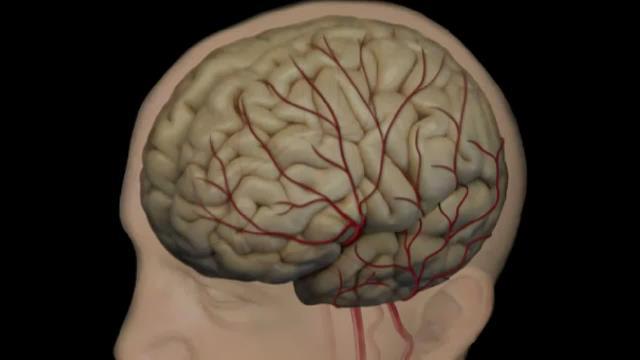
9 CEREBRAL CIRCULATION The mvement f bld thrugh the netwrk f bld vessels t supply the brain. The arteries carry xygenated bld and ther nutrients t the brain. The veins carry dexygenated bld back t the heart remving carbn dixide and ther metablic prducts. The mvement f bld in the cerebral circulatin is called cerebral bld flw.

10 CEREBRAL ARTERIAL SUPPLY The arterial cerebral circulatin is divided int anterir and psterir cerebral circulatins. The anterir and psterir cerebral circulatins are intercnnected via bilateral psterir cmmunicating arteries. Psterir cmmunicating arteries are part f Circle f Willis. Lcated n the base f the brain. It Encircles: Optic chiasma Hypthalamus Midbrain The cerebral arterial supply is prvided by tw systems: Cartid System Supply anterir prtin f the brain. Vertebr-Basilar System Supply psterir prtin f the brain.
, an English physician It")


11 CIRCULUS ARTERIOSUS (CIRCLE OF WILLIS) Named after Thmas Willis ( ), an English physician It is Frmed by: Tw Anterir cerebral arteries Tw Internal cartid arteries Tw Psterir cerebral arteries Tw Psterir cmmunicating arteries One Anterir cmmunicating artery It Gives numerus small vessels that penetrate the surface f the brain Perfrating arteries Anterir perfrating arteries Psterir perfrating arteries
12 ANTERIOR PERFORATING ARTERIES Arise frm: Anterir cerebral artery Anterir cmmunicating artery Middle cerebral artery Enter brain thrugh: Anterir perfrated substance irregularly quadrilateral area in frnt f the ptic tract and behind the lfactry trigne. Supply: Large part f basal ganglia Optic chiasma Internal capsule Hypthalamus


13 POSTERIOR PERFORATING ARTERIES Arise frm: Psterir cerebral artery Psterir cmmunicating artery Enter brain thrugh: Psterir Perfrated substance Supply: Ventral prtin f Midbrain Parts f Subthalamus and Hypthalamus
14 ANTERIOR CEREBRAL ARTERY Supplies: Orbital and medial surfaces f frntal and parietal lbes


15 MIDDLE CEREBRAL ARTERY Supplies: Entire Superlateral surface: Smatsensry Crtex Mtr Crtex Brca's Area linked t speech prductin Heschl s Gyrus t prcess incming auditry infrmatin Wernicke s Area It is invlved in the understanding f written and spken language

16 POSTERIOR CEREBRAL ARTERY Supplies: Anterir and inferir tempral lbes Uncus Lcated n the tip end f the medial surface f the parahippcampal gyrus. Part f the lfactry crtex that prcesses infrmatin frm the sense f smell. Inferir tempral gyri Inferir and Medial Occipital lbe

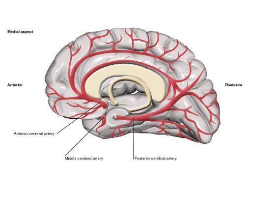
17 CEREBRAL ARTERIES

18 DISTRIBUTION OF CEREBRAL ARTERIES

19 BASILAR ARTERY Supplies: Midbrain and Cerebellum. Branches: Anterir inferir cerebellar artery Pntine branches Superir cerebellar artery

 r derived frm cells f the tissues")
20 ARTERIAL DISORDER Strke Sudden cclusin Hemrrhage Aneurysm lcalized, bld-filled ballnlike bulge in the wall f a bld vessel. Angima is benign tumrs derived frm cells f the vascular r lymphatic vessel walls (epithelium) r derived frm cells f the tissues surrunding these vessels.
21 ACCLUSION OF ACA Manifestatins: Mtr disturbance in cntralateral distal leg Difficulty in Prefrntal lbe Functins: Cgnitive thinking Judgment Mtr initiatin Self mnitring
22 ACCLUSION OF MCA Manifestatins: Cntralateral weakness f: face, arm, and hand mre than legs Cntralateral sensry lss f: face, arm, and hand mre than legs visual field cut (damage t ptic radiatin) Aphasia: language disturbances Brca's: prductin Wernicke's: cmprehensin

 Memry impairment If tempral lbe is")
23 ACCLUSION OF PCA Manifestatins: Visual disturbances Cntralateral hmnymus hemianpsia Bilateral lesins: crtical blindness patients unaware they cannt see (Antn's syndrme) Memry impairment If tempral lbe is affected
24 HOW WE ARE DOING..? Which statement(s) f the fllwing is NOT Wrng? Anterir cerebral arteries supply Brca's and Wernicke s Area..!! Occlusin f MCA causes difficulty in Prefrntal lbe s functins..!! Middle cerebral arteries are part f Willis Circle..!! Aneurysm is benign tumrs derived frm cells f the vascular r lymphatic vessel walls..!! Psterir cerebral arteries supply anterir and inferir tempral lbes..!!
25 CEREBRAL VENOUS DRAINAGE It invlves: Superficial (crtical) veins: Drain the crtical surface Deep veins: Drain the deep structures These veins ultimately drain int: Dural Venus Sinuses The Veins are thin walled and are devid f valves.
26 SUPERFICIAL CORTICAL VEINS Lie n the brain surface, in the Subarchnid space. They are divided int: Superir cerebral veins Inferir cerebral veins Superficial middle cerebral vein


27 SUPERFICIAL CORTICAL VEINS Superir Cerebral Veins 6 t 12 veins Drain lateral surface f brain abve the lateral sulcus Terminate mainly int the Superir Sagittal sinus, and partly int superficial middle cerebral vein.

28 SUPERFICIAL CORTICAL VEINS Inferir Cerebral Veins Run belw the lateral sulcus Drain the lateral surface f the tempral lbe Terminate partly int superficial middle cerebral vein & partly int Transverse sinus.
29 SUPERFICIAL CORTICAL VEINS Superficial Middle Cerebral Vein Runs alng the lateral sulcus Terminates int the Cavernus sinus Cnnected psterirly by Superir & Inferir anastmtic veins t Superir Sagittal & Transverse sinuses respectively.

30 DEEP CEREBRAL VEINS They drain the internal structures; Basal ganglia Internal capsule Thalamus They merge t frm the Internal Cerebral Veins. The tw veins unite in the midline t frm the Great Cerebral vein. This shrt vessel is cntinuus with the Straight Sinus.
31 DURAL VENOUS SINUSES Paired Single Transverse Sigmid Cavernus Petrsal Superir sagittal Inferir sagittal Straight Occipital Bld flws frm transverse & sigmid sinuses int IJV
32 WATCH
 and raised")
33 VENOUS DISORDER Infarctin refers t tissue death (necrsis) that is caused by a lcal lack f xygen due t bstructin f the tissue's bld supply Sinus thrmbsis: SSS thrmbsis Superir Sagittal Sinus Can cmplicates ear infectin Cavernus Sinus thrmbsis As a cmplicatin f infectin in the dangerus area f the face Obstructin f venus drainage f the brain leads t Cerebral swelling (edema) and raised Intracranial Pressure.
34 ALSO, HOW WE ARE DOING..? Which statement(s) f the fllwing is Wrng? 1. Superir Cerebral Veins terminate mainly int the Superir Sagittal sinus, and partly int superficial middle cerebral vein..!! 2. Infarctin refers t tissue death (necrsis)..!! 3. Superir Cerebral Veins drain lateral surface f brain abve the lateral sulcus..!! 4. Inferir Cerebral Veins terminate partly int superficial middle cerebral vein & partly int Transverse sinus..!! 5. Superficial Middle Cerebral Vein drains the lateral surface f the tempral lbe..!!

35 QUESTION?
OBJECTIVES. At the end of the lecture, students should be able to: List the cerebral arteries.
 DR JAMILA EL MEDANY OBJECTIVES At the end of the lecture, students should be able to: List the cerebral arteries. Describe the cerebral arterial supply regarding the origin, distribution and branches.
Al Balqa Applied University. Collage of Medicine. Anatomy Lab Check List
 Al Balqa Applied University Cllage f Medicine Anatmy Lab Check List Blck: Nervus System and Special Senses (31500311) Lab Title: Neuranatmy 1 Lab Objectives: Identify majr cmpnents f brain. Knw majr lbes,
Materials Dissecting pan, dissecting kit, safety glasses, lab apron, pig heart, & gloves
 Heart Dissectin Intrductin Mammals have fur-chambered hearts and duble circulatin. The heart f a bird r mammal has tw atria and tw cmpletely separated ventricles. The dublelp circulatin is similar t amphibians
CARDIOVASCULAR SYSTEM. Khaleel Alyahya, PhD, MEd King Saud University School of
 CARDIOVASCULAR SYSTEM Khaleel Alyahya, PhD, MEd King Saud University Schl f Medicine @khaleelya OBJECTIVES At the end f the lecture, students shuld be able t: Identify the cmpnents f the cardivascular
Principles Arteries & Veins of the CNS LO14
 Principles Arteries & Veins of the CNS LO14 14. Identify (on cadaver specimens, models and diagrams) and name the principal arteries and veins of the CNS: Why is it important to understand blood supply
Medical Neuroscience Tutorial Notes
 Medical Neuroscience Tutorial Notes Blood Supply to the Brain MAP TO NEUROSCIENCE CORE CONCEPTS 1 NCC1. The brain is the body's most complex organ. LEARNING OBJECTIVES After study of the assigned learning
Lecture 17 (03/28/2011) (Lateralization in the Brain) PSY 215. Lecture 17 Topic: Lateralization in the Brain Chapter 14.
 (Lateralization in the Brain) PSY 215. Lecture 17 Topic: Lateralization in the Brain Chapter 14.") PSY 215 Lecture 17 (03/28/2011) (Lateralizatin in the Brain) Dr. Achtman PSY 215 Lecture 17 Tpic: Lateralizatin in the Brain Chapter 14.1, pages 404-413 Crrectins: Nne needed Annuncements: Wednesday will
PSY 215 Lecture 17 (03/28/2011) (Lateralizatin in the Brain) Dr. Achtman PSY 215 Lecture 17 Tpic: Lateralizatin in the Brain Chapter 14.1, pages 404-413 Crrectins: Nne needed Annuncements: Wednesday will
The brain stem functions in homeostasis, coordination of movement, and conduction of information to and from higher brain centers.
 Chapter 49 Nervus Systems Lecture Outline Cncept 49.2 The vertebrate brain is reginally specialized. In all vertebrates, three bilaterally symmetrical, anterir bulges f the neural tube the frebrain, midbrain,
Group Members: Date Period
 Dissectin Kit Number: Grup Members: Date Perid Dissectins Objective: In this tw-week investigatin, we will be lking at an invetebrate (grasshpper) and a vertebrate (frg). Yu will be fcusing n the external
CARDIOVASCULAR SYSTEM: OVERVIEW & ANATOMY Cathy Proenza
 CV 03-20-17 08AM CVPR Overview-Anatmy - Prenza CARDIOVASCULAR SYSTEM: OVERVIEW & ANATOMY Cathy Prenza catherine.prenza@ucdenver.edu Recmmended Reading Lilly p 1-12 Other learning resurces: http://www.cvphysilgy.cm/
Brain ميهاربا لض اف دمح ا د The Meninges 1- Dura Mater of the Brain endosteal layer does not extend meningeal layer falx cerebri tentorium cerebelli
 .احمد د فاضل ابراهيم Lecture 15 Brain The Meninges Three protective membranes or meninges surround the brain in the skull: the dura mater, the arachnoid mater, and the pia mater 1- Dura Mater of the Brain
Blood Supply of the CNS
 Blood Supply of the CNS Lecture Objectives Describe the four arteries supplying the CNS. Follow up each artery to its destination. Describe the circle of Willis and its branches. Discuss the principle
Topic 11: Nervous System
 Tpic 11: Nervus System Functin: Imprtant Definitins Neurns: Stimulus: anything that causes a nerve impulse t be sent Ex: Receptrs: sense rgans that detect Impulse: change alng the neurn Effectrs: any structure
Chapter 20 The Heart
 Chapter 20 The Heart ANATOMY OF THE HEART The adult heart is apprximately the size f. The heart is lcated in the cavity, between. Base is directed, and the apex is directed. The membrane surrunding and
Al Balqa Applied University. Collage of Medicine. Anatomy Lab Check List
 Al Balqa Applied University Cllage f Medicine Anatmy Lab Check List Blck: Nervus System and Special Senses (31500311) Lab Title: Neuranatmy 3 Lab Objectives: Study majr parts f the brainstem, rigin f the
Dissection of the Sheep Brain
 Dissection of the Sheep Brain Laboratory Objectives After completing this lab, you should be able to: 1. Identify the main structures in the sheep brain and to compare them with those of the human brain.
Organization of The Nervous System PROF. MOUSAED ALFAYEZ & DR. SANAA ALSHAARAWY
 Organization of The Nervous System PROF. MOUSAED ALFAYEZ & DR. SANAA ALSHAARAWY Objectives At the end of the lecture, the students should be able to: List the parts of the nervous system. List the function
Group D: Central nervous system yellow
 Group D: Central nervous system yellow Central nervous system 1. General structure of nervous system (neuron, glia, synapsis, mediators, receptors) Main points: types of neurons and glial cells, synapses,
PTA 106 Unit 1 Lecture 3
 PTA 106 Unit 1 Lecture 3 The Basics Arteries: Carry blood away from the heart toward tissues. They typically have thicker vessels walls to handle increased pressure. Contain internal and external elastic
Announcement. Danny to schedule a time if you are interested.
 Announcement If you need more experiments to participate in, contact Danny Sanchez (dsanchez@ucsd.edu) make sure to tell him that you are from LIGN171, so he will let me know about your credit (1 point).
INTRODUCTION TO THE CIRCULATORY SYSTEM
 INTRODUCTION TO THE CIRCULATORY SYSTEM What des bld d? 5. What makes this pssible? : In rder fr there t be an efficient exchange f xygen, waste and nutrients there must be a high surface area between the
M555 Medical Neuroscience Lab 1: Gross Anatomy of Brain, Crainal Nerves and Cerebral Blood Vessels
 M555 Medical Neuroscience Lab 1: Gross Anatomy of Brain, Crainal Nerves and Cerebral Blood Vessels Anatomical Directions Terms like dorsal, ventral, and posterior provide a means of locating structures
Neuroanatomy Dr. Maha ELBeltagy Assistant Professor of Anatomy Faculty of Medicine The University of Jordan 2018
 Neuroanatomy Dr. Maha ELBeltagy Assistant Professor of Anatomy Faculty of Medicine The University of Jordan 2018 Blood Supply of Brain and Spinal Cord Arterial Supply of Brain The brain receives blood
Inside Your Patient s Brain Michelle Peterson, APRN, CNP Centracare Stroke and Vascular Neurology
 Inside Your Patient s Brain Michelle Peterson, APRN, CNP Centracare Stroke and Vascular Neurology Activity Everyone stand up, raise your right hand, tell your neighbors your name 1 What part of the brain
Blood Supply. Allen Chung, class of 2013
 Blood Supply Allen Chung, class of 2013 Objectives Understand the importance of the cerebral circulation. Understand stroke and the types of vascular problems that cause it. Understand ischemic penumbra
BIOL Dissection of the Sheep and Human Brain
 BIOL 2401 Dissection of the Sheep and Human Brain Laboratory Objectives After completing this lab, you should be able to: Identify the main structures in the sheep brain and to compare them with those
CEREBRUM. Dr. Jamila EL Medany
 CEREBRUM Dr. Jamila EL Medany Objectives At the end of the lecture, the student should be able to: List the parts of the cerebral hemisphere (cortex, medulla, basal nuclei, lateral ventricle). Describe
Organization of The Nervous System PROF. SAEED ABUEL MAKAREM
 Organization of The Nervous System PROF. SAEED ABUEL MAKAREM Objectives By the end of the lecture, you should be able to: List the parts of the nervous system. List the function of the nervous system.
DEMENTIA. DESCRIPTION: a progressive, degenerative disease of the brain, which causes impairment of thinking and memory
 DEMENTIA Dementia a syndrme cnsisting f a number f symptms including lss f memry, judgment and reasning, and changes in md and behavir the changes may affect a persn's ability t functin at wrk, in scial
Nervous Systems. Chapter 49. Lecture Outline. Overview: Command and Control System
 Chapter 49 Nervus Systems Lecture Outline Overview: Cmmand and Cntrl System The human brain cntains an estimated 10 11 (100 billin) neurns. The circuits that intercnnect these brain cells are enrmusly
CEREBRUM Dr. Jamila Elmedany Dr. Essam Eldin Salama
 CEREBRUM Dr. Jamila Elmedany Dr. Essam Eldin Salama Objectives At the end of the lecture, the student should be able to: List the parts of the cerebral hemisphere (cortex, medulla, basal nuclei, lateral
Exercise Physiology CardioRespiratory Trimester 2. Exercise Physiology Cardiorespiratory Study Guide
 Exercise Physilgy CardiRespiratry Trimester 2 Cardivascular System Exercise Physilgy Cardirespiratry Study Guide 1. Be able t label the structures f the heart (chambers, vessels, valves) and the pattern
b. The groove between the two crests is called 2. The neural folds move toward each other & the fuse to create a
 Chapter 13: Brain and Cranial Nerves I. Development of the CNS A. The CNS begins as a flat plate called the B. The process proceeds as: 1. The lateral sides of the become elevated as waves called a. The
For Emergency Doctors. Dr Suzanne Smallbane November 2011
 For Emergency Doctors Dr Suzanne Smallbane November 2011 A: Orbit B: Sphenoid Sinus C: Temporal Lobe D: EAC E: Mastoid air cells F: Cerebellar hemisphere A: Frontal lobe B: Frontal bone C: Dorsum sellae
Lab Activity 25. Blood Vessels & Circulation. Portland Community College BI 232
 Lab Activity 25 Blood Vessels & Circulation Portland Community College BI 232 Artery and Vein Histology Walls have 3 layers: Tunica intima Tunica media Tunica externa 2 Tunica Intima Is the innermost layer
Stroke School for Internists Part 1
 Stroke School for Internists Part 1 November 4, 2017 Dr. Albert Jin Dr. Gurpreet Jaswal Disclosures I receive a stipend for my role as Medical Director of the Stroke Network of SEO I have no commercial
YOU MUST BRING GLOVES FOR THIS ACTIVITY
 ACTIVITY 10: VESSELS AND CIRCULATION OBJECTIVES: 1) How to get ready: Read Chapter 23, McKinley et al., Human Anatomy, 5e. All text references are for this textbook. 2) Observe and sketch histology slide
The Excretory System. 4 The Excretory System.notebook. May 24, 2017
 4 The Excretry System.ntebk The Excretry System OSMOREGULATION (WATER BALANCE) Mst marine invertebrates are smcnfrmers, meaning the cncentratin f slutes in their bdy fluid is equal t that f their envirnment.
VESSELS: GROSS ANATOMY
 ACTIVITY 10: VESSELS AND CIRCULATION OBJECTIVES: 1) How to get ready: Read Chapter 23, McKinley et al., Human Anatomy, 4e. All text references are for this textbook. 2) Observe and sketch histology slide
Lecture 4 The BRAINSTEM Medulla Oblongata
 Lecture 4 The BRAINSTEM Medulla Oblongata Introduction to brainstem 1- Medulla oblongata 2- Pons 3- Midbrain - - - occupies the posterior cranial fossa of the skull. connects the narrow spinal cord
Cerebral hemisphere. Parietal Frontal Occipital Temporal
 Cerebral hemisphere Sulcus / Fissure Central Precental gyrus Postcentral gyrus Lateral (cerebral) Parieto-occipital Cerebral cortex Frontal lobe Parietal lobe Temporal lobe Insula Amygdala Hippocampus
HEAD/NECK VESSELS. Objectives
 Objectives Arterial Supply to Head and Neck Arteries to Head Surrounding Brain Common carotid arteries Arteries to Head Surrounding Brain External carotid arteries Arteries to Head Surrounding Brain External
Anatomy & Physiology Central Nervous System Worksheet
 1. What are the two parts of the CNS? 2. What are the four functions of the CNS Anatomy & Physiology Central Nervous System Worksheet 3. What are the four functions of the meninges? (p430) 4. Starting
Unit 18: Cranial Cavity and Contents
 Unit 18: Cranial Cavity and Contents Dissection Instructions: The calvaria is to be removed without damage to the dura mater which is attached to the inner surface of the calvaria. Cut through the outer
Autonomic. Nervous System
 Autnmic Nervus System Cmpiled by Campbell M Gld (2006) CMG Archives http://campbellmgld.cm IMPORTANT The health infrmatin cntained herein is nt meant as a substitute fr advice frm yur physician, r ther
The dura is sensitive to stretching, which produces the sensation of headache.
 Dural Nerve Supply Branches of the trigeminal, vagus, and first three cervical nerves and branches from the sympathetic system pass to the dura. Numerous sensory endings are in the dura. The dura is sensitive
ASFYT Part I: The Skeletal System S1: Intro to Kinesiology
 S1: Intr t Kinesilgy (1) Intr t Kinesilgy Majr Divisins f the Human Bdy Jints Between Majr Bdy Parts Describing Mvement in the Bdy True Mvement vs. Ging Alng fr the Ride Anatmic Psitin Directinal Terms
[(PHY-3a) Initials of MD reviewing films] [(PHY-3b) Initials of 2 nd opinion MD]
![[(PHY-3a) Initials of MD reviewing films] [(PHY-3b) Initials of 2 nd opinion MD]](/thumbs/89/98619893.jpg "[(PHY-3a) Initials of MD reviewing films] [(PHY-3b) Initials of 2 nd opinion MD]") 2015 PHYSICIAN SIGN-OFF (1) STUDY NO (PHY-1) CASE, PER PHYSICIAN REVIEW 1=yes 2=no [strictly meets case definition] (PHY-1a) CASE, IN PHYSICIAN S OPINION 1=yes 2=no (PHY-2) (PHY-3) [based on all available
2015 PHYSICIAN SIGN-OFF (1) STUDY NO (PHY-1) CASE, PER PHYSICIAN REVIEW 1=yes 2=no [strictly meets case definition] (PHY-1a) CASE, IN PHYSICIAN S OPINION 1=yes 2=no (PHY-2) (PHY-3) [based on all available
Downloaded from
 POINTS TO REMEMBER : Dwnladed frm www.studiestday.cm 21. Neural Cntrl and Crdinatin HUMAN NEURAL SYSTEM : The human neural system divided int tw parts The central nervus system (CNS) The peripheral nervus
2. As a caregiver, it s important to build a team/ support network around you (choose one):
:") Test Questins (Part 1, Chapter 1) 1. Of thse affected by dementia: 50% are nt aware they are changing and 50% are aware they are changing. Of the 50% wh are aware, they will always respnd by becming anxius
Test Questins (Part 1, Chapter 1) 1. Of thse affected by dementia: 50% are nt aware they are changing and 50% are aware they are changing. Of the 50% wh are aware, they will always respnd by becming anxius
TRANSVERSE SECTION PLANE Scalp 2. Cranium. 13. Superior sagittal sinus
 TRANSVERSE SECTION PLANE 1 1. Scalp 2. Cranium 3. Superior sagittal sinus 4. Dura mater 5. Falx cerebri 6. Frontal lobes of the cerebrum 7. Middle meningeal artery 8. Cortex, grey matter 9. Cerebral vessels
Slide 1. Slide 2. Slide 3. Tomography vs Topography. Computed Tomography (CT): A simplified Topographical review of the Brain. Learning Objective
: A simplified Topographical review of the Brain. Learning Objective") Slide 1 Computed Tomography (CT): A simplified Topographical review of the Brain Jon Wheiler, ACNP-BC Slide 2 Tomography vs Topography Tomography: A technique for displaying a representation of a cross
Slide 1 Computed Tomography (CT): A simplified Topographical review of the Brain Jon Wheiler, ACNP-BC Slide 2 Tomography vs Topography Tomography: A technique for displaying a representation of a cross
NSB603 Introduction to Cardiothoracic Nursing
 NSB603 Intrductin t Cardithracic Nursing Lecture 1a HEART OBJECTIVES Describe pathphysilgy f heart failure, CAD, angina/mi List Rx fr angina/mi Evaluate effectiveness f interventins HEART PATHWAY Frm Bdy
Essentials of Clinical MR, 2 nd edition. 14. Ischemia and Infarction II
 14. Ischemia and Infarction II Lacunar infarcts are small deep parenchymal lesions involving the basal ganglia, internal capsule, thalamus, and brainstem. The vascular supply of these areas includes the
meninges Outermost layer of the meninge dura mater arachnoid mater pia mater membranes located between bone and soft tissue of the nervous system
 membranes located between bone and soft tissue of the nervous system meninges Outermost layer of the meninge dura mater middle layer of the meninges, contains no blood vessels arachnoid mater Innermost
Lab Photo Review Sheet
 9 8 0. Posterior Median Sulcus. Central Canal. Dorsal (Posterior) Horn. Ventral (Anterior) Horn. Grey Matter. White Matter. Anterior Median Fissure 8. Ventral (Anterior) Root (ramus) 9. Dorsal (Posterior)
Brain and Cranial Nerves (Ch. 15) Human Anatomy lecture. caudal = toward the spinal cord)
 Human Anatomy lecture. caudal = toward the spinal cord)") Insight: Some cranial nerve disorders Brain and Cranial Nerves (Ch. 15) Human Anatomy lecture I. Overview (Directional terms: rostral = toward the forehead caudal = toward the spinal cord) A. 3 Major parts
Insight: Some cranial nerve disorders Brain and Cranial Nerves (Ch. 15) Human Anatomy lecture I. Overview (Directional terms: rostral = toward the forehead caudal = toward the spinal cord) A. 3 Major parts
Enhancement of Cranial US: Utility of Supplementary Acoustic Windows and Doppler Harriet J. Paltiel, MD
 Enhancement of Cranial US: Utility of Supplementary Acoustic Windows and Doppler Harriet J. Paltiel, MD Boston Children s Hospital Harvard Medical School None Disclosures Conventional US Anterior fontanelle
Anatomy, Histology, & Embryology of the Pancreas
 Anatmy, Histlgy, & Embrylgy f the Pancreas *Pancreas is secndary retrperitneal, with the exceptin f the tail, the fregut. Anatmy f Pancreas Lcatin: Within the curve f the dudenum, lcated in the epigastric
4The head basic anatomy and physiology
 Hene_Ch04.qxd 8/30/04 2:47 AM Page 108 108 THE HEAD BASIC ANATOMY AND PHYSIOLOGY 4The head basic anatomy and physiology The scalp Anatomists describe the SCALP as having five layers: Skin, Subcutaneous
Chapter 13 Brain and Cranial Nerves
 Chapter 13 Brain and Cranial Nerves 13-1 Brain and Cranial Nerves Brain Part of CNS contained in cranial cavity Control center for many of body s functions Much like a complex computer but more Parts of
Central Nervous System (CNS) -> brain and spinal cord. Major Divisions of the nervous system:
 -> brain and spinal cord. Major Divisions of the nervous system:") Central Nervous System (CNS) -> brain and spinal cord Major Divisions of the nervous system: Afferent (sensory input) -> cell bodies outside of the central nervous system (CNS), carry info into the CNS
Central Nervous System (CNS) -> brain and spinal cord Major Divisions of the nervous system: Afferent (sensory input) -> cell bodies outside of the central nervous system (CNS), carry info into the CNS
Biology 30S Unit Test Review: Digestion
 Bilgy 30S Unit Test Review: Digestin Test utline: Multiple Chice: 10 Questins, 1 mark each. Shrt Answer: 5 Questins (answer 3 f them), 5 marks each. Lng Answer: 2 Lng Answer Questins, 10 marks each. Tasks
Overview of the Nervous System (some basic concepts) Steven McLoon Department of Neuroscience University of Minnesota
 Steven McLoon Department of Neuroscience University of Minnesota") Overview of the Nervous System (some basic concepts) Steven McLoon Department of Neuroscience University of Minnesota 1 Coffee Hour Tuesday (Sept 11) 10:00-11:00am Friday (Sept 14) 8:30-9:30am Surdyk s
Overview of the Nervous System (some basic concepts) Steven McLoon Department of Neuroscience University of Minnesota 1 Coffee Hour Tuesday (Sept 11) 10:00-11:00am Friday (Sept 14) 8:30-9:30am Surdyk s
Topic 11: Senses. Sensory Receptors
 Tpic 11: Senses Sensry Receptrs Part f the nervus system that detects a stimulus A sensry receptr culd be: Types f Sensry Receptrs: Chemreceptrs: detects chemicals ( ) Phtreceptrs: detects light (fund
Anatomy and Physiology (Bio 220) The Brain Chapter 14 and select portions of Chapter 16
 The Brain Chapter 14 and select portions of Chapter 16") Anatomy and Physiology (Bio 220) The Brain Chapter 14 and select portions of Chapter 16 I. Introduction A. Appearance 1. physical 2. weight 3. relative weight B. Major parts of the brain 1. cerebrum 2.
Anatomy and Physiology (Bio 220) The Brain Chapter 14 and select portions of Chapter 16 I. Introduction A. Appearance 1. physical 2. weight 3. relative weight B. Major parts of the brain 1. cerebrum 2.
NEURO IMAGING 2. Dr. Said Huwaijah Chairman of radiology Dep, Damascus Univercity
 NEURO IMAGING 2 Dr. Said Huwaijah Chairman of radiology Dep, Damascus Univercity I. EPIDURAL HEMATOMA (EDH) LOCATION Seventy to seventy-five percent occur in temporoparietal region. CAUSE Most likely caused
I Before You Read. I Read to Learn. What is an arthropod? How does an exoskeleton provide protection?
 I Befre Yu Read Have yu seen a drawing r picture f a knight in armr? What is the purpse f armr? On the lines belw list the advantages and disadvantages f wearing a heavy suit f armr. As yu read this sectin
If I Only Had a Brain
 If I Only Had a Brain A Heart. (The Nerve!) Regions of the Brain Cerebral hemisphere Diencephalon Cerebellum (b) Adult brain Brain stem Regions of the Brain: Cerebrum Precentral gyrus Frontal lobe Central
ANAT2011 Anatomy Notes
 ANAT2011 Anatmy Ntes Mdule 1: Hw t Speak Anatmy... 2 Mdule 2: Nervus System... 4 Mdule 3: Sniff, Swallw, Speak!... 14 Mdule 4: Just Breathe... 35 Mdule 5: Wh are the Great 8?... 43 Mdule 6: N Guts, N Glry...
Harold P. Adams, Jr., MD Department of Neurology Carver College of Medicine UIHC Comprehensive Stroke Center University of Iowa
 Harld P. Adams, Jr., MD Department f Neurlgy Carver Cllege f Medicine UIHC Cmprehensive Strke Center University f Iwa D nt receive persnal cmpensatin frm cmmercial interests D receive grant supprt frm
Neuroanatomy lecture (1)
") Neuroanatomy lecture (1) Introduction: Neuroanatomy has two parts: the central and peripheral nervous system. The central nervous system is composed of brain and spinal cord. The brain has the following
Neuroanatomy lecture (1) Introduction: Neuroanatomy has two parts: the central and peripheral nervous system. The central nervous system is composed of brain and spinal cord. The brain has the following
Name: Date: Period: Notes: The Blood and Lymphatic System
 Name: Date: Perid: Cmpsitin f Bld and their Functins Red Bld Cells (aka ) Structure Ntes: The Bld and Lymphatic System D nt have a like ther cells d Cntain a specialized prtein called Hemglbin cntains
Test 3 Study Guide: Photosynthesis, Respiration, and the Cell Membrane
 Name Blck Date Test 3 Study Guide: Phtsynthesis, Respiratin, and the Cell Membrane Test Dates: December 13 (1 st and 7 th blck) and 12 (6 th blck) SOL: BIO.2d, 3d-e Related Ntes Phtsynthesis Respiratin
Cerebrum-Cerebral Hemispheres. Cuneyt Mirzanli Istanbul Gelisim University
 Cerebrum-Cerebral Hemispheres Cuneyt Mirzanli Istanbul Gelisim University The largest part of the brain. Ovoid shape. Two incompletely separated cerebral hemispheres. The outer surface of the cerebral
Neuro Nugget: Stroke. Free Additional Board Exam Preparation Resources
 Neur Nugget: Strke Free Additinal Bard Exam Preparatin Resurces www.beatthebards.cm 877-225-8384 Neur Nugget: Strke By Jack Krasuski, MD cpyright 2007-2008 American Physician Institute fr Advanced Prfessinal
DEVELOPMENT OF BRAIN
 Ahmed Fathalla OBJECTIVES At the end of the lecture, students should: List the components of brain stem. Describe the site of brain stem. Describe the relations between components of brain stem & their
-Zeina Assaf. -Omar Odeh. - Maha Beltagy
 -3 -Zeina Assaf -Omar Odeh - Maha Beltagy 1 P a g e The Inferior Surface Of The Brain The inferior surface of the brain is divide by the stem of the lateral fissure into 2 parts : The orbital surface and
Close to spine/ point of attachment
 Terms, Tissues and Imaging INTRODUCTION TO ANATOMY AND ANATOMICAL TERMINOLOGY 1. Anatmical Facts; islated bservatins r statements abut structures (bdy parts) r events (happenings) Anatmical cncepts; structures
Meninges and Ventricles
 Meninges and Ventricles Irene Yu, class of 2019 LEARNING OBJECTIVES Describe the meningeal layers, the dural infolds, and the spaces they create. Name the contents of the subarachnoid space. Describe the
BRAIN PART I (A & B): VENTRICLES & MENINGES
: VENTRICLES & MENINGES") BRAIN PART I (A & B): VENTRICLES & MENINGES Cranial Meninges Cranial meninges are continuous with spinal meninges Dura mater: inner layer (meningeal layer) outer layer (endosteal layer) fused to periosteum
BRAIN PART I (A & B): VENTRICLES & MENINGES Cranial Meninges Cranial meninges are continuous with spinal meninges Dura mater: inner layer (meningeal layer) outer layer (endosteal layer) fused to periosteum
Chapter 13 The Occipital Lobe Anatomy of the Occipital Lobe Subdivision of the occipital cortex Connections of the Visual Cortex
 Chapter 13 The Occipital Lbe Anatmy f the Occipital Lbe - Frm the psterir ple f the cerebral hemisphere - Subdivisin f the ccipital crtex Mnkey s crtex was first divided by Brdmann int 3 regins area 17,
Edinburgh Imaging Academy online distance learning courses. Neuroanatomy
 Neuroanatomy Semester 1 / Autumn 10 credits (IMSc) / 20 Credits (N14R) Each Course is composed of Modules & Activities. Modules: Major Lobes and Fissures IMSc NI4R MIAA Ventricles and CSF IMSc NI4R MIAA
1A Human Biology Food, Digestion and Associated Body Systems
 1A Human Bilgy Fd, Digestin and Assciated Bdy Systems Human Bilgy Fd, Digestin and Assciated Bdy Systems Fd Fd is ne f the basic human needs and the digestive system enables the bdy t cnvert it int a frm
Bio 103 Tissues and Skin 35
 Bi 103 Tissues and Skin 35 Lecture Outline: Tissues & Skin [Chapters 5, 6, 12 (p. 440441)] Chapter 5: Tissues Def.: Tissue = Fur basic tissue types 1. 2. 3. 4. Epithelial Tissues General Characteristics
Chapter 13 Lecture Outline *
 Anatomy and Physiology, Seventh Edition Rod R. Seeley Idaho State University Trent D. Stephens Idaho State University Philip Tate Phoenix College Chapter 13 Lecture Outline * *See PowerPoint Image Slides
Skull-2. Norma Basalis Interna. Dr. Heba Kalbouneh Assistant Professor of Anatomy and Histology
 Skull-2 Norma Basalis Interna Dr. Heba Kalbouneh Assistant Professor of Anatomy and Histology Norma basalis interna Base of the skull- superior view The interior of the base of the skull is divided into
Chapter 18 The Urinary System Renal Function
 Chapter 18 The Urinary System Renal Functin Chapter Outline Functins f the Urinary System Anatmy f the Urinary System Basic Renal Exchange Prcesses Reginal Specializatin f the Renal Tubules Excretin 18.1.
PROPERTY OF ELSEVIER SAMPLE CONTENT - NOT FINAL. Gross Anatomy and General Organization of the Central Nervous System
 3 Gross Anatomy and General Organization of the Central Nervous System C h a p t e r O u t l i n e The Long Axis of the CNS Bends at the Cephalic Flexure Hemisecting a Brain Reveals Parts of the Diencephalon,
Sheep Brain Dissection
 Sheep Brain Dissection Mammalian brains have many features in common. Human brains may not be available, so sheep brains often are dissected as an aid to understanding the mammalian brain since he general
Guide to Draw It to Know It Neuroanatomy (relative to Medical Neuro, UI-COM Urbana)
") Guide to Draw It to Know It Neuroanatomy (relative to Medical Neuro, UI-COM Urbana) Note: Sometimes DITKI goes into far more detail than is necessary for the course, and in other cases not enough. As helpful
Guide to Draw It to Know It Neuroanatomy (relative to Medical Neuro, UI-COM Urbana) Note: Sometimes DITKI goes into far more detail than is necessary for the course, and in other cases not enough. As helpful
Impact of developmental trauma on brain function and connectivity. Presented by Carl A. Armes, BS. & Robert Coben, Ph.D.
 Impact f develpmental trauma n brain functin and cnnectivity. Presented by Carl A. Armes, BS. & Rbert Cben, Ph.D. Ace Study EEG Measures OUTLINE Previus Findings Results Methds Discussin WHAT IS DEVELOPMENTAL
Neurology study of the nervous system. nervous & endocrine systems work together to maintain homeostasis
 Nervous System Neurology study of the nervous system nervous & endocrine systems work together to maintain homeostasis Nervous System works very fast Uses electrical signals called nerve impulses Short-lived
Homework Week 2. PreLab 2 HW #2 Synapses (Page 1 in the HW Section)
") Homework Week 2 Due in Lab PreLab 2 HW #2 Synapses (Page 1 in the HW Section) Reminders No class next Monday Quiz 1 is @ 5:30pm on Tuesday, 1/22/13 Study guide posted under Study Aids section of website
Homework Week 2 Due in Lab PreLab 2 HW #2 Synapses (Page 1 in the HW Section) Reminders No class next Monday Quiz 1 is @ 5:30pm on Tuesday, 1/22/13 Study guide posted under Study Aids section of website
Cairns Base Hospital Emergency Department Part 1 FACEM MCQs
 airns ase Hspital mergency epartment Part 1 FM MQs 1 ncerning the cardiac actin ptential Phase 1 is due t rapid sdium influx Resting membrane ptential is typically -90mV Phase 2 results frm slw prlnged
NQF 0075 Ischemic Vascular Disease (IVD): Complete Lipid Panel and LDL Control
: Complete Lipid Panel and LDL Control") NQF 0075 Ischemic Vascular Disease (IVD): Cmplete Lipid Panel and LDL Cntrl Initial Patient Ppulatin: Numeratr(1): Numeratr(2): N Exclusin: Denminatr: D Exceptin: D Exclusin: Patients 18 years f age and
NQF 0075 Ischemic Vascular Disease (IVD): Cmplete Lipid Panel and LDL Cntrl Initial Patient Ppulatin: Numeratr(1): Numeratr(2): N Exclusin: Denminatr: D Exceptin: D Exclusin: Patients 18 years f age and
Biology 12 Review sheet Urinary and Reproductive Systems ANSWERS
 Bilgy 12 Review sheet Urinary and Reprductive Systems ANSWERS 1. What is the main excretry rgan? Kidneys 2. What are the functins f the kidney? helps maintain hmestasis thrugh regulatin f H 2O and ins
Cranial Cavity REFERENCES: OBJECTIVES OSTEOLOGY. Stephen A. Gudas, PT, PhD
 Stephen A. Gudas, PT, PhD Cranial Cavity REFERENCES: Moore and Agur, Essential Clinical Anatomy (ECA), 3rd ed., pp. 496 498; 500 507; 512 514 Grant s Atlas 12 th ed., Figs 7.6; 7.19 7.30. Grant s Dissector
Downloaded from
 POINTS TO REMEMBER : 20. Lcmtin and Mvement Types f Mvement : Amebid mvement: This mvement takes place in phagcytes where leuccytes and macrphages migrate thrugh tissue. It is affected by pseudpdia frmed
stored information, making decisions, and taking action. 1. It is also the center for intellect, emotions, behavior, and memory.
 Chapter 14 - Outline I. INTRODUCTION A. The brain is the center for registering sensations, correlating them with one another and with stored information, making decisions, and taking action. 1. It is