Point-of-Care Ultrasound Closer look at the Inferior Vena Cavae &
|
|
|
- Harry Herbert Gordon
- 5 years ago
- Views:
Transcription
1 Point-of-Care Ultrasound Closer look at the Inferior Vena Cavae & Brief Introduction to Gross Systolic Function Omar S. Darwish, MS, DO Certified in Point-of-Care Ultrasound Hospitalist University of California, Irvine
2 Objectives Know the Terminology and Basic Concepts of Bedside Ultrasound Learn how to acquire the IVC image Learn how to interpret the diameter and collapsibility of the IVC Know which view is best to assess systolic function Know how to objectively determine the degree of systolic function
3 Literature Indications for IVC Ultrasound Literature says: Estimation of intravascular status; i.e. any patient undergoing fluid resuscitation in order to monitor for response Differentiate causes of shock Shock States Cardiac Tamponade Acute Pulmonary Embolism Acute Congestive Heart Failure Pneumothorax Sepsis IVC DIAMETER Dilated, low collapsibility index Dilated, low collapsibility index Dilated, low collapsibility index Dilated, low collapsibility index Small, high collapsibility index
4 My Indications for IVC Ultrasound Unknown history of CHF, but with risk factors requiring high IV fluid rates Known history of CHF who require high IV fluid rates CHF exacerbation à assess response to diuretics Some Examples of patients who require IV fluid rates Rhabdomyolysis (particularly CPK > 5000) Hypercalcemia Acute Pancreatitis Severe Sepsis Diabetic Ketoacidosis


5 Terminology. Imaging Plans Transverse or Axial Longitudinal or Sagittal

6 Concept 1. Probes with Different Frequencies Many different types of probes that vary in frequency and foot print size For us you need to know only 3 (The letter represents the type of probe and the number represents the length in millimeters) P21 (phase array, 21 mm) à 1-5 MHz L38 (linear probe, 38 mm) à 5-10 MHz C60 (curvalinear probe, 60 mm) à MHz Increasing the frequency, improves the resolution At higher frequencies it becomes more difficult to see deeper structures On the machine you can adjust the frequency of a probe. For SonoSite, words are used to describe the different frequencies: RES, GEN, PEN E.g. For a P21 RES: 5 MHz, 3 GEN, 1 PEN C60 P21
7 Concept 2. Physiology for Spontaneous Breathing Inspiration Expiration Pleural Pressure Pleural Pressure Increase Venous Return IVC collapses Decrease Venous Return IVC expands
8 Mechanically Ventilated Patients Inspiratory Phase Expiratory Phase Pleural Pressure Pleural Pressure Decrease Venous Return IVC expands Increase Venous Return IVC collapses
9 Concept 3. Temporal Resolution Effects of Temporal Resolution: the number of frames/second (i.e. cardiac setting has a high temporal resolution) Software setting (cardiac, abdominal) à use the right setting i.e. make sure you are on the cardiac setting when performing a cardiac ultrasound Depth of the image à worsens resolution Using Color flow doppler à worsens resolution
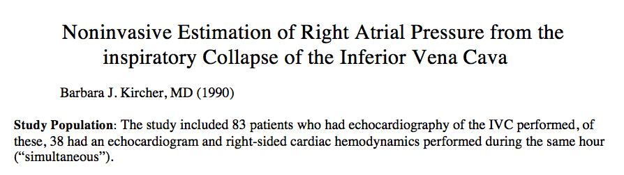
10 Acquiring the IMAGE


11 Step 1. Find the Xiphoid Process. Xiphoid Process Acoustic Shadowing Place Probe in the Transverse Position Below Xiphoid Process
12 Step 2. Rotate Probe 90 degrees clockwise


13 You can skip the first step and place the probe longitudinally first but make sure you are looking at the IVC and not the aorta
14
15 IVC Diameter (cm) Collapsibility (%) Estimated CVP (mm Hg) < > < >2.5 < >2.5 0 >20 Measure IVC 2 cm inferior to hepatic vein Otto CM. Echocardiographic evaluation of left and right ventricular systolic function. Textbook of clinical echocardiography. 2 nd ed. Philadelphia: WB Saunders; 2000.

16 Measuring IVC in Supine vs Left-Lateral Decubitis Position IVC diameter is larger in the supine position than in the left lateral

17 European Association of Echocardiography, 2005 Description Small IVC (cm < 1.2 cm Normal Dilated Markedly Dilated cm cm >2.6 cm IVC athletes: cm vs controls cm Swimmers: 2.66 vs 2.17 cm in other athletes Collapsibility index 58% in athletes compared to 70% in controls
 If it does not fit both criteria, then an intermediate estimated right atrial pressure of 8 mm Hg")
18 IVC Diameter (cm) Collapsibility Index with Sniff Estimated Right Atrial Pressure (mm Hg) < 2.1 >50% 3 mm Hg (0-5) >2.1 <50% 15 mm Hg (10-20) If it does not fit both criteria, then an intermediate estimated right atrial pressure of 8 mm Hg (5-10) is given
19 Interpretation of the IVC Diameter Size Only Average People Fluid Overload, Normal Variant in Athletes IVC Diameter Small Dilated 1.0 cm 2.0 cm 2.7 cm Normal Range Dilated My Interpretation
20
21 Elevated Right Atrial Pressure Normal Right Atrial Pressure IVC Collapsibility Index 0% 20%** 50% 80% 100% Highly Likely Fluid Tolerant Highly Likely Fluid Intolerant
22 Bottom Line. The IVC is overall considered dilated > cm, however, this by itself does not mean that with 100% specificity that the patient is fluid overloaded. The IVC collapsibility index has a better predictability value than the diameter of the IVC regarding a patient s fluid status. As to the degree of collapsibility the reference point of 50% has been reported many times to be the best cut off in regards to providing the best combined sensitivity and specificity.


23 /2.37 x 100%= 5% During inspiration, IVC measured at 2.25 cm
24
25 How to interpret collapsibility of the IVC? Fluid responsiveness vs Fluid tolerance Fluid unresponsiveness vs Fluid intolerance
26 Fluid Tolerance IVC Diameter (cm) Collapsibility (%) Estimated CVP (mm Hg) < > < >2.5 < >2.5 0 >20 Fluid Intolerance
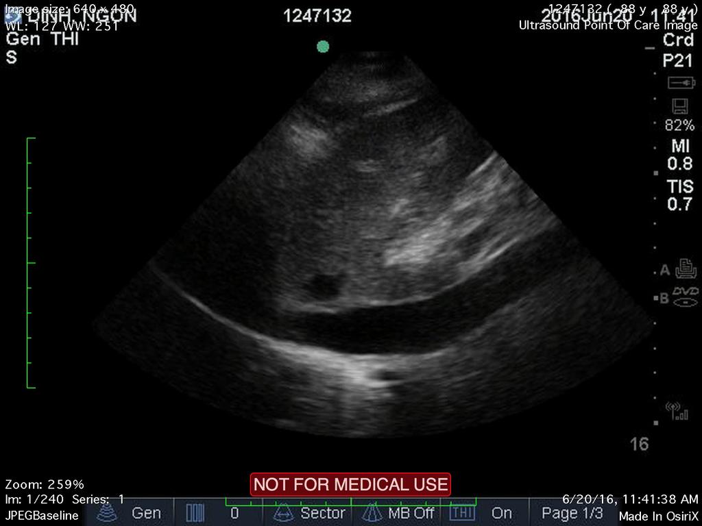
27 How to Assess Systolic Function

28 Parasternal Long Axis View Place P21 probe on the left side of the sternum around the 3-4 intercostal space Make sure you stay close to the sternum Indicator should be pointing to the Left Elbow with the indicator on the screen to be on the left

29 Parasternal Long Axis View

30 What is the systolic Function?
31 Conclusion IVC ultrasound is common data point that can help you in determining fluid status in a patient. Diameter and collapsibility information should then be applied to the clinical context of the patient. The Parasternal long axis view is the best view to determine gross systolic function when using bedside cardiac ultrasound.

32 Ventilated Patient 1) Sinus rhythm 2) PEEP > 4 mm Hg 3) Tv 8 ml/kg We recommend critical care practitioners consider measuring IVC collapsibility in patients on positive pressure ventilation by bedside cardiac ultrasound to assess fluid responsiveness prior to undergoing large volume fluid resuscitation. Any patient who has more than 15% change in vena caval diameter should be considered preload responsive. Patients with a smaller change in IVC diameter may not respond favorably to fluid resuscitation. Grade 1B, 2016
33 Measuring IVC in Ventilated Patient Meausuring the difference between the max and min diameters/mean of the two values = Respiration Variation > 12% Meausuring the difference between the max and min diameters/minumum diameter = Distensibility Index > 18% Not perfect, but absence of respiratory variation in a shock patient suggest that volume expansion will be ineffective 90% of cases.
34 Guidelines Supported goes back to ACEP Policy Statement on Emergency Ultrasounds includes evaluation of intravascular volume status and estimation of CVP based on imaging of the IVC.

35 85 year-old woman admitted 4 days ago before I started service on Monday for Severe Sepsis due to Strep Pneumonia with positive blood cultures improving overall, but requiring oxygen 2 liters of oxygen, desaturates to high 80s on room air. Hospitalist Options 1) d/c on home oxygen 2) See if there is alternative explanation of her hypoxia
 2 = 36.48 36.48 + RAP (est 15 mm Hg) = RVSP =51.5 mm Hg")
36 Pulmonary Pressures are effected by the Intravascular fluid volumes of a patient P = 4v 2 = RVSP - RA --> 4(3.02) 2 = RAP (est 15 mm Hg) = RVSP =51.5 mm Hg

37 After removal of nearly 6 liters of fluid At Discharge: O2 saturation 95% on RA
Background: Bedside ultrasound is emerging as a useful tool in the assessment of
 Abstract: Background: Bedside ultrasound is emerging as a useful tool in the assessment of intravascular volume status by examining measurements of the inferior vena cava (IVC). Many previous studies do
AAENP US WORKSHOP 2/25/17
 Know the components of the Rapid Ultrasound for Shock & Hypotension & Extended Focused Assessment Sonography in Trauma & how they can help quickly determine diagnosis. Be comfortable obtaining and interpreting
Prof. Dr. Iman Riad Mohamed Abdel Aal
 The Use of New Ultrasound Indices to Evaluate Volume Status and Fluid Responsiveness in Septic Shock Patients Thesis Submitted for partial fulfillment of MD degree in Anesthesiology, Surgical Intensive
ORIGINAL ARTICLE. Role of Ultrasound in Evaluation of Undifferentiated Shock in ICU Settings
 Journal of The Association of Physicians of India Vol. 66 August 2018 13 Role of Ultrasound in Evaluation of Undifferentiated Shock in ICU Settings Tanvi Vaidya 1*, Pradeep D costa 2, Satish Pande 3 ORIGINAL
Identification of congestive heart failure via respiratory variation of inferior vena cava diameter,
 American Journal of Emergency Medicine (2009) 27, 71 75 www.elsevier.com/locate/ajem Brief Report Identification of congestive heart failure via respiratory variation of inferior vena cava diameter, David
Certificate in Clinician Performed Ultrasound (CCPU) Syllabus. Rapid Cardiac Echo (RCE)
 Syllabus. Rapid Cardiac Echo (RCE)") Certificate in Clinician Performed Ultrasound (CCPU) Syllabus Rapid Cardiac Echo (RCE) Purpose: Rapid Cardiac Echocardiography (RCE) This unit is designed to cover the theoretical and practical curriculum
Certificate in Clinician Performed Ultrasound (CCPU) Syllabus Rapid Cardiac Echo (RCE) Purpose: Rapid Cardiac Echocardiography (RCE) This unit is designed to cover the theoretical and practical curriculum
The role of bedside ultrasound in the diagnosis of pericardial effusion and cardiac tamponade
 Symposium The role of bedside ultrasound in the diagnosis of pericardial effusion and cardiac tamponade Adam Goodman, Phillips Perera, Thomas Mailhot, Diku Mandavia Department of Emergency Medicine, Los
Echocardiography Volume assessment. Justin Mandeville 2014
 Echocardiography Volume assessment Justin Mandeville 2014 Volume assessment and the intensivist Hypovolaemic shock Fluid tolerance Optimising cardiac output Avoiding overloading Guided fluid removal Add
The Vigileo monitor by Edwards Lifesciences supports both the FloTrac Sensor for continuous cardiac output and the Edwards PreSep oximetry catheter
 1 2 The Vigileo monitor by Edwards Lifesciences supports both the FloTrac Sensor for continuous cardiac output and the Edwards PreSep oximetry catheter for continuous central venous oximetry (ScvO2) 3
Adel Hasanin Ahmed 1
 Adel Hasanin Ahmed 1 PERICARDIAL DISEASE The pericardial effusion ends anteriorly to the descending aorta and is best visualised in the PLAX. PSAX is actually very useful sometimes for looking at posterior
Echocardiographic Cardiovascular Risk Stratification: Beyond Ejection Fraction
 Echocardiographic Cardiovascular Risk Stratification: Beyond Ejection Fraction October 4, 2014 James S. Lee, M.D., F.A.C.C. Associates in Cardiology, P.A. Silver Spring, M.D. Disclosures Financial none
Bedside Ultrasound. US Guided Fluid Resuscitation. Michiel J. van Veelen, Emergency Physician, DTM&H
 Bedside Ultrasound US Guided Fluid Resuscitation Michiel J. van Veelen, Emergency Physician, DTM&H Outline Shock and Fluid Resuscitation in ICU Ultrasound in Shock Ultrasound Guided Fluid Resuscitation
ENDPOINTS OF RESUSCITATION
 ENDPOINTS OF RESUSCITATION Fred Pieracci, MD, MPH Acute Care Surgeon Denver Health Medical Center Assistant Professor of Surgery University of Colorado Health Science Center OUTLINE Recognition and characterization
Dr. Rami M. Adil Al-Hayali Assistant Professor in Medicine
 Dr. Rami M. Adil Al-Hayali Assistant Professor in Medicine Venous thromboembolism: pulmonary embolism (PE) deep vein thrombosis (DVT) 1% of all patients admitted to hospital 5% of in-hospital mortality
Echocardiography as a diagnostic and management tool in medical emergencies
 Echocardiography as a diagnostic and management tool in medical emergencies Frank van der Heusen MD Department of Anesthesia and perioperative Care UCSF Medical Center Objective of this presentation Indications
Shock, Monitoring Invasive Vs. Non Invasive
 Shock, Monitoring Invasive Vs. Non Invasive Paula Ferrada MD Assistant Professor Trauma, Critical Care and Emergency Surgery Virginia Commonwealth University Shock Fluid Pressors Ionotrope Intervention
Sepsis Wave II Webinar Series. Sepsis Reassessment
 Sepsis Wave II Webinar Series Sepsis Reassessment Presenters Nova Panebianco, MD Todd Slesinger, MD Fluid Reassessment in Sepsis Todd L. Slesinger, MD, FACEP, FCCM, FCCP, FAAEM Residency Program Director
Background & Indications Probe Selection
 Teresa S. Wu, MD, FACEP Director, EM Ultrasound Program & Fellowship Co-Director, Simulation Based Training Program & Fellowship Associate Program Director, EM Residency Program Maricopa Medical Center
Estimation of Right Atrial Pressure from the Inspiratory Collapse of the Inferior Vena cava in Pediatric Patients
 Original Article Iran J Pediatr Jun 2010; Vol 20 (No 2), Pp:206-210 Estimation of Right Atrial Pressure from the Inspiratory Collapse of the Inferior Vena cava in Pediatric Patients Hamid Amoozgar*, MD;
Pericardial Diseases. Smonporn Boonyaratavej, MD. Division of Cardiology, Department of Medicine Chulalongkorn University
 Pericardial Diseases Smonporn Boonyaratavej, MD Division of Cardiology, Department of Medicine Chulalongkorn University Cardiac Center, King Chulalongkorn Memorial Hospital 21 AUGUST 2016 Pericardial
Ultrasound. FAST Focused Assessment with Sonography in Trauma
 Ultrasound FAST Focused Assessment with Sonography in Trauma Rohit Patel, MD University of Florida Health Director, Critical Care Ultrasound Surgical ICU Center for Intensive Care Gainesville, Florida
Perioperative Ultrasonography Ehab Farag, MD, FRCA Hesham Elsharkawy David G. Anthony, M.D.
 Perioperative Ultrasonography Ehab Farag, MD, FRCA Hesham Elsharkawy David G. Anthony, M.D. Cleveland Clinic, Cleveland OH 1 Complications during central venous catheterization (CVC) occur 2% -15% of the
Transthoracic Echocardiography:
 Transthoracic Echocardiography: An essential tool for the obstetric anaesthetist? Brendan Carvalho MBBCh, FRCA Department of Anesthesiology Stanford University, California Focused TTE Stethoscope of the
Intro to Bedside Ultrasound. Cardiac Ultrasound
 Intro to Bedside Ultrasound Cardiac Ultrasound TEACHERS University of California-Irvine School of Medicine Nathan Molina nathan.d.molina@gmail.com Trevor Plescia taplescia90@gmail.com Jack Silva jpsilva42@gmail.com
Department of General Medicine, Kilpauk Medical College and Hospital, Chennai, Tamil Nadu, India * Corresponding author
 Original Research Article Study on clinical assessment of volume status and correlation to the respiratory variation in inferior vena cava diameter by echocardiography, a non-invasive method of measuring
Doppler Basic & Hemodynamic Calculations
 Doppler Basic & Hemodynamic Calculations August 19, 2017 Smonporn Boonyaratavej MD Division of Cardiology, Department of Medicine Chulalongkorn University Cardiac Center, King Chulalongkorn Memorial Hospital
Hemodynamic Assessment. Assessment of Systolic Function Doppler Hemodynamics
 Hemodynamic Assessment Matt M. Umland, RDCS, FASE Aurora Medical Group Milwaukee, WI Assessment of Systolic Function Doppler Hemodynamics Stroke Volume Cardiac Output Cardiac Index Tei Index/Index of myocardial
Session 2: Ultrasonography for Primary Care Clinicians Learning Objectives
 Session 2: Ultrasonography for Primary Care Clinicians Learning Objectives 1. Assess the main components and functions of a portable ultrasound unit. 2. Identify three clinical applications of portable
Hemodynamic Monitoring
 Perform Procedure And Interpret Results Hemodynamic Monitoring Tracheal Tube Cuff Pressure Dean R. Hess PhD RRT FAARC Hemodynamic Monitoring Cardiac Rate and Rhythm Arterial Blood Pressure Central Venous
Atrial Septal Defects
 Supplementary ACHD Echo Acquisition Protocol for Atrial Septal Defects The following protocol for echo in adult patients with atrial septal defects (ASDs) is a guide for performing a comprehensive assessment
Original Research Article
 ASSESSMENT AND COMPARISON OF CARDIAC FUNCTION AND VOLUME STATUS USING BEDSIDE ECHOCARDIOGRAPHY IN CRITICALLY ILL PATIENTS WITH OTHER CONVENTIONAL METHODS Priyanka Dwivedi, Shahbaz Ahmad, Vivek Pushp 3,
A Practical Approach to Ultrasound Assessment of Respiratory Distress
 A Practical Approach to Ultrasound Assessment of Respiratory Distress Yanick Beaulieu, MD, FRCPC Director, Bedside Ultrasound Curriculum Division of Cardiology and Critical Care Hôpital du Sacré-Coeur
University of Cape Town
 The copyright of this thesis vests in the author. No quotation from it or information derived from it is to be published without full acknowledgement of the source. The thesis is to be used for private
Echo in Pulmonary HTN
 Echo in Pulmonary HTN Steven A. Goldstein MD FACC FASE Professor of Medicine Georgetown University Medical Center MedStar Heart Institute Washington Hospital Center Monday, October 10, 2017 Pulmonary Artery
Diagnostic Bedside Ultrasound for the Hospitalist
 Diagnostic Bedside Ultrasound for the Hospitalist Trevor Jensen MD MS Assistant Professor, UCSF Nima Afshar MD Associate Professor, UCSF Diagnostic Bedside Ultrasound AKA Point-of-Care Ultrasound (POCUS)
Breakout Session: Transesophageal Echocardiography
 Breakout Session: Transesophageal Echocardiography Doris Ockert, MD Andrew Schroeder, MD University of Wisconsin School of Medicine and Public Health Jutta Novalija, MD, PhD Medical College of Wisconsin
Outline. Echocardiographic Assessment of Pericardial Effusion/Tamponade: The Essentials
 Echocardiographic Assessment of Pericardial Effusion/Tamponade: The Essentials John R Schairer DO FACC Henry Ford Heart and Vascular Institute No Disclosures Outline Normal Anatomy and Physiology Pathophysiology
Certificate in Clinician Performed Ultrasound (CCPU) Syllabus. Basic Echocardiography in Life Support
 Syllabus. Basic Echocardiography in Life Support") Certificate in Clinician Performed Ultrasound (CCPU) Syllabus Basic Echocardiography in Life Support Page 1 of 7 05/18 ACN 001 679 161 ABN 64 001 679 Basic Echocardiography in Life Support (BELS) Syllabus
Certificate in Clinician Performed Ultrasound (CCPU) Syllabus Basic Echocardiography in Life Support Page 1 of 7 05/18 ACN 001 679 161 ABN 64 001 679 Basic Echocardiography in Life Support (BELS) Syllabus
2/4/2011. Nathan Kerner, M.D.
 Nathan Kerner, M.D. Definition Elevated pressures - cut off usually >40 mmhg pulmonary artery systolic pressure (PASP) Usually associated with elevated pulmonary vascular resistance (PVR) measured in dynessec/cm
ASCeXAM / ReASCE. Practice Board Exam Questions Monday Morning
 ASCeXAM / ReASCE Practice Board Exam Questions Monday Morning Ultrasound Physics Artifacts Doppler Physics Imaging, Knobology, and Artifacts Echocardiographic Evaluation of the RV Tricuspid and Pulmonary
Echo Emergencies. Outline. Michael H. Picard, MD Massachusetts General Hospital Harvard Medical School No disclosures
 Echo Emergencies Michael H. Picard, MD Massachusetts General Hospital Harvard Medical School No disclosures Outline Common emergency / on call scenarios Tamponade Pulmonary embolism/rv strain Cardiogenic
Lung ultrasound in the critically ill patient BASICS
 Lung ultrasound in the critically ill patient BASICS Rohit Patel, MD University of Florida Health Director, Critical Care Ultrasound Surgical ICU Center for Intensive Care Gainesville, Florida Introduction
Handling Common Problems & Pitfalls During. Oxygen desaturation in patients receiving mechanical ventilation ACUTE SEVERE RESPIRATORY FAILURE
 Handling Common Problems & Pitfalls During ACUTE SEVERE RESPIRATORY FAILURE Pravit Jetanachai, MD QSNICH Oxygen desaturation in patients receiving mechanical ventilation Causes of oxygen desaturation 1.
Questions on Chamber Quantitation
 Questions on Chamber Quantitation @RobertoMLang Which of the following statements is true? 1. The aortic annulus should be measured in midsystole. 2. The aortic annulus should be measured in enddiastole.
Adult Echocardiography Examination Content Outline
 Adult Echocardiography Examination Content Outline (Outline Summary) # Domain Subdomain Percentage 1 2 3 4 5 Anatomy and Physiology Pathology Clinical Care and Safety Measurement Techniques, Maneuvers,
Pathophysiology. Tutorial 3 Hemodynamic Disorders
 Pathophysiology Tutorial 3 Hemodynamic Disorders ILOs Recall different causes of thrombosis. Explain different types of embolism and their predisposing factors. Differentiate between hemorrhage types.
Resident & Student Association. Activities
 AAEM/SA esident Journal eview: Ultrasound Measurements of the Inferior Vena Cava Collapse as a Determinate of Intravascular Volume Status Michael Allison, MD; Ali Farzad, MD; David Wacker, MD; Dan Boutsikaris,
Competency Title: Continuous Positive Airway Pressure
 Competency Title: Continuous Positive Airway Pressure Trainee Name: ------------------------------------------------------------- Title: ---------------------------------------------------------------
Index. Note: Page numbers of article titles are in boldface type.
 Index Note: Page numbers of article titles are in boldface type. A Acute coronary syndrome(s), anticoagulant therapy in, 706, 707 antiplatelet therapy in, 702 ß-blockers in, 703 cardiac biomarkers in,
TACO CASE STUDIES RTC JUNE Kerry Dowling Blood Transfusion Laboratory Manager Jonathan Ricks Blood Transfusion Nurse Practitioner
 TACO CASE STUDIES RTC JUNE 2017 Kerry Dowling Blood Transfusion Laboratory Manager Jonathan Ricks Blood Transfusion Nurse Practitioner RISK FACTORS - TACO Age over 70 years although also seen in younger
Review Article. Interactive Physiology in Critical Illness : Pulmonary and Cardiovascular Systems. Introduction
 310 Indian Deepak J Physiol Shrivastava Pharmacol 2016; 60(4) : 310 314 Indian J Physiol Pharmacol 2016; 60(4) Review Article Interactive Physiology in Critical Illness : Pulmonary and Cardiovascular Systems
Volume Responsiveness in Critically Ill Patients
 SOUND JUDGMENT SERIES Volume Responsiveness in Critically Ill Patients Use of Sonography to Guide Management David Evans, MD, Giovanna Ferraioli, MD, John Snellings, MD, Alexander Levitov, MD Invited paper
Contraindications to time critical surgery; when not to proceed from the perspective of: The Physician A/Prof Peter Morley
 Contraindications to time critical surgery; when not to proceed from the perspective of: The Physician A/Prof Peter Morley British Journal of Surgery 2013; 100: 1045 1049 The risk of 30 day mortality
Ultrasound basics Part 1
 Ultrasound basics Part 1 'Ultrasound enhanced critical care medicine' Rohit Patel, MD University of Florida Health Director, Critical Care Ultrasound Surgical ICU Center for Intensive Care Gainesville,
Learning Objectives. Ultrasound for the Primary Care Provider. Portable Ultrasound: Laptops, Tablets, Plug-in Probes, and Pocket devices
 Learning Objectives Ultrasound for the Primary Care Provider Richard Hoppmann, MD, FACP University of South Carolina School of Medicine Assess the main components and functions of a portable ultrasound
suggested by Katz and Gauchat (3) for the ex- diaphragm during inspiration, traction is applied Dornhorst, Howard, and Leathart (2), using an
 for the ex- diaphragm during inspiration, traction is applied Dornhorst, Howard, and Leathart (2), using an") Journal of Clinical Investigation Vol. 42, No. 2, 1963 THE MECHANISM OF PULSUS PARADOXUS DURING ACUTE PERICARDIAL TAMPONADE * By RICHARD J. GOLINKO,t NEVILLE KAPLAN, AND ABRAHAM M. RUDOLPH t (From the
Journal of Clinical Investigation Vol. 42, No. 2, 1963 THE MECHANISM OF PULSUS PARADOXUS DURING ACUTE PERICARDIAL TAMPONADE * By RICHARD J. GOLINKO,t NEVILLE KAPLAN, AND ABRAHAM M. RUDOLPH t (From the
BEDSIDE ULTRASOUND BEDSIDE ULTRASOUND. Deep Vein Thrombosis. Probe used
 BEDSIDE ULTRASOUND Part 2 Diagnosis of deep vein thrombosis Kishore Kumar Pichamuthu, Professor, Department of Critical Care, CMC, Vellore Summary: Deep vein thrombosis (DVT) is a problem encountered in
Intro Case. Outline What We ll Cover. What we won t cover. Cardiac Ultrasound and The RUSH Exam: Bedside Ultrasound in Resuscitation and Shock
 Cardiac Ultrasound and The RUSH Exam: Bedside Ultrasound in Resuscitation and Shock Justin Davis, MD, MPH, RDMS Associate Physician Subchief for Emergency Ultrasound Services Kaiser Oakland Medical Center
Shock. Undifferentiated Shock: Beyond Blood Pressure. Shock. Epidemiology. Matthew Strehlow, MD Stanford University
 Shock Undifferentiated Shock: Beyond Blood Pressure Matthew Strehlow, MD Stanford University Shock Shock - The rude unhinging of the machinery of life -SD Gross 1872 Epidemiology Shock - inadequate tissue
Patients with sustained hypotension and shock are at
 Bedside Ultrasound Reduces Diagnostic Uncertainty and Guides Resuscitation in Patients With Undifferentiated Hypotension* Hamid Shokoohi, MD, MPH, RDMS, RDCS, FACEP 1 ; Keith S. Boniface, MD, RDMS, RDCS
Abdominal Ultrasonography
 Abdominal Ultrasonography David A. Masneri, DO, FACEP, FAAEM Assistant Professor of Emergency Medicine Assistant Director, Emergency Medicine Residency Medical Director, Operational Medicine Division Center
COMPREHENSIVE EVALUATION OF FETAL HEART R. GOWDAMARAJAN MD
 COMPREHENSIVE EVALUATION OF FETAL HEART R. GOWDAMARAJAN MD Disclosure No Relevant Financial Relationships with Commercial Interests Fetal Echo: How to do it? Timing of Study -optimally between 22-24 weeks
Right Heart Hemodynamics: Echo-Cath Discrepancies
 Department of cardiac, thoracic and vascular sciences University of Padua, School of Medicine Padua, Italy Right Heart Hemodynamics: Echo-Cath Discrepancies Luigi P. Badano, MD, PhD, FESC, FACC **Dr. Badano
Case Scenario 3: Shock and Sepsis
 Name: Molly Boyle 1. Define the term shock (Lewis textbook): Shock is a syndrome characterized by decreased perfusion and impaired metabolism. Shock can have a number of causes that result in damage to
Bedside Ultrasound for DVT. Linear Probe. Leg Veins
 Bedside Ultrasound for DVT J. Christian Fox, MD, RDMS, FAAEM, FAIUM Director of Emergency Ultrasound Fellowship University of California, Irvine Jchrsitianfox@gmail.com Linear Probe High frequency transducer
DESIGNER RESUSCITATION: TITRATING TO TISSUE NEEDS
 DESIGNER RESUSCITATION: TITRATING TO TISSUE NEEDS R. Phillip Dellinger MD, MSc, MCCM Professor and Chair of Medicine Cooper Medical School of Rowan University Chief of Medicine Cooper University Hospital
ΚΑΡΔΙΟΛΟΓΟΣ EUROPEAN ACCREDITATION IN TRANSTHORACIC AND TRANSESOPHAGEAL ECHOCARDIOGRAPHY
 1 ΚΑΡΔΙΟΛΟΓΟΣ EUROPEAN ACCREDITATION IN TRANSTHORACIC AND TRANSESOPHAGEAL ECHOCARDIOGRAPHY 2 Constrictive pericarditis (CP) is characterized by impaired ventricular filling due to a stiffened or noncompliant
Fig MHz cm/s. Table 1 Fig. 2. Fig. 3, 4. Fig. 5
 GE Fig. 1 3. 5 MHz 7 10 MHz 3. 5 5. 0 MHz B 10 20 cm/s Table 1 Fig. 2 Fig. 1 1 2 3 3 3 : 1 2 3 Fig. 3, 4 Fig. 5 Table 1 a b c Fig. 2 a B b B c Fig. 6 Table 1 Fig. 7 a b c Fig. 3 a AV b A VV c 1 cm 2 1
CHAPTER 13. Fluid Responsiveness
 CHAPTER 13 Fluid Responsiveness SECTION 1 Introduction Administration of an intravenous fluid challenge is a common medical intervention in the hypotensive or hypovolemic patient. Ideally, a fluid challenge
Terminology Tissue Appearance
 By Marc Nielsen, MD Advantages/Disadvantages Generation of Image Ultrasound Machine/Transducer selection Modes of Ultrasound Terminology Tissue Appearance Scanning Technique Real-time Portable No ionizing
Point of Care Ultrasound (PoCUS)
") Point of Care Ultrasound (PoCUS) Competency Assessment Forms AORTA Competency A Focussed Assessment of the Aorta (AAA) Guidance Please follow this guidance as closely as possible to ensure consistency
Point of Care Ultrasound (PoCUS) Competency Assessment Forms AORTA Competency A Focussed Assessment of the Aorta (AAA) Guidance Please follow this guidance as closely as possible to ensure consistency
POLICY STATEMENT. Emergency Ultrasound Imaging Criteria Compendium. Approved by the ACEP Board of Directors October 2014
 POLICY Approved by the ACEP Board of Directors October 2014 Emergency Ultrasound Imaging Criteria Compendium Developed by the following members of ACEP's Emergency Ultrasound Section: John L. Kendall,
Derivation of a pediatric growth curve for inferior vena caval diameter in healthy pediatric patients: brief report of initial curve development
 Haines et al. Critical Ultrasound Journal 22, 4:2 ORIGINAL ARTICLE Open Access Derivation of a pediatric growth curve for inferior vena caval diameter in healthy pediatric patients: brief report of initial
Guide to Small Animal Vascular Imaging using the Vevo 770 Micro-Ultrasound System
 Guide to Small Animal Vascular Imaging using the Vevo 770 Micro-Ultrasound System January 2007 Objectives: After completion of this module, the participant will be able to accomplish the following: Understand
Extended FAST Exam. Goal of Trauma Care. Golden Hour of Trauma
 Extended FAST Exam Goal of Trauma Care Golden Hour of Trauma Best INITIAL screening modality in trauma efast 2014 LLSA Article (ACEP Policy Statement) Level B Recommendation: In hemodynamically unstable
Troubleshooting Audio
 Welcome! Audio for this event is available via ReadyTalk Internet Streaming. No telephone line is required. Computer speakers or headphones are necessary to listen to streaming audio. Limited dial-in lines
Anatomy & Physiology
 1 Anatomy & Physiology Heart is divided into four chambers, two atrias & two ventricles. Atrioventricular valves (tricuspid & mitral) separate the atria from ventricles. they open & close to control flow
Patrick C. Cullinan, DO, NBPNS, FCCM, FACOEP, FACOI Associate Clinical Professor, UIWSOM, San Antonio, Texas Adjunct Assistant Professor, University
 Patrick C. Cullinan, DO, NBPNS, FCCM, FACOEP, FACOI Associate Clinical Professor, UIWSOM, San Antonio, Texas Adjunct Assistant Professor, University of Texas Health Science Center, Department of Emergency
Department of Cardiac, Thoracic and Vascular Sciences University of Padua Cardiac Tamponade. Echocardiography in Diagnosis and Management
 Department of Cardiac, Thoracic and Vascular Sciences University of Padua Cardiac Tamponade. Echocardiography in Diagnosis and Management Luigi P. Badano, MD, FESC, FACC Declaration of interest **Dr. Badano
Paramedic Rounds. Pre-Hospital Continuous Positive Airway Pressure (CPAP)
") Paramedic Rounds Pre-Hospital Continuous Positive Airway Pressure (CPAP) Morgan Hillier MD Class of 2011 Dr. Mike Peddle Assistant Medical Director SWORBHP Objectives Outline evidence for pre-hospital
Paramedic Rounds Pre-Hospital Continuous Positive Airway Pressure (CPAP) Morgan Hillier MD Class of 2011 Dr. Mike Peddle Assistant Medical Director SWORBHP Objectives Outline evidence for pre-hospital
Right Ventricle. Interaction with the pulmonary circulation. Fellowship Intensive Care Nijmegen
 Right Ventricle Interaction with the pulmonary circulation Fellowship Intensive Care Nijmegen Normal right ventricle PAP 25/10 EF 62 ± 8% Constant low pressure perfusion of the lung Maintain low RA pressure
POLICY ON CREDENTIALING FOR FOCUSSED ECHOCARDIOGRAPHY IN LIFE SUPPORT
 POLICY Document No: P61 Approved: Jul 2000 Last Revised: Feb 2016 Version No: 03 POLICY ON CREDENTIALING FOR FOCUSSED ECHOCARDIOGRAPHY IN LIFE SUPPORT 1. PURPOSE AND SCOPE This document is a policy of
ICU Volume 12 - Issue 4 - Winter 2012/ Matrix Features
 ICU Volume 12 - Issue 4 - Winter 2012/2013 - Matrix Features Fluid Management in Critically Ill Patients: A Guided Approach Prof. Antonio Artigas, MD, PhD ICU Management & Practice Editorial Board Member
Emergency department bedside echocardiography diagnosis of massive pulmonary embolism with direct visualization of thrombus in the pulmonary artery
 Crit Ultrasound J (2011) 3:155 160 DOI 10.1007/s13089-011-0081-4 CASE REPORT Emergency department bedside echocardiography diagnosis of massive pulmonary embolism with direct visualization of thrombus
Upper Extremity Venous Duplex Evaluation
 VASCULARTECHNOLOGY PROFESSIONAL PERFORMANCE GUIDELINES Upper Extremity Venous Duplex Evaluation This Guideline was prepared by the Professional Guidelines Subcommittee of the Society for Vascular Ultrasound
Hepatobiliary Ultrasound Rimon Bengiamin, MD, RDMS Assistant Clinical Professor Director of Emergency Ultrasound UCSF Fresno. Objectives. Why?
 Hepatobiliary Ultrasound Rimon Bengiamin, MD, RDMS Assistant Clinical Professor Director of Emergency Ultrasound UCSF Fresno Objectives Discuss the goals of point-of-care biliary ultrasound Review the
Focused Assessment Sonography of Trauma (FAST) Scanning Protocol
 Scanning Protocol") Focused Assessment Sonography of Trauma (FAST) Scanning Protocol Romolo Gaspari CHAPTER 3 GOAL OF THE FAST EXAM Demonstrate free fluid in abdomen, pleural space, or pericardial space. EMERGENCY ULTRASOUND
Focused Assessment Sonography of Trauma (FAST) Scanning Protocol Romolo Gaspari CHAPTER 3 GOAL OF THE FAST EXAM Demonstrate free fluid in abdomen, pleural space, or pericardial space. EMERGENCY ULTRASOUND
Impedance Cardiography (ICG) Application of ICG in Intensive Care and Emergency
 Application of ICG in Intensive Care and Emergency") Impedance Cardiography (ICG) Application of ICG in Intensive Care and Emergency Aim of haemodynamic monitoring in ICU and ED Detection and therapy of insufficient organ perfusion Answers to common cardiovascular
Impedance Cardiography (ICG) Application of ICG in Intensive Care and Emergency Aim of haemodynamic monitoring in ICU and ED Detection and therapy of insufficient organ perfusion Answers to common cardiovascular
Learning Objectives. Frequency: resolution and depth. The Evolution of Ultrasound Technology. Systems are smaller and portable
 9:45 10:45am Ultrasound for the PCP SPEAKER Richard Hoppmann, MD, FACP Presenter Disclosure Information The following relationships exist related to this presentation: Richard Hoppmann, MD, FACP, has no
Writing with purpose. Make IT real and simple. On the Medicine Wards for Medical Students, Interns, and Residents
 Writing with purpose Make IT real and simple On the Medicine Wards 2017-2018 for Medical Students, Interns, and Residents Omar S. Darwish, MS, DO Health Science Assistant Professor Coordinator of the M&M
Non-Invasive Bed-Side Assessment of Pulmonary Vascular Resistance in Critically Ill Pediatric Patients with Acute Respiratory Distress Syndrome
 Aim of the Work This study aimed to evaluate the degree of pulmonary hypertension as well as alterations in the pulmonary vascular resistance in critically ill children with ARDS using bed- side echocardiography.
HISTORY. Question: What category of heart disease is suggested by the fact that a murmur was heard at birth?
 HISTORY 23-year-old man. CHIEF COMPLAINT: Decreasing exercise tolerance of several years duration. PRESENT ILLNESS: The patient is the product of an uncomplicated term pregnancy. A heart murmur was discovered
Guidelines, Policies and Statements D5 Statement on Abdominal Scanning
 Guidelines, Policies and Statements D5 Statement on Abdominal Scanning Disclaimer and Copyright The ASUM Standards of Practice Board have made every effort to ensure that this Guideline/Policy/Statement
ECHO HAWAII. My home. Pulmonary Hypertension and Pulmonary Embolism: Role of Echo U.S.A. Japan. Hawaii Island 1/9/2018
 Pulmonary Hypertension and Pulmonary Embolism: Role of Echo ECHO HAWAII January 15 19, 2018 Kenya Kusunose, MD, PhD, FASE Tokushima University Hospital Japan My home Japan U.S.A Hawaii Island 1 Economy
Normal Pericardial Physiology
 Normal Pericardial Physiology Normal pericardium contains 20-30 ml of lymphoid fluid lubricating function that facilitates normal myocardial rotation and translation during each cardiac cycle in that the
The Effect of Weight-based Volume Loading on the Inferior Vena Cava in Fasting Subjects: A Prospective Randomized Double-blinded Trial
 ORIGINAL RESEARCH CONTRIBUTION The Effect of Weight-based Volume Loading on the Inferior Vena Cava in Fasting Subjects: A Prospective Randomized Double-blinded Trial Anthony J. Weekes, MD, Margaret R.
Landmark articles on ventilation
 Landmark articles on ventilation Dr Shrikanth Srinivasan MD,DNB,FNB,EDIC Consultant, Critical Care Medicine Medanta, The Medicity ARDS AECC DEFINITION-1994 ALI Acute onset Bilateral chest infiltrates PCWP
Patient Management Code Blue in the CT Suite
 Patient Management Code Blue in the CT Suite David Stultz, MD November 28, 2001 Case Presentation A 53-year-old woman experienced acute respiratory distress during an IV contrast enhanced CT scan of the
Emergency department diagnosis of atrial and ventricular septal defects, bicuspid aortic valve and pulmonary hypertension
 Crit Ultrasound J (2011) 3:35 39 DOI 10.1007/s13089-011-0061-8 CASE REPORT Emergency department diagnosis of atrial and ventricular septal defects, bicuspid aortic valve and pulmonary hypertension David
Point of Care Ultrasound in the ICU
 Point of Care Ultrasound in the ICU JENNIFER P. KANAAN, M.D. ASSISTANT PROFESSOR OF MEDICINE UNIVERSITY OF CONNECTICUT I have no disclosures 1 Ultrasound Ultrasound imaging is among the fastest, safest