Sistemul cardiovascular. Szederjesi Janos
|
|
|
- Esmond Allen
- 5 years ago
- Views:
Transcription
1 Sistemul cardiovascular Szederjesi Janos 1
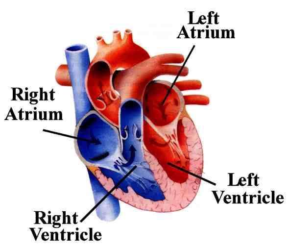
2 2
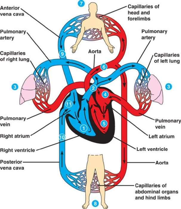
3 3


4 Pulsul A. carotidă, A. brahială Tipuri: Parvus Tardus Altus Bisferiens Alternans Paradoxus Pulsarea V. jugula Șocul apexian 4
5 5
6 ECG P < 0,10 sec, < 0,3 mv Segment PR <0,20 sec QRS <0,12 sec, mv QT <0,47 sec ST < 2 mv T 1/3 R U < 0,1 mv 6

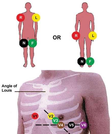
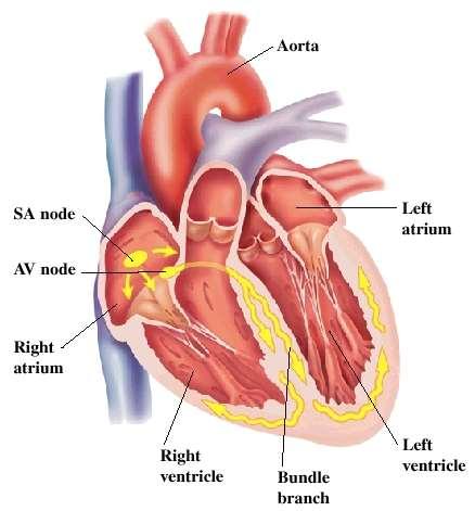
7 Derivații ECG Bipolar standard: 3 electrozi L, R, F D1: L-R D2: F-R D3: F-L Unipolar avr: Membru sup. drept avl: Membru sup. stâng avf: Membru inf. stâng 7

8 Derivații ECG Precordial V1: spațiu ic IV drept, parasternal V2: spațiu ic IV stâng, parasternal V3: între V2 și V4 V4: spațiu ic V stâng, medioclavicular V5: spațiu ic V stâng, axilar ant. V6: spațiu ic V stâng, axilar med. V7-V9: posterior 8
9 9


10 Derivații ECG Derivații dr. Situs inversus, dextrocardie 10


11 EKG Frecvență: / minut Apreciere rapidă (Dubin): distanța RR
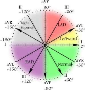
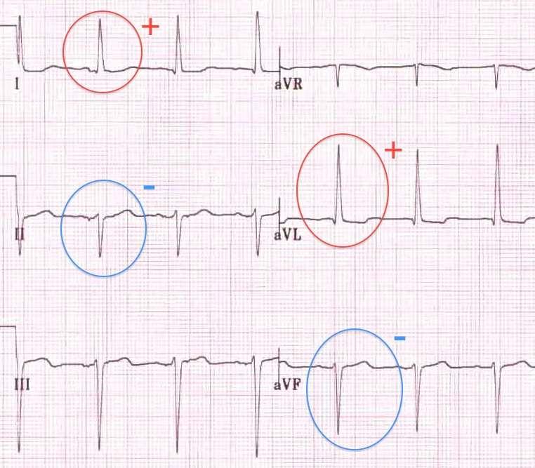
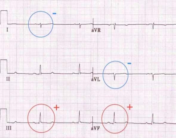
12 Ax ECG 12
13 13
14 14
15 15
16 16
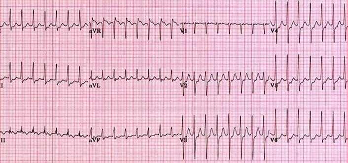
17 Aritmii Supraventricular - îngust (normal) QRS < 0,12 sec Tahicardie sinusală: > 100/min Bradicardie sinusală: < 60/min Aritmie sinusală Extrasistole atriale SVT /min Flutter atrial: /min Fibrilație atrială: /min Nod AV Extrasistole joncționale 17
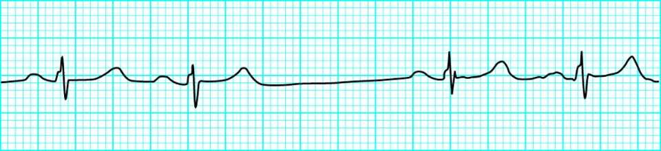
18 18
19 19
20 20
21 ESA 21
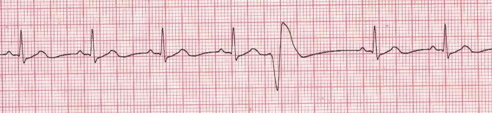
22 TPSV 22

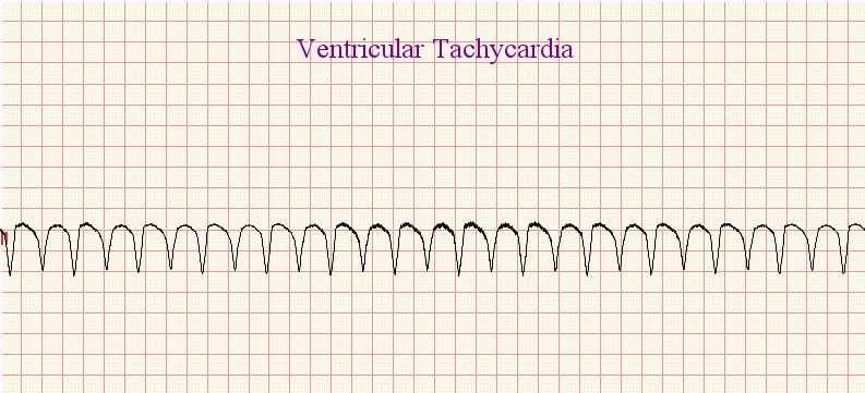
23 FLUTTER 23

24 FIBRILAȚIE ATRIALĂ 24

25 ESJ 25
26 Aritmii Ventriculare (QRS larg) >0,12 sec Extrasistole ventriculare Tahicardie ventriculară /min Torsada vârfurilor (vandering pacemaker) /min Flutter ventricular: /min Fibrilație ventriculară: /min 26
27 ESV 27
28 28

29 TORSADA VÂRFURILOR 29

30 TAHICARDIE VENTRICULARĂ 30

31 FIBRILAȚIE VENTRICULARĂ 31
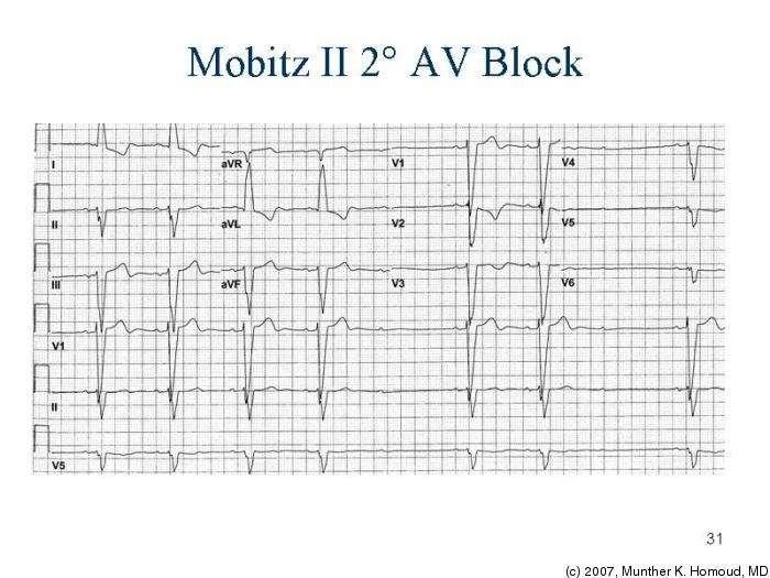
32 Defecte de conducere: blocuri Sino-atrial I ECG normal II Mobitz I, Mobitz II III ECG greu de apreciat Atrio-ventricular I PR>0,2 sec II Mobitz I, Mobitz II III Bloc de ramură Drept Stâng 32
33 BLOC SINOATRIAL 33

34 AV GRD 1 34

35 AV MOBITZ I 35
36 36

37 AV GRD 3 37

38 AV GRD 3 38
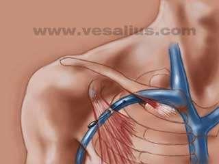
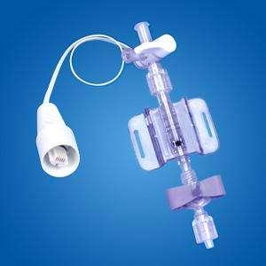
39 39
40 40
")
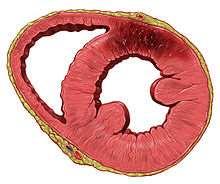
41 IMA (infarct miocardic acut) 41
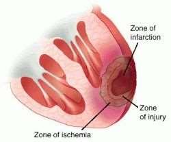
42 42
43 IMA T negativ Primele 3-4 ore: supradenivelare ST 2-3 zile: subdenivelare ST, Q Localizare: Antero-lateral: DI, avl, V5-V6 Antero-apical: DI, DII, V4-V5, avl Antero-septal: V1-V3 Lateral: DI, avl, V4-V8 Inferior: DII, DIII, avf Infero-lateral DII, DIII, avf, V4-V6 Posterior: DIII, V1-V2 43
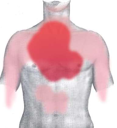
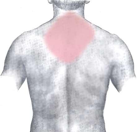
44 Durere 44
45 Nursing Durere Imobilizare Oxigen (SpO2) Monitorizare hemodinamică (tensiune arterială, puls) Respectarea tratamentului specific Anticoagulant!!! (monitorizare, hemoragie tub digestiv) Se notează toate modificările (ECG) Monitorizarea diurezei Dietă Fumat Poziție de comfort 45

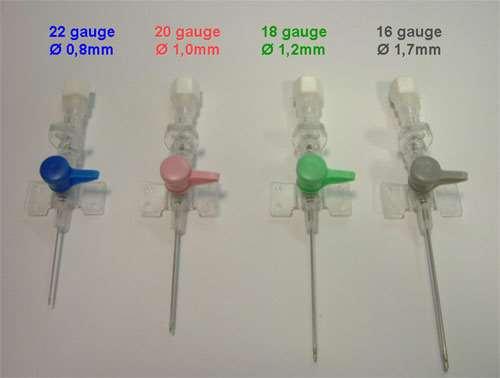
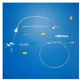
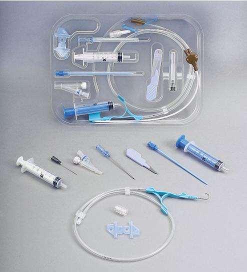
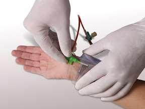
46 Puncție și cateterizare venoasă Periferic Central 46
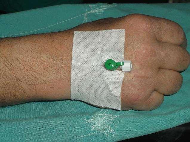
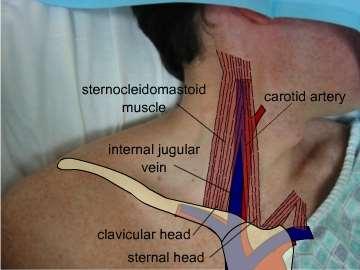
47 47
48 film 48
49 49
50 Cateter venos central VCS, VCI, atriu drept (5-12 mmhg, 8-15 cm H2O) Indicații Umplere volemică Alimentație parenterală (lipide) Pace-maker Epurare extrarenală (dializă) Recoltare de sânge Autotransfuzie Cateter Swan-Ganz Lipsa abordului venos periferic Șoc Administrare de inotropice Hipo Na, K Intervenție chirurgicală: risc de hemoragie Tratament intravenos îndelungat 50
51 51
52 52
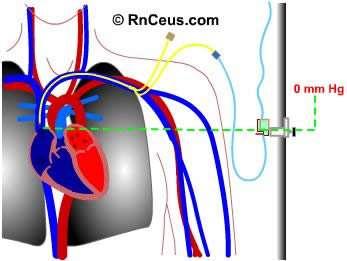
53 53
54 Materiale necesare Cateter Trusă chirurgivală Mască de protecție Oxigen Halat și mănuși sterile Monitor Antiseptic Pansament steril Leucoplast (Operfix) Lidocaină, ser fiziologic 54
55 Pregătirea pacientului Informare, acord Analize de laborator Monitor: ECG, SpO2, TA Trendelenburg, membru superior lângă corp, capul întors în direcția opusă Mască de oxigen Loc lângă pacient Asepsia și antisepsia locului de puncție, izolare Fixarea cateterului, bandajare Măsurarea presiunii venoase centrale În foaia de observație se notează tipul cateterului, locul de puncție, adâncimea de fixare, data și ora montării 55
56 56
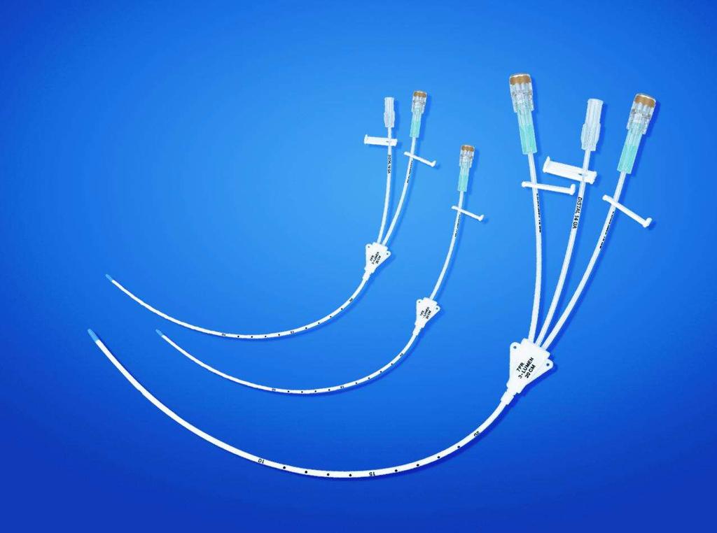
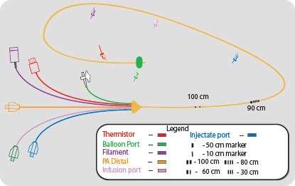
57 57
58 58

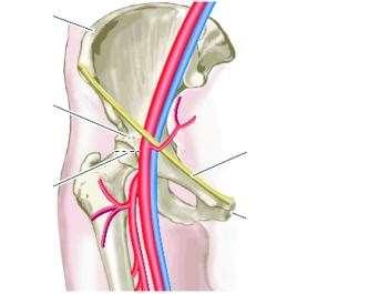
59 Tehnica Seldinger 59
60 Poziționarea cateterului Adâncime Subclavicular dr: cm Subclavicular stg: cm Jugular dr: cm Jugular stg: cm Femural: cm Brahial: cm Verificare ecografică Rtg toracic Măsurarea presiunii: transductor, cmh2o (ser fiziologic) 60
61 Întreținerea cateterului venos central PEEP, Valsalva Anticoagulant Disfuncții de coagulare Punkcie: jugulară, subclavie Schimbare după 3 săptămâni Verificare poziție: rtg, presiune, întoarcere venoasă Heparinizare, perfuzie continuă Verificarea firelor de fixare Pansament zilnic Sterilitate Măsurarea presiunii: 0 în atriul drept, linia medio-axilară, 1/3 61
62 62
63 63
64 Complicații Puncție arterială Pneumotorace Emboli Infecție Puncție de vas limfatic Tromboză de cateter, trombembolie Mobilizarea cateterului Leziune de nerv Embolie de cateter Cateterizarea venei greșite Imposibilitate de cateterizare Hematom, hemoragie Catetrizarea atriului drept Puncția traheei 64

65 Cateter Swan-Ganz 65
66 66

67 Puncția venei jugulare externe 67


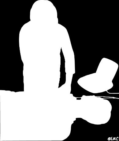
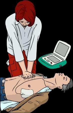
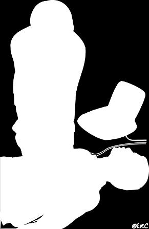
68 Cateterizare venoasă la copii Epicranian Ombilical (5-7 zile) Puncție osoasă Sedare!!! 68
69 Măsurarea tensiunii arteriale Normal: sistolic , diastolic mmhg Valoare medie: (sist + 2 x diast)/3 mmhg (65) Noninvaziv Puls: sist Korotkoff Oscilometru Invaziv 69
70 Indicațiile tensiunii arteriale invazive Măsurare continuă Tensiune arterială scăzută Pacient instabil Urmărirea tratamentului Recoltare de sânge arterial 70

71 Vase Radială Ulnară Brahială Testul Allen 71
72 Materiale necesare Cateter arterial Cameră de presiune (transductor) Kit de măsurarea presiunii Pansament steril Antiseptic Leucoplast Perfuzie sub presiune Robinet cu 3 căi Monitor invaziv 72
73 73
74 74
75 Măsurarea debitului cardiac 4-8 l/min Termodiluție Fick (O2) Substanță de contrast Pletismografie (PICCO, PULSECO, LIDCO) ECHO Doppler Bioimpedanță NICO (CO2) 75


76 Compresie toracică 4-5 cm 100/min Mijlocul sternului 76

77 Puncție pericardică Tamponadă cardiacă 2 degete lângă stern Matitate Aspirare Steril 77
78 78

79 Complicații Pneumotorace Puncție miocardică Puncție coronariană Puncție ventriculară Aritmii Puncția splinei, ficatului Stop cardiac 79
 Torsada vârfurilor Asistolie?")
80 Defibrilare Fibrilație ventriculară Tahicardie ventriculară (fără puls) Torsada vârfurilor Asistolie?!!!!! 80
")
 150 300 J")
81 Defibrilare Timp Poziția electrodului Contact (gel) Siguranță Sincron/asincron Energie (bifazic) J 81
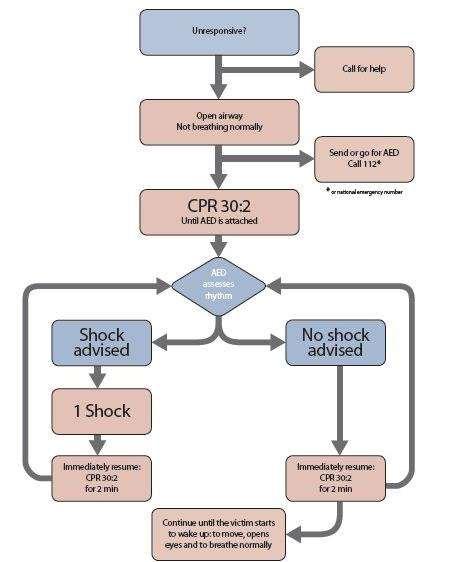
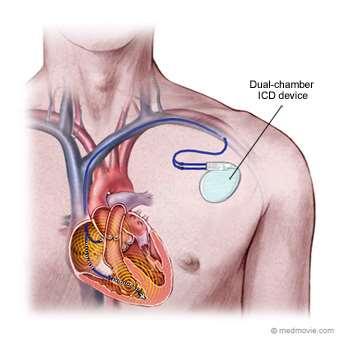
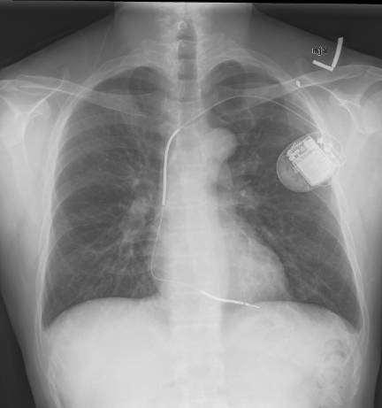
82 82
83 Protocol de defibrilare Asigurarea mediului Conștiență Solicitare de ajutor Eliberarea căilor aeriene Respirație 112 Montarea defibrilatorului Urmărirea instrucțiunilor 83


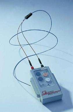
84 SWITCH ON AED Unele defibrilatoare automate externe se vor activa automat la deschiderea capacului 84


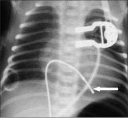
85 ATTACH PADS TO CASUALTY S BARE CHEST 85

86 ANALYSING RHYTHM DO NOT TOUCH VICTIM 86


87 SHOCK INDICATED Vă îndepărtați Livrați șocul 87
88 SHOCK DELIVERED FOLLOW AED INSTRUCTIONS


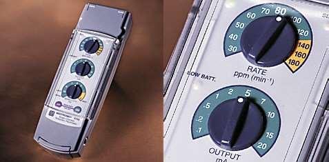
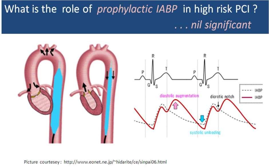
89 NO SHOCK ADVISED FOLLOW AED INSTRUCTIONS
90 Conversie sincron Sincron Aritmii Tahicardie cu instabilitate hemodinamică Aritmie refractară la tratament medicamentos (fibrilație atrială, tahicardie supraventriculară) Sedare (propofol, benzodiazepine, barbiturice, ketamină) apnee Interzis după digoxin (lidocaină 1,5 mg/kg) Energie J Materiale necesare pentru intubație Fibrilație atrială prima dată anticoagulant 90
91 Complicațiile defibrilării și electroconversiei Leziune de miocard!!! Fibrilație ventriculară Embolie Modificări de segment ST Bradicardie Tahicardie Extrasistole Edem pulmonar acut Scăderea tensiunii arteriale Leziune de țesut cardiac 91
92 Defibrilator automat extern 92

93 Stimularea electrică a cordului Pacemaker Temporar permanent Extern intern Extern: Transtoracal Transesofagian Intern Temporar Permanent 93
94 Indicații de pacemaker Bracdicardie simptomatică Bloc AV Boală de nod sinusal Tahicardie supraventriculară Tahiaritmii ventriculare Torsada vârfurilor IMA Asistolie!!! Hipotermie nu este indicat 94


95 Stimulare externă Electrod toracic Sedare Contracție musculară pectorală Transesofagian 95
96 Pacing transvenos 96
 Epicardiac")
97 Intern permanent VVI, DDD, DRR Ventricular, atrial, ambele Cu resincronizare (ambii ventriculi) Epicardiac 97

98 98

99 Complicații Stimulare diafragmatică (sughiț) Aritmii Lipsa contracturii Complicațiile puncției venoase Leziune miocardică Trombembolie Infecție Durere (pectorală) 99


100 Balon de contrapulsație aortică (IABP) Balon aortă Expansionare diastolică Scop: Scăderea sarcinii ventriculului stâng Creșterea capacității cardiace Ameliorarea circulației coronariene și sistemice 100
101 101
102 Indicații IABP Șoc cardiogen Insuficiență cardiacă IMA Defecte mecanice severe (insuficiență mitrală, defect septal ventricular, anevrism de VS) Profilaxie după PTCA Ocluzie severă de arteră coronariană Instabilitate hemodinamică Înainte și după intervenție chirurgicală cardiacă Înainte și după transplant cardiac 102
103 Contraindicații IABP Fără posibilitate de recuperare Modificări aterosclerotice severe Insuficiență aortică Anevrism de aortă, disecție 103
104 Complicații IABP Ruptură de vas, disecție Mobilizarea plăcii ateromatoase Obstrucție arterială Cateterism eșuat Poziție greșită Embolie Trombocitopenie Infecție Hemoragie Creștere inadecvată de capacitate cardiacă 104

105 MAST (Military Antishock Trousers) 105
106 Îngrijirea pacientului cardiovascular Monitorizare Oxigen Poziție ECG Tratament medicamentos Analiza gazelor sanguine Imobilizare Echilibru acido-bazic Ionogramă Acces venos Prevenirea infecțiilor 106


107 107
Tematica cursului an IV MG aparat cardiovascular, aparat respirator:
 Tematica cursului an IV MG aparat cardiovascular, aparat respirator: TEMATICA CURSULUI Explorarea aparatului cardiovascular. Cardiopatii congenitale. Reumatismul articular acut. Valvulopatii. Endocardita
Radiant warming table with servo-control Incubator Skin temperature C Temperature with 1.5 C > child temperature
 Figure no. 1. Pre-transport thermo-equilibration (post-resuscitated) Newborn Central temperature 36.5-37.5 C < 36.5 C Normal Hypothermia Radiant warming table with servo-control Incubator Skin temperature
12 LEAD EKG BASICS. By: Steven Jones, NREMT P CLEMC
 12 LEAD EKG BASICS By: Steven Jones, NREMT P CLEMC ECG Review Waves and Intervals P wave: the sequential activation (depolarization) of the right and left atria QRS complex: right and left ventricular
By the end of this lecture, you will be able to: Understand the 12 lead ECG in relation to the coronary circulation and myocardium Perform an ECG
 By the end of this lecture, you will be able to: Understand the 12 lead ECG in relation to the coronary circulation and myocardium Perform an ECG recording Identify the ECG changes that occur in the presence
Appendix D Output Code and Interpretation of Analysis
 Appendix D Output Code and Interpretation of Analysis 8 Arrhythmia Code No. Description 8002 Marked rhythm irregularity 8110 Sinus rhythm 8102 Sinus arrhythmia 8108 Marked sinus arrhythmia 8120 Sinus tachycardia
Basic electrocardiography reading. R3 lee wei-chieh
 Basic electrocardiography reading R3 lee wei-chieh The Normal Conduction System Lead Placement avf Limb Leads Precordial Leads Interpretation Rate Rhythm Interval Axis Chamber abnormality QRST change What
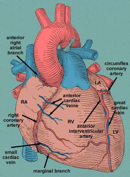
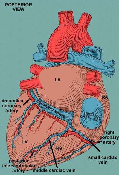
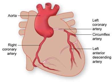
CORONARY ARTERIES HEART
 CARDIAC/ECG MODULE THE HEART CORONARY ARTERIES FIBRILLATING HEART CORONARY ARTERIES HEART PRACTICE RHYTHMS PRACTICE RHYTHMS ELECTRICAL CONDUCTION SA Node (60 100) Primary pacemaker AV Node (40 60) ***Creates
ECG INTERPRETATION MANUAL
 Lancashire & South Cumbria Cardiac Network ECG INTERPRETATION MANUAL THE NORMAL ECG Lancashire And South Cumbria Cardiac Physiologist Training Manual THE NORMAL ECG E.C.G CHECKLIST 1) Name, Paper Speed,
12 Lead ECG Interpretation: Color Coding for MI s
 12 Lead ECG Interpretation: Color Coding for MI s Anna E. Story, RN, MS Director, Continuing Professional Education Critical Care Nurse Online Instructional Designer 2004 Anna Story 1 Objectives review
BASIC CONCEPT OF ECG
 BASIC CONCEPT OF ECG Electrocardiogram The electrocardiogram (ECG) is a recording of cardiac electrical activity. The electrical activity is readily detected by electrodes attached to the skin. After the
Family Medicine for English language students of Medical University of Lodz ECG. Jakub Dorożyński
 Family Medicine for English language students of Medical University of Lodz ECG Jakub Dorożyński Parts of an ECG The standard ECG has 12 leads: six of them are considered limb leads because they are placed
Relax and Learn At the Farm 2012
 Relax and Learn At the Farm 2012 Session 2: 12 Lead ECG Fundamentals 101 Cynthia Webner DNP, RN, CCNS, CCRN-CMC, CHFN Though for Today Mastery is not something that strikes in an instant, like a thunderbolt,
Electrocardiogram ECG. Hilal Al Saffar FRCP FACC College of medicine,baghdad University
 Electrocardiogram ECG Hilal Al Saffar FRCP FACC College of medicine,baghdad University Tuesday 29 October 2013 ECG introduction Wednesday 30 October 2013 Abnormal ECG ( ischemia, chamber hypertrophy, heart
Figure 2. Normal ECG tracing. Table 1.
 Figure 2. Normal ECG tracing that navigates through the left ventricle. Following these bundle branches the impulse finally passes to the terminal points called Purkinje fibers. These Purkinje fibers are
ELECTROCARDIOGRAPHY (ECG)
") ELECTROCARDIOGRAPHY (ECG) The heart is a muscular organ, which pumps blood through the blood vessels of the circulatory system. Blood provides the body with oxygen and nutrients, as well as assists in
ELECTROCARDIOGRAPHY (ECG) The heart is a muscular organ, which pumps blood through the blood vessels of the circulatory system. Blood provides the body with oxygen and nutrients, as well as assists in
Diploma in Electrocardiography
 The Society for Cardiological Science and Technology Diploma in Electrocardiography The Society makes this award to candidates who can demonstrate the ability to accurately record a resting 12-lead electrocardiogram
12 Lead ECG Interpretation: The Basics and Beyond
 12 Lead ECG Interpretation: The Basics and Beyond Cindy Weston, DNP, RN, CCRN, CNS-CC, FNP-BC Assistant Professor Texas A&M University College of Nursing cweston@tamhsc.edu Objectives Review the basics
ECG pre-reading manual. Created for the North West Regional EMET training program
 ECG pre-reading manual Created for the North West Regional EMET training program Author:- Dr Juan Carlos Ascencio-Lane juan.ascencio-lane@ths.tas.gov.au 1 Disclaimer This handbook has been created for
ECG Basics Sonia Samtani 7/2017 UCI Resident Lecture Series
 ECG Basics Sonia Samtani 7/2017 UCI Resident Lecture Series Agenda I. Introduction II.The Conduction System III.ECG Basics IV.Cardiac Emergencies V.Summary The Conduction System Lead Placement avf Precordial
Birmingham Regional Emergency Medical Services System
 Birmingham Regional Emergency Medical Services System 2018 ALCTE Summer Conference EKG Basics Brian Gober, MAT, ATC, NRP, CSCS Education Services Manager ECC Training Center Coordinator Birmingham Regional
INTRODUCTION TO ECG. Dr. Tamara Alqudah
 INTRODUCTION TO ECG Dr. Tamara Alqudah Excitatory & conductive system of the heart + - The ECG The electrocardiogram, or ECG, is a simple & noninvasive diagnostic test which records the electrical
Electrocardiography Abnormalities (Arrhythmias) 7. Faisal I. Mohammed, MD, PhD
 7. Faisal I. Mohammed, MD, PhD") Electrocardiography Abnormalities (Arrhythmias) 7 Faisal I. Mohammed, MD, PhD 1 Causes of Cardiac Arrythmias Abnormal rhythmicity of the pacemaker Shift of pacemaker from sinus node Blocks at different
Electrocardiography Abnormalities (Arrhythmias) 7 Faisal I. Mohammed, MD, PhD 1 Causes of Cardiac Arrythmias Abnormal rhythmicity of the pacemaker Shift of pacemaker from sinus node Blocks at different
Please check your answers with correct statements in answer pages after the ECG cases.
 ECG Cases ECG Case 1 Springer International Publishing AG, part of Springer Nature 2018 S. Okutucu, A. Oto, Interpreting ECGs in Clinical Practice, In Clinical Practice, https://doi.org/10.1007/978-3-319-90557-0
Understanding basics of EKG
 Understanding basics of EKG By Alula A.(R III) www.le.ac.uk Topic for discussion Understanding of cellular electrophysiology Basics Rate Rhythm Axis Intervals P wave QRS ST/T wave Abnormal EKGs Understanding
ECG CONVENTIONS AND INTERVALS
 1 ECG Waveforms and Intervals ECG waveforms labeled alphabetically P wave== represents atrial depolarization QRS complex=ventricular depolarization ST-T-U complex (ST segment, T wave, and U wave)== V repolarization.
ECG Interpretation Made Easy
 ECG Interpretation Made Easy Dr. A Tageldien Abdellah, MSc MD EBSC Lecturer of Cardiology- Hull University Hull York Medical School 2007-2008 ECG Interpretation Made Easy Synopsis Benefits Objectives Process
Denumire Pret Specialitate Consult 50 chirurgie Control 30 chirurgie Interv.0 chirurgicala 150 chirurgie Interv.I chirurgicala 300 chirurgie
 Denumire Pret Specialitate Consult 50 chirurgie Control 30 chirurgie Interv.0 chirurgicala 150 chirurgie Interv.I chirurgicala 300 chirurgie Interv.II chirurgicala 500 chirurgie Interv.III chirurgicala
Aortic Dissection Causes of Death
 Aortic Dissection Causes of Death Rupture aorta 33.3% Unspecified 33.3% Neurological l deficit it 13.9% Visceral ischemia/kidney failure 11.5% Cardiac tamponade 7.9% (Circulation 2002;105:200-6) Medical
CH; Multiple-choice questions. Cardiology questions for the MRCP (UK) examination QUESTION 4. Mark Appleby, Derek Rowlands QUESTION 1 QUESTION 3
 examination QUESTION 4. Mark Appleby, Derek Rowlands QUESTION 1 QUESTION 3") Postgrad MedJ7 1998;74:643-648 ) The Fellowship of Postgraduate Medicine, 1998 Multiple-choice questions Yorkshire Heart Centre, Leeds LS1 3EX, UK M Appleby Manchester Heart Centre, Manchester M13 9WL,
Postgrad MedJ7 1998;74:643-648 ) The Fellowship of Postgraduate Medicine, 1998 Multiple-choice questions Yorkshire Heart Centre, Leeds LS1 3EX, UK M Appleby Manchester Heart Centre, Manchester M13 9WL,
DAY1_CARDIOVASCULAR PRACTICE QUESTIONS
 DAY1_CARDIOVASCULAR PRACTICE QUESTIONS 1 P age 1. A 59-year-old male is admitted complaining of chest pain and dyspnea. ST elevation and T-wave inversion were seen on the ECG in V2, V3, and V4. IV thrombolytic
10 ECGs No Practitioner Can Afford to Miss. Objectives
 10 ECGs No Practitioner Can Afford to Miss Mary L. Dohrmann, MD Professor of Clinical Medicine Division of Cardiovascular Medicine University of Missouri School of Medicine No disclosures Objectives 1.
Electrocardiography Normal 5. Faisal I. Mohammed, MD, PhD
 Electrocardiography Normal 5 Faisal I. Mohammed, MD, PhD 1 Objectives 2 1. Describe the different waves in a normal electrocardiogram. 2. Recall the normal P-R and Q-T interval time of the QRS wave. 3.
DR QAZI IMTIAZ RASOOL OBJECTIVES
 PRACTICAL ELECTROCARDIOGRAPHY DR QAZI IMTIAZ RASOOL OBJECTIVES Recording of electrical events in heart Established electrode pattern results in specific tracing pattern Health of heart i. e. Anatomical
REtrive. REpeat. RElearn Design by. Test-Enhanced Learning based ECG practice E-book
 Test-Enhanced Learning Test-Enhanced Learning Test-Enhanced Learning Test-Enhanced Learning based ECG practice E-book REtrive REpeat RElearn Design by S I T T I N U N T H A N G J U I P E E R I Y A W A
12 Lead ECG. Presented by Rebecca Sevigny BSN, RN Professional Practice & Development Dept.
 12 Lead ECG Presented by Rebecca Sevigny BSN, RN Professional Practice & Development Dept. Two Main Coronary Arteries RCA LCA which branches into Left Anterior Descending Circumflex Artery Two Main Coronary
Ekg pra pr c a tice D.HAMMOUDI.MD
 Ekg practice D.HAMMOUDI.MD Anatomy Revisited RCA (Right Coronary Artery) Right ventricle Inferior wall of LV Posterior wall of LV (75%) SA Node (60%) AV Node (>80%) LCA (Left Coronary Artery) Septal wall
EKG. Danil Hammoudi.MD
 EKG Danil Hammoudi.MD What is an EKG? The electrocardiogram (EKG) is a representation of the electrical events of the cardiac cycle. Each event has a distinctive waveform, the study of which can lead to
Return to Basics. ECG Rate and Rhythm. Management of the Hospitalized Patient September 25, 2009
 Management of the Hospitalized Patient September 25, 2009 ECG Refresher and Update 2009 Return to Basics Determine rate and rhythm Determine intervals and axes Define morphology of P-QRS-T-U Compare with
ECG interpretation basics
 ECG interpretation basics Michał Walczewski, MD Krzysztof Ozierański, MD 21.03.18 Electrical conduction system of the heart Limb leads Precordial leads 21.03.18 Precordial leads Precordial leads 21.03.18
12 Lead Electrocardiogram (ECG) PFN: SOMACL17. Terminal Learning Objective. References
 PFN: SOMACL17. Terminal Learning Objective. References") 12 Lead Electrocardiogram (ECG) PFN: SOMACL17 Slide 1 Terminal Learning Objective Action: Communicate knowledge of 12 Lead Electrocardiogram (ECG) Condition: Given a lecture in a classroom environment
12 Lead Electrocardiogram (ECG) PFN: SOMACL17 Slide 1 Terminal Learning Objective Action: Communicate knowledge of 12 Lead Electrocardiogram (ECG) Condition: Given a lecture in a classroom environment
Pennsylvania Academy of Family Physicians Foundation & UPMC 43rd Refresher Course in Family Medicine CME Conference March 10-13, 2016
 Pennsylvania Academy of Family Physicians Foundation & UPMC 43rd Refresher Course in Family Medicine CME Conference March 10-13, 2016 Disclosures: EKG Workshop Louis Mancano, MD Speaker has no disclosures
ECG in coronary artery disease. By Sura Boonrat Central Chest Institute
 ECG in coronary artery disease By Sura Boonrat Central Chest Institute EKG P wave = Atrium activation PR interval QRS = Ventricle activation T wave= repolarization J-point EKG QT interval Abnormal repolarization
5- The normal electrocardiogram (ECG)
") 5- The (ECG) Introduction Electrocardiography is a process of recording electrical activities of heart muscle at skin surface. The electrical current spreads into the tissues surrounding the heart, a small
5- The (ECG) Introduction Electrocardiography is a process of recording electrical activities of heart muscle at skin surface. The electrical current spreads into the tissues surrounding the heart, a small
Acute Coronary Syndromes. Disclosures
 Acute Coronary Syndromes Disclosures I work for Virginia Garcia Memorial Health Center, Beaverton, OR. Jon Tardiff, BS, PA-C OHSU Clinical Assistant Professor And I am a medical editor for Jones & Bartlett
ECG Interpretation. Best to have a system to methodically evaluate ECG (from Dubin) * Rate * Rhythm * Axis * Intervals * Hypertrophy * Infarction
 * Rate * Rhythm * Axis * Intervals * Hypertrophy * Infarction") ECG to save Babies ECG Interpretation Best to have a system to methodically evaluate ECG (from Dubin) * Rate * Rhythm * Axis * Intervals * Hypertrophy * Infarction Electrical Activity in the heart 5 events
ECG to save Babies ECG Interpretation Best to have a system to methodically evaluate ECG (from Dubin) * Rate * Rhythm * Axis * Intervals * Hypertrophy * Infarction Electrical Activity in the heart 5 events
June 2009 CE. Site code # E-1209
 1 June 2009 CE Site code # 107200E-1209 Region X SOP Review ACS, Asystole/PEA, Bradycardia, VF/Pulseless VT, Conscious Sedation, Region X Field Triage Criteria Prepared by: Sharon Hopkins, RN, BSN, EMT-P
This presentation will deal with the basics of ECG description as well as the physiological basics of
 Snímka 1 Electrocardiography basics This presentation will deal with the basics of ECG description as well as the physiological basics of Snímka 2 Lecture overview 1. Cardiac conduction system functional
Acute Coronary Syndromes Unstable Angina Non ST segment Elevation MI (NSTEMI) ST segment Elevation MI (STEMI)
 ST segment Elevation MI (STEMI)") Leanna R. Miller, RN, MN, CCRN-CSC, PCCN-CMC, CEN, CNRN, CMSRN, NP Education Specialist LRM Consulting Nashville, TN Objectives Evaluate common abnormalities that mimic myocardial infarction. Identify
Leanna R. Miller, RN, MN, CCRN-CSC, PCCN-CMC, CEN, CNRN, CMSRN, NP Education Specialist LRM Consulting Nashville, TN Objectives Evaluate common abnormalities that mimic myocardial infarction. Identify
Introduction to Electrocardiography
 Introduction to Electrocardiography Class Objectives: Introduction to ECG monitoring Discuss principles of interpretation Identify the components and measurements of the ECG ECG analysis ECG Monitoring
BEDSIDE ECG INTERPRETATION
 BEDSIDE ECG INTERPRETATION Presented by: Ryan Dean, RN, MSN, CCRN, CCNS, CFRN Flight Nurse 2017 Based on presentations originally by Gennifer DePaoli, RN Objectives Hospital policies Electrical conduction
Electrocardiography. Hilal Al Saffar College of Medicine,Baghdad University
 Electrocardiography Hilal Al Saffar College of Medicine,Baghdad University Which of the following is True 1. PR interval, represent the time taken for the impulse to travel from SA node to AV nose. 2.
Return to Basics. Normal Intervals & Axes. ECG Rate and Rhythm
 Return to Basics Management of the Hospitalized Patient October 15, 2010 ECG Refresher and Update 2010 Determine rate and rhythm Determine intervals and axes Define morphology of P-QRS-T-U Compare with
12-Lead ECG Interpretation. Kathy Kuznar, RN, ANP
 12-Lead ECG Interpretation Kathy Kuznar, RN, ANP The 12-Lead ECG Objectives Identify the normal morphology and features of the 12- lead ECG. Perform systematic analysis of the 12-lead ECG. Recognize abnormalities
The Fundamentals of 12 Lead EKG. ECG Recording. J Point. Reviewing the Cardiac Conductive System. Dr. E. Joe Sasin, MD Rusty Powers, NRP
 The Fundamentals of 12 Lead EKG Dr. E. Joe Sasin, MD Rusty Powers, NRP SA Node Intranodal Pathways AV Junction AV Fibers Bundle of His Septum Bundle Branches Purkinje System Reviewing the Cardiac Conductive
12 Lead ECG Skills: Building Confidence for Clinical Practice. Presented By: Cynthia Webner, BSN, RN, CCRN-CMC. Karen Marzlin, BSN, RN,CCRN-CMC
 12 Lead ECG Skills: Building Confidence for Clinical Practice NTI 2009 Preconference Session 803 Presented By: Karen Marzlin, BSN, RN,CCRN-CMC 1 12 Lead ECG Fundamentals: The Starting Place for Linking
Cardiology/Cardiothoracic
 Cardiology/Cardiothoracic ICD-9-CM to ICD-10-CM Code Mapper 800-334-5724 www.contexomedia.com 2013 ICD-9-CM 272.0 Pure hypercholesterolemia 272.2 Mixed hyperlipidemia 272.4 Other and hyperlipidemia 278.00
ECG ABNORMALITIES D R. T AM A R A AL Q U D AH
 ECG ABNORMALITIES D R. T AM A R A AL Q U D AH When we interpret an ECG we compare it instantaneously with the normal ECG and normal variants stored in our memory; these memories are stored visually in
15 16 September Seminar W10O. ECG for General Practice
 15 16 September 2012 Seminar W10O ECG for General Practice Speaker: Ms Natasha Eaton ECG for General Practice Speaker: Natasha Eaton Cardiac CNC Executive Representative Electrocardiography The graphic
12 Lead ECGs: Ischemia, Injury & Infarction. Kevin Handke NRP, FP-C, CCP, CMTE STEMI Coordinator Flight Paramedic
 12 Lead ECGs: Ischemia, Injury & Infarction Kevin Handke NRP, FP-C, CCP, CMTE STEMI Coordinator Flight Paramedic None Disclosures Objectives Upon completion of this program the learner will be able to
Debitul cardiac. Dr. Kovacs Judit CEEA 2013
 Debitul cardiac Dr. Kovacs Judit CEEA 2013 Definiţie Cantitatea de sânge ejectat/ minut DC = VB x FC Vincent JL, Crit Care 2009 Contracţia atrială Presarcină Întoarcerea Ven. Volemie Compl. Ventr. P.intrator.
The ABC of Pediatric ECG
 The ABC of Pediatric ECG Mohamed Hamdan, MD, FAAP, FACC Assistant Professor of Pediatrics Columbia University College of Physicians and Surgeons, NY, USA Consultant Pediatric Cardiologist & Co-Director
402 Index. B β-blockers, 4, 5 Bradyarrhythmias, 76 77
 Index A Acquired immunodeficiency syndrome (AIDS), 126, 163 Action potentials, 1, 5, 27 Acute coronary syndromes, 123t, 129 Adenosine, intravenous, 277 Alcohol abuse, as T wave inversion cause, 199 Aneurysm,
ELECTROCARDIOGRAPH. General. Heart Rate. Starship Children s Health Clinical Guideline
 General Heart Rate QRS Axis T Wave Axis PR Interval according to Heart Rate & Age P Wave Duration and Amplitude QRS Duration according to Age QT Interval R & S voltages according to Lead & Age R/S ratio
EKG Practice. Homan Wai
 EKG Practice Homan Wai Objectives Learn EKG by Examples Learn by repetition Learn by repetition Learn by repetition Learn by repetition Learn by repetition Learn by repetition Learn by repetition Details
ECG and Cardiac Electrophysiology
 ECG and Cardiac Electrophysiology Simon Some very basic electrophysiology Intracellular fluid: 10 mm Na, 140 mm K, etc. K Na-K ATPase Extracellular fluid: 140mM Na, 4mM K, etc. Na Ion gradient plus selective
Myocardial Infarction. Reading Assignment (p66-78 in Outline )
") Myocardial Infarction Reading Assignment (p66-78 in Outline ) Objectives 1. Why do ST segments go up or down in ischemia? 2. STEMI locations and culprit vessels 3. Why 15-lead ECGs? 4. What s up with avr?
Myocardial Infarction Reading Assignment (p66-78 in Outline ) Objectives 1. Why do ST segments go up or down in ischemia? 2. STEMI locations and culprit vessels 3. Why 15-lead ECGs? 4. What s up with avr?
EHRA Accreditation Exam - Sample MCQs Invasive cardiac electrophysiology
 EHRA Accreditation Exam - Sample MCQs Invasive cardiac electrophysiology Dear EHRA Member, Dear Colleague, As you know, the EHRA Accreditation Process is becoming increasingly recognised as an important
Section V. Objectives
 Section V Landscape of an MI Objectives At the conclusion of this presentation the participant will be able to Outline a systematic approach to 12 lead ECG interpretation Demonstrate the process for determining
Return to Basics. ECG Rate and Rhythm. Management of the Hospitalized Patient October 4, 2007
 Management of the Hospitalized Patient October 4, 2007 ECG Refresher for the Hospitalists Return to Basics Determine rate and rhythm Determine intervals and axes Define morphology of P-QRS-T-U Compare
CORONARY ARTERIES. LAD Anterior wall of the left vent Lateral wall of left vent Anterior 2/3 of interventricluar septum R & L bundle branches
 CORONARY ARTERIES RCA Right atrium Right ventricle SA node 55% AV node 90% Posterior wall of left ventricle in 90% Posterior third of interventricular septum 90% LAD Anterior wall of the left vent Lateral
ECG Interactive Session
 SA HEART 2018 PRE-CONGRESS WORKSHOP 4 OCTOBER 2018 ECG Interactive Session Ashley Chin Cardiologist/Electrophysiologist University of Cape Town Groote Schuur Hospital ECG 1 Which ONE of the following is
Arrhythmic Complications of MI. Teferi Mitiku, MD Assistant Clinical Professor of Medicine University of California Irvine
 Arrhythmic Complications of MI Teferi Mitiku, MD Assistant Clinical Professor of Medicine University of California Irvine Objectives Brief overview -Pathophysiology of Arrhythmia ECG review of typical
THE ELECTROCARDIOGRAM A UBIQUITOUS AND COST-EFFECTIVE DIAGNOSTIC TOOL FOR THE FAMILY MEDICINE REFRESHER COURSE MARCH 8, 2019
 THE ELECTROCARDIOGRAM A UBIQUITOUS AND COST-EFFECTIVE DIAGNOSTIC TOOL FOR THE FAMILY MEDICINE REFRESHER COURSE MARCH 8, 2019 Major Clinical Disorders Pulmonary Embolism 69 y/o woman with dyspnea and an
-RHYTHM PRACTICE- By Dr.moanes Msc.cardiology Assistant Lecturer of Cardiology Al Azhar University. OBHG Education Subcommittee
 -RHYTHM PRACTICE- By Dr.moanes Msc.cardiology Assistant Lecturer of Cardiology Al Azhar University The Normal Conduction System Sinus Node Normal Sinus Rhythm (NSR) Sinus Bradycardia Sinus Tachycardia
4. The two inferior chambers of the heart are known as the atria. the superior and inferior vena cava, which empty into the left atrium.
 Answer each statement true or false. If the statement is false, change the underlined word to make it true. 1. The heart is located approximately between the second and fifth ribs and posterior to the
A Review of Cardiac Pathophysiology and EKG. Jamie Dyson PT, DPT Kathy Swanick PT, DPT, OCS
 A Review of Cardiac Pathophysiology and EKG Jamie Dyson PT, DPT Kathy Swanick PT, DPT, OCS Cardiac Pathophysiology Coronary Artery Disease Congestive Heart Failure Valvular Heart Disease Athletic Heart
The Cardiovascular System: The Heart
 The Cardiovascular System: The Heart 1 2 3 4 5 6 7 8 9 10 11 12 Across 1. The spontaneously changing pacemaker membranes. 7. The distinguishing feature of heart muscle. 9. What allows heart cells to beat
CRC 431 ECG Basics. Bill Pruitt, MBA, RRT, CPFT, AE-C
 CRC 431 ECG Basics Bill Pruitt, MBA, RRT, CPFT, AE-C Resources White s 5 th ed. Ch 6 Electrocardiography Einthoven s Triangle Chest leads and limb leads Egan s 10 th ed. Ch 17 Interpreting the Electrocardiogram
Arrhythmias. Sarah B. Murthi Department of Surgery University of Maryland Medical School R. Adams Cowley Shock Trauma Center
 Arrhythmias Sarah B. Murthi Department of Surgery University of Maryland Medical School R. Adams Cowley Shock Trauma Center 2012 Clinical Congress Presenter Disclosure Slide American College of Surgeons
All About STEMIs. Presented By: Brittney Urvand, RN, BSN, CCCC. Essentia Health Fargo Cardiovascular Program Manager.
 All About STEMIs Presented By: Brittney Urvand, RN, BSN, CCCC Essentia Health Fargo Cardiovascular Program Manager Updated 10/2/2018 None Disclosures Objectives Identify signs and symptoms of a heart attack
Blocks & Dissociations. Reading Assignment (p47-52 in Outline )
") Blocks & Dissociations Reading Assignment (p47-52 in Outline ) Objectives Who are Wenckebach and Mobitz? Review SA and AV Blocks AV Dissociations: learning who s the boss and why 2 nd degree SA Block:
Blocks & Dissociations Reading Assignment (p47-52 in Outline ) Objectives Who are Wenckebach and Mobitz? Review SA and AV Blocks AV Dissociations: learning who s the boss and why 2 nd degree SA Block:
Basic Dysrhythmia Interpretation
 Basic Dysrhythmia Interpretation Objectives 2 To understand the Basic ECG To understand the meaning of Dysrhythmia To describe the normal heart conduction system. To describe the normal impulse pathways.
12 Lead ECG Interpretation
 12 Lead ECG Interpretation Julie Zimmerman, MSN, RN, CNS, CCRN Significant increase in mortality for every 15 minutes of delay! N Engl J Med 2007;357:1631-1638 Who should get a 12-lead ECG? Also include
OBJECTIVE. 1. Define defibrillation. 2. Describe Need and history of defibrillation. 3. Describe the principle and mechanism of defibrillation.
 Defibrillators OBJECTIVE 1. Define defibrillation. 2. Describe Need and history of defibrillation. 3. Describe the principle and mechanism of defibrillation. 4. Types and classes of defibrillator 5. Describe
Ask Mish. EKG INTERPRETATION part i
 EKG INTERPRETATION part i What is EKG? EKG or ECG= electrocardiogram(~graphy) means the recording of the heart electrical activity from Greek kardio= heart, graphein= to write cardiac cell physiology Cardiac
CARDIOVASCULAR PHYSIOLOGY ECG. Dr. Ana-Maria Zagrean
 CARDIOVASCULAR PHYSIOLOGY ECG Dr. Ana-Maria Zagrean Electrocardiogram (ECG) ECG is a non-invasive method to record at the body surface the electrical activity of the heart. - the rate and regularity of
PEDIATRIC SVT MANAGEMENT
 PEDIATRIC SVT MANAGEMENT 1 INTRODUCTION Supraventricular tachycardia (SVT) can be defined as an abnormally rapid heart rhythm originating above the ventricles, often (but not always) with a narrow QRS
Basic ECG Interpretation Module Notebook
 Basic ECG Interpretation Module Notebook ECG_Notebook_04.27.05 Page 1 of 142 Basic ECG Interpretation Table of Contents Module Objectives... 3 Module Outline... 6 Lesson I... 6 Lesson II... 8 Lesson III...
Electrocardiography for Healthcare Professionals. Chapter 14 Basic 12-Lead ECG Interpretation
 Electrocardiography for Healthcare Professionals Chapter 14 Basic 12-Lead ECG Interpretation 2012 The Companies, Inc. All rights reserved. Learning Outcomes 14.1 Discuss the anatomic views seen on a 12-lead
CARDIOVASCULAR SYSTEM
 CARDIOVASCULAR SYSTEM Overview Heart and Vessels 2 Major Divisions Pulmonary Circuit Systemic Circuit Closed and Continuous Loop Location Aorta Superior vena cava Right lung Pulmonary trunk Base of heart
Chapter 12: Cardiovascular Physiology System Overview
 Chapter 12: Cardiovascular Physiology System Overview Components of the cardiovascular system: Heart Vascular system Blood Figure 12-1 Plasma includes water, ions, proteins, nutrients, hormones, wastes,
Supraventricular Tachycardia: From Fetus to Adult. Mohamed Hamdan, MD
 Supraventricular Tachycardia: From Fetus to Adult Mohamed Hamdan, MD Learning Objectives Define type of SVT by age Describe clinical approach Describe prenatal and postnatal management of SVT 2 SVT Across
ECG. Prepared by: Dr.Fatima Daoud Reference: Guyton and Hall Textbook of Medical Physiology,12 th edition Chapters: 11,12,13
 ECG Prepared by: Dr.Fatima Daoud Reference: Guyton and Hall Textbook of Medical Physiology,12 th edition Chapters: 11,12,13 The Concept When the cardiac impulse passes through the heart, electrical current
Cardiac Electrical Therapies. By Omar AL-Rawajfah, PhD, RN
 Cardiac Electrical Therapies By Omar AL-Rawajfah, PhD, RN Outlines What are cardiac electrical therapies Ablation Defibrillation Cardioversion What are the nursing considerations for each type of therapy
ECG (MCQs) In the fundamental rules of the ECG all the following are right EXCEP:
 In the fundamental rules of the ECG all the following are right EXCEP:") ECG (MCQs) 2010 1- In the fundamental rules of the ECG all the following are right EXCEP: a- It is a biphasic record of myocardial action potential fluctuations. b- Deflection record occurs only during
ECG (MCQs) 2010 1- In the fundamental rules of the ECG all the following are right EXCEP: a- It is a biphasic record of myocardial action potential fluctuations. b- Deflection record occurs only during
Cardiac Cycle. Each heartbeat is called a cardiac cycle. First the two atria contract at the same time.
 The Heartbeat Cardiac Cycle Each heartbeat is called a cardiac cycle. First the two atria contract at the same time. Next the two ventricles contract at the same time. Then all the chambers relax. http://www.youtube.com/watch?v=frd3k6lkhws
PECTUS EXCAVATUM WITH SPONTANEOUS TYPE 1 ECG BRUGADA PATTERN OR BRUGADA LIKE PHENOTYPE: ANOTHER BRUGADA ECG PHENOCOPY
 PECTUS EXCAVATUM WITH SPONTANEOUS TYPE 1 ECG BRUGADA PATTERN OR BRUGADA LIKE PHENOTYPE: ANOTHER BRUGADA ECG PHENOCOPY ANDRÉS RICARDO PÉREZ RIERA MD Chief of the Sector of Electro-Vectocardiography of the
EHRA Accreditation Exam - Sample MCQs Cardiac Pacing and ICDs
 EHRA Accreditation Exam - Sample MCQs Cardiac Pacing and ICDs Dear EHRA Member, Dear Colleague, As you know, the EHRA Accreditation Process is becoming increasingly recognised as an important step for
also aid the clinician in recognizing both the obvious and subtle abnormalities that may help guide therapy.
 Karen Lieberman, MS, CRNP f the many diagnostic tools used to screen for and evaluate cardiac abnormalities, the 12-lead electrocardiogram (ECG) is among the most basic. This inexpensive and noninvasive
Step by step approach to EKG rhythm interpretation:
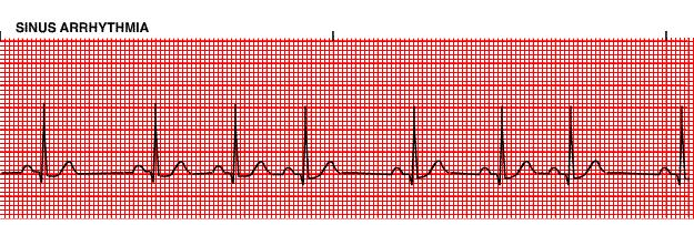
 Sinus Rhythms Normal sinus arrhythmia Small, slow variation of the R-R interval i.e. variation of the normal sinus heart rate with respiration, etc. Sinus Tachycardia Defined as sinus rhythm with a rate