Cath Lab Essentials: Basic Hemodynamics for the Cath Lab and ICU
|
|
|
- Blanche Snow
- 5 years ago
- Views:
Transcription


1 Cath Lab Essentials: Basic Hemodynamics for the Cath Lab and ICU Ailin Barseghian El-Farra, MD, FACC Assistant Professor, Interventional Cardiology University of California, Irvine Department of Cardiology
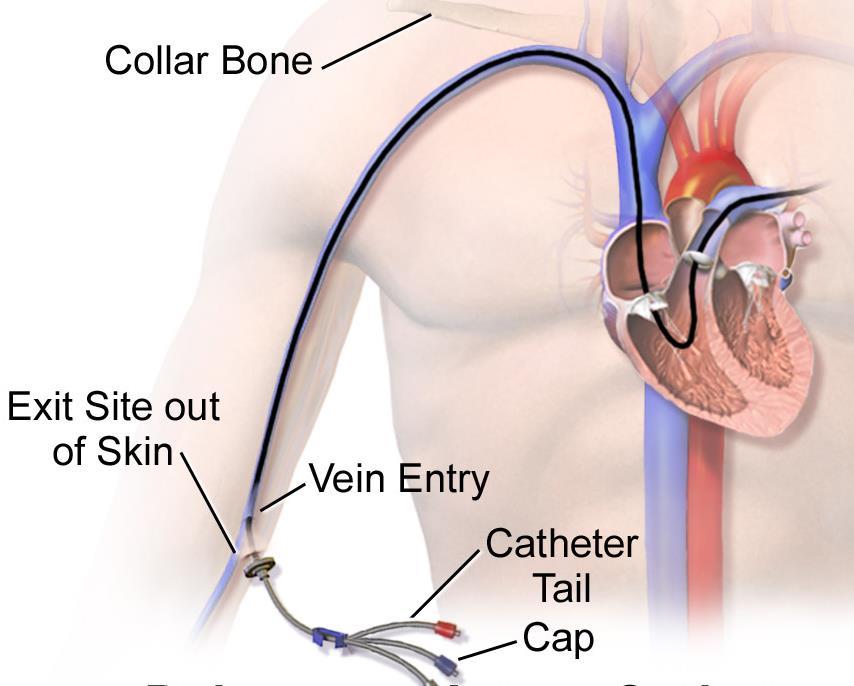
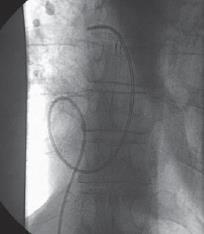
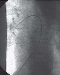
2 Right Heart Catheterization

3 By Don Ramey Logan - Own work, CC BY-SA 4.0,
4 INDICATIONS Cause of shock Pulmonary hypertension Fluid management and hemodynamic monitoring Guidance for pericardial tamponade Constrictive versus restrictive cardiomyopathy Diagnosis of left to right shunt

5 CONTRAINDICATIONS ABSOLUTE contraindications: None CAUTION: Pulmonary hypertension Elderly Left bundle branch block
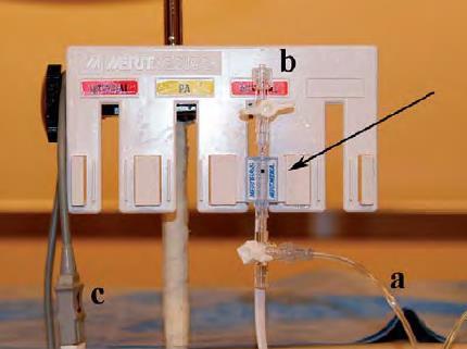
6 EQUIPMENT


7 PULMONARY ARTERY CATHETER EXTRA PORT DISTAL PORT PROXIMAL PORT THERMISTOR BALLOON
8 TECHNIQUE
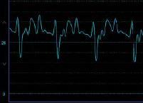
9 A Systematic Approach to Hemodynamic Interpretation 1. Establish the zero level and balance transducer. 2. Confirm the scale of the recording. -40 mmhg for RHC, 200 mmhg for LHC 3. Collect hemodynamics in a systematic method using established protocols. 4. Critically assess the pressure waveforms for proper fidelity. 5. Carefully time pressure events with the ECG. 6. Review the tracings for common artifacts
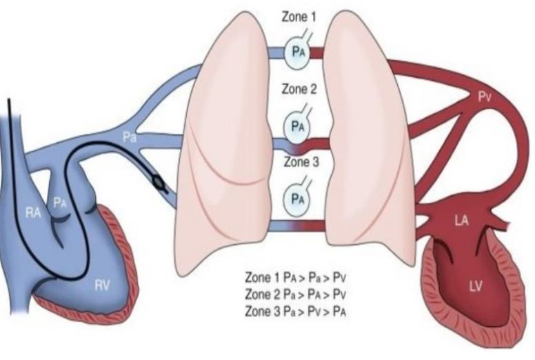
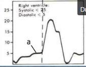
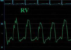
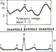
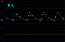
10 Components of a Right Heart 1.Right atrium Mean (1-5 mmhg) 2.Right ventricle Phasic (25/5 mmhg) Catheterization 3.Pulmonary capillary wedge Mean (7-12 mmhg) 4.Pulmonary artery Pulm HTN: mean PA pressure > 25mmHg PCWP < 15mmhg Phasic and mean (25/10 mmhg; mean mmhg)

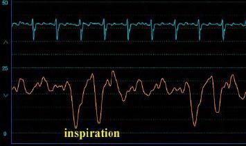
11 Precautions Always record pressures at end-expiration During inspiration, pressures will be lower due to decrease in intrathoracic pressure Always zero and reference the system
12 SAT RUN SVC to RA STEP UP If highest values are used, at least 11% If average of multiple samples, then 7% SVC IVC RA to PA STEP UP highest or average values 5% RIGHT ATRIUM LUNGS RA to RV STEP UP highest values are used, at least 10% If average of multiple samples, then 5% (for L-> R shunt)
13 1
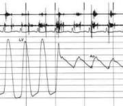
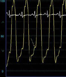
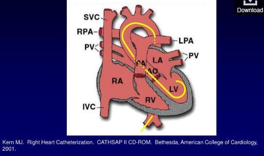
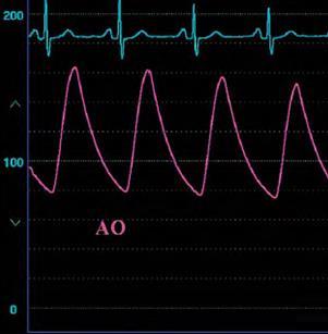
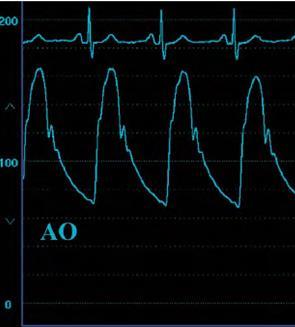
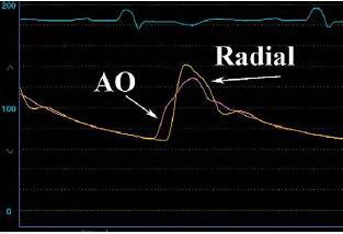
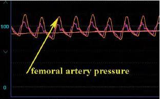
14 SIMULTANEOUS RIGHT- and LEFT- HEART CATHETERIZATION 1. Pulmonary artery (PA) catheter to pulmonary artery 2. Measure cardiac output by measuring oxygen saturation in PA and AO blood samples to determine Fick output or by thermodilution (x3); screen for shunt. 3. Record aortic pressures with AO catheter. Cross the AV into the ventricle -> Wedge the PA catheter -> Measure simultaneous LV- PCWP (mitral valve assessment). 4. Pull back from PCWP to PA. 5. Pull back from PA to right ventricle (RV) (to screen for pulmonic stenosis) and record RV. 6. Record simultaneous LV-RV (constriction vs restriction). 7. Pull back from RV to right atrium (RA) (to screen for tricuspid stenosis) and record RA 8. Pull back from LV to AO (to screen for aortic stenosis).
15 CARDIAC CYCLE
 5: Isovolumic Relaxation (PV/AV closure to TV/MV opening) 6: Rapid Ventricular Filling 7: Reduced Ventricular Filling (TV/MV opening to TV/MV")
16 PHASES 1: Atrial Contraction 2: Isovolumic Contraction (TV/MV closure to PV/AV opening) 3: Rapid Ejection 4: Reduced Ejection (PV/AV opening to PV/AV closure) 5: Isovolumic Relaxation (PV/AV closure to TV/MV opening) 6: Rapid Ventricular Filling 7: Reduced Ventricular Filling (TV/MV opening to TV/MV closure)
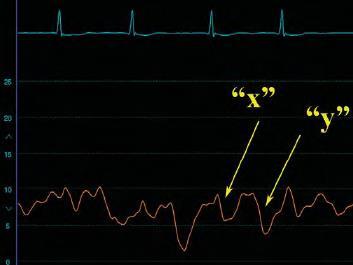
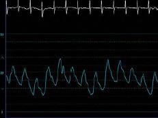
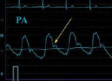
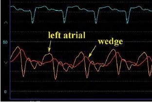
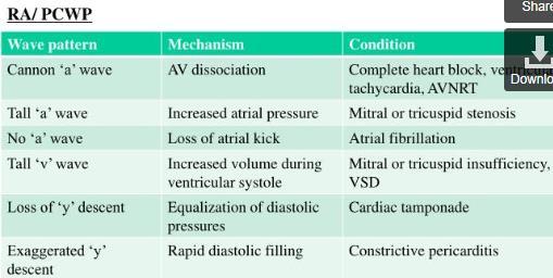
17 PRESSURE WAVE INTERPRETATION
18
19
20
21 LEFT HEART CATHETERIZATION
22
23
24 PITFALLS
25
26
27 ARTIFACTS

28
29 CARDIAC OUTPUT
30 Cardiac Output Thermodilution Fick Method


31 Thermodilution Bolus injection of saline into the proximal port Change in temperature is measured by thermistor in the distal portion of the catheter

32 Fick Principle Described in 1870 Assumes rate of O2 consumption is a function of rate of blood flow times the rate of O2 pick up by the RBC Oxygen consumption 1. Direct Fick: -Directly measured 2. Indirect Fick: --3 ml O2/kg X 10
33 Limitations Thermodilution Not accurate in tricuspid regurgitation Overestimated cardiac output at low output states Fick Oxygen consumption is often estimated by body weight (indirect method) rather than measured directly Large errors possible with small differences in saturations and hemoglobin. Measurements on room air
34 THANK YOU
35 Site Right Atrium (or CVP) Normal Pressures Normal Value (mmhg) Mean Pressure (mmhg) % Right Ventricle 25/5 75% Saturation Pulmonary Artery 25/ % PCWP % LV 120/ % Aorta 120/ %
36 Normal Values Site Value Sv Stroke Volume ml/beat Stroke Index ml/beat/m2 Cardiac Output 4-8 L/min Cardiac Index L/min/m2 SVR PVR dynes sec/-cm5 <250 dynes sec/-cm5 MAP mmhg
37
38 References Bangalore and Bhatt. Right heart catheterization, coronary angiography and percutaneous coronary intervention. Circulation, 2011; 124: e428-e433. Kern, Morton J. The Cardiac Catheterization Handbook. Philadelphia, PA: Saunders Elsevier, Print. Ragosta, Michael. Textbook of Clinical Hemodynamics. Philadelphia, PA: Saunders/Elsevier, Print.
Technique. Technique. Technique. Monitoring 1. Local anesthetic? Aseptic technique Hyper-extend (if radial)
") Critical Care Monitoring Hemodynamic Monitoring Arterial Blood Pressure Cannulate artery Uses 2 Technique Sites Locate artery, prep 3 1 Technique Local anesthetic? Aseptic technique Hyper-extend (if radial)
Critical Care Monitoring Hemodynamic Monitoring Arterial Blood Pressure Cannulate artery Uses 2 Technique Sites Locate artery, prep 3 1 Technique Local anesthetic? Aseptic technique Hyper-extend (if radial)
FUNDAMENTALS OF HEMODYNAMICS, VASOACTIVE DRUGS AND IABP IN THE FAILING HEART
 FUNDAMENTALS OF HEMODYNAMICS, VASOACTIVE DRUGS AND IABP IN THE FAILING HEART CINDY BITHER, MSN, ANP, ANP, AACC, CHFN CHIEF NP, ADV HF PROGRAM MEDSTAR WASHINGTON HOSPITAL CENTER CONFLICTS OF INTEREST NONE
Calculations the Cardiac Cath Lab. Thank You to: Lynn Jones RN, RCIS, FSICP Jeff Davis RCIS, FSICP Wes Todd, RCIS CardioVillage.
 Calculations the Cardiac Cath Lab Thank You to: Lynn Jones RN, RCIS, FSICP Jeff Davis RCIS, FSICP Wes Todd, RCIS CardioVillage.com Disclosure Information Calculations the Cardiac Cath Lab Darren Powell,
Καθετηριασμός δεξιάς κοιλίας. Σ. Χατζημιλτιάδης Καθηγητής Καρδιολογίας ΑΠΘ
 Καθετηριασμός δεξιάς κοιλίας Σ. Χατζημιλτιάδης Καθηγητής Καρδιολογίας ΑΠΘ The increasing interest in pulmonary arterial hypertension (PAH), the increasing interest in implantation of LVADs, and the evolution
Brief View of Calculation and Measurement of Cardiac Hemodynamics
 Cronicon OPEN ACCESS EC CARDIOLOGY Review Article Brief View of Calculation and Measurement of Cardiac Hemodynamics Samah Alasrawi* Pediatric Cardiologist, Al Jalila Children Heart Center, Dubai, UAE *
Right Heart Catheterization. Franz R. Eberli MD Chief of Cardiology Stadtspital Triemli, Zurich
 Right Heart Catheterization Franz R. Eberli MD Chief of Cardiology Stadtspital Triemli, Zurich Right Heart Catheterization Pressure measurements Oxygen saturation measurements Cardiac output, Vascular
Hemodynamics: Cardiac and Vascular Jeff Davis, RRT, RCIS
 Hemodynamics: Cardiac and Vascular Jeff Davis, RRT, RCIS Program Director, Cardiovascular Technology Florida SouthWestern State College Fort Myers, FL Disclosures Speaker s Bureau: None Stockholder: None
PVDOMICS: Right Heart Catheterization Training
 PVDOMICS: Right Heart Catheterization Training Cardiovascular Physiology Core Cleveland Clinic, Cleveland OH November 7, 2016 NHLBI Pulmonary Vascular Disease Phenomics Program Funded by the National Heart,
Georgios C. Bompotis Cardiologist, Director of Cardiological Department, Papageorgiou Hospital,
 Georgios C. Bompotis Cardiologist, Director of Cardiological Department, Papageorgiou Hospital, Disclosure Statement of Financial Interest I, Georgios Bompotis DO NOT have a financial interest/arrangement
Relax and Learn At the Farm 2012
 Relax and Learn At the Farm Session 9: Invasive Hemodynamic Assessment and What to Do with the Data Carol Jacobson RN, MN Cardiovascular Nursing Education Associates Function of CV system is to deliver
Cardiac Cycle MCQ. Professor of Cardiovascular Physiology. Cairo University 2007
 Cardiac Cycle MCQ Abdel Moniem Ibrahim Ahmed, MD Professor of Cardiovascular Physiology Cairo University 2007 1- Regarding the length of systole and diastole: a- At heart rate 75 b/min, the duration of
CATCH A WAVE.. INTRODUCTION NONINVASIVE HEMODYNAMIC MONITORING 4/12/2018
 WAVES CATCH A WAVE.. W I S C O N S I N P A R A M E D I C S E M I N A R A P R I L 2 0 1 8 K E R I W Y D N E R K R A U S E R N, C C R N, E M T - P Have you considered that if you don't make waves, nobody
Hemodynamic Monitoring and Circulatory Assist Devices
 Hemodynamic Monitoring and Circulatory Assist Devices Speaker: Jana Ogden Learning Unit 2: Hemodynamic Monitoring and Circulatory Assist Devices Hemodynamic monitoring refers to the measurement of pressure,
The Cardiac Cycle Clive M. Baumgarten, Ph.D.
 The Cardiac Cycle Clive M. Baumgarten, Ph.D. OBJECTIVES: 1. Describe periods comprising cardiac cycle and events within each period 2. Describe the temporal relationships between pressure, blood flow,
Hemodynamic Monitoring
 Perform Procedure And Interpret Results Hemodynamic Monitoring Tracheal Tube Cuff Pressure Dean R. Hess PhD RRT FAARC Hemodynamic Monitoring Cardiac Rate and Rhythm Arterial Blood Pressure Central Venous
Coronary Anomalies & Hemodynamic Identification
 Coronary Anomalies & Hemodynamic Identification David Stultz, MD Cardiology Fellow, PGY 6 May 2, 2006 Anomaly #1 Anomaly #2 Anomaly #3 Figure 18-27 Anomalous origin of the left circumflex artery.
เอกราช อร ยะช ยพาณ ชย
 30 July 2016 เอกราช อร ยะช ยพาณ ชย Heart Failure and Transplant Cardiology aekarach.a@chula.ac.th Disclosure Speaker, CME service: Merck, Otsuka, Servier Consultant, non-cme service: Novartis, Menarini
5 consecutive cases of PH I wish I never saw
 5 consecutive cases of PH I wish I never saw Abubakr A Bajwa. MD, FCCP Associate Professor of Medicine Division Chief Pulmonary, Critical Care and Sleep Medicine Director Pulmonary Hypertension and Interstitial
Shunt Detection and Quantification. September 2007 Joe M. Moody, Jr, MD UTHSCSA and STVAHCS
 Shunt Detection and Quantification September 2007 Joe M. Moody, Jr, MD UTHSCSA and STVAHCS Normal Physiology - Overview Right heart saturations (oxygen content) are generally about 75% and are equal in
Basic Hemodynamics. July 19, 2006 Joe M. Moody, Jr, MD UTHSCSA and STVHCS
 Basic Hemodynamics July 19, 2006 Joe M. Moody, Jr, MD UTHSCSA and STVHCS The Cardiac Cycle Types of Measures Pressure Outline Flow (Calculated from Temperature, O 2 Saturation, Indicator Concentration,
Introduction. Invasive Hemodynamic Monitoring. Determinants of Cardiovascular Function. Cardiovascular System. Hemodynamic Monitoring
 Introduction Invasive Hemodynamic Monitoring Audis Bethea, Pharm.D. Assistant Professor Therapeutics IV January 21, 2004 Hemodynamic monitoring is necessary to assess and manage shock Information obtained
Revision of 10/27/2017 Form #280 Page 1 of 12 PVDOMICS STUDY Clinical Center Right Heart Catheterization (RHC) Results Form #280
 Results Form #280") Revision of 10/27/2017 Form #280 Page 1 of 12 PVDOMICS STUDY Clinical Center Right Heart Catheterization (RHC) Results Form #280 Instructions: Review PVDOMICS MOP Chapter 100 prior to completing right
Revision of 10/27/2017 Form #280 Page 1 of 12 PVDOMICS STUDY Clinical Center Right Heart Catheterization (RHC) Results Form #280 Instructions: Review PVDOMICS MOP Chapter 100 prior to completing right
Adult Echocardiography Examination Content Outline
 Adult Echocardiography Examination Content Outline (Outline Summary) # Domain Subdomain Percentage 1 2 3 4 5 Anatomy and Physiology Pathology Clinical Care and Safety Measurement Techniques, Maneuvers,
Hemodynamic Assessment. Assessment of Systolic Function Doppler Hemodynamics
 Hemodynamic Assessment Matt M. Umland, RDCS, FASE Aurora Medical Group Milwaukee, WI Assessment of Systolic Function Doppler Hemodynamics Stroke Volume Cardiac Output Cardiac Index Tei Index/Index of myocardial
HISTORY. Question: What category of heart disease is suggested by this history? CHIEF COMPLAINT: Heart murmur present since early infancy.
 HISTORY 18-year-old man. CHIEF COMPLAINT: Heart murmur present since early infancy. PRESENT ILLNESS: Although normal at birth, a heart murmur was heard at the six week check-up and has persisted since
Disclosures. Objectives 6/16/2016. A Look at the Other Side: Focus on the Right Ventricle and Pulmonary Hypertension
 A Look at the Other Side: Focus on the Right Ventricle and Pulmonary Hypertension Susan P. D Anna MSN, APN-BC, CHFN June 24, 2016 Disclosures Objectives Differentiate structure and function of RV and LV
Hemodynamics of Exercise
 Hemodynamics of Exercise Joe M. Moody, Jr, MD UTHSCSA and ALMMVAH, STVAHCS Exercise Physiology - Acute Effects Cardiac Output (Stroke volume, Heart Rate ) Oxygen Extraction (Arteriovenous O 2 difference,
The Hemodynamics of PH Interpreting the numbers
 The Hemodynamics of PH Interpreting the numbers Todd M Bull MD Associate Professor of Medicine Division of Pulmonary Sciences and Critical Care Medicine Pulmonary Hypertension Center University of Colorado
CARDIOVASCULAR PHYSIOLOGY
 CARDIOVASCULAR PHYSIOLOGY LECTURE 4 Cardiac cycle Polygram - analysis of cardiac activity Ana-Maria Zagrean MD, PhD The Cardiac Cycle - definitions: the sequence of electrical and mechanical events that
The Doppler Examination. Katie Twomley, MD Wake Forest Baptist Health - Lexington
 The Doppler Examination Katie Twomley, MD Wake Forest Baptist Health - Lexington OUTLINE Principles/Physics Use in valvular assessment Aortic stenosis (continuity equation) Aortic regurgitation (pressure
Cardiac output and Venous Return. Faisal I. Mohammed, MD, PhD
 Cardiac output and Venous Return Faisal I. Mohammed, MD, PhD 1 Objectives Define cardiac output and venous return Describe the methods of measurement of CO Outline the factors that regulate cardiac output
CARDIOVASCULAR MONITORING. Prof. Yasser Mostafa Kadah
 CARDIOVASCULAR MONITORING Prof. Yasser Mostafa Kadah Introduction Cardiovascular monitoring covers monitoring of heart and circulatory functions It makes it possible to commence interventions quickly in
Outline. Echocardiographic Assessment of Pericardial Effusion/Tamponade: The Essentials
 Echocardiographic Assessment of Pericardial Effusion/Tamponade: The Essentials John R Schairer DO FACC Henry Ford Heart and Vascular Institute No Disclosures Outline Normal Anatomy and Physiology Pathophysiology
Pulmonary Artery Catheter Helpful Hints 2017
 Pulmonary Artery Catheter Helpful Hints 2017 Swan Ganz Catheter 1) Gather Equipment Hint1: Introducer is the actual catheter [Cordis is a brand name: at GW we use Arrow brand] Hint 2: 9 Fr Introducer for
SECTION I FUNDAMENTALS AND CLINICAL APPLICATIONS OF HEMODYNAMICS: COPYRIGHTED MATERIAL
 SECTION I FUNDAMENTALS AND CLINICAL APPLICATIONS OF HEMODYNAMICS: COPYRIGHTED MATERIAL UNDERSTANDING THE PRESSURE WAVES IN THE HEART: THE WIGGER S DIAGRAM Everything you want to know about hemodynamics
Cardiovascular Physiology. Heart Physiology. Introduction. The heart. Electrophysiology of the heart
 Cardiovascular Physiology Heart Physiology Introduction The cardiovascular system consists of the heart and two vascular systems, the systemic and pulmonary circulations. The heart pumps blood through
Cardiovascular Nursing Practice: A Comprehensive Resource Manual and Study Guide for Clinical Nurses 2 nd Edition
 Cardiovascular Nursing Practice: A Comprehensive Resource Manual and Study Guide for Clinical Nurses 2 nd Edition Table of Contents Volume 1 Chapter 1: Cardiovascular Anatomy and Physiology Basic Cardiac
Topics to be Covered. Cardiac Measurements. Distribution of Blood Volume. Distribution of Pulmonary Ventilation & Blood Flow
 Topics to be Covered MODULE F HEMODYNAMIC MONITORING Cardiac Output Determinants of Stroke Volume Hemodynamic Measurements Pulmonary Artery Catheterization Control of Blood Pressure Heart Failure Cardiac
Cardiovascular Structure & Function
 Cardiovascular Structure & Function Cardiovascular system: The heart Arteries Veins Capillaries Lymphatic vessels Weighting of the heart ceremony: Ancient Egyptians William Harvey and Blood Flow April
Disclosures. Objectives. RV vs LV. Structure and Function 9/25/2016. A Look at the Other Side: Focus on the Right Ventricle and Pulmonary Hypertension
 Disclosures A Look at the Other Side: Focus on the Right Ventricle and Pulmonary Hypertension No financial relationships Susan P. D Anna MSN, APN BC, CHFN September 29, 2016 Objectives RV vs LV Differentiate
Echo Doppler Assessment of Right and Left Ventricular Hemodynamics.
 Echo Doppler Assessment of Right and Left Ventricular Hemodynamics. Itzhak Kronzon, MD, FASE, FACC, FESC, FAHA, FACP, FCCP Northwell, Lenox Hill Hospital, New York Professor of Cardiology Hofstra University
PVDOMICS: Right Heart Catheterization
 PVDOMICS: Right Heart Catheterization Cardiovascular Physiology Core Cleveland Clinic, Cleveland OH March 16, 2018 NHLBI Pulmonary Vascular Disease Phenomics Program Funded by the National Heart, Lung,
Impedance Cardiography (ICG) Method, Technology and Validity
 Method, Technology and Validity") Method, Technology and Validity Hemodynamic Basics Cardiovascular System Cardiac Output (CO) Mean arterial pressure (MAP) Variable resistance (SVR) Aortic valve Left ventricle Elastic arteries / Aorta
Method, Technology and Validity Hemodynamic Basics Cardiovascular System Cardiac Output (CO) Mean arterial pressure (MAP) Variable resistance (SVR) Aortic valve Left ventricle Elastic arteries / Aorta
Ejection across stenotic aortic valve requires a systolic pressure gradient between the LV and aorta. This places a pressure load on the LV.
 Valvular Heart Disease Etiology General Principles Cellular and molecular mechanism of valve damage Structural pathology Functional pathology - stenosis/regurgitation Loading conditions - pressure/volume
Congenital Heart Defects
 Normal Heart Congenital Heart Defects 1. Patent Ductus Arteriosus The ductus arteriosus connects the main pulmonary artery to the aorta. In utero, it allows the blood leaving the right ventricle to bypass
HEMODYNAMIC ASSESSMENT
 HEMODYNAMIC ASSESSMENT INTRODUCTION Conventionally hemodynamics were obtained by cardiac catheterization. It is possible to determine the same by echocardiography. Methods M-mode & 2D echo alone can provide
MASSACHUSETTS INSTITUTE OF TECHNOLOGY
 Harvard-MIT Division of Health Sciences and Technology HST.542J: Quantitative Physiology: Organ Transport Systems Instructors: Roger Mark and Jose Venegas MASSACHUSETTS INSTITUTE OF TECHNOLOGY Departments
Ejection across stenotic aortic valve requires a systolic pressure gradient between the LV and aorta. This places a pressure load on the LV.
 Valvular Heart Disease General Principles Etiology Cellular and molecular mechanism of valve damage Structural pathology Functional pathology - stenosis/regurgitation Loading conditions - pressure/volume
Cardiovascular System
 Cardiovascular System The Heart Cardiovascular System The Heart Overview What does the heart do? By timed muscular contractions creates pressure gradients blood moves then from high pressure to low pressure
Cardiogenic Shock Protocol
 Cardiogenic Shock Protocol Impella Devices Best Practices in AMI Cardiogenic Shock Identify 1-3 SBP < 90 mmhg or on inotropes /pressors Cold, clammy, tachycardia Lactate elevated > 2 mmoi /L Stabilize
There has been a striking evolution in the role of the
 Contemporary Reviews in Cardiovascular Medicine Hemodynamics in the Cardiac Catheterization Laboratory of the 21st Century Rick A. Nishimura, MD; Blase A. Carabello, MD There has been a striking evolution
Mechanics of Cath Lab Support Devices
 Mechanics of Cath Lab Support Devices Issam D. Moussa, MD Chief Medical Officer First Coast Cardiovascular Institute, Jacksonville, FL Professor of Medicine, UCF, Orlando, FL None DISCLOSURE Percutaneous
Shock, Monitoring Invasive Vs. Non Invasive
 Shock, Monitoring Invasive Vs. Non Invasive Paula Ferrada MD Assistant Professor Trauma, Critical Care and Emergency Surgery Virginia Commonwealth University Shock Fluid Pressors Ionotrope Intervention
Ray Matthews MD Professor of Clinical Medicine Chief of Cardiology University of Southern California
 High Risk PCI Making Possible the Impossible Ray Matthews MD Professor of Clinical Medicine Chief of Cardiology University of Southern California Disclosures Abiomed Research Support Consulting Agreement
Maternal Cardiac Disease In Pregnancy. August 25, 2017 PREGNANCY ECHO CONFERENCE
 Maternal Cardiac Disease In Pregnancy August 25, 2017 PREGNANCY ECHO CONFERENCE Maternal Physiology Cardiac Output = HR x SV Non-pregnant: 4.5 L/min Pregnant: 6.0 L/min Increase most acute in first 10
Principles of Biomedical Systems & Devices. Lecture 8: Cardiovascular Dynamics Dr. Maria Tahamont
 Principles of Biomedical Systems & Devices Lecture 8: Cardiovascular Dynamics Dr. Maria Tahamont Review of Cardiac Anatomy Four chambers Two atria-receive blood from the vena cave and pulmonary veins Two
Outline. Electrical Activity of the Human Heart. What is the Heart? The Heart as a Pump. Anatomy of the Heart. The Hard Work
 Electrical Activity of the Human Heart Oguz Poroy, PhD Assistant Professor Department of Biomedical Engineering The University of Iowa Outline Basic Facts about the Heart Heart Chambers and Heart s The
Congenital Heart Disease: Physiology and Common Defects
 Congenital Heart Disease: Physiology and Common Defects Jamie S. Sutherell, M.D, M.Ed. Associate Professor, Pediatrics Division of Cardiology Director, Medical Student Education in Pediatrics Director,
(D) (E) (F) 6. The extrasystolic beat would produce (A) increased pulse pressure because contractility. is increased. increased
 (E) (F) 6. The extrasystolic beat would produce (A) increased pulse pressure because contractility. is increased. increased") Review Test 1. A 53-year-old woman is found, by arteriography, to have 5% narrowing of her left renal artery. What is the expected change in blood flow through the stenotic artery? Decrease to 1 2 Decrease
Review Test 1. A 53-year-old woman is found, by arteriography, to have 5% narrowing of her left renal artery. What is the expected change in blood flow through the stenotic artery? Decrease to 1 2 Decrease
QUIZ 1. Tuesday, March 2, 2004
 Harvard-MIT Division of Health Sciences and Technology HST.542J: Quantitative Physiology: Organ Transport Systems Instructors: Roger Mark and Jose Venegas MASSACHUSETTS INSTITUTE OF TECHNOLOGY Departments
HAEMODYNAMIC IN THE CATH LABORATORY INTRO TO BASICS
 HAEMODYNAMIC IN THE CATH LABORATORY INTRO TO BASICS BY NOOR FADZLY ALIAS CARDIOVASCULAR TECHNOLOGIST NCL Department National Heart Institute Kuala Lumpur INTRODUCTION ROLES OF HAEMODYNAMIC MONITORING PURPOSE
Chapter 9, Part 2. Cardiocirculatory Adjustments to Exercise
 Chapter 9, Part 2 Cardiocirculatory Adjustments to Exercise Electrical Activity of the Heart Contraction of the heart depends on electrical stimulation of the myocardium Impulse is initiated in the right
DOPPLER HEMODYNAMICS (1) QUANTIFICATION OF PRESSURE GRADIENTS and INTRACARDIAC PRESSURES
 QUANTIFICATION OF PRESSURE GRADIENTS and INTRACARDIAC PRESSURES") THORAXCENTRE DOPPLER HEMODYNAMICS (1) QUANTIFICATION OF PRESSURE GRADIENTS and INTRACARDIAC PRESSURES J. Roelandt DOPPLER HEMODYNAMICS Intracardiac pressures and pressure gradients Volumetric measurement
THORAXCENTRE DOPPLER HEMODYNAMICS (1) QUANTIFICATION OF PRESSURE GRADIENTS and INTRACARDIAC PRESSURES J. Roelandt DOPPLER HEMODYNAMICS Intracardiac pressures and pressure gradients Volumetric measurement
Goal-directed vs Flow-guidedresponsive
 Goal-directed vs Flow-guidedresponsive therapy S Magder Department of Critical Care, McGill University Health Centre Flow-directed vs goal directed strategy for management of hemodynamics S Magder Curr
PROBLEM SET 2. Assigned: February 10, 2004 Due: February 19, 2004
 Harvard-MIT Division of Health Sciences and Technology HST.542J: Quantitative Physiology: Organ Transport Systems Instructors: Roger Mark and Jose Venegas MASSACHUSETTS INSTITUTE OF TECHNOLOGY Departments
Appendix II: ECHOCARDIOGRAPHY ANALYSIS
 Appendix II: ECHOCARDIOGRAPHY ANALYSIS Two-Dimensional (2D) imaging was performed using the Vivid 7 Advantage cardiovascular ultrasound system (GE Medical Systems, Milwaukee) with a frame rate of 400 frames
Comprehensive Hemodynamics By Doppler Echocardiography. The Echocardiographic Swan-Ganz Catheter.
 Comprehensive Hemodynamics By Doppler Echocardiography. The Echocardiographic Swan-Ganz Catheter. Itzhak Kronzon, MD, FASE, FACC, FESC, FAHA, FACP, FCCP North Shore HS, LIJ/Lenox Hill Hospital, New York
Rhythm Disorders 2017 TazKai LLC and NRSNG.com
 Rhythm Disorders 1. Outline the conduction system of the heart. 2. What do the different portions of the EKG represent? 3. Define the following terms: a. Automaticity b. Conductivity c. Excitability d.
Dr. Md. Rajibul Alam Prof. of Medicine Dinajpur Medical college
 Dr. Md. Rajibul Alam Prof. of Medicine Dinajpur Medical college PULMONARY HYPERTENSION Difficult to diagnose early Because Not detected during routine physical examination and Even in advanced cases symptoms
Cardiac MRI in ACHD What We. ACHD Patients
 Cardiac MRI in ACHD What We Have Learned to Apply to ACHD Patients Faris Al Mousily, MBChB, FAAC, FACC Consultant, Pediatric Cardiology, KFSH&RC/Jeddah Adjunct Faculty, Division of Pediatric Cardiology
Atrial Fibrillaton. Key: RA: right atrium RV: right ventricle PA: pulmonic artery LA: left atrium LV: left ventricle AO: aorta
 Atrial Fibrillaton How does the heart work? The heart is the organ responsible for pumping blood to and from all tissues of the body. The heart is divided into right and left sides. The job of the right
Cath Lab Essentials: Pericardial effusion & tamponade
 Cath Lab Essentials: Pericardial effusion & tamponade Pranav M. Patel, MD, FACC, FSCAI Chief, Division of Cardiology Director, Cardiac Cath Lab & CCU University of California, Irvine Division of Cardiology
Dr.Fayez EL Shaer Consultant cardiologist Assistant professor of cardiology KKUH
 Pulmonary Hypertension in patients with Heart Failure with Preserved Ejection Fraction Dr.Fayez EL Shaer Consultant cardiologist Assistant professor of cardiology KKUH Recent evaluation of available data
Echocardiography as a diagnostic and management tool in medical emergencies
 Echocardiography as a diagnostic and management tool in medical emergencies Frank van der Heusen MD Department of Anesthesia and perioperative Care UCSF Medical Center Objective of this presentation Indications
PVDOMICS: Right Heart Catheterization
 PVDOMICS: Right Heart Catheterization Cardiovascular Physiology Core Cleveland Clinic, Cleveland OH November 7, 2016 NHLBI Pulmonary Vascular Disease Phenomics Program Funded by the National Heart, Lung,
Acute Mechanical Circulatory Support Right Ventricular Support Devices
 Acute Mechanical Circulatory Support Right Ventricular Support Devices Navin K. Kapur, MD, FACC, FSCAI, FAHA Associate Professor, Department of Medicine Interventional Cardiology & Advanced Heart Failure
4/21/2018. The Role of Cardiac Catheterization in Pediatric PVD. The Role(s) of Cath in PVD. Pre Cath Management. Catheterization Mechanics in PVD
 of Cath in PVD. Pre Cath Management. Catheterization Mechanics in PVD") UCSF Pediatric Heart Center Benioff Children s Hospitals Oakland & San Francisco April 19, 2018 The Role of Cardiac Catheterization in Pediatric PVD Phillip Moore MD, MBA The Role(s) of Cath in PVD Diagnosis
UCSF Pediatric Heart Center Benioff Children s Hospitals Oakland & San Francisco April 19, 2018 The Role of Cardiac Catheterization in Pediatric PVD Phillip Moore MD, MBA The Role(s) of Cath in PVD Diagnosis
IB TOPIC 6.2 THE BLOOD SYSTEM
 IB TOPIC 6.2 THE BLOOD SYSTEM TERMS TO KNOW circulation ventricle artery vein THE BLOOD SYSTEM 6.2.U1 - Arteries convey blood at high pressure from the ventricles to the tissues of the body Circulation
Hemodynamic Monitoring in the CCU. Edward G. Hamaty Jr., D.O. FACCP, FACOI
 Hemodynamic Monitoring in the CCU Edward G. Hamaty Jr., D.O. FACCP, FACOI Waveform Review Left Ventricular Pressure Normal left ventricular pressures are: Systolic 100 to 140 mm of mercury End-diastolic
Can SCMR CMR protocol recommendations
 Can SCMR CMR protocol recommendations V1.3 - April 2009 CanSCMR CMR Protocol and SOP Recommendation 2009 (15 minutes) 2 Planning of LV fct. real time multiple axes Realtime 3 cine long axis 6 long axes
Heart Pump and Cardiac Cycle. Faisal I. Mohammed, MD, PhD
 Heart Pump and Cardiac Cycle Faisal I. Mohammed, MD, PhD 1 Objectives To understand the volume, mechanical, pressure and electrical changes during the cardiac cycle To understand the inter-relationship
Pulmonary hypertension in clinical practice: are we focusing on the problem?
 Pulmonary hypertension in clinical practice: are we focusing on the problem? Odd Bech-Hanssen, MD, PhD Cardiology/Clinical Physiology Sahlgrenska University Hospital Gothenburg, Sweden Definition Mean
SIKLUS JANTUNG. Rahmatina B. Herman
 SIKLUS JANTUNG Rahmatina B. Herman The Cardiac Cycle Definition: The cardiac events that occur from the beginning of one heartbeat to the beginning of the next The cardiac cycle consists of: - Diastole
Mechanics of Cath Lab Support Devices
 Mechanics of Cath Lab Support Devices Issam D. Moussa, MD Professor of Medicine Mayo Clinic College of Medicine Chair, Division of Cardiovascular Diseases Mayo Clinic Jacksonville, Florida DISCLOSURE Presenter:
Adel Hasanin Ahmed 1
 Adel Hasanin Ahmed 1 PERICARDIAL DISEASE The pericardial effusion ends anteriorly to the descending aorta and is best visualised in the PLAX. PSAX is actually very useful sometimes for looking at posterior
THE CARDIOVASCULAR SYSTEM
 THE CARDIOVASCULAR SYSTEM AND RESPONSES TO EXERCISE Mr. S. Kelly PSK 4U North Grenville DHS THE HEART: A REVIEW Cardiac muscle = myocardium Heart divided into two sides, 4 chambers (L & R) RS: pulmonary
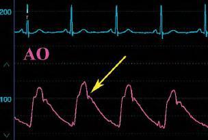
The V Wave. January, 2007 Joe M. Moody, Jr, MD UTHSCSA and ALMMVAH. Ref: Kern MJ. Hemodynamic Rounds, 2 nd ed
 The V Wave January, 2007 Joe M. Moody, Jr, MD UTHSCSA and ALMMVAH Ref: Kern MJ. Hemodynamic Rounds, 2 nd ed. 1999. Normal Hemodynamic Values Cardiac index 2.8-4.2 (mean 3.4 L/min/m 2 ) Stroke volume 30-65
Cath Lab Essentials : LV Assist Devices for Hemodynamic Support (IABP, Impella, Tandem Heart, ECMO)
") Cath Lab Essentials : LV Assist Devices for Hemodynamic Support (IABP, Impella, Tandem Heart, ECMO) Michael A. Gibson, MD Assistant Professor of Medicine University of California, Irvine Division of Cardiology
Cath Lab Essentials : LV Assist Devices for Hemodynamic Support (IABP, Impella, Tandem Heart, ECMO) Michael A. Gibson, MD Assistant Professor of Medicine University of California, Irvine Division of Cardiology
BME 5742 Bio-Systems Modeling and Control. Lecture 41 Heart & Blood Circulation Heart Function Basics
 BME 5742 Bio-Systems Modeling and Control Lecture 41 Heart & Blood Circulation Heart Function Basics Dr. Zvi Roth (FAU) 1 Pumps A pump is a device that accepts fluid at a low pressure P 1 and outputs the
National Imaging Associates, Inc. Clinical guidelines CARDIAC CATHETERIZATION -LEFT HEART CATHETERIZATION. Original Date: October 2015 Page 1 of 5
 National Imaging Associates, Inc. Clinical guidelines CARDIAC CATHETERIZATION -LEFT HEART CATHETERIZATION CPT Codes: 93451, 93452, 93453, 93454, 93455, 93456, 93457, 93458, 93459, 93460, 93461 LCD ID Number:
CPT Code Details
 CPT Code 93572 Details Code Descriptor Intravascular Doppler velocity and/or pressure derived flow reserve measurement ( vessel or graft) during angiography pharmacologically induced stress; each additional
Large Arteries of Heart
 Cardiovascular System (Part A-2) Module 5 -Chapter 8 Overview Arteries Capillaries Veins Heart Anatomy Conduction System Blood pressure Fetal circulation Susie Turner, M.D. 1/5/13 Large Arteries of Heart
ΔΙΑΧΕΙΡΙΣΗ ΑΣΘΕΝΩΝ ΜΕ ΜΕΣΟΚΟΛΠΙΚΗ ΕΠΙΚΟΙΝΩΝΙΑ ΖΑΧΑΡΑΚΗ ΑΓΓΕΛΙΚΗ ΚΑΡΔΙΟΛΟΓΟΣ ΗΡΑΚΛΕΙΟ - ΚΡΗΤΗ
 ΔΙΑΧΕΙΡΙΣΗ ΑΣΘΕΝΩΝ ΜΕ ΜΕΣΟΚΟΛΠΙΚΗ ΕΠΙΚΟΙΝΩΝΙΑ ΖΑΧΑΡΑΚΗ ΑΓΓΕΛΙΚΗ ΚΑΡΔΙΟΛΟΓΟΣ ΗΡΑΚΛΕΙΟ - ΚΡΗΤΗ European Accreditation in TTE, TEE and CHD Echocardiography NOTHING TO DECLARE ATRIAL SEPTAL DEFECT TYPES SECUNDUM
Lab 16. The Cardiovascular System Heart and Blood Vessels. Laboratory Objectives
 Lab 16 The Cardiovascular System Heart and Blood Vessels Laboratory Objectives Describe the anatomical structures of the heart to include the pericardium, chambers, valves, and major vessels. Describe
Pre-discussion questions
 Amanda Bartlett, PA-C Dustin Bartlett, PA-C Andrea Applegate, PA-C Leslie Yearta Brown, NP CHF Round Table Discussion Objectives ANDREA- Discuss the definition and different categories of CHF DUSTIN- Define
IP: Regulation of Cardiac Output
 ANP 1105D Winter 2013 Assignment 9: The Heart, part 2: Chap... Assignment 9: The Heart, part 2: Chapter 18 Signed in as Alex Sokolowski Help Close Resources Due: 11:59pm on Monday, March 25, 2013 Note:
Cardiology. the Sounds: #7 HCM. LV Outflow Obstruction: Aortic Stenosis. (Coming Soon - HCM)
") A Cardiology HCM LV Outflow Obstruction: Aortic Stenosis (Coming Soon - HCM) the Sounds: #7 Howard J. Sachs, MD www.12daysinmarch.com E-mail: Howard@12daysinmarch.com Aortic Valve Disorders Stenosis Regurgitation
A Cardiology HCM LV Outflow Obstruction: Aortic Stenosis (Coming Soon - HCM) the Sounds: #7 Howard J. Sachs, MD www.12daysinmarch.com E-mail: Howard@12daysinmarch.com Aortic Valve Disorders Stenosis Regurgitation