Gene annotation for heart rhythm. 1. Control of heart rate 2. Action Potential 3. Ion channels and transporters 4. Arrhythmia 5.
|
|
|
- Ambrose Lynch
- 5 years ago
- Views:
Transcription

1 Gene annotation for heart rhythm 1. Control of heart rate 2. Action Potential 3. Ion channels and transporters 4. Arrhythmia 5. EC coupling
2 Control of heart rate
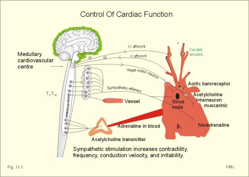
3 Autonomic regulation of heart function

4 Autonomic Regulation II Central integration of blood pressure and respiratory control Afferents via baroreceptors, chemoreceptors etc Integrated in brainstem centres


5 Effector arm Autonomic Regulation III Proteins involved in presynaptic vesicle release Proteins involved in signal transduction in the SA node

6 Heart Rate Variability The heart beat is not quite regular subject to small variations e.g. sinus arrhythmia Indicative of health. Correlates inversely with outcome after MI etc Time domain: Tachograms, SD of R-R or ΔR-R Frequency domain:- Potentially more revealing. HF=vagal\respiration, LF=sympathetic\BP control


7 What ionic mechanisms are responsible? Intrinsic rhythm set by SA node Modulation of pacemaker depolarisation β receptor activation Gs Adenylate cyclase Increased camp I f activation M2 receptor activation Gi\o Adenylate cyclase Decreased camp I f inhibition G βγ liberation I KAch activation
8 What is the intrinsic pacemaker? Spontaneous activity in the absence of innervation (intrinsic heart rate) Actually currently quite controversial Two hypotheses I f current is centrally important and\or Ca 2+ cycling


9 Activated by hyperpolarisation Cation but otherwise nonselective Directly opened by camp HCN1-4, mainly HCN4 in heart Largely expressed in SA node Ivabradine used for the treatment of angina I f \HCN channels

10 Action potential
11 Cardiac Action Potential I


12 Anatomy Conduction system AP in heart regions
13 Cardiac action potential II I Kur Kv1.5 I KACh Kir3.1\3.4 I KATP SUR1\Kir6.1\Kir6.2 vs SUR2A\Kir6.2 Cx40 in atria. Cx43 in ventricle SK channels
14 Ion Channels and Transporters


15 What is happening at the molecular level? Ion channels predominantly control membrane excitability
16 Sodium channels SCN5A in the heart. Both beta subunits present.

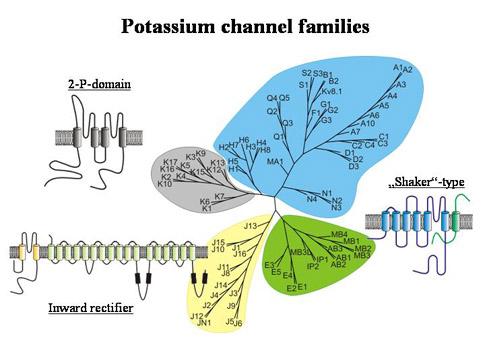
17 Potassium channels
18 Lots of genes underlying K+ channels Current I to,f (I to1 ) Molecular composition α-subunit Kv4.3 and β-subunit KChiP2. I to,s (I to1 ) α-subunit Kv1.4 and possibly β- subunits (Kvβ1.2, Kvβ1.3 and I Kur I Kr I Ks I K1 Kvβ2) α-subunit Kv1.5 and β-subunit Kvβ1.2. α-subunit Kv11.1 (HERG) and probably β-subunit KCNE2. α subunit Kv7.1 (KCNQ1) and β subunit KCNE1. Kir2.1 and perhaps Kir2.2 and Kir2.3. Channel structure Function Location Reference(s) Octameric complex of a tetramer of 6 TMD α- subunits and 4 β subunits. A tetramer of 6 TMD α subunits may coassemble with 4 β-subunits. I KACh Kir3.1 and Kir3.4. Tetrameric complex of 2 Kir3.1 and 2 Kir3.4 2 TMD subunits. (During development channel may be formed by a homotetramer of Kir3.4) I KATP (Ventricular) I KATP (Atrial) Provides the rapid component of the transient outward current that contributes to early rapid repolarization during Phase 1. Provides the slow component of the transient outward current that contributes to early rapid repolarization during Phase 1. Plays an important role in early phase (1-2) atrial repolarization. Repolarisation, outward rectifier during Phase 2 and 3. Repolarisation, outward rectifier during Phase 2 and 3. A tetramer of 6 TMD α subunits associates with 4 β-subunits to form an octameric complex. A tetramer of 6 TMD α-subunits and an unknown number of 1 TMD β-subunits. Tetramer of 6 TMD α-subunits assembles with probably two 1 TMD β-subunits. Tetramer of 2 TMD α subunits. Contributes to late repolarisation, late phase 3, and helps to set membrane potential. During late phase 3 and phase 4 activation of I KACh by acetylcholine acts to hyperpolarise the membrane potential, slow the firing rate of pacemaker cells in the SA and AV nodes and delays AV conduction. Kir6.2 and SUR2A. Octameric complex formed by coassembly of 4 2 TMD pore subunits and 4 17 TMD SUR subunits. Kir6.2 and SUR1. (Kir6.1?) Octameric complex formed by coassembly of 4 2 TMD pore subunits and 4 17 TMD SUR subunits. During late phase 3 and phase 4 this channel acts to link cellular metabolism and membrane excitability. During late phase 3 and phase 4 this channel acts to link cellular metabolism and membrane excitability. Atrial and Ventricular. Atrial and Ventricular. Atrial. Atrial and Ventricular. Atrial and Ventricular. Ventricular and Atrial Predominantly Atrial and nodal tissue expression. Ventricular. Atrial. [23], [24], (a), (b) [25], [26], (a), (c) [27], [28], (d), (e) (f), (g), (h), (i) [20], [21], [22], (h), (i) [30], [31], (j), (k), (l) [32], [33], (j), (m), (n), (o), (p) [10], (q), (r), (s) [9], [10], (q), (r), (s) Also SK channels and twin pore channels
19 K channels in Long QT Voltage-gated (6-TM) KCNE family Extracellular H5 N Extracellular Intracellular N C Intracellular C alpha beta current KCNQ1 (KvLQT1) KCNE1 (IsK) Iks HERG KCNE2 (MIRP1) Ikr


20 Na + \K + ATPase Member of the P type ATPase pumps α1, α2 and α3 subunits. β1 and β2 auxiliary subunits Electrogenic 3Na + for 2K + but transport rate ~4 four fold less than the Na channel (100 ions\second)
21 Arrhythmia
22 This carefully orchestrated activity can go wrong Classification of arrhythmia Site of origin e.g. atrial, nodal, ventricular Rate e.g. bradycardia, tachycardia Process\Substrate e.g. fibrillation, heart block, ectopic etc
23 Electrocardiogram (ECG) The benchmark of clinical diagnosis is the ECG P wave= atrial depolarisation QRS= ventricular depolarisation T wave=ventricular repolarisation

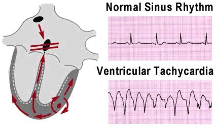
24 Examples Atrial Fibrillation Ventricular Tachycardia
25 Repolarisation and K + currents

26 Excitation-contraction coupling
27 Cardiac excitation-contraction coupling
 Ca v 1 = L-type, Ca v 2 = N-, P\Q and R type and Ca v 3 = T type")
28 Calcium channels Gene = CACNAx for alpha subunits (CACNA1C = Ca v 1.2) Ca v 1 = L-type, Ca v 2 = N-, P\Q and R type and Ca v 3 = T type


29 Ryanodine receptor RyR2 in heart Calcium induced calcium release LTCC and RyR2 opposed in T- tubule Large tetrameric complex Protein interactions


30 Sodium\calcium exchanger Major mechanism for calcium extrusion from the heart Electrogenic 3 Na + for single Ca 2+ Passive coupled counter transport system NCX1 in the heart (3 isoforms in total) Also P type ATPase Ca 2+ pump present in heart which actively extrudes Ca 2+ (PMCA)


31 SERCA2a and phospholamban Major mechanism for calcium uptake into SR P-type ATPase that transports Ca 2+ actively driven by ATP hydrolysis Regulated by phospholamban
Ionchannels and channelopaties in the heart. Viktória Szőts
 Ionchannels and channelopaties in the heart Viktória Szőts Action of membrane transport protein ATP-powered pump Ion chanels Transporters 10 1-10 3 ions/s 10 7-10 8 ions/s 10 2-10 4 ions/s Cardiac K +
Ionchannels and channelopaties in the heart
 Ionchannels and channelopaties in the heart Csatorna müködés Több betegség Drugok kapcsolodása csat.hoz Sejtekbe ioncsat.expresszios módszerek, bemutatása Viktória Szőts Action of membrane transport protein
Differences in cardiac atrial and ventricular ion channels
 Differences in cardiac atrial and ventricular ion channels Norbert Jost, PhD Department of Pharmacology & Pharmacotherapy, University of Szeged Division for Cardiovascular Pharmacology, Hungarian Academy
Cardiac physiology. b. myocardium -- cardiac muscle and fibrous skeleton of heart
 I. Heart anatomy -- general gross. A. Size/orientation - base/apex B. Coverings D. Chambers 1. parietal pericardium 2. visceral pericardium 3. Layers of heart wall a. epicardium Cardiac physiology b. myocardium
The Electrocardiogram
 The Electrocardiogram Chapters 11 and 13 AUTUMN WEDAN AND NATASHA MCDOUGAL The Normal Electrocardiogram P-wave Generated when the atria depolarizes QRS-Complex Ventricles depolarizing before a contraction
Where are the normal pacemaker and the backup pacemakers of the heart located?
 CASE 9 A 68-year-old woman presents to the emergency center with shortness of breath, light-headedness, and chest pain described as being like an elephant sitting on her chest. She is diagnosed with a
The action potential and the underlying ionic currents. Norbert Jost, PhD
 The action potential and the underlying ionic currents Norbert Jost, PhD The propagation of the stimulation in the heart Sinus node Left atria His Bundle Conduction velocity in m/s Time to arrive from
a lecture series by SWESEMJR
 Electrolyte disturbances Hypokalaemia Decreased extracellular potassium increases excitability in the myocardial cells and consequently the effect of very severe hypokalaemia is ventricular arrhythmia.
Collin County Community College
 Collin County Community College BIOL. 2402 Anatomy & Physiology WEEK 5 The Heart 1 The Heart Beat and the EKG 2 1 The Heart Beat and the EKG P-wave = Atrial depolarization QRS-wave = Ventricular depolarization
PART I. Disorders of the Heart Rhythm: Basic Principles
 PART I Disorders of the Heart Rhythm: Basic Principles FET01.indd 1 1/11/06 9:53:05 AM FET01.indd 2 1/11/06 9:53:06 AM CHAPTER 1 The Cardiac Electrical System The heart spontaneously generates electrical
FIBER TYPES - oxidative metabolism is the main form here - ATPase activity is relatively low
 Cardiac Muscle Physiology Special characteristics of cardiac muscle - Branching and interdigitating cells - At their ends, they are connected by INTERCALATED DISCS - The discs are always at the Z-lines
Rhythmical Excitation of the Heart
 Rhythmical Excitation of the Heart KALEB HOOD AND JIMMY JOHNSON Special Excitory and Conductive System of the Heart Sinus Node (or sinoatrial node or S-A): A small node with almost no contractile muscle,
Introduction. Circulation
 Introduction Circulation 1- Systemic (general) circulation 2- Pulmonary circulation carries oxygenated blood to all parts of the body carries deoxygenated blood to the lungs From Lt. ventricle aorta From
Chapter 13 The Cardiovascular System: Cardiac Function
 Chapter 13 The Cardiovascular System: Cardiac Function Overview of the Cardiovascular System The Path of Blood Flow through the Heart and Vasculature Anatomy of the Heart Electrical Activity of the Heart
Genetics of Sudden Cardiac Death. Geoffrey Pitt Ion Channel Research Unit Duke University. Disclosures: Grant funding from Medtronic.
 Genetics of Sudden Cardiac Death Geoffrey Pitt Ion Channel Research Unit Duke University Disclosures: Grant funding from Medtronic Duke U N I V E R S I T Y Sudden Cardiac Death High incidence 50-100 per
Cardiac Properties MCQ
 Cardiac Properties MCQ Abdel Moniem Ibrahim Ahmed, MD Professor of Cardiovascular Physiology Cairo University 2007 1- Cardiac Valves: a- Prevent backflow of blood from the ventricles to the atria during
QUIZ/TEST REVIEW NOTES SECTION 1 CARDIAC MYOCYTE PHYSIOLOGY [CARDIOLOGY]
![QUIZ/TEST REVIEW NOTES SECTION 1 CARDIAC MYOCYTE PHYSIOLOGY [CARDIOLOGY]](/thumbs/96/126998162.jpg "QUIZ/TEST REVIEW NOTES SECTION 1 CARDIAC MYOCYTE PHYSIOLOGY [CARDIOLOGY]") QUIZ/TEST REVIEW NOTES SECTION 1 CARDIAC MYOCYTE PHYSIOLOGY [CARDIOLOGY] Learning Objectives: Describe the ionic basis of action potentials in cardiac contractile and autorhythmic cells Explain the relationship
QUIZ/TEST REVIEW NOTES SECTION 1 CARDIAC MYOCYTE PHYSIOLOGY [CARDIOLOGY] Learning Objectives: Describe the ionic basis of action potentials in cardiac contractile and autorhythmic cells Explain the relationship
Arrhythmias. 1. beat too slowly (sinus bradycardia). Like in heart block
. Like in heart block") Arrhythmias It is a simple-dysfunction caused by abnormalities in impulse formation and conduction in the myocardium. The heart is designed in such a way that allows it to generate from the SA node electrical
Arrhythmias It is a simple-dysfunction caused by abnormalities in impulse formation and conduction in the myocardium. The heart is designed in such a way that allows it to generate from the SA node electrical
Chapter 20 (2) The Heart
 The Heart") Chapter 20 (2) The Heart ----------------------------------------------------------------------------------------------------------------------------------------- Describe the component and function of
Chapter 20 (2) The Heart ----------------------------------------------------------------------------------------------------------------------------------------- Describe the component and function of
Ask Mish. EKG INTERPRETATION part i
 EKG INTERPRETATION part i What is EKG? EKG or ECG= electrocardiogram(~graphy) means the recording of the heart electrical activity from Greek kardio= heart, graphein= to write cardiac cell physiology Cardiac
CASE 10. What would the ST segment of this ECG look like? On which leads would you see this ST segment change? What does the T wave represent?
 CASE 10 A 57-year-old man presents to the emergency center with complaints of chest pain with radiation to the left arm and jaw. He reports feeling anxious, diaphoretic, and short of breath. His past history
Full file at
 MULTIPLE CHOICE. Choose the one alternative that best completes the statement or answers the question. 1) What electrical event must occur for atrial kick to occur? 1) A) Atrial repolarization B) Ventricular
Cardiac arrhythmias. Janusz Witowski. Department of Pathophysiology Poznan University of Medical Sciences. J. Witowski
 Cardiac arrhythmias Janusz Witowski Department of Pathophysiology Poznan University of Medical Sciences A 68-year old man presents to the emergency department late one evening complaining of increasing
ECG interpretation basics
 ECG interpretation basics Michał Walczewski, MD Krzysztof Ozierański, MD 21.03.18 Electrical conduction system of the heart Limb leads Precordial leads 21.03.18 Precordial leads Precordial leads 21.03.18
THE CARDIOVASCULAR SYSTEM. Heart 2
 THE CARDIOVASCULAR SYSTEM Heart 2 PROPERTIES OF CARDIAC MUSCLE Cardiac muscle Striated Short Wide Branched Interconnected Skeletal muscle Striated Long Narrow Cylindrical PROPERTIES OF CARDIAC MUSCLE Intercalated
آالء العجرمي أسامة الخضر. Faisal Muhammad
 16 آالء العجرمي أسامة الخضر Faisal Muhammad 1. Summary for what taken : *changes in permeability of ions: 1. During phase 0: changes happen due to the influx of Na+, the permeability of Na ions increase
Collin County Community College. ! BIOL Anatomy & Physiology! WEEK 5. The Heart
 Collin County Community College! BIOL. 2402 Anatomy & Physiology! WEEK 5 The Heart 1 (1578-1657) A groundbreaking work in the history of medicine, English physician William Harvey s Anatomical Essay on
Molecular Physiology of Ion Channels That Control Cardiac Repolarization
 Chapter 2 / Ion Channels Underlying Repolarization 13 2 Molecular Physiology of Ion Channels That Control Cardiac Repolarization Jeanne M. Nerbonne, PhD and Robert S. Kass, PhD CONTENTS INTRODUCTION INWARD
Basics of Structure/Function of Sodium and Potassium Channels Barry London, MD PhD
 Basics of Structure/Function of Sodium and Potassium Channels Barry London, MD PhD University of Pittsburgh Medical Center Pittsburgh, PA International Symposium of Inherited Arrhythmia Disorders and Hypertrophic
ABCs of ECGs. Shelby L. Durler
 ABCs of ECGs Shelby L. Durler Objectives Review the A&P of the cardiac conduction system Placement and obtaining 4-lead and 12-lead ECGs Overview of the basics of ECG rhythm interpretation Intrinsic
BIPN100 F15 Human Physiology I (Kristan) Problem set #5 p. 1
 Problem set #5 p. 1") BIPN100 F15 Human Physiology I (Kristan) Problem set #5 p. 1 1. Dantrolene has the same effect on smooth muscles as it has on skeletal muscle: it relaxes them by blocking the release of Ca ++ from the
BIPN100 F15 Human Physiology I (Kristan) Problem set #5 p. 1 1. Dantrolene has the same effect on smooth muscles as it has on skeletal muscle: it relaxes them by blocking the release of Ca ++ from the
Conduction System of the Heart. Faisal I. Mohammed, MD, PhD
 Conduction System of the Heart Faisal I. Mohammed, MD, PhD 1 Objectives l List the parts that comprise the conduction system l Explain the mechanism of slow response action potential (pacemaker potential)
EKG Abnormalities. Adapted from:
 EKG Abnormalities Adapted from: http://www.bem.fi/book/19/19.htm Some key terms: Arrhythmia-an abnormal rhythm or sequence of events in the EKG Flutter-rapid depolarizations (and therefore contractions)
Chapter 12: Cardiovascular Physiology System Overview
 Chapter 12: Cardiovascular Physiology System Overview Components of the cardiovascular system: Heart Vascular system Blood Figure 12-1 Plasma includes water, ions, proteins, nutrients, hormones, wastes,
Cardiovascular system
 BIO 301 Human Physiology Cardiovascular system The Cardiovascular System: consists of the heart plus all the blood vessels transports blood to all parts of the body in two 'circulations': pulmonary (lungs)
Cardiovascular System: The Heart
 Cardiovascular System: The Heart I. Anatomy of the Heart (See lab handout for terms list) A. Describe the size, shape and location of the heart B. Describe the structure and function of the pericardium
Mechanism of Action of the Antiarrhythmic Agent AZD7009
 Department of Medical Biophysics Institute of Neuroscience and Physiology Göteborg University Mechanism of Action of the Antiarrhythmic Agent AZD7009 Frida Persson Göteborg 2007 ISBN 978-91-628-7047-8
AnS SI 214 Practice Exam 2 Nervous, Muscle, Cardiovascular
 AnS SI 214 Practice Exam 2 Nervous, Muscle, Cardiovascular Select the best answer choice in the questions below. 1) On the electrocardiogram, repolarization of the atria is represented by the: A) P wave
Cardiac Telemetry Self Study: Part One Cardiovascular Review 2017 THINGS TO REMEMBER
 Please review the above anatomy of the heart. THINGS TO REMEMBER There are 3 electrolytes that affect cardiac function o Sodium, Potassium, and Calcium When any of these electrolytes are out of the normal
Electrocardiography Abnormalities (Arrhythmias) 7. Faisal I. Mohammed, MD, PhD
 7. Faisal I. Mohammed, MD, PhD") Electrocardiography Abnormalities (Arrhythmias) 7 Faisal I. Mohammed, MD, PhD 1 Causes of Cardiac Arrythmias Abnormal rhythmicity of the pacemaker Shift of pacemaker from sinus node Blocks at different
Electrocardiography Abnormalities (Arrhythmias) 7 Faisal I. Mohammed, MD, PhD 1 Causes of Cardiac Arrythmias Abnormal rhythmicity of the pacemaker Shift of pacemaker from sinus node Blocks at different
Deposited on: 29 October 2009
 Workman, A.J. (2009) Mechanisms of postcardiac surgery atrial fibrillation: more pieces in a difficult puzzle. Heart Rhythm, 6 (10). pp. 1423-1424. ISSN 1547-5271 http://eprints.gla.ac.uk/7847/ Deposited
Lab 2. The Intrinsic Cardiac Conduction System. 1/23/2016 MDufilho 1
 Lab 2 he Intrinsic Cardiac Conduction System 1/23/2016 MDufilho 1 Figure 18.13 Intrinsic cardiac conduction system and action potential succession during one heartbeat. Superior vena cava ight atrium 1
Electrical Conduction
 Sinoatrial (SA) node Electrical Conduction Sets the pace of the heartbeat at 70 bpm AV node (50 bpm) and Purkinje fibers (25 40 bpm) can act as pacemakers under some conditions Internodal pathway from
Chapter 20: Cardiovascular System: The Heart
 Chapter 20: Cardiovascular System: The Heart I. Functions of the Heart A. List and describe the four functions of the heart: 1. 2. 3. 4. II. Size, Shape, and Location of the Heart A. Size and Shape 1.
UNDERSTANDING YOUR ECG: A REVIEW
 UNDERSTANDING YOUR ECG: A REVIEW Health professionals use the electrocardiograph (ECG) rhythm strip to systematically analyse the cardiac rhythm. Before the systematic process of ECG analysis is described
EKG Competency for Agency
 EKG Competency for Agency Name: Date: Agency: 1. The upper chambers of the heart are known as the: a. Atria b. Ventricles c. Mitral Valve d. Aortic Valve 2. The lower chambers of the heart are known as
Paroxysmal Supraventricular Tachycardia PSVT.
 Atrial Tachycardia; is the name for an arrhythmia caused by a disorder of the impulse generation in the atrium or the AV node. An area in the atrium sends out rapid signals, which are faster than those
HTEC 91. Performing ECGs: Procedure. Normal Sinus Rhythm (NSR) Topic for Today: Sinus Rhythms. Characteristics of NSR. Conduction Pathway
 Topic for Today: Sinus Rhythms. Characteristics of NSR. Conduction Pathway") HTEC 91 Medical Office Diagnostic Tests Week 3 Performing ECGs: Procedure o ECG protocol: you may NOT do ECG if you have not signed up! If you are signed up and the room is occupied with people who did
HTEC 91 Medical Office Diagnostic Tests Week 3 Performing ECGs: Procedure o ECG protocol: you may NOT do ECG if you have not signed up! If you are signed up and the room is occupied with people who did
Department of medical physiology 7 th week and 8 th week
 Department of medical physiology 7 th week and 8 th week Semester: winter Study program: Dental medicine Lecture: RNDr. Soňa Grešová, PhD. Department of medical physiology Faculty of Medicine PJŠU Cardiovascular
Shock-induced termination of cardiac arrhythmias
 Shock-induced termination of cardiac arrhythmias Group members: Baltazar Chavez-Diaz, Chen Jiang, Sarah Schwenck, Weide Wang, and Jinglei Zhang Abstract: Cardiac arrhythmias occur when blood flow to the
Chapter 14. Agents used in Cardiac Arrhythmias
 Chapter 14 Agents used in Cardiac Arrhythmias Cardiac arrhythmia Approximately 50% of post-myocardial infarction fatalities result from ventricular tachycarida (VT) or ventricular fibrillation (VF). These
The Heart. Size, Form, and Location of the Heart. 1. Blunt, rounded point; most inferior part of the heart.
 12 The Heart FOCUS: The heart is composed of cardiac muscle cells, which are elongated, branching cells that appear striated. Cardiac muscle cells behave as a single electrical unit, and the highly coordinated
Genetic testing in Cardiomyopathies
 Genetic testing in Cardiomyopathies Silvia Giuliana Priori Cardiovascular Genetics, Langone Medical Center, New York University School of Medicine, New York, USA and Molecular Cardiology, IRCCS Fondazione
TEST BANK FOR ECGS MADE EASY 5TH EDITION BY AEHLERT
 Link download full: http://testbankair.com/download/test-bank-for-ecgs-made-easy-5thedition-by-aehlert/ TEST BANK FOR ECGS MADE EASY 5TH EDITION BY AEHLERT Chapter 5 TRUE/FALSE 1. The AV junction consists
Arrhythmias. Simple-dysfunction cause abnormalities in impulse formation and conduction in the myocardium.
 Arrhythmias Simple-dysfunction cause abnormalities in impulse formation and conduction in the myocardium. However, in clinic it present as a complex family of disorders that show variety of symptoms, for
Electrocardiography Biomedical Engineering Kaj-Åge Henneberg
 Electrocardiography 31650 Biomedical Engineering Kaj-Åge Henneberg Electrocardiography Plan Function of cardiovascular system Electrical activation of the heart Recording the ECG Arrhythmia Heart Rate
Slide 1. Slide 2. Slide 3. Muscles general information. Smooth Muscle - introduction. Smooth muscles - introduction
 Slide 1 Muscles general information Vertebrates and many invertebrates have three main classes of muscle Skeletal muscle Smooth muscles surround internal organs such as the large and small intestines,
Signal Processing of Stress Test ECG Using MATLAB
 Signal Processing of Stress Test ECG Using MATLAB Omer Mukhtar Wani M. Tech ECE Geeta Engineering College, Panipat Abstract -Electrocardiography is used to record the electrical activity of the heart over
Basic Dysrhythmia Interpretation
 Basic Dysrhythmia Interpretation Objectives 2 To understand the Basic ECG To understand the meaning of Dysrhythmia To describe the normal heart conduction system. To describe the normal impulse pathways.
Cardiac Cycle. Each heartbeat is called a cardiac cycle. First the two atria contract at the same time.
 The Heartbeat Cardiac Cycle Each heartbeat is called a cardiac cycle. First the two atria contract at the same time. Next the two ventricles contract at the same time. Then all the chambers relax. http://www.youtube.com/watch?v=frd3k6lkhws
PITTMed Cardiology. Pharmacology Modules. Learning Objectives. Site Contents. Fall 2018
 PITTMed Cardiology Fall 2018 Site Contents Pharmacology Modules Please complete the following modules during the first week of class: Adrenergics Cholinergics Adrenergic Drugs in Cardiology Hypercalcemia
Cardiac Muscle Physiology. Physiology Sheet # 8
 15 8 1 We have three types of muscles in our body: 1. Skeletal muscles. 2. Cardiac muscle. 3. Smooth muscles. The cardiovascular system consists of : Heart, cardiac vessels. The wall of the Heart has three
Objectives. Functions of smooth muscle. Smooth muscle. Smooth Muscle Contraction: Mechanism. Latch state. Smooth muscle contraction
 Objectives Functions of smooth muscle Sompol Tapechum,, M.D., Ph.D. Department of Physiology Faculty of Medicine Siriraj hospital อธ บายล กษณะการหดต วของกล ามเน อเร ยบได อธ บายกลไกและป จจ ยท ม ผลต อการหดต
WHAT S THAT RHYTHM I AM HEARING? GUIDE TO AUSCULTATION OF ARRHYTHMIAS IN HORSES
 WHAT S THAT RHYTHM I AM HEARING? GUIDE TO AUSCULTATION OF ARRHYTHMIAS IN HORSES Michelle Henry Barton DVM, PhD, DACVIM University of Georgia, Athens, GA INTRODUCTION The purpose of this talk is to review
CARDIOVASCULAR SYSTEM
 CARDIOVASCULAR SYSTEM Overview Heart and Vessels 2 Major Divisions Pulmonary Circuit Systemic Circuit Closed and Continuous Loop Location Aorta Superior vena cava Right lung Pulmonary trunk Base of heart
Comparison of Different ECG Signals on MATLAB
 International Journal of Electronics and Computer Science Engineering 733 Available Online at www.ijecse.org ISSN- 2277-1956 Comparison of Different Signals on MATLAB Rajan Chaudhary 1, Anand Prakash 2,
4. The two inferior chambers of the heart are known as the atria. the superior and inferior vena cava, which empty into the left atrium.
 Answer each statement true or false. If the statement is false, change the underlined word to make it true. 1. The heart is located approximately between the second and fifth ribs and posterior to the
Electrocardiography for Healthcare Professionals
 Electrocardiography for Healthcare Professionals Kathryn A. Booth Thomas O Brien Chapter 5: Rhythm Strip Interpretation and Sinus Rhythms Learning Outcomes 5.1 Explain the process of evaluating ECG tracings
Step by step approach to EKG rhythm interpretation:
 Sinus Rhythms Normal sinus arrhythmia Small, slow variation of the R-R interval i.e. variation of the normal sinus heart rate with respiration, etc. Sinus Tachycardia Defined as sinus rhythm with a rate
current, and acting like
 Heart 10 IV. HEART PHYSIOLOGY - How the heart beats. How the heart depolarizes the myocardium, which leads to a contraction. A) INTRINSIC CONTROL - Heart controls its own rhythm. HOW? The presence of gap
10/23/2017. Muscular pump Two atria Two ventricles. In mediastinum of thoracic cavity 2/3 of heart's mass lies left of midline of sternum
 It beats over 100,000 times a day to pump over 1,800 gallons of blood per day through over 60,000 miles of blood vessels. During the average lifetime, the heart pumps nearly 3 billion times, delivering
Modeling of Anatomy, Electrophysiology and Tension Development in the Human Heart
 European Functional Cardiac Modeling Meeting Modeling of Anatomy, Electrophysiology and Tension Development in the Human Heart Dr.-Ing. Gunnar Seemann Overview Electrophysiology Tension development Anatomy
االء العجرمي. Not corrected. Faisal Muhammad
 61 االء العجرمي Not corrected Faisal Muhammad 1. Summary for what taken : *changes in permeability of ions : 1. During phase 0 : changes happen due to the influx of Na+, the permeability of Na ions increase
Defining new insight into fatal human arrhythmia: a mathematical analysis
 University of Iowa Iowa Research Online Theses and Dissertations Spring 2012 Defining new insight into fatal human arrhythmia: a mathematical analysis Roseanne Marie Wolf University of Iowa Copyright 2012
Investigation of human cardiovascular physiology is very interesting, but many
 6 E X E R C I S E Frog Cardiovascular Physiology O B J E C T I V E S 1. To list the properties of cardiac muscle as automaticity and rhythmicity, and to define each. 2. To explain the statement, Cardiac
Systems Biology Across Scales: A Personal View XXVII. Waves in Biology: Cardiac Arrhythmia. Sitabhra Sinha IMSc Chennai
 Systems Biology Across Scales: A Personal View XXVII. Waves in Biology: Cardiac Arrhythmia Sitabhra Sinha IMSc Chennai The functional importance of biological waves Spiral Waves Cardiac Arrhythmias Arrhythmias:
Strength and weakness of genetic testing in clinical routine.
 Strength and weakness of genetic testing in clinical routine. Silvia G Priori MD PhD Molecular Cardiology, IRCCS Fondazione Maugeri Pavia, Italy AND Leon Charney Division of Cardiology, Cardiovascular
ECGs: Everything a finalist needs to know. Dr Amy Coulden As part of the Simply Finals series
 ECGs: Everything a finalist needs to know Dr Amy Coulden As part of the Simply Finals series Aims and objectives To be able to interpret basic ECG abnormalities To be able to recognise commonly tested
Chapter 26. Media Directory. Dysrhythmias. Diagnosis/Treatment of Dysrhythmias. Frequency in Population Difficult to Predict
 Chapter 26 Drugs for Dysrythmias Slide 33 Slide 35 Media Directory Propranolol Animation Amiodarone Animation Upper Saddle River, New Jersey 07458 All rights reserved. Dysrhythmias Abnormalities of electrical
The Cardiovascular System
 Chapter 18 Part A The Cardiovascular System 1/19/16 1 Annie Leibovitz/Contact Press Images Similarities of Cardiac and Skeletal Muscle RMP Ion concentration Deploarization Action Potential Repolarization
2017 BDKA Review. Regularity Rate P waves PRI QRS Interpretation. Regularity Rate P waves PRI QRS Interpretation 1/1/2017
 1. 2017 BDKA Review 2. 3. 4. Interpretation 5. QT 6. 7. 8. 9. 10. QT 11. 12. 13. 14. 15. 16. 17. 18. QT 19. 20. QT 21. 22. QT 23. 24. Where are pacer spikes? Before the P wave or before the QRS complex?
Lab 16. The Cardiovascular System Heart and Blood Vessels. Laboratory Objectives
 Lab 16 The Cardiovascular System Heart and Blood Vessels Laboratory Objectives Describe the anatomical structures of the heart to include the pericardium, chambers, valves, and major vessels. Describe
*Generating blood pressure *Routing blood: separates. *Ensuring one-way blood. *Regulating blood supply *Changes in contraction
 *Generating blood pressure *Routing blood: separates pulmonary and systemic circulations *Ensuring one-way blood flow: valves *Regulating blood supply *Changes in contraction rate and force match blood
Chapter 2 Background. 2.1 The Heart and the Circulatory System Gross Anatomy of the Heart
 Chapter 2 Background 2.1 The Heart and the Circulatory System In mammals, the circulatory system has two components: the pulmonary circuit, whose function is to oxygenate the blood via the lungs, and the
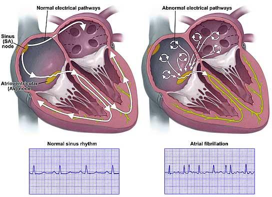
Compendium. Cellular and Molecular Electrophysiology of Atrial Fibrillation Initiation, Maintenance, and Progression
 Compendium Circulation Research Compendium on Atrial Fibrillation: Atrial Fibrillation Compendium: Historical Context and Detailed Translational Perspective on an Important Clinical Problem The Clinical
ANTI - ARRHYTHMIC DRUGS
 ANTI - ARRHYTHMIC DRUGS CARDIAC ACTION POTENTIAL K Out Balance Ca in/k out Na in K Out GENERATION OF ARRHYTHMIAS Four mechanisms of arrhythmia generation; Increased normal automaticity Abnormal automaticity
ELECTROCARDIOGRAPHY (ECG)
") ELECTROCARDIOGRAPHY (ECG) The heart is a muscular organ, which pumps blood through the blood vessels of the circulatory system. Blood provides the body with oxygen and nutrients, as well as assists in
ELECTROCARDIOGRAPHY (ECG) The heart is a muscular organ, which pumps blood through the blood vessels of the circulatory system. Blood provides the body with oxygen and nutrients, as well as assists in
We are IntechOpen, the world s leading publisher of Open Access books Built by scientists, for scientists. International authors and editors
 We are IntechOpen, the world s leading publisher of Open Access books Built by scientists, for scientists 3,350 108,000 1.7 M Open access books available International authors and editors Downloads Our
EXAM II Animal Physiology ZOO 428 Fall 2006
 V Eq EXAM II Animal Physiology ZOO 428 Fall 2006 = RT X o. ln( [ zf [ X ) RT p K[K o pna[na o pcl[cl i V = m ln i F pk[k i pna[na i pcl[cl o I = g(v m V eq. ) Q = C m V m Q Driving Force = V m V eq. 10
University of Groningen. Atrial electrical remodeling from barn to bedside Tieleman, Robert George
 University of Groningen Atrial electrical remodeling from barn to bedside Tieleman, Robert George IMPORTANT NOTE: You are advised to consult the publisher's version (publisher's PDF) if you wish to cite
Antiarrhythmic Drugs 1/31/2018 1
 Antiarrhythmic Drugs 1/31/2018 1 Normal conduction pathway: 1- SA node generates action potential and delivers it to the atria and the AV node 2- The AV node delivers the impulse to purkinje fibers Other
CARDIAC POTASSIUM CHANNEL SUBTYPES: NEW ROLES IN REPOLARIZATION AND ARRHYTHMIA
 CARDIAC POTASSIUM CHANNEL SUBTYPES: NEW ROLES IN REPOLARIZATION AND ARRHYTHMIA Nicole Schmitt, Morten Grunnet, and Søren-Peter Olesen Physiol Rev 94: 609 653, 2014 doi:10.1152/physrev.00022.2013 The Danish
Rhythm and Blues Drugs and QT Prolongation
 Rhythm and Blues Drugs and QT Prolongation Dr Martin Quinn St Vincents University Hospital Irish Medication Safety Network conference Farmleigh 18 Oct 2013 Drugs and QT Prolongation Anti-psychotic, antidepressant,
Electrocardiography I Laboratory
 Introduction The body relies on the heart to circulate blood throughout the body. The heart is responsible for pumping oxygenated blood from the lungs out to the body through the arteries and also circulating
Interpreting Electrocardiograms (ECG) Physiology Name: Per:
 Physiology Name: Per:") Interpreting Electrocardiograms (ECG) Physiology Name: Per: Introduction The heart has its own system in place to create nerve impulses and does not actually require the brain to make it beat. This electrical
Interpreting Electrocardiograms (ECG) Physiology Name: Per: Introduction The heart has its own system in place to create nerve impulses and does not actually require the brain to make it beat. This electrical
Cardiovascular System Notes: Heart Disease & Disorders
 Cardiovascular System Notes: Heart Disease & Disorders Interesting Heart Facts The Electrocardiograph (ECG) was invented in 1902 by Willem Einthoven Dutch Physiologist. This test is still used to evaluate
CRC 431 ECG Basics. Bill Pruitt, MBA, RRT, CPFT, AE-C
 CRC 431 ECG Basics Bill Pruitt, MBA, RRT, CPFT, AE-C Resources White s 5 th ed. Ch 6 Electrocardiography Einthoven s Triangle Chest leads and limb leads Egan s 10 th ed. Ch 17 Interpreting the Electrocardiogram
Mathematical modeling of ischemia and infarction
 Mathematical modeling of ischemia and infarction Mostly based on Cimponeriu, Starmer and Bezerianos: A theoretical analysis of acute ischemia and infarction using ECG reconstruction on a 2-D model of myocardium
Approximately the size of your fist Location. Pericardial physiology
 Heart Anatomy Approximately the size of your fist Location Superior surface of diaphragm Left of the midline Anterior to the vertebral column, posterior to the sternum Wednesday, March 28, 2012 Muscle