Critical Heart Disease in the Newborn. What you need to know
|
|
|
- Margaret Todd
- 5 years ago
- Views:
Transcription
1 Critical Heart Disease in the Newborn What you need to know
2 DISCLOSURES Nothing to report
3 OBJECTIVES DESCRIBE NEONATAL CARDIOVASCULAR PHYSIOLOGY RECOGNIZE NEONATAL CARDIAC EMERGENCIES FORMULATE TREATMENT PLANS
4 DEFINITION Critical congenital heart disease all cardiovascular lesions that would result in neonatal demise unless immediate intervention to palliate or correct the anatomic defect is undertaken Signs and symptoms of severe heart disease in the newborn cyanosis discrepant pulses and blood pressures congestive heart failure cardiogenic shock
5 GENERAL PRINCIPLES OF TREATMENT Early diagnosis is the key High index of suspicion is very important ABCs trump other considerations, take care of the airway and treat shock Start prostaglandins early, don t wait for cardiologist or echocardiogram Volume resuscitate Correct acidosis Inotropic support Keep sats in the range, be gentle with oxygen
6 EPIDEMIOLOGY In the United States congenital heart disease (CHD) occurs in 8/1000 live births the subpopulation with critical CHD is roughly 3.5/1000 live births approximately 32,000 children each year are born with CHD, and 14,000 of them are born with critical heart lesions
7 FETAL CIRCULATION critical heart disease is well tolerated in utero but is uniformly fatal postnatally without intervention the ventricles work together to deliver blood to systemic tissues placenta serves as the organ of oxygen delivery the umbilical vein carries oxygenated blood from the placenta to the inferior vena cava through the ductus venosus a series of central shunts and preferential streaming patterns, directs oxygenated blood to vital organs deoxygenated blood is diverted to organs with lower oxygen consumption and to the placenta
8
9
10 TRANSITIONAL CIRCULATION at birth, with separation of the umbilical cord, the responsibility of oxygenation shifts from the placenta to the lungs in order for this to occur, PVR must fall rapidly physical expansion of the lungs and the replacement of fluid-filled alveoli with gas promotes the dilation and distention of the pulmonary arteries decreasing PVR mechanical distention of the lungs promote local production of prostacyclin, a pulmonary artery vasodilator, further decreasing PVR increased oxygen tension in the pulmonary artery acts as a vasodilator both directly and through its ability to stimulate nitric oxide production
11 TRANSITIONAL CIRCULATION when the placenta is removed from the circulation, blood return to the heart through the inferior vena cava is significantly diminished, causing right atrial pressures to fall the increase in Qp brings about an increase in pulmonary venous return and subsequent elevation in left atrial pressures, the flap over the foramen ovale closes there is a dramatic reduction in the production of prostaglandin E2 (by the placenta) and an increase in its metabolism (by the lungs) in combination with increased oxygen content in the blood, provides the stimulus for the PDA to constrict the neonatal circulation transitions to a series configuration, thereby establishing separate systemic and pulmonary circulations
12 NEONATAL CIRCULATION cardiac output (CO) is directly proportional to the heart rate (HR) and stroke volume (SV):CO = SV HR stroke volume is dependent on three determinants (1) preload, or the distention of the ventricle prior to systole (2) afterload, or the resistance to ejection from the ventricle, and (3) myocardial contractility neonatal myocardium is relatively stiff and has fewer contractile myofibrils compared to adults the newborn cannot increase cardiac output by increasing stroke volume, and relies mainly on increases in heart rate neonatal myocytes are deficient in sarcoplasmic reticulum calcium stores, newborn cardiac output is exquisitely sensitive to calcium calcium is an inotrope for the newborn
13 Basic Evaluation of the Newborn for Congenital Heart Disease EXAM: Systolic murmur: valvar stenosis (pulmonic or aortic) (Ejection Systolic) tricuspid or mitral regurgitation (Holosystolic) Diminished pulses and BP: Diminished lower extremity pulse and BP: Coarctation Diminished four-extremity pulse and BP: Left-sided obstructive lesions Tachypnea: High Qp:Qs Diminished left ventricular function
14 Basic Evaluation of the Newborn for Congenital Heart Disease ECG: Check for sinus rhythm Superior axis: consider atrioventricular septal defect or tricuspid atresia
15 EKG AXIS DETERMINATION
16 Basic Evaluation of the Newborn for Congenital Heart Disease CXR Severe cardiomegaly: consider neonatal Ebstein s or cardiomyopathy Look for right aortic arch Look for normal abdominal situs (stomach on left) Hypoxemia with normal lung fields: Consider congenital heart disease Progressive interstitial pattern: Consider obstruction to pulmonary venous return
17 Basic Evaluation of the Newborn for Congenital Heart Disease OXYGEN SATURATIONS: Differential cyanosis: Consider pulmonary hypertension (PPHN), coarctation, or interrupted arch Reverse differential cyanosis: Consider the above with transposition of the great arteries ARTERIAL BLOOD GAS: Hyperoxia test: PaO2 in right radial artery on 100% FiO2 with less than 150 mmhg: Consider intracardiac mixing Hypoxemia that improves markedly with oxygen: Consider lung disease
18 PROBLEM 1 A newborn infant with prenatal diagnosis of hypoplastic left heart syndrome is on PGE1 infusion, D3 of life, not intubated, waiting for Norwood procedure. He is on 2L/min nasal canula oxygen, sats are 90%. He is not being fed, is on maintenance D10 iv fluids. You notice that his BP has been progressively decreasing, his extremities are cold, cap refill is prolonged. What is going on? What should you do now? 1. start TPN 2. intubate 3. start Dopamine 4. discontinue nasal canula O2 5. take him to the OR immediately
19 Qp:Qs Ratio of Pulmonary to Systemic Perfusion Single Ventricle Physiology newborns with critical CHD whose PDA is kept open with PGE1, balancing Qs and Qp is essential particularly important for patients with single-ventricle physiology and complete intracardiac mixing systemic venous return (desaturated blood) and pulmonary venous return (saturated blood) usually completely mix within the heart there is competitive Qp and Qs blood flow and the relative resistances to flow govern the ratio of distribution of flow between the two circuits
20 Qp:Qs and Oxygen oxygen must be used with caution in the neonate with congenital heart disease, particularly those with single-ventricle physiology oxygen is a potent pulmonary vasodilator and will increase pulm circulation (Qp) at the expense of the systemic circulation (Qs) oxygen should be minimized in ductal-dependent congenital heart disease should be given when there is concurrent underlying lung disease may be required in newborns with cyanotic right-sided obstructive lesions and cyanotic transposition of the great arteries In general, arterial oxygen saturations should be maintained between 80% and 85%,which in a neonate with single-ventricle physiology and good cardiac output translates to a balanced circulation with a Qp:Qs ratio of 1:1.
21 Qp:Qs Ratio of Pulmonary to Systemic Perfusion Single Ventricle Physiology management goal of patients with single ventricle physiology provide adequate pulmonary blood flow without compromising systemic oxygen delivery and tissue perfusion Qp:Qs = Aortic Sats Mixed venous Sat / Pulm v sat Pulm a sat Qp:Qs = 25/ 95 Ao sat Ao sat = 90 Qp:Qs = 25/95-90 = 25/5 = 5:1
22 Qp:Qs Ratio of Pulmonary to Systemic Perfusion Single Ventricle Physiology
23 The Cost of a High Systemic Saturation in Single Ventricle Physiology
24 PROBLEM 2 Term newborn in delivery room, called to see for low sats. Difficult resuscitation, sats in the 50s and 60s at 10 mins, even after bagging with 100% O2, poor peripheral pulses, hypotensive, prolonged capillary refill. Intubated and transferred to the NICU. Lines placed, cultures sent, antibiotics given. ABG ph 7.1 PaCO2 29 PaO2 27 HCO3 12, BD -16. Lactate 8. CXR shows bilateral pulm edema. Cardiac lesion suspected, awaiting Echocardiogram. Prostaglandin infusion started, but no improvement. What is going on?
25 Timing of Presentation of Critical CHD Shock in delivery room Symptoms on first day of life Symptoms in the first week of life
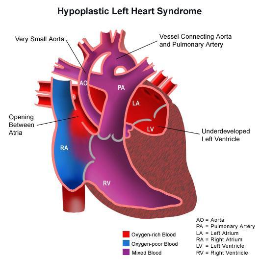
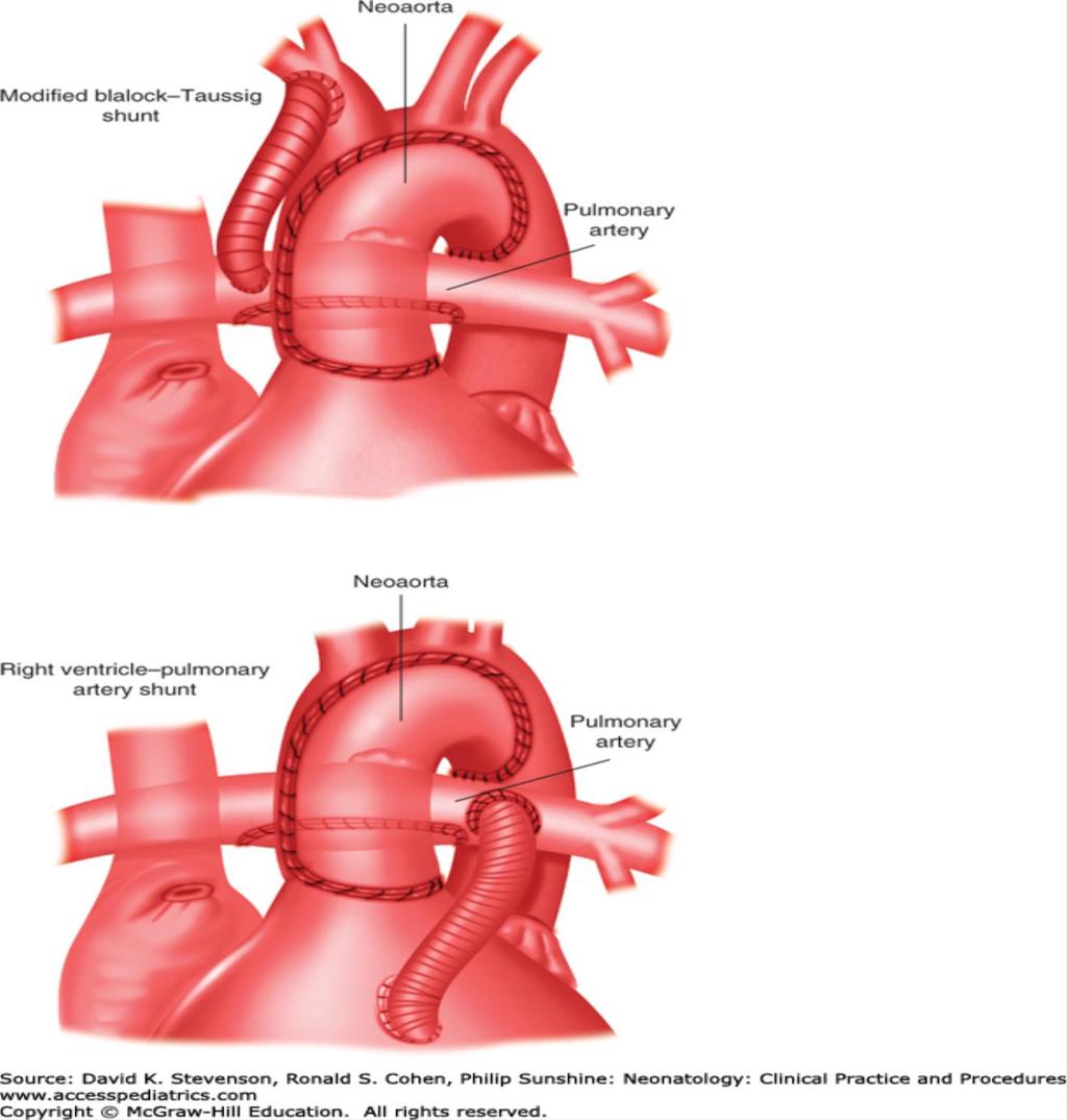
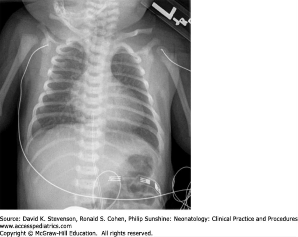
26 Shock in delivery room Cardiac lesions that are unstable in the delivery room represent severe abnormalities of oxygen delivery that are often not stabilized by PGE1 alone and require immediate intervention in order to sustain life Hypoplastic Left Heart Syndrome with Intact Atrial Septum Transposition of the Great Arteries with Restrictive or Intact Atrial Septum
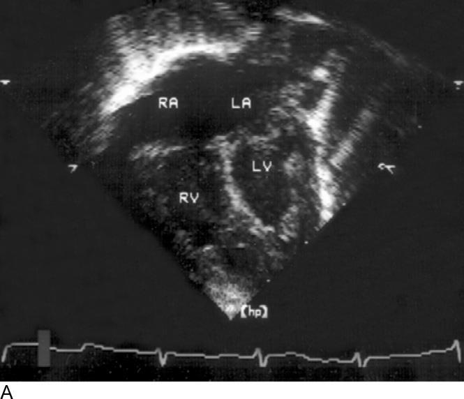
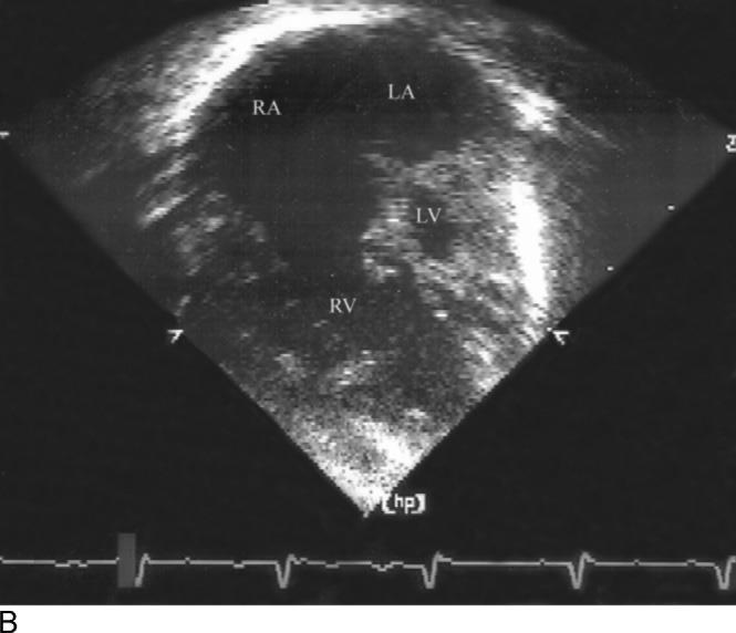
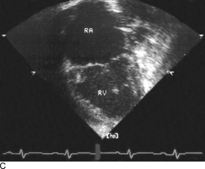
27 Hypoplastic Left Heart Syndrome underdevelopment of the mitral valve, LV, left ventricular outflow tract, aortic valve, and aorta the right ventricle is responsible for maintaining both pulmonary and systemic circulation, systemic perfusion is dependent on the PDA single ventricle physiology majority present within the first week, when the PDA closes, with signs and symptoms of shock If an intact or restrictive atrial septum (IAS) is present, effective egress from the left atrium is not possible presents at birth pulmonary venous obstruction develops and causes pulmonary hypertension
28
29
30 Hypoplastic Left Heart Syndrome with Intact Atrial Septum at delivery, infants with HLHS/IAS present with profound cyanosis, metabolic acidosis, respiratory distress, and cardiovascular collapse patients are critically ill, markedly tachypneic, and often have a PaO2 of less than 20 mmhg prostaglandin should be administered immediately to ensure ductal patency and systemic perfusion emergent transcatheter (balloon and blade atrial septostomy) intervention must be performed decompress the left atrium allow for oxygenated blood to reach the circulation
31
32 Transposition of the Great Arteries aorta arises from the RV and pulm artery arises from the LV systemic and pulmonary circulations are arranged in parallel rather than in series causes recirculated Qp and a deficiency of oxygen supply to the tissues survival with the systemic and pulmonary circulations arranged in parallel is not possible requires some blood to exit the pulmonary circuit in order to enter the systemic circuit to provide oxygen (QES), and for blood to similarly exit the systemic circuit in order to enter the pulmonary circuit to pick up oxygen (QEP) In a completely normal heart, Q P = Q EP, Q S = Q ES, and Q P = Q S

33 TRANSPOSITION OF THE GREAT VESSELS

34 TRANSPOSITION OF THE GREAT VESSELS
35 .
36 Transposition of the Great Arteries with Restrictive or Intact Atrial Septum a newborn baby with transposition of the great arteries, both the PFO and the PDA are open in the first few minutes or hours of life. the two parallel circuits consisting of the pulmonary and systemic circulations are expected to have two possible areas of communication between them when the atrial septum is intact or very restrictive, mixing between the two circulations only can occur at the level of the PDA significantly impaired O2 delivery PaO2 in the 20s presents at delivery with profound cyanosis and metabolic acidosis pre- and postductal saturations may demonstrate reverse differential cyanosis PGE1 should be initiated immediately to maintain ductal patency emergent balloon atrial septostomy must be performed to allow for adequate mixing at the atrial level

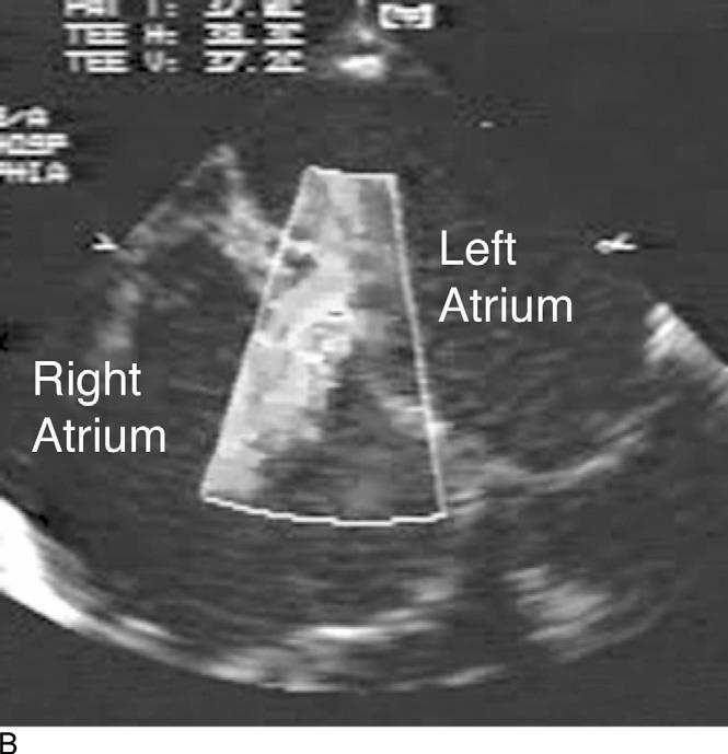
37 Balloon Atrial Septostomy
38 SYMPTOMS ON FIRST DAY OF LIFE Airway compromise Severe Ebstein's anomaly of the tricuspid valve Tetralogy of Fallot with absent pulmonary valve Obstruction to pulmonary venous return Total anomalous pulmonary venous return (TAPVR) with obstruction infracardiac type
39 Severe Ebstein s Anomaly of the Tricuspid Valve the septal and posterior leaflets are deformed and displaced inferiorly into the RV the tricuspid valve is severely incompetent (TR), resulting in profound right atrial enlargement Holosystolic murmur audible newborns often present with respiratory failure due to airway compression from the profound cardiomegaly and cyanosis there may be functional pulmonary atresia, because of severe TR causing right-to-left atrial-level shunt, cyanosis and ductal-dependent pulmonary blood flow
40 EBSTEIN S
41 Tetralogy of Fallot with absent pulmonary valve the four components tetralogy of Fallot (TOF) are a VSD, an overriding aorta, pulmonary stenosis, and right ventricular hypertrophy a variant, TOF with absent pulmonary valve, is associated with markedly dilated central(proximal) pulmonary arteries in utero characteristic to-and-fro murmur at the left upper sternal border, consistent with pulmonary outflow obstruction and regurgitation severe bronchomalacia can result from compression of the bronchi by the large central pulmonary arteries require immediate intubation and ventilation prognosis of these patients may be limited by their ventilatory difficulties

42 TETRALOGY OF FALLOT

43 TETRALOGY OF FALLOT WITH ABSENT PULMONARY VALVE
44 TOTAL ANOMALOUS PULMONARY VENOUS RETURN (TAPVR) in TAPVR there is no connection between the pulmonary veins and the left atrium pulmonary veins form a confluence behind the left atrium which decompresses through a vertical vein inferiorly below the diaphragm empties either into the portal system or into the ductus venosus before ultimately returning to the right atrium complete mixing of systemic and pulmonary venous return occur within the right atrium
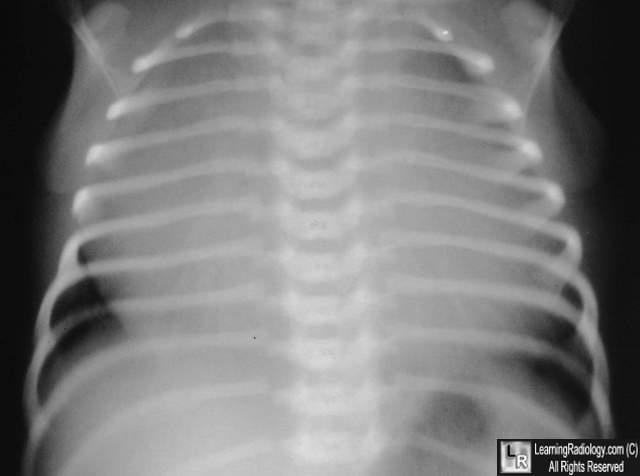
45 Obstruction to Pulmonary Venous Return neonates with TAPVR with obstruction usually present within the first hours to days of life true surgical emergency cyanosis and respiratory distress secondary to pulmonary venous congestion with pulm edema and small heart size on CXR surgical repair should be performed as soon as possible if critically ill and immediate surgery is not an option, may be stabilized with veno arterial extracorporeal membrane oxygenation (VA ECMO) prostaglandin does not help, may worsen the hemodynamic state by further increasing pulmonary blood flow
46 Obstruction to Pulmonary Venous Return
47
48 PROBLEM 3 A 6 day old, male term infant presents to the ER with 2 day history of not eating well, increasing lethargy, and tachypnea. HR 190, Sats are 88%, pulses are difficult to feel, extremities are cool, cap refill is 5 seconds, the ER physician is not sure whether he is feeling his own pulses. Glucose is 40, ph is 7.1, BUN is 20 and Creatinine 1.9. Mother s GBS status is unknown. Had a previous child who died early. He has been given 20 ml/kg fluid bolus, and antibiotics are ordered. Your institution is one of the few who will admit a neonate up to 7 days of age into the NICU. Lucky you! What is going on? 1. Sepsis 2. Metabolic disorder 3. Non accidental trauma 4. Duct dependent circulation 5. acute renal failure from dehydration
49 SYMPTOMS IN THE FIRST WEEK OF LIFE Lesions with Ductal-Dependent Systemic Blood Flow Hypoplastic Left Heart Syndrome Critical Aortic Stenosis Critical Coarctation of the Aorta Lesions with Ductal-Dependent Pulmonary Blood Flow Critical Pulmonary Valve Stenosis Pulmonary Atresia, Including RV-Dependent Coronary Circulation Severe Tetralogy of Fallot Lesions with Large Left-to-Right Shunts Truncus Arteriosus VSD with Arch Obstruction
50 Critical Coarctation of the Aorta newborns with critical coarctation usually present in the first 6 weeks (typically in the first 7 10 days) of life with tachypnea, tachycardia, and severe hypotension if the PDA is partially open preductal saturations will be higher than postductal saturations evidence of upper extremity hypertension and diminished palpable femoral pulses if the PDA has closed no discrepancy between saturations will be noted and femoral pulses often will be absent.
51 Critical Coarctation of the Aorta occurs at the insertion site of the PDA into the descending aorta can be difficult to appreciate in the face of a widely patent PDA with closure of PDA, the entire cardiac output must cross the area of coarctation(narrowing) to enter the descending aorta in the case of critical coarctation, severe obstruction is present and the LV cannot supply adequate flow to the descending aorta congestive heart failure and cardiogenic shock occurs
52 COARCTATION AND BLOOD PRESSURE BP should be measured in all four extremities. A difference of greater than 10 mmhg in upper compared to lower-extremity blood pressure suggests the presence of aortic coarctation, aortic arch hypoplasia, or interrupted aortic arch there are 2 caveats in the event of low cardiac output and systemic hypotension, blood pressure differences are diminished. Coarctation may be present in the absence of significant blood pressure gradient. Hypotension should be corrected, and cardiac output should be maximized prior to interpretation of blood pressure differences if the PDA is widely patent, a blood pressure difference between upper and lower extremities may not be noted, despite underlying coarctation. A complete assessment of the newborn with cyanosis includes preductal and postductal measurements of oxygen saturation
53 Coarctation of the Aorta

54 Coarctation of the Aorta
55 SUMMARY As the newborn separates from the placenta, oxygenation and ventilation are dependent on the infant; PVR falls, and the PFO and PDA begin to close Cardiac output is proportional to heart rate and stroke volume. The infant s stroke volume is fairly fixed, and cardiac output is predominantly heart rate-dependent Newborns are deficient in calcium stores in their sarcoplasmic reticulum. They are very responsive to calcium administration Potential side effects of PGE1 are hypotension, apnea and fever Reverse differential cyanosis is only seen with the physiology of transposition of the great arteries A failed hyperoxia test (PaO 2 <150 mmhg in the right radial artery) is consistent with an intracardiac mixing lesion. Congenital heart lesions with normal hyperoxia tests include coarctation, aortic stenosis, and isolated interrupted aortic arch
56 SUMMARY Many factors impact Qp:Qs. Oxygen, hyperventilation, alkalosis,and inspired nitric oxide increase Qp:Qs. Hypoventilation and inspired carbon dioxide decrease Qp:Qs Cardiac lesions that are unstable in the delivery room represent abnormalities of oxygen delivery that are often not stabilized by PGE1 alone and require immediate intervention (HLHS with intact atrial septum, TGA with intact atrial septum, or TAPVR with severe obstruction) Neonatal Ebstein s anomaly and TOF with absent pulmonary valve syndrome can result in immediate respiratory compromise caused by compression of the airways by the large right atrium or large central pulmonary arteries, respectively
57 SUMMARY Patients with left-sided obstructive lesions (e.g., critical AS, HLHS,or critical coarctation of the aorta) can present in shock in the first few weeks of life with diminished pulses and a profound metabolic acidosis. Coarctation can present in the first several months. Patients with ductal-dependent Qp on PGE 1 are also at risk for pulmonary overcirculation and systemic hypoperfusion. Patients with truncus arteriosus often have low systemic diastolic pressures and are at risk for pulmonary overcirculation. They have an increased preoperative incidence of ventricular fibrillation.
58 THANK YOU FOR LISTENING

59 TOTAL ANOMALOUS PULMONARY VENOUS RETURN - SUPRACARDIAC

60 TOTAL ANOMALOUS PULMONARY VENOUS RETURN - INFRACARDIAC
61 TRANSPOSITION OF THE GREAT VESSELS
62
Cardiovascular Pathophysiology: Right to Left Shunts aka Cyanotic Lesions
 Cardiovascular Pathophysiology: Right to Left Shunts aka Cyanotic Lesions Ismee A. Williams, MD, MS iib6@columbia.edu Pediatric Cardiology Learning Objectives To discuss the hemodynamic significance of
Cardiovascular Pathophysiology: Right to Left Shunts aka Cyanotic Lesions Ismee A. Williams, MD, MS Pediatric Cardiology
 Cardiovascular Pathophysiology: Right to Left Shunts aka Cyanotic Lesions Ismee A. Williams, MD, MS iib6@columbia.edu Pediatric Cardiology Learning Objectives To discuss the hemodynamic significance of
Screening for Critical Congenital Heart Disease
 Screening for Critical Congenital Heart Disease Caroline K. Lee, MD Pediatric Cardiology Disclosures I have no relevant financial relationships or conflicts of interest 1 Most Common Birth Defect Most
Cardiac Emergencies in Infants. Michael Luceri, DO
 Cardiac Emergencies in Infants Michael Luceri, DO October 7, 2017 I have no financial obligations or conflicts of interest to disclose. Objectives Understand the scope of congenital heart disease Recognize
Congenital Heart Defects
 Normal Heart Congenital Heart Defects 1. Patent Ductus Arteriosus The ductus arteriosus connects the main pulmonary artery to the aorta. In utero, it allows the blood leaving the right ventricle to bypass
Anatomy & Physiology
 1 Anatomy & Physiology Heart is divided into four chambers, two atrias & two ventricles. Atrioventricular valves (tricuspid & mitral) separate the atria from ventricles. they open & close to control flow
Congenital Heart Disease: Physiology and Common Defects
 Congenital Heart Disease: Physiology and Common Defects Jamie S. Sutherell, M.D, M.Ed. Associate Professor, Pediatrics Division of Cardiology Director, Medical Student Education in Pediatrics Director,
How to Recognize a Suspected Cardiac Defect in the Neonate
 Neonatal Nursing Education Brief: How to Recognize a Suspected Cardiac Defect in the Neonate https://www.seattlechildrens.org/healthcareprofessionals/education/continuing-medical-nursing-education/neonatalnursing-education-briefs/
When is Risky to Apply Oxygen for Congenital Heart Disease 부천세종병원 소아청소년과최은영
 When is Risky to Apply Oxygen for Congenital Heart Disease 부천세종병원 소아청소년과최은영 The Korean Society of Cardiology COI Disclosure Eun-Young Choi The author have no financial conflicts of interest to disclose
Objectives Part 1. Objectives Part 2. Fetal Circulation Transition to Postnatal Circulation Normal Cardiac Anatomy Ductal Dependence and use of PGE1
 Cardiac Physiology Gia Marzano, AC PNP Pediatric Cardiac Surgery Rush Center for Congenital Heart Disease Rush University Medical Center Objectives Part 1 Fetal Circulation Transition to Postnatal Circulation
Congenital Heart Disease
 Congenital Heart Disease Mohammed Alghamdi, MD, FRCPC, FAAP, FACC Associate Professor and Consultant Pediatric Cardiology, Cardiac Science King Fahad Cardiac Centre King Saud University INTRODUCTION CHD
3/14/2011 MANAGEMENT OF NEWBORNS CARDIAC INTENSIVE CARE CONFERENCE FOR HEALTH PROFESSIONALS IRVINE, CA. MARCH 7, 2011 WITH HEART DEFECTS
 CONFERENCE FOR HEALTH PROFESSIONALS IRVINE, CA. MARCH 7, 2011 MANAGEMENT OF NEWBORNS WITH HEART DEFECTS A NTHONY C. CHANG, MD, MBA, MPH M E D I C AL D I RE C T OR, HEART I N S T I T U T E C H I LDRE N
Foetal Cardiology: How to predict perinatal problems. Prof. I.Witters Prof.M.Gewillig UZ Leuven
 Foetal Cardiology: How to predict perinatal problems Prof. I.Witters Prof.M.Gewillig UZ Leuven Cardiopathies Incidence : 8-12 / 1000 births ( 1% ) Most frequent - Ventricle Septum Defect 20% - Atrium Septum
Heart and Lungs. LUNG Coronal section demonstrates relationship of pulmonary parenchyma to heart and chest wall.
 Heart and Lungs Normal Sonographic Anatomy THORAX Axial and coronal sections demonstrate integrity of thorax, fetal breathing movements, and overall size and shape. LUNG Coronal section demonstrates relationship
Introduction. Pediatric Cardiology. General Appearance. Tools of Assessment. Auscultation. Vital Signs
 Introduction Pediatric Cardiology An introduction to the pediatric patient with heart disease: M-III Lecture Douglas R. Allen, M.D. Assistant Professor and Director of Community Pediatric Cardiology at
The Blue Baby. Network Stabilisation of the Term Infant Study Day 15 th March 2017 Joanna Behrsin
 The Blue Baby Network Stabilisation of the Term Infant Study Day 15 th March 2017 Joanna Behrsin Session Structure Definitions and assessment of cyanosis Causes of blue baby Structured approach to assessing
Duct Dependant Congenital Heart Disease
 Children s Acute Transport Service Clinical Guidelines Duct Dependant Congenital Heart Disease Document Control Information Author CATS/NTS Author Position CC Transport Services Document Owner E. Polke
ECHOCARDIOGRAPHIC APPROACH TO CONGENITAL HEART DISEASE: THE UNOPERATED ADULT
 ECHOCARDIOGRAPHIC APPROACH TO CONGENITAL HEART DISEASE: THE UNOPERATED ADULT Karen Stout, MD, FACC Divisions of Cardiology University of Washington Medical Center Seattle Children s Hospital NO DISCLOSURES
Duct Dependant Congenital Heart Disease
 Children s Acute Transport Service Clinical Guidelines Duct Dependant Congenital Heart Disease This guideline has been agreed by both NTS & CATS Document Control Information Author CATS/NTS Author Position
Neonatal Cardiac Anomalies
 Objectives Neonatal Cardiac Anomalies Karen Knuth, RNC, MN, NNP-BC, ARNP Seattle Childrens Hospital What is CHD? Normal anatomy and circulation Clinical presentation: signs and symptoms Diagnostics Common
The Physiology of the Fetal Cardiovascular System
 The Physiology of the Fetal Cardiovascular System Jeff Vergales, MD, MS Department of Pediatrics Division of Pediatric Cardiology jvergales@virginia.edu Disclosures I serve as the medical director for
Pediatric Board Review Congenital Heart Disease. Steven H. Todman, M.D. Pediatric Cardiologist Louisiana State University
 Pediatric Board Review Congenital Heart Disease Steven H. Todman, M.D. Pediatric Cardiologist Louisiana State University Our Mission To discuss various types of congenital heart disease that are commonly
CONGENITAL HEART DISEASE (CHD)
") CONGENITAL HEART DISEASE (CHD) DEFINITION It is the result of a structural or functional abnormality of the cardiovascular system at birth GENERAL FEATURES OF CHD Structural defects due to specific disturbance
CONGENITAL HEART DISEASE (CHD) DEFINITION It is the result of a structural or functional abnormality of the cardiovascular system at birth GENERAL FEATURES OF CHD Structural defects due to specific disturbance
DORV: The Great Chameleon. Heart Conference October 15, 2016 Tina Kwan, MD
 DORV: The Great Chameleon Heart Conference October 15, 2016 Tina Kwan, MD Kenneth Maehara, Ph.D. May 7, 1942 - August 26, 2013 A.R. A classic case of broken heart 38 week AGA F born at an OSH to
Patent ductus arteriosus PDA
 Patent ductus arteriosus PDA Is connecting between the aortic end just distal to the origin of the LT sub clavian artery& the pulmonary artery at its bifurcation. Female/male ratio is 2:1 and it is more
Congenital Heart Disease: Cyanotic Lesions. Amitesh Aggarwal
 Congenital Heart Disease: Cyanotic Lesions Amitesh Aggarwal 12 y/o male admitted because of dyspnea and cyanosis Patient has been cyanotic since few months after birth Has episodes of tachypnea and worsening
Congenital heart disease: When to act and what to do?
 Leading Article Congenital heart disease: When to act and what to do? Duminda Samarasinghe 1 Sri Lanka Journal of Child Health, 2010; 39: 39-43 (Key words: Congenital heart disease) Congenital heart disease
By Dickens ATURWANAHO & ORIBA DAN LANGOYA MAKchs, MBchB CONGENTAL HEART DISEASE
 By Dickens ATURWANAHO & ORIBA DAN LANGOYA MAKchs, MBchB CONGENTAL HEART DISEASE Introduction CHDs are abnormalities of the heart or great vessels that are present at birth. Common type of heart disease
Approach to a baby with cyanosis
 Approach to a baby with cyanosis Objectives Cyanosis : types Differentials: cardiac vs. non cardiac Approach Case scenarios Cyanosis Greek word kuaneos meaning dark blue Bluish discolouration of skin,
The blue baby. Case 4
 Case 4 The blue baby Mrs Smith has brought her baby to A&E because she says he has started turning blue. What are your immediate differential diagnoses? 1 Respiratory causes: Congenital respiratory disorder.
Paediatric Cardiology. Acyanotic CHD. Prof F F Takawira
 Paediatric Cardiology Acyanotic CHD Prof F F Takawira Aetiology Chromosomal Down syndrome, T13, T18 Genetic syndromes (gene defects) Velo-Cardio-facial (22 del) Genetic syndromes (undefined aetiology)
Slide 1. Slide 2. Slide 3 CONGENITAL HEART DISEASE. Papworth Hospital NHS Trust INTRODUCTION. Jakub Kadlec/Catherine Sudarshan INTRODUCTION
 Slide 1 CONGENITAL HEART DISEASE Jakub Kadlec/Catherine Sudarshan NHS Trust Slide 2 INTRODUCTION Most common congenital illness in the newborn Affects about 4 9 / 1000 full-term live births in the UK 1.5
Congenital heart disease. By Dr Saima Ali Professor of pediatrics
 Congenital heart disease By Dr Saima Ali Professor of pediatrics What is the most striking clinical finding in this child? Learning objectives By the end of this lecture, final year student should be able
Cardiac Catheterization Cases Primary Cardiac Diagnoses Facility 12 month period from to PRIMARY DIAGNOSES (one per patient)
") PRIMARY DIAGNOSES (one per patient) Septal Defects ASD (Atrial Septal Defect) PFO (Patent Foramen Ovale) ASD, Secundum ASD, Sinus venosus ASD, Coronary sinus ASD, Common atrium (single atrium) VSD (Ventricular
PRIMARY DIAGNOSES (one per patient) Septal Defects ASD (Atrial Septal Defect) PFO (Patent Foramen Ovale) ASD, Secundum ASD, Sinus venosus ASD, Coronary sinus ASD, Common atrium (single atrium) VSD (Ventricular
Pediatric Echocardiography Examination Content Outline
 Pediatric Echocardiography Examination Content Outline (Outline Summary) # Domain Subdomain Percentage 1 Anatomy and Physiology Normal Anatomy and Physiology 10% 2 Abnormal Pathology and Pathophysiology
Notes by Sandra Dankwa 2009 HF- Heart Failure DS- Down Syndrome IE- Infective Endocarditis ET- Exercise Tolerance. Small VSD Symptoms -asymptomatic
 Congenital Heart Disease: Notes. Condition Pathology PC Ix Rx Ventricular septal defect (VSD) L R shuntsdefect anywhere in the ventricle, usually perimembranous (next to the tricuspid valve) 30% 1)small
NCC Review Cardiac 8/22/12. Intrauterine Blood Flow. Topics
 NCC Review Cardiac Tracey Buckley MSN,RNC, NNP-BC Cape Fear Valley Health System Topics Transition to Extrauterine Life Cyanosis Congenital Heart Disease (CHD) Clinical Manifestations of CHD Therapeutic
Neonatal/Pediatric Cardiopulmonary Care. Persistent Pulmonary Hypertension of the Neonate (PPHN) PPHN. Other. Other Diseases
 PPHN. Other. Other Diseases") Neonatal/Pediatric Cardiopulmonary Care Other Diseases Persistent Pulmonary Hypertension of the Neonate (PPHN) PPHN 3 Also known as Persistent Fetal Circulation (PFC) Seen most frequently in term, post-term
Neonatal/Pediatric Cardiopulmonary Care Other Diseases Persistent Pulmonary Hypertension of the Neonate (PPHN) PPHN 3 Also known as Persistent Fetal Circulation (PFC) Seen most frequently in term, post-term
Paediatrics Revision Session Cardiology. Emma Walker 7 th May 2016
 Paediatrics Revision Session Cardiology Emma Walker 7 th May 2016 Cardiovascular Examination! General:! Make it fun!! Change how you act depending on their age! Introduction! Introduce yourself & check
Absent Pulmonary Valve Syndrome
 Absent Pulmonary Valve Syndrome Fact sheet on Absent Pulmonary Valve Syndrome In this condition, which has some similarities to Fallot's Tetralogy, there is a VSD with narrowing at the pulmonary valve.
Neonatal Single Ventricle Heart Disease Recognition, Management, Counseling
 Neonatal Single Ventricle Heart Disease Recognition, Management, Counseling Christopher J. Petit MD Assistant Professor, Pediatric Cardiology Director, Single Ventricle Program Baylor College of Medicine,
Heart and Soul Evaluation of the Fetal Heart
 Heart and Soul Evaluation of the Fetal Heart Ivana M. Vettraino, M.D., M.B.A. Clinical Associate Professor, Michigan State University College of Human Medicine Objectives Review the embryology of the formation
SURGICAL TREATMENT AND OUTCOME OF CONGENITAL HEART DISEASE
 SURGICAL TREATMENT AND OUTCOME OF CONGENITAL HEART DISEASE Mr. W. Brawn Birmingham Children s Hospital. Aims of surgery The aim of surgery in congenital heart disease is to correct or palliate the heart
5.8 Congenital Heart Disease
 5.8 Congenital Heart Disease Congenital heart diseases (CHD) refer to structural or functional heart diseases, which are present at birth. Some of these lesions may be discovered later. prevalence of Chd
Pathophysiology: Left To Right Shunts
 Pathophysiology: Left To Right Shunts Daphne T. Hsu, MD dh17@columbia.edu Learning Objectives Learn the relationships between pressure, blood flow, and resistance Review the transition from fetal to mature
The fetal circulation
 Peter John Murphy MB ChB DA FRCA The fetal circulation (Fig. 1) is markedly different from the adult circulation. In the fetus, gas exchange does not occur in the lungs but in the placenta. The placenta
Data Collected: June 17, Reported: June 30, Survey Dates 05/24/ /07/2010
 Job Task Analysis for ARDMS Pediatric Echocardiography Data Collected: June 17, 2010 Reported: Analysis Summary For: Pediatric Echocardiography Exam Survey Dates 05/24/2010-06/07/2010 Invited Respondents
Echocardiography in Adult Congenital Heart Disease
 Echocardiography in Adult Congenital Heart Disease Michael Vogel Kinderherz-Praxis München CHD missed in childhood Subsequent lesions after repaired CHD Follow-up of cyanotic heart disease CHD missed in
Adult Echocardiography Examination Content Outline
 Adult Echocardiography Examination Content Outline (Outline Summary) # Domain Subdomain Percentage 1 2 3 4 5 Anatomy and Physiology Pathology Clinical Care and Safety Measurement Techniques, Maneuvers,
Total Anomalous Pulmonary Venous Connections: Anatomy and Diagnostic Imaging
 Total Anomalous Pulmonary Venous Connections: Anatomy and Diagnostic Imaging Timothy Slesnick, MD March 12, 2015 Congenital Cardiac Anesthesia Society Annual Meeting Disclosures I will discuss the use
Pathophysiology: Left To Right Shunts
 Pathophysiology: Left To Right Shunts Daphne T. Hsu, MD dh17@columbia.edu Learning Objectives Learn the relationships between pressure, blood flow, and resistance Review the transition from fetal to mature
Upon completion of this presentation, the participant will be able to:
 B12 Neonatal Cardiology Review Nicole Bowie, NNP-/BC, PNP Neonatal Nurse Practitioner Jackson Memorial Hospital, Miami, FL The speaker has signed a disclosure form and indicated she has no significant
Introduction to Fetal Medicine. Lloyd R. Feit M.D. Associate Professor of Pediatrics Warren Alpert Medical School Brown University
 Associate Professor of Pediatrics Warren Alpert Medical School Brown University Fetal Cardiology Important in evaluation of high risk pregnancies. Information obtainable in > 95% of patients attempted.
Congenital Heart Disease. Mohamed Waheed Elsharief.
 Congenital Heart Disease Mohamed Waheed Elsharief. Objectives l By the end of this lecture you should be able to Fetal Circulation l For the fetus the placenta is the oxygenator so the lungs do little
PATENT DUCTUS ARTERIOSUS (PDA)
") PATENT DUCTUS ARTERIOSUS (PDA) It is a channel that connect the pulmonary artery with the descending aorta (isthumus part). It results from the persistence of patency of the fetal ductus arteriosus after
PATENT DUCTUS ARTERIOSUS (PDA) It is a channel that connect the pulmonary artery with the descending aorta (isthumus part). It results from the persistence of patency of the fetal ductus arteriosus after
Transposition of the Great Arteries Preoperative Diagnostic Considerations. John Simpson Evelina Children s Hospital London, UK
 Transposition of the Great Arteries Preoperative Diagnostic Considerations John Simpson Evelina Children s Hospital London, UK Areas to be covered Definitions Scope of occurrence of transposition of the
Karen Corlett, RN, MSN, CPNP-AC/PC Pediatric Nurse Practitioner Congenital Heart Surgery Unit Pediatric Cardiac Intensivists of North Texas Medical
 Karen Corlett, RN, MSN, CPNP-AC/PC Pediatric Nurse Practitioner Congenital Heart Surgery Unit Pediatric Cardiac Intensivists of North Texas Medical City Children s Hospital, Dallas Hypoxia Shortage of
FANNP 28TH NATIONAL NNP SYMPOSIUM: CLINICAL UPDATE AND REVIEW OCTOBER 17-21, 2017
 Neonatal Cardiology Review Nicole Bowie, NNP/BC, PNP Neonatal Nurse Practitioner Jackson Memorial Hospital, Miami, FL B12 The speaker has signed a disclosure statement indicating that she has no significant
Coarctation of the aorta
 T H E P E D I A T R I C C A R D I A C S U R G E R Y I N Q U E S T R E P O R T Coarctation of the aorta In the normal heart, blood flows to the body through the aorta, which connects to the left ventricle
Ummeenatrbilaoiasetptiwmsaiiri
 atrial This This atrial CIRCULATORY CHANGES My My pressure In the foetus the left atrial is low as relatively Ummeenatrbilaoiasetptiwmsaiiri ze@fgffmftheyubsidtritupyeiirieminfyifjjtajefjjieiminylntentiiiarmmnitnteimiiiinc1udingfromthepl9centaj
Uptofate Study Summary
 CONGENITAL HEART DISEASE Uptofate Study Summary Acyanotic Atrial septal defect Ventricular septal defect Patent foramen ovale Patent ductus arteriosus Aortic coartation Pulmonary stenosis Cyanotic Tetralogy
Objective 2/9/2012. Blood Gas Analysis In The Univentricular Patient: The Need For A Different Perspective. VENOARTERIAL CO2 GRADIENT
 Blood Gas Analysis In The Univentricular Patient: The Need For A Different Perspective. Gary Grist RN CCP Chief Perfusionist The Children s Mercy Hospitals and Clinics Kansas City, Mo. Objective The participant
ULTRASOUND OF THE FETAL HEART
 ULTRASOUND OF THE FETAL HEART Cameron A. Manbeian, MD Disclosure Statement Today s faculty: Cameron Manbeian, MD does not have any relevant financial relationships with commercial interests or affiliations
Notes: 1)Membranous part contribute in the formation of small portion in the septal cusp.
Membranous part contribute in the formation of small portion in the septal cusp.") Embryology 9 : Slide 16 : There is a sulcus between primitive ventricular and bulbis cordis that will disappear gradually and lead to the formation of one chamber which is called bulboventricular chamber.
Embryology 9 : Slide 16 : There is a sulcus between primitive ventricular and bulbis cordis that will disappear gradually and lead to the formation of one chamber which is called bulboventricular chamber.
Adult Congenital Heart Disease: What All Echocardiographers Should Know Sharon L. Roble, MD, FACC Echo Hawaii 2016
 1 Adult Congenital Heart Disease: What All Echocardiographers Should Know Sharon L. Roble, MD, FACC Echo Hawaii 2016 DISCLOSURES I have no disclosures relevant to today s talk 2 Why should all echocardiographers
Cases in Adult Congenital Heart Disease
 Cases in Adult Congenital Heart Disease Sabrina Phillips, MD FACC FASE Associate Professor of Medicine The University of Oklahoma Health Sciences Center No Disclosures I Have Palpitations 18 Year old Man
9/8/2009 < 1 1,2 3,4 5,6 7,8 9,10 11,12 13,14 15,16 17,18 > 18. Tetralogy of Fallot. Complex Congenital Heart Disease.
 Current Indications for Pediatric CTA S Bruce Greenberg Professor of Radiology Arkansas Children s Hospital University of Arkansas for Medical Sciences greenbergsbruce@uams.edu 45 40 35 30 25 20 15 10
Born Blue. Anesthesia and CHD. Kristine Faust, CRNA, MS, MBA, DNAP
 Born Blue Anesthesia and CHD Kristine Faust, CRNA, MS, MBA, DNAP Disclosures Disclosures None to Report Objectives Review all congenital defects in which the patient is blue Describe physiology of the
Index. Note: Page numbers of article titles are in boldface type.
 Index Note: Page numbers of article titles are in boldface type. A Acute coronary syndrome(s), anticoagulant therapy in, 706, 707 antiplatelet therapy in, 702 ß-blockers in, 703 cardiac biomarkers in,
CongHeartDis.doc. Андрій Миколайович Лобода
 CongHeartDis.doc Андрій Миколайович Лобода 2015 Зміст 3 Зміст Зміст 4 A child with tetralogy of Fallot is most likely to exhibit: -Increased pulmonary blood flow -Increased pressure in the right ventricle
Congenital Heart Disease An Approach for Simple and Complex Anomalies
 Congenital Heart Disease An Approach for Simple and Complex Anomalies Michael D. Pettersen, MD Director, Echocardiography Rocky Mountain Hospital for Children Denver, CO None Disclosures 1 ASCeXAM Contains
MEDICAL MANAGEMENT WITH CAVEATS 1. In one study of 50 CHARGE patients with CHD, 75% required surgery. 2. Children with CHARGE may be resistant to chlo
 CARDIOLOGY IN CHARGE SYNDROME: FOR THE PHYSICIAN Angela E. Lin, M.D. Teratology Program/Active Malformation Surveillance, Brigham and Women's Hospital, Old PBBH-B501, 75 Francis St., Boston, MA 02115 alin@partners.org
1st Annual Clinical Simulation Conference
 1st Annual Clinical Simulation Conference Newborns with Acute Respiratory Distress: Diagnosis and Management Ma Teresa C. Ambat, MD Assistant Professor Division of Neonatology, Department of Pediatrics
The Chest X-ray for Cardiologists
 Mayo Clinic & British Cardiovascular Society at the Royal College of Physicians, London : 21-23-October 2013 Cases-Controversies-Updates 2013 The Chest X-ray for Cardiologists Michael Rubens Royal Brompton
Chapter 2 Cardiac Interpretation of Pediatric Chest X-Ray
 Chapter 2 Cardiac Interpretation of Pediatric Chest X-Ray Ra-id Abdulla and Douglas M. Luxenberg Key Facts The cardiac silhouette occupies 50 55% of the chest width on an anterior posterior chest X-ray
Surgical Management Of TAPVR. Daniel A. Velez, M.D. Congenital Cardiac Surgeon Phoenix Children s Hospital
 Surgical Management Of TAPVR Daniel A. Velez, M.D. Congenital Cardiac Surgeon Phoenix Children s Hospital No Disclosures Goals Review the embryology and anatomy Review Surgical Strategies for repair Discuss
Congenital Heart Disease. CCCHD In WI. Critical Congenital Heart Disease. Why Screen? 4/20/2018. Early Detection = Better Outcomes
 Congenital Heart Disease A Positive Screen? What Does it Mean? A Review of Pulse Oximetry Screening for Critical Congenital Heart Disease Elizabeth Goetz MD MPH 8-10/1000 livebirths 3% of all infant mortality
DEVELOPMENT OF THE CIRCULATORY SYSTEM L E C T U R E 5
 DEVELOPMENT OF THE CIRCULATORY SYSTEM L E C T U R E 5 REVIEW OF CARDIAC ANATOMY Heart 4 chambers Base and apex Valves Pericardial sac 3 layers: epi, myo, endo cardium Major blood vessels Aorta and its
The sinus venosus represent the venous end of the heart It receives 3 veins: 1- Common cardinal vein body wall 2- Umbilical vein from placenta 3-
 1 2 The sinus venosus represent the venous end of the heart It receives 3 veins: 1- Common cardinal vein body wall 2- Umbilical vein from placenta 3- Vitelline vein from yolk sac 3 However!!!!! The left
Index. cardiology.theclinics.com. Note: Page numbers of article titles are in boldface type.
 Index Note: Page numbers of article titles are in boldface type. A ACHD. See Adult congenital heart disease (ACHD) Adult congenital heart disease (ACHD), 503 512 across life span prevalence of, 504 506
PAEDIATRIC EMQs. Andrew A Mallick Paediatrics.info.
 PAEDIATRIC EMQs Andrew A Mallick Paediatrics.info www.paediatrics.info Paediatric EMQs Paediatrics.info First published in the United Kingdom in 2012. While the advice and information in this book is believed
Adults with Congenital Heart Disease
 Adults with Congenital Heart Disease Edward K. Rhee, MD, FACC Director, Pediatric-Adult Congenital Arrhythmia Service SJHMC Disclosures & Disclaimer I have no lucrative financial relationships with industry
Perioperative Management of TAPVC
 Perioperative Management of TAPVC Professor Andrew Wolf Rush University Medical Center,Chicago USA Bristol Royal Children s Hospital UK I have no financial disclosures relevant to this presentation TAPVC
CMR for Congenital Heart Disease
 CMR for Congenital Heart Disease * Second-line tool after TTE * Strengths of CMR : tissue characterisation, comprehensive access and coverage, relatively accurate measurements of biventricular function/
"Lecture Index. 1) Heart Progenitors. 2) Cardiac Tube Formation. 3) Valvulogenesis and Chamber Formation. 4) Epicardium Development.
 Heart Progenitors. 2) Cardiac Tube Formation. 3) Valvulogenesis and Chamber Formation. 4) Epicardium Development.") "Lecture Index 1) Heart Progenitors. 2) Cardiac Tube Formation. 3) Valvulogenesis and Chamber Formation. 4) Epicardium Development. 5) Septation and Maturation. 6) Changes in Blood Flow during Development.
"Lecture Index 1) Heart Progenitors. 2) Cardiac Tube Formation. 3) Valvulogenesis and Chamber Formation. 4) Epicardium Development. 5) Septation and Maturation. 6) Changes in Blood Flow during Development.
HISTORY. Question: What category of heart disease is suggested by this history? CHIEF COMPLAINT: Heart murmur present since early infancy.
 HISTORY 18-year-old man. CHIEF COMPLAINT: Heart murmur present since early infancy. PRESENT ILLNESS: Although normal at birth, a heart murmur was heard at the six week check-up and has persisted since
Disclosures. Practical Aspects of Pediatric Cardiology for the General Practitioner. Objectives. Outlines. CCHD Neonatal Screening
 Practical Aspects of Pediatric Cardiology for the General Practitioner Majd Makhoul, MD 48 th Annual Family Medicine Review and Contemporary Pediatrics Conference November 11, 2016 Lexington, KY None Disclosures
Hypoplastic Left Heart Syndrome: Echocardiographic Assessment
 Hypoplastic Left Heart Syndrome: Echocardiographic Assessment Craig E Fleishman, MD, FACC, FASE Director, Non-invasive Cardiac Imaging The Hear Center at Arnold Palmer Hospital for Children, Orlando SCAI
Cardiovascular Nursing Practice: A Comprehensive Resource Manual and Study Guide for Clinical Nurses 2 nd Edition
 Cardiovascular Nursing Practice: A Comprehensive Resource Manual and Study Guide for Clinical Nurses 2 nd Edition Table of Contents Volume 1 Chapter 1: Cardiovascular Anatomy and Physiology Basic Cardiac
Stabilization and Transportation guidelines for Neonates and infants with Heart disease:
 Stabilization and Transportation guidelines for Neonates and infants with Heart disease: Background: Referral Pediatric Cardiac Units, frequently receive neonates and infants referred and transported from
Cardiology Competency Based Goals and Objectives
 Cardiology Competency Based Goals and Objectives COMPETENCY 1. Patient Care. Provide family centered patient care that is developmentally and age appropriate, compassionate, and effective for the treatment
B11. Cardiology Review. Session Summary. Session Objectives. Test Questions
 B11 Cardiology Review Lyn Vargo, PhD, NNP-BC Clinical Assistant Professor Stony Brook University NNP Program University of Missouri, Kansas City The speaker has signed a disclosure form and indicated she
Anomalous Systemic Venous Connection Systemic venous anomaly
 World Database for Pediatric and Congenital Heart Surgery Appendix B: Diagnosis (International Paediatric and Congenital Cardiac Codes (IPCCC) and definitions) Anomalous Systemic Venous Connection Systemic
Large Arteries of Heart
 Cardiovascular System (Part A-2) Module 5 -Chapter 8 Overview Arteries Capillaries Veins Heart Anatomy Conduction System Blood pressure Fetal circulation Susie Turner, M.D. 1/5/13 Large Arteries of Heart
Children with Single Ventricle Physiology: The Possibilities
 Children with Single Ventricle Physiology: The Possibilities William I. Douglas, M.D. Pediatric Cardiovascular Surgery Children s Memorial Hermann Hospital The University of Texas Health Science Center
Fetal Tetralogy of Fallot
 36 Fetal Tetralogy of Fallot E.D. Bespalova, R.M. Gasanova, O.A.Pitirimova National Scientific and Practical Center of Cardiovascular Surgery, Moscow Elena D. Bespalova, MD Professor, Director Rena M,
The approach to the infant with a cardiac emergency
 Abstract: The approach to the infant with a cardiac emergency begins with identification of the unstable or critically ill child and proceeds rapidly into stabilization and provision of immediate therapies.
Leitlinien. Hypoplastisches Linksherzsyndrom. Hypoplastic left heart syndrome (HLHS)
") 1.Title Hypoplastic left heart syndrome (HLHS) N.A. Haas, Bad Oeynhausen Ch. Jux, Giessen J. Photiadis, Berlin H.-H. Kramer, Kiel Typical forms: Mitral atresia/aortic atresia (MA/AoA) Mitral stenosis/aortic
1.Title Hypoplastic left heart syndrome (HLHS) N.A. Haas, Bad Oeynhausen Ch. Jux, Giessen J. Photiadis, Berlin H.-H. Kramer, Kiel Typical forms: Mitral atresia/aortic atresia (MA/AoA) Mitral stenosis/aortic
Surgical Procedures. Direct suture of small ASDs Patch repair Transcatheter closure with a prosthetic device called occluder
 PEDIATRIC Review Surgical Procedures Atrial Septal Defect repair: Direct suture of small ASDs Patch repair Transcatheter closure with a prosthetic device called occluder Balloon atrial septostomy (Rashkind)
Hybrid Stage I Palliation / Bilateral PAB
 Hybrid Stage I Palliation / Bilateral PAB Jeong-Jun Park Dept. of Thoracic & Cardiovascular Surgery Asan Medical Center, University of Ulsan CASE 1 week old neonate with HLHS GA 38 weeks Birth weight 3.0Kg