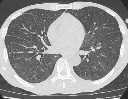
Case 1: Question. 1.1 What is the main pattern of this HRCT? 1. Intralobular line 2. Groundglass opacity 3. Perilymphatic nodule
|
|
|
- Alexandra Pierce
- 5 years ago
- Views:
Transcription
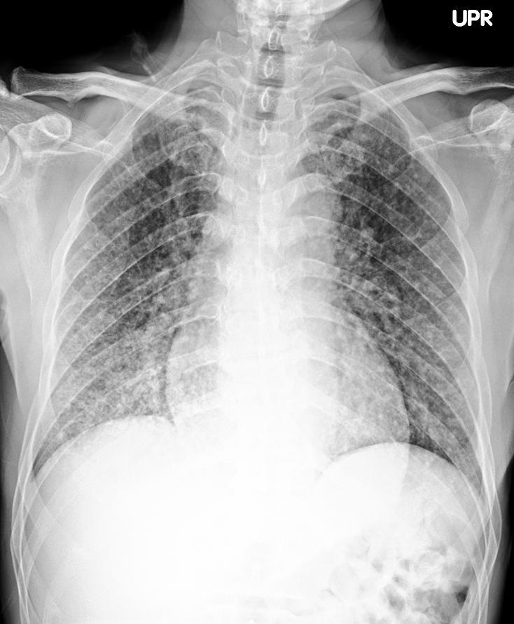
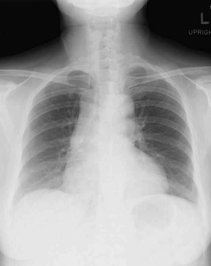
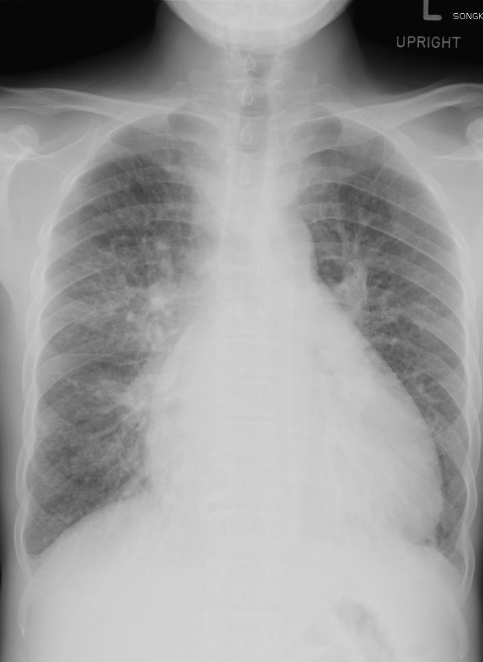
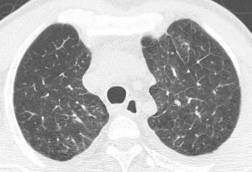
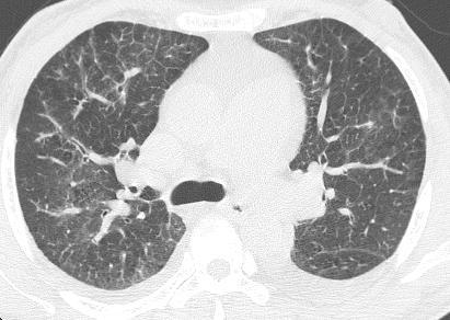
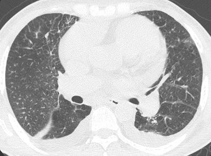
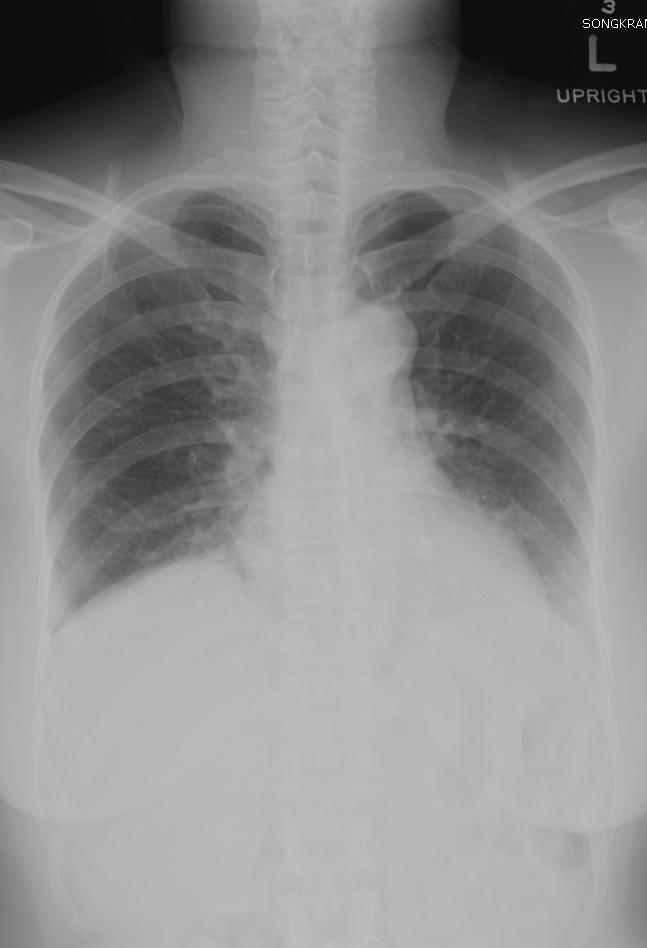
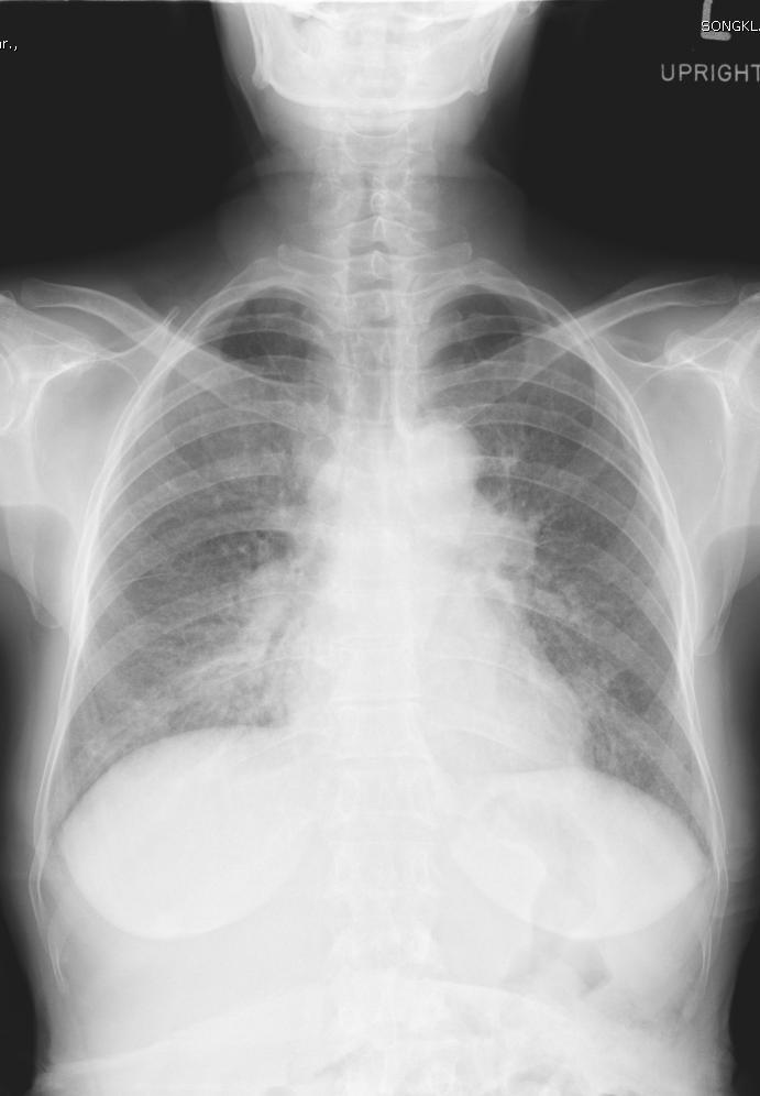
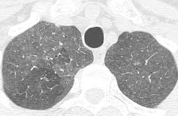
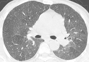
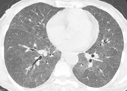
1 HRCT WORK SHOP
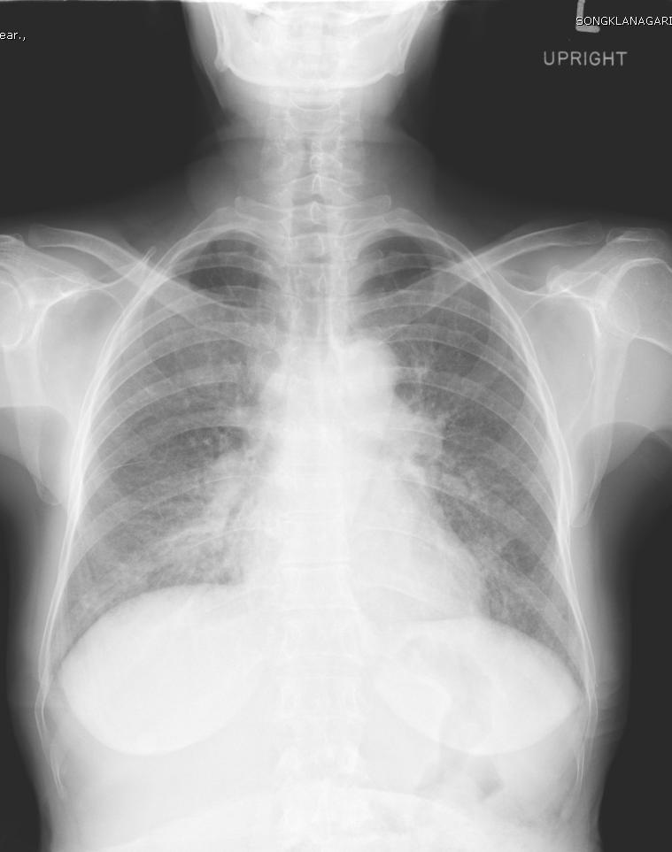
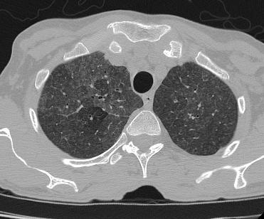
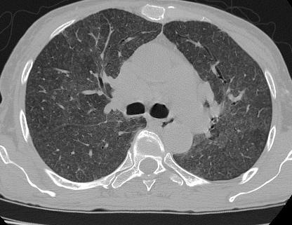
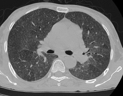
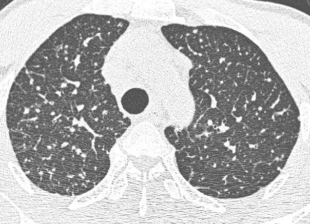
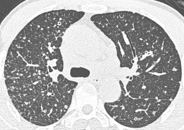
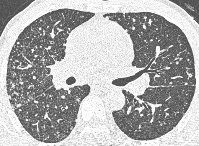
2 Case 1
3

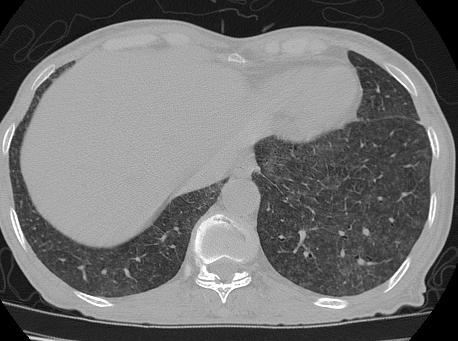
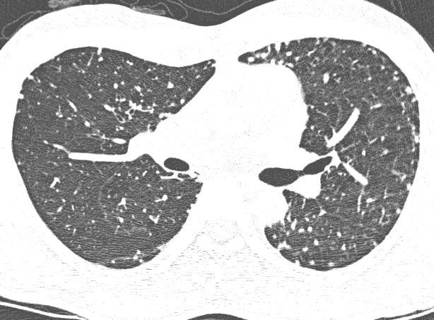
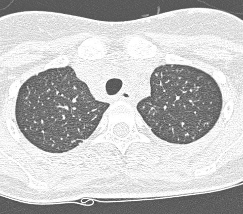
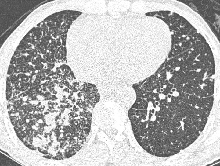
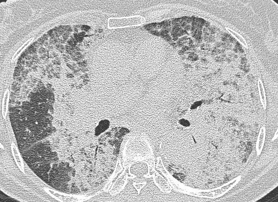
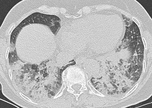
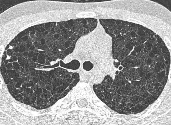
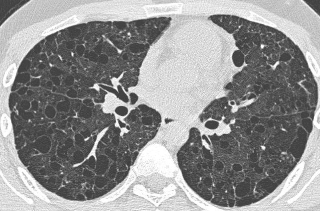
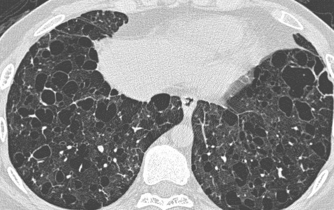
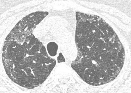
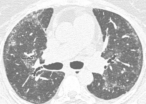
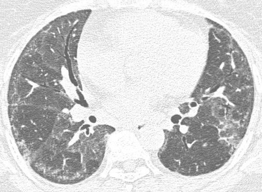
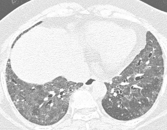
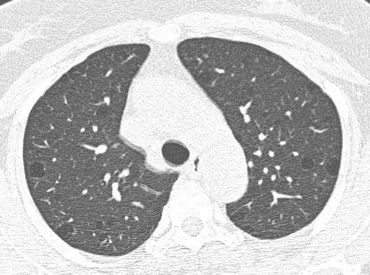
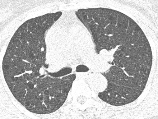
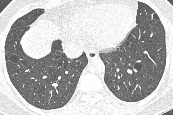
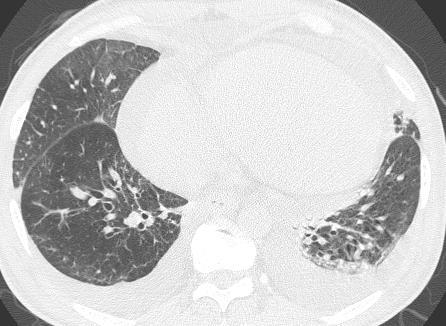
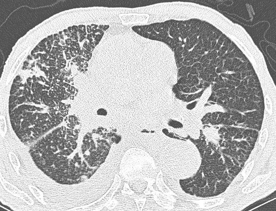
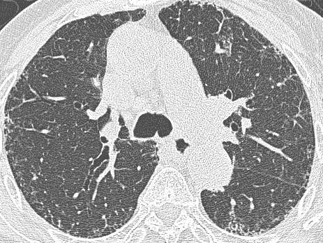
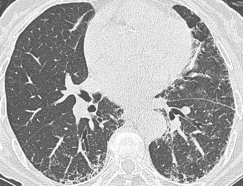
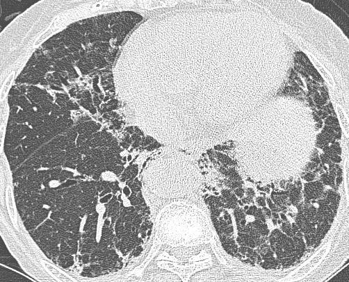
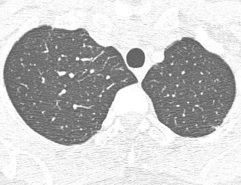
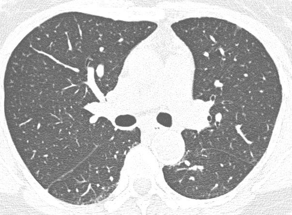
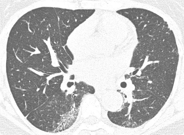
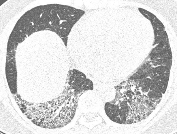
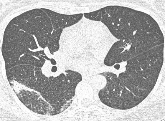
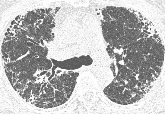
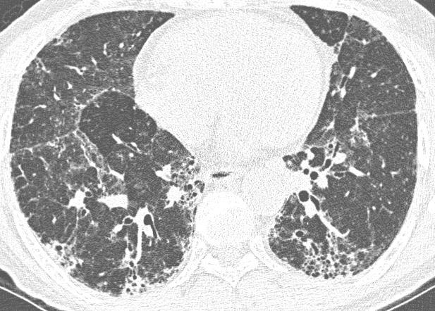
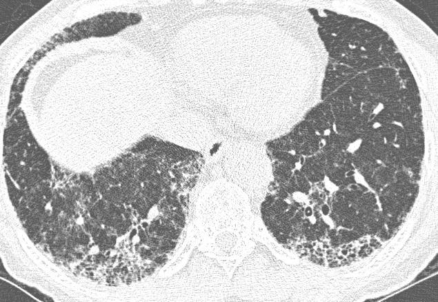
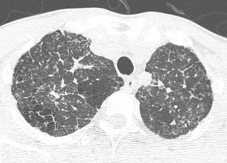
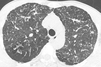
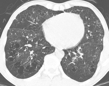
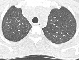
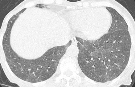
4 Case 1: Question 1.1 What is the main pattern of this HRCT? 1. Intralobular line 2. Groundglass opacity 3. Perilymphatic nodule
5 Case 1: Question 1.2 What is the diagnosis? 1. Hypersensitivity pneumonitis 2. Silicosis 3. Alveolar proteinosis 4. Miliary tuberculosis
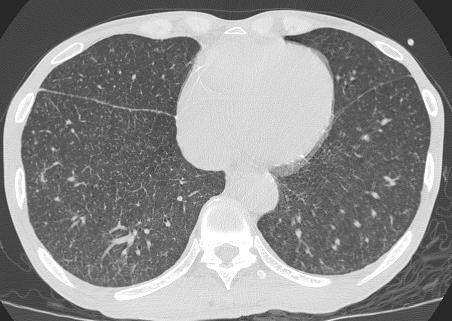
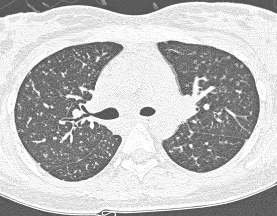
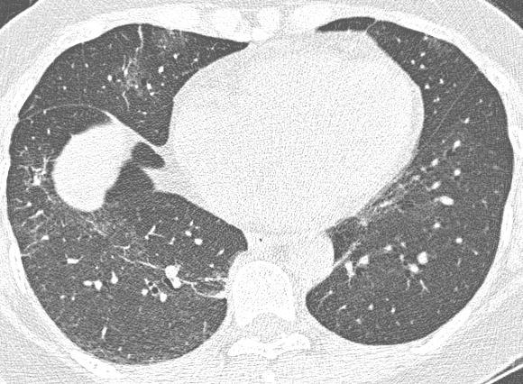
6 Case 2
7

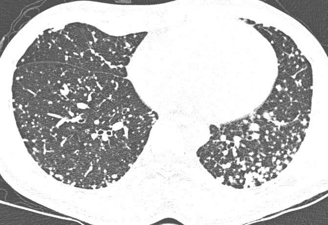
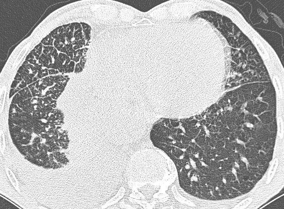
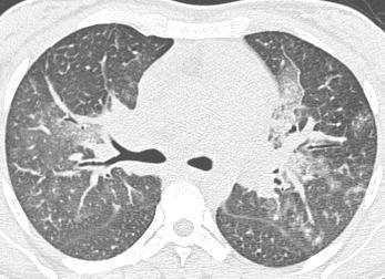
8 Case 2 : Question 2.1 What is the main pattern of this HRCT? 1. Nodule 2. Groundglass opacity 3. Reticular line
9 Case 2 : Question 2.2 What is the intralobular distribution? 1. Centrilobular 2. Perilymphatic 3. Random
10 Case 2 : Question 2.3 What is the diagnosis? 1. Hypersensitivity pneumonitis 2. Silicosis 3. Lymphangitic carcinomatosis 4. Miliary tuberculosis
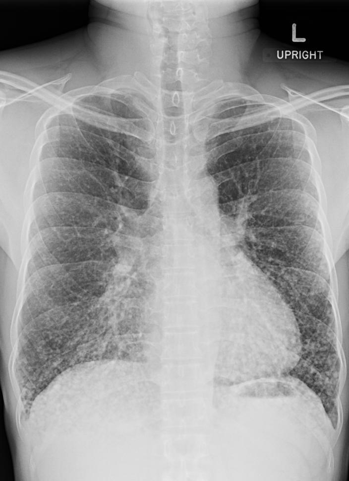
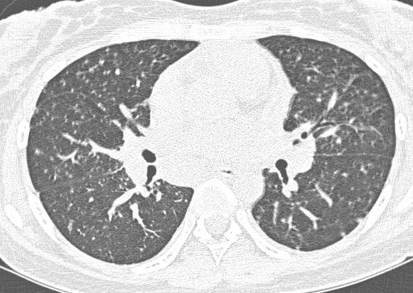
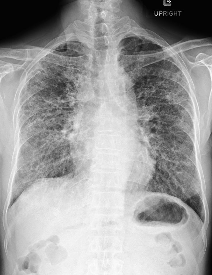
11 Case 3
12

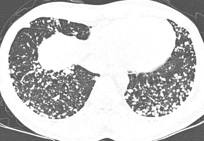
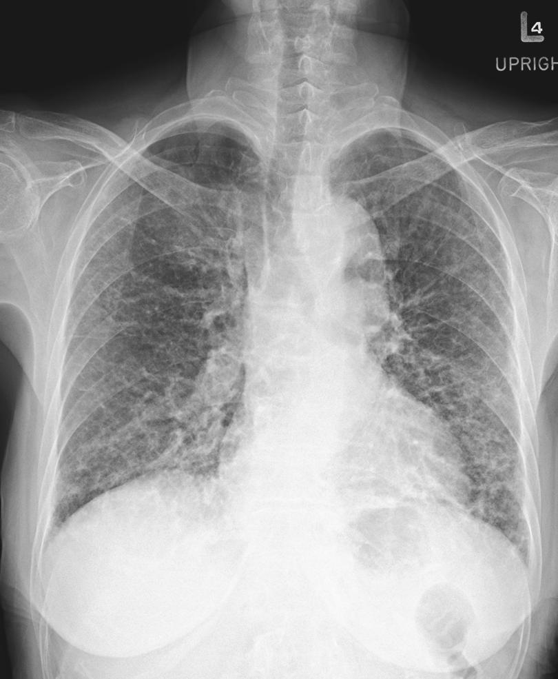
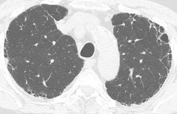
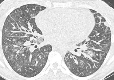
13 Case 3 : Question 3.1 What about the lung volume? 1. Increase 2. Normal 3. Decrease

14 Case 3 : Question 3.2 What is the intralobular distribution? 1. Centrilobular 2. Perilymphatic 3. Random
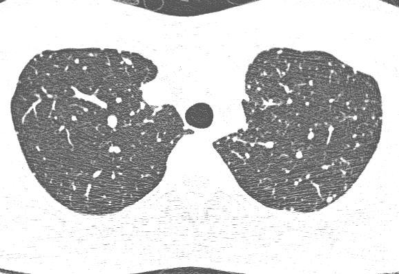
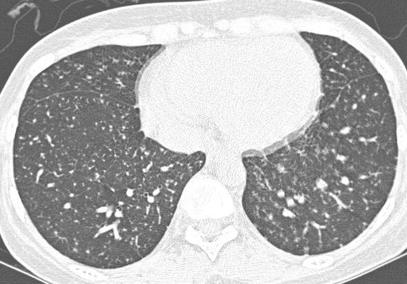
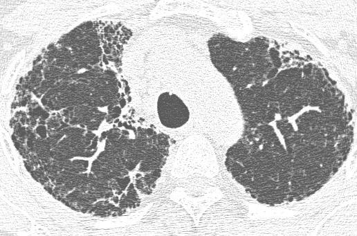
15 Case 4
16
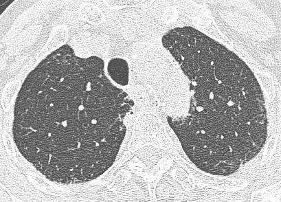
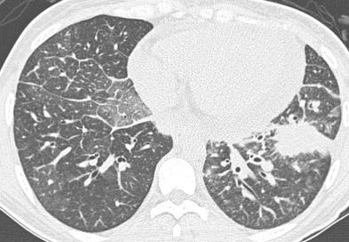
17 Case 4: Question 4.1 What is the intralobular distribution? 1. Centrilobular 2. Perilymphatic 3. Random

18 Case 4: Question 4.2 What is the associated finding? 1. Bronchiectasis 2. Lymphadenopathy 3. Pleural effusion
19 Case 4: Question 4.3 What is the diagnosis? 1. Bronchial spreading tuberculosis 2. Miliary tuberculosis 3. Lymphangitic carcinomatosis
20 Case 5
21

22 Case 5: Question 5.1 What is the main pattern of this HRCT? 1. Micronodule 2. Line 3. Both 1 and 2

23 Case 5: Question 5.2 What is the intralobular distribution? 1. Centrilobular 2. Perilymphatic 3. Random
24 Case 6
25

26 Case 6: Question 6.1 What is this CT sign? 1. Crazy paving 2. Mosaic pattern
27 Case 7
28

29 Case 7: Question 7.1 What is the craniocaudal distribution? 1. Upper + middle 2. Lower + middle 3. Diffuse
 3.")
30 Case 7: Question 7.2 What is the diagnosis? 1. Emphysema 2. Langerhans cell histocytosis (LCH) 3. Lymphangiomyomatosis (LAM)
31 Case 8
32

33 Case 8: Question 8.1 What is the main pattern of this HRCT? 1. Honeycomb 2. Groundglass opacity 3. Consolidation
34 Case 9
35
 3.")
36 Case 9: Question 9.1 What is the diagnosis? 1. Emphysema 2. Langerhans cell histocytosis(lch) 3. Lymphangiomyomatosis (LAM)
37 Case 10
38

39 Case 10: Question 10.1 What is the main pattern of this HRCT? 1. Smooth septal thickening 2. Nodular septal thickening 3. Centrilobular nodule

40 Case 10: Question 10.2 What is the associated finding? 1. Pleural effusion 2. Pulmonary arterial hypertension 3. Lymphadenopathy
41 Case 11
42
43 Case 11: Question 11.1 What is the main pattern of this HRCT? 1. Smooth interlobular septal thickening 2. Nodule 3. Ground glass opacity

44 Case 11: Question 11.2 What is the pattern of disease spreading? 1. Perilymphatic spreading 2. Bronchial spreading

45 Case 11: Question 11.3 What is the associated finding? 1. Pleural effusion 2. Enlarged lymph node 3. 1 and 2

46 Case 11: Question 11.4 What is the diagnosis? 1. PAP 2. Hypersensitivity pneumonitis 3. Lymphangitic carcinomatosis
47 Case11 : Question 11.5 What is the best further investigation? 1. Surgical Bx 2. Thoracoscopic Bx 3. Bronchoscopic BX
48 Case 12
49

50 Case12 : Question 12.1 What is the main pattern of this HRCT? 1. Interlobular line 2. Intralobular line 3. GGO

51 Case 12 : Question 12.2 What is the axial distribution? 1. Peripheral 2. Central 3. Diffuse

52 Case 12 : Question 12.3 What is the intralobular distribution? 1. Perilobular 2. Centrilobular 3. Panlobular
53 Case 13
54

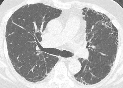
55 Case 13 : Question 13.1 What about the lung volume? 1. Increase 2. Normal 3. Decrease

56 Case 13 : Question 13.2 What is the main pattern of this HRCT? 1. Reticulation 2. Nodule 3. Honeycombing
57 Case 13 : Question 13.3 What is the craniocaudal distribution? 1. Upper 2. Diffuse 3. Lower

58 Case 13 : Question 13.4 What is the axial distribution? 1. Peripheral 2. Diffuse 3. Central

59 Case 13 : Question 13.5 What is the intralobular distribution? 1. Perilobular 2. Centrilobular 3. Panlobular
60 Case 14
61

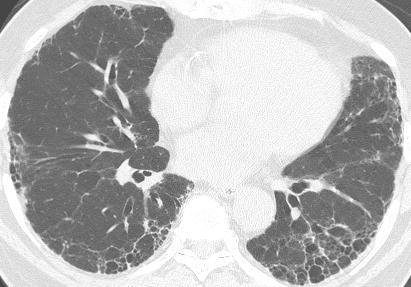
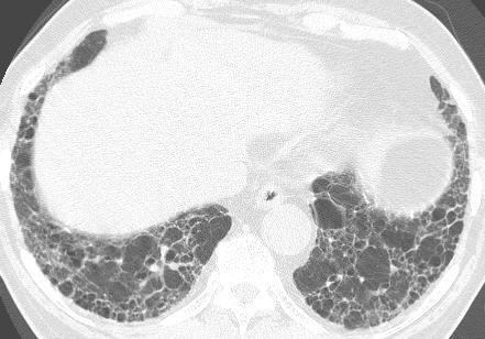
62 Case 14 : Question 14.1 What is the main pattern of this HRCT? 1. Reticulation 2. Groundglass opacity 3. Honeycombing

63 Case 14 : Question 14.2 What is the craniocaudal distribution? 1. Upper 2. Diffuse 3. Lower

64 Case 14 : Question 14.3 What is the axial distribution? 1. Peripheral 2. Diffuse 3. Central

65 Case 14 : Question 14.4 What is the intralobular distribution? 1. Panlobular 2. Centrilobular 3. Perilobular
66 Case 14 : Question 14.5 A patient known to have systemic sclerosis for 5 years develops dry cough, dyspnea on exertion and shortness of breath. What is the proper management? 1. Surgical biopsy 2. Pulmonary function test 3. Bronchoscopic biopsy
67 Case 15
68

69 Case 15 : Question 15.1 What is the main pattern of this HRCT? 1. Groundglass opacity 2. Nodule 3. Reticulation


70 Case 15 : Question 15.2 What is the most likely diagnosis? 1. BOOP 2. Lymphoma 3. NSIP
71 Case 15 : Question 15.3 What is the best further management? 1. Bronchoscopic biopsy 2. Surgical biopsy 3. Steroid
72 Case 16
73

74 Case 16: Question 16.1 What is the main pattern of this HRCT? 1. Groundglass opacity 2. Nodule 3. reticulation

75 Case 16: Question 16.2 What is the craniocaudal distribution? 1. Upper 2. Diffuse 3. Lower
76 Case 16 : Question 16.3 What is the axial distribution? 1. Peripheral 2. Diffuse 3. Central
77 Case 17

78

79 Case 17 : Question 17.1 What about the lung volume? 1. Increased 2. Normal 3. Decreased

80 Case 17 : Question 17.2 What is the axial distribution? 1. Peripheral 2. Diffuse 3. Central

81 Case 17 : Question 17.3 What is the intralobular distribution? 1. Perilymphatic 2. Centrilobular 3. Random
82 Case 18
83
84 Case 18 : Question 18.1 What is the main pattern of this HRCT? 1. Ground glass opacity 2. Septal thickening 3. 1 and 2

85 Case 18: Question 18.2 What is the craniocaudal distribution? 1. Upper 2. Diffuse 3. Lower

86 Case 18: Question 18.3 What is the axial distribution? 1. Peripheral 2. Diffuse 3. Central

87 Case 18 : Question 18.4 What is intralobular distribution? 1. Perilymphatic 2. Centrilobular 3. Random

88 Case 18 : Question 18.5 What is the associated finding? 1. Lymph node enlargement 2. Lung mass 3. Pleural thickening

89 Case 18 : Question 18.6 What is the diagnosis? 1. Lymphangitic carcinomatosis 2. Pulmonary alveolar proteinosis 3. Pulmonary edema
90 Case 19
91

92 Case 19 : Question 19.1 What is the main pattern of this HRCT? 1. Ground glass opacity 2. Ill-defined nodule and 2.

93 Case 19: Question 19.2 What is the axial distribution? 1. Peripheral 2. Diffuse 3. Central

94 Case 19 : Question 19.3 What is the intralobular distribution? 1. Perilymphatic 2. Centrilobular 3. Random
95 Case 19 : Question 19.4 What is the diagnosis? 1. Hypersensitivity pneumonitis 2. NSIP 3. PAP
96 Case 20


97

98 Case 20 : Question 20.1 What is the main pattern of this HRCT? 1. Nodule 2. Ground glass opacity 3. Septal thickening 4. 2 and 3.

99 Case 20 : Question 20.2 What is the distribution of this HRCT in axial view? 1. Peripheral 2. Diffuse 3. Central

100 Case 20 : Question 20.3 What is the intralobular distribution? 1. Perilymphatic 2. Centrilobular 3. Random

101 Case 20: Question 20.4 What is the associated finding? 1. Lymph node enlargement 2. Pericardial thickening and 2.

102 Case 20: Question 20.5 What is the diagnosis? 1. Lymphangitic carcinomatosis 2. PAP 3. Pulmonary edema
103 Suspected diffused lung disease Chest radiograph Normal or equivocal Abnormal Suggestive of sarcoidosis HRCT with prone views HRCT Trans bronchial Bx Normal Abnormal See next slide Bx if clinical/physiologic evidence of disease See next slide
 Thoracosopic biopsy (CT")
104 Suspected diffused lung disease Specific CT pattern(uip,eg,lam,h P, Lipoid Pneumonia) CT pattern suggestive of HP sarcoid, lymphagitic carcinoma,pap, alveolar carcinoma,eosinophillic pneumonia, BOOP Other CT pattern Accept CT diagnosis if clinical scenario is consistent Trans bronchial biopsy and/or brochoalveolar lavage (CT directed) Thoracosopic biopsy (CT directed)
105 Thank you
Case 1 : Question. 1.1 What is the intralobular distribution? 1. Centrilobular 2. Perilymphatic 3. Random
 Interesting case Case 1 Case 1 : Question 1.1 What is the intralobular distribution? 1. Centrilobular 2. Perilymphatic 3. Random Case 1: Answer 1.1 What is the intralobular distribution? 1. Centrilobular
HRCT in Diffuse Interstitial Lung Disease Steps in High Resolution CT Diagnosis. Where are the lymphatics? Anatomic distribution
 Steps in High Resolution CT Diagnosis Pattern of abnormality Distribution of disease Associated findings Clinical history Tomás Franquet MD What is the diagnosis? Hospital de Sant Pau. Barcelona Secondary
Daria Manos RSNA 2016 RC 401. https://medicine.dal.ca/departments/depar tment-sites/radiology/contact/faculty/dariamanos.html
 Daria Manos RSNA 2016 RC 401 https://medicine.dal.ca/departments/depar tment-sites/radiology/contact/faculty/dariamanos.html STEP1: Is this fibrotic lung disease? STEP 2: Is this a UIP pattern? If yes:
Acute and Chronic Lung Disease
 KATHOLIEKE UNIVERSITEIT LEUVEN Faculty of Medicine Acute and Chronic Lung Disease W De Wever, JA Verschakelen Department of Radiology, University Hospitals Leuven, Belgium Clinical utility of HRCT To detect
Financial disclosure COMMON DIAGNOSES IN HRCT. High Res Chest HRCT. HRCT Pre test. I have no financial relationships to disclose. Anatomy Nomenclature
 Financial disclosure I have no financial relationships to disclose. Douglas Johnson D.O. Cardiothoracic Imaging Gaston Radiology COMMON DIAGNOSES IN HRCT High Res Chest Anatomy Nomenclature HRCT Sampling
Manish Powari Regional Training Day 10/12/2014
 Manish Powari Regional Training Day 10/12/2014 Large number of different types of Interstitial Lung Disease (ILD). Most are very rare Most patients present with one of a smaller number of commoner diseases
Mimics in chest disease: interstitial opacities
 Insights Imaging (2013) 4:9 27 DOI 10.1007/s13244-012-0207-7 PICTORIAL REVIEW Mimics in chest disease: interstitial opacities Anastasia Oikonomou & Panos Prassopoulos Received: 19 June 2012 / Revised:
The crazy-paving pattern: A radiological-pathological correlated and illustrated overview
 The crazy-paving pattern: A radiological-pathological correlated and illustrated overview Poster No.: C-0827 Congress: ECR 2010 Type: Educational Exhibit Topic: Chest Authors: W. F. M. De Wever, J. Coolen,
Interesting Cases. Pulmonary
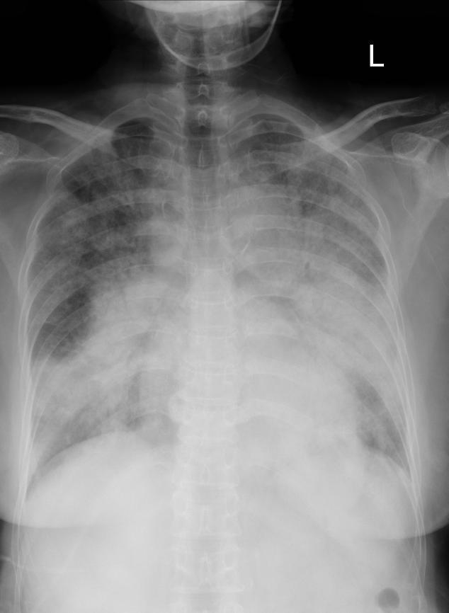
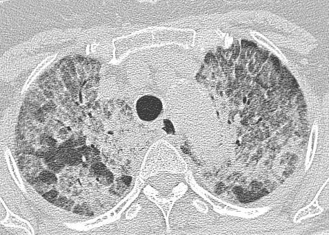
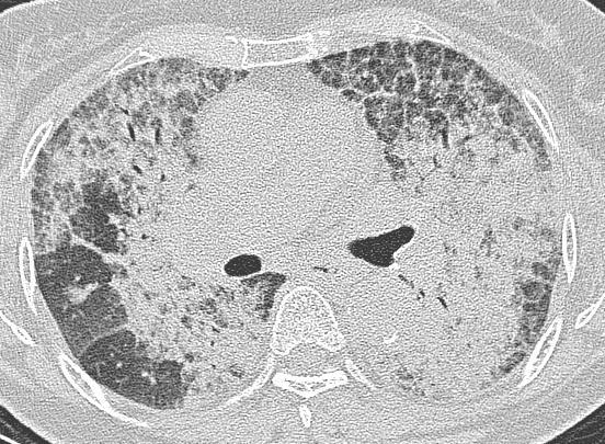
 Interesting Cases Pulmonary 54M with prior history of COPD, hep B/C, and possible history of TB presented with acute on chronic dyspnea, and productive cough Hazy opacity overlying the left hemithorax
Differential diagnosis
 Differential diagnosis Idiopathic pulmonary fibrosis (IPF) is part of a large family of idiopathic interstitial pneumonias (IIP), one of four subgroups of interstitial lung disease (ILD). Differential
Outline Definition of Terms: Lexicon. Traction Bronchiectasis
 HRCT OF IDIOPATHIC INTERSTITIAL PNEUMONIAS Disclosures Genentech, Inc. Speakers Bureau Tadashi Allen, MD University of Minnesota Assistant Professor Diagnostic Radiology 10/29/2016 Outline Definition of
Radiologists toolbox to differentiate alveolar versus interstitial lung diseases
 Radiologists toolbox to differentiate alveolar versus interstitial lung diseases Dr Sumer Shikhare, Dr Trishna Shimpi, Dr Ashish Chawla Khoo Teck Puat Hospital Singapore. Relevant financial disclosures
I have no relevant conflicts of interest to disclose
 I have no relevant conflicts of interest to disclose Diffuse parenchymal lung disease (DPLD) and its associations Secondary lobular anatomy DPLD History, clinical findings, temporal evolution, and exposures
Micronodular Lung Disease an algorithm
 Micronodular Lung Disease an algorithm H. Page McAdams, MD Department of Radiology Duke University Medical Center Durham, NC USA page.mcadams@duke.edu Question Which of the following lung diseases is MOST
11/10/2014. Multi-disciplinary Approach to Diffuse Lung Disease: The Imager s Perspective. Radiology
 Multi-disciplinary Approach to Diffuse Lung Disease: The Imager s Perspective Radiology Pathology Clinical 1 Role of HRCT Diagnosis Fibrosis vs. inflammation Next step in management Response to treatment
A Review of Interstitial Lung Diseases. Paul J. Wolters, MD Associate Professor Department of Medicine University of California San Francisco
 A Review of Interstitial Lung Diseases Paul J. Wolters, MD Associate Professor Department of Medicine University of California San Francisco Outline Overview of diagnosis in ILD Why it is important Definition/Classification
10/17/2016. Nuts and Bolts of Thoracic Radiology. Objectives. Techniques
 Nuts and Bolts of Thoracic Radiology October 20, 2016 Carleen Risaliti Objectives Understand the basics of chest radiograph Develop a system for interpreting chest radiographs Correctly identify thoracic
A Review of Interstitial Lung Diseases
 Outline A Review of Interstitial Lung Diseases Paul J. Wolters, MD Associate Professor Department of Medicine University of California San Francisco Overview of diagnosis in ILD Why it is important Definition/Classification
An Image Repository for Chest CT
 An Image Repository for Chest CT Francesco Frajoli for the Chest CT in Antibody Deficiency Group An Image Repository for Chest CT he Chest CT in Antibody Deficiency Group is an international and interdisciplinary
Resident Case Review CHEST. Daria Manos CAR 2016
 Resident Case Review CHEST CAR 2016 Daria Manos Disclosure Speakers bureau, Roche CAR 2016 Daria Manos 1. Recognize common and critical chest radiograph and computed tomography signs and use these clues
Typical and atypical findings of pulmonary sarcoidosis at high resolution CT
 Typical and atypical findings of pulmonary sarcoidosis at high resolution CT Poster No.: C-0169 Congress: ECR 2013 Type: Educational Exhibit Authors: L. Raposo Rodríguez, C. Mejía, B. Escobar Mallada,
Radiologic-pathologic correlation of pulmonary diseases
 The 1578 th Chest Conference/ 3 rd Biennial Clinical- Radiologic-Pathologic Correlation Radiologic-pathologic correlation of pulmonary diseases Harumi Itoh, M.D. University of Fukui, Japan Centriacinar
Imaging Spectrum of Allergic Lung Disease: Hypersensitivity Reactions on the Lung Parenchyma
 Imaging Spectrum of Allergic Lung Disease: Hypersensitivity Reactions on the Lung Parenchyma Moon Sung Kim 1, Ki-Nam Lee 1, Won Jin Choi 1, Bo Ra Kim 1, Eun-Ju Kang 1 1 Department of Radiology, Dong-A
IPF: Epidemiologia e stato dell arte
 IPF: Epidemiologia e stato dell arte Clinical Classification Diffuse parenchimal lung diseases Exposure-related: - occupational - environmental - medication Desquamative interstitial pneumonia Idiopathic
Diagnostic Imaging of Diffuse Infiltrative Disease of the Lung
 Thematic Review Series Respiration 2004;71:4 19 DOI: 10.1159/000075642 Diagnostic Imaging of Diffuse Infiltrative Disease of the Lung Maurizio Zompatori a Claudio Bnà a Venerino Poletti c Enrica Spaggiari
Pneumocystis jirovecci pneumonia: from mild disease to a real disaster. A pictorial review of the different radiologic patterns in acute settings
 Pneumocystis jirovecci pneumonia: from mild disease to a real disaster. A pictorial review of the different radiologic patterns in acute settings Poster No.: C-1425 Congress: ECR 2017 Type: Educational
Pulmonary Manifestations of Systemic Lupus Erythematosus 1
 Pulmonary Manifestations of Systemic Lupus Erythematosus 1 Kee Hyuk Yang, M.D., Yo Won Choi, M.D., Seok Chol Jeon, M.D., Choong Ki Park, M.D., Kyung in Joo, M.D., Chang Kok Hahm, M.D., Seung Ro Lee, M.D.
Thoracic lung involvement in rheumatoid arthritis: Findings on HRCT
 Thoracic lung involvement in rheumatoid arthritis: Findings on HRCT Poster No.: C-2488 Congress: ECR 2015 Type: Educational Exhibit Authors: R. E. Correa Soto, M. J. Martín Sánchez, J. M. Fernandez 1 1
INTERSTITIAL LUNG DISEASE. Radhika Reddy MD Pulmonary/Critical Care Long Beach VA Medical Center January 5, 2018
 INTERSTITIAL LUNG DISEASE Radhika Reddy MD Pulmonary/Critical Care Long Beach VA Medical Center January 5, 2018 Interstitial Lung Disease Interstitial Lung Disease Prevalence by Diagnosis: Idiopathic Interstitial
Thoracic Sarcoidosis Imaging Updated: Jul 19, 2013
 Thoracic Sarcoidosis Imaging Updated: Jul 19, 2013 Overview Radiography Computed Tomography Magnetic Resonance Imaging Nuclear Imaging Show All Multimedia Library References Overview For patients with
Non-neoplastic Lung Disease II
 Pathobasic Non-neoplastic Lung Disease II Spasenija Savic Prince Pathology Program Systematic approach to surgical lung biopsies with ILD Examples (chronic ILD): Idiopathic interstitial pneumonias: UIP,
Idiopathic interstitial pneumonias (IIPs) are a group of
 are a group of") SYMPOSIA C. Isabela S. Silva, MD, PhD and Nestor L. Müller, MD, PhD Abstract: The idiopathic interstitial pneumonias (IIPs) are a group of diffuse parenchymal lung diseases of unknown etiology characterized
SYMPOSIA C. Isabela S. Silva, MD, PhD and Nestor L. Müller, MD, PhD Abstract: The idiopathic interstitial pneumonias (IIPs) are a group of diffuse parenchymal lung diseases of unknown etiology characterized
Pulmonary Sarcoidosis - Radiological Evaluation
 Original Research Article Pulmonary Sarcoidosis - Radiological Evaluation Jayesh Shah 1, Darshan Shah 2*, C. Raychaudhuri 3 1 Associate Professor, 2 1 st Year Resident, 3 Professor and HOD Radiology Department,
Radiologic Approach to Smoking Related Interstitial Lung Disease
 Radiologic Approach to Smoking Related Interstitial Lung Disease Poster No.: C-1854 Congress: ECR 2013 Type: Educational Exhibit Authors: K.-N. Lee, J.-Y. Han, E.-J. Kang, J. Kang; Busan/KR Keywords: Toxicity,
August 2018 Imaging Case of the Month: Dyspnea in a 55-Year-Old Smoker. Michael B. Gotway, MD
 August 2018 Imaging Case of the Month: Dyspnea in a 55-Year-Old Smoker Michael B. Gotway, MD Department of Radiology Mayo Clinic Arizona Scottsdale, AZ USA Clinical History: A 55 year old woman presented
CT findings in multifocal or diffuse non-mucinous bronchioloalveolar carcinoma (BAC)
") CT findings in multifocal or diffuse non-mucinous bronchioloalveolar carcinoma (BAC) Poster No.: C-2192 Congress: ECR 2014 Type: Educational Exhibit Authors: I. Sandu, A. R. Popita, I.-A. Brumboiu; Cluj-Napoca/RO
CT findings in multifocal or diffuse non-mucinous bronchioloalveolar carcinoma (BAC) Poster No.: C-2192 Congress: ECR 2014 Type: Educational Exhibit Authors: I. Sandu, A. R. Popita, I.-A. Brumboiu; Cluj-Napoca/RO
Usual Interstitial pneumonia and Nonspecific Interstitial Pneumonia. Nitra and the Gangs.
 Usual Interstitial pneumonia and Nonspecific Interstitial Pneumonia Nitra and the Gangs. บทน ำและบทท ๓, ๑๐, ๑๒, ๑๓, ๑๔, ๑๕, ๑๗ Usual Interstitial Pneumonia (UIP) Most common & basic pathologic pattern
Bronkhorst colloquium Interstitiële longziekten. Katrien Grünberg, klinisch patholoog
 Bronkhorst colloquium 2013-2014 Interstitiële longziekten De pathologie achter de CT Katrien Grünberg, klinisch patholoog K.grunberg@vumc.nl Preparing: introduction and 3 cases The introduction on microscopic
Workshop Cyst & Lucency. How to Approach
 Workshop Cyst & Lucency How to Approach To Approach Cystic Lung Disease True cysts? Cavitary disease Cystic bronchiectasis Mosaic attenuation Subpleural cysts Bullae Paraseptal emphysema Honeycombing Birt
HYPERSENSITIVITY PNEUMONITIS
 HYPERSENSITIVITY PNEUMONITIS A preventable fibrosis MOSAVIR ANSARIE MB., FCCP INTERSTITIAL LUNG DISEASES A heterogeneous group of non infectious, non malignant diffuse parenchymal disorders of the lower
The radiological differential diagnosis of the UIP pattern
 5th International Conference on Idiopathic Pulmonary Fibrosis, Modena, 2015, June 12th The radiological differential diagnosis of the UIP pattern Simon Walsh King s College Hospital Foundation Trust London,
Case Presentations in ILD. Harold R. Collard, MD Department of Medicine University of California San Francisco
 Case Presentations in ILD Harold R. Collard, MD Department of Medicine University of California San Francisco Outline Overview of diagnosis in ILD Definition/Classification High-resolution CT scan Multidisciplinary
How to Analyse Difficult Chest CT
 How to Analyse Difficult Chest CT Complex diseases are:- - Large lesion - Unusual or atypical pattern - Multiple discordant findings Diffuse diseases are:- - Numerous findings in both sides 3 basic steps
5/9/2015. Multi-disciplinary Approach to Diffuse Lung Disease: The Imager s Perspective. No, I am not a pulmonologist! Radiology
 Multi-disciplinary Approach to Diffuse Lung Disease: The Imager s Perspective No, I am not a pulmonologist! Radiology Pathology Clinical 1 Everyone needs a CT Confidence in diagnosis Definitive HRCT +
Diagnosis of TB: Radiology David Finlay, MD
 TB Intensive Tyler, Texas June 2-4, 2010 Diagnosis of TB: Radiology David Finlay, MD June 3, 2010 2stages stages- Tuberculosis 1. primary infection 2. reactivation, or post primary disease 2 1 Primary
September 2014 Imaging Case of the Month. Michael B. Gotway, MD. Department of Radiology Mayo Clinic Arizona Scottsdale, AZ
 September 2014 Imaging Case of the Month Michael B. Gotway, MD Department of Radiology Mayo Clinic Arizona Scottsdale, AZ Clinical History: A 57-year-old non-smoking woman presented to her physician as
Cystic Lung Disease. Cristopher A. Meyer, MD
 Cystic Lung Disease Cristopher A. Meyer, MD Air filled structure with definable wall typically less than 1 mm thick Cris A. Meyer, M.D. Professor of Radiology University of Wisconsin School of Medicine
Role of Computed Tomography in Diagnosis of Diffuse Lung Diseases Chauhan Jayant 1*, Panchal Pankaj 2, Faruqui Tehzeeb 3
 ORIGINAL ARTICLE Role of Computed Tomography in Diagnosis of Diffuse Lung Diseases Chauhan Jayant 1*, Panchal Pankaj 2, Faruqui Tehzeeb 3 1 MD,DTCD,Additional Professor& HOD, 2,3 MBBS, 3 rd year resident
RADIOLOGICALL ANALYSIS OF INTERSTITIAL LUNG DISEASES
 Original Research Article RADIOLOGICALL ANALYSIS OF INTERSTITIAL LUNG DISEASES Meraj Rentia 1*, Himanshu Singla 1, Divya Malpani 2, Tushar Vaishnav 3, Pradeep Jhala 3 1 1 st year Resident, 2 3 rd year
Epidemiology and classification of smoking related interstitial lung diseases
 Epidemiology and classification of smoking related interstitial lung diseases Šterclová M. Department of Respiratory Diseases, Thomayer Hospital, Prague, Czech Republic Supported by an IGA Grant No G 1207
American Thoracic Society European Respiratory Society Classification of the Idiopathic Interstitial Pneumonias: Advances in Knowledge since 20021
 This copy is for personal use only. To order printed copies, contact reprints@rsna.org American Thoracic Society European Respiratory Society Classification of the Idiopathic Interstitial Pneumonias: Advances
Cryptogenic Organizing Pneumonia Diagnosis Approach Based on a Clinical-Radiologic-Pathologic Consensus
 Cryptogenic Organizing Pneumonia Diagnosis Approach Based on a Clinical-Radiologic-Pathologic Consensus Poster No.: C-1622 Congress: ECR 2012 Type: Scientific Exhibit Authors: C. Cordero Lares, E. Zorita
Micronodular lung pattern - Differential diagnosis
 Micronodular lung pattern - Differential diagnosis Poster No.: P-0074 Congress: ESTI 2015 Type: Educational Poster Authors: P. Ninitas, F. Marinho, P. Campos, I. Távora ; Lisbon/PT, 1 2 2 3 1 1 3 Funchal/PT,
Web Chapter 3. Image Gallery: Lesion detection on low dose chest CT
 Web Chapter 3 Image Gallery: Lesion detection on low dose chest CT Sarabjeet Singh, MD Mannudeep K. Kalra, MD *Eugene J. Mark, MD *James Stone, MD James H. Thrall, MD Department of Radiology and *Department
Diffuse Interstitial Lung Diseases: Is There Really Anything New?
 : Is There Really Anything New? Sujal R. Desai, MBBS, MD ESTI SPEAKER SUNDAY Society of Thoracic Radiology San Antonio, Texas March 2014 Diffuse Interstitial Lung Disease The State of Play DILDs Is There
Idiopathic Pulmonary of Care
 Chapter 6.1 Living Medical etextbook A Digital Tool at the Point of Care From Projects In Knowledge Pulmonology Idiopathic Pulmonary Fibrosis @Point of Care IPF Case Study: Typical Presentation, Role of
IPF - Inquadramento clinico
 IPF - Inquadramento clinico Sergio Harari Unità Operativa di Pneumologia UTIR Servizio di Fisiopat. Resp. e Emodinamica Polmonare Ospedale S. Giuseppe, Milano Clinical Classification Diffuse parenchimal
The Imaging Analysis of Pulmonary Sarcodiosis
 www.cancercellresearch.org ISSN: 2161-2609 Article The Imaging Analysis of Pulmonary Sarcodiosis Xin He, Chuanyu Zhang* Department of Radiology, Affiliated Hospital of Qingdao University, Qingdao, China
Liebow and Carrington's original classification of IIP
 Liebow and Carrington's original classification of IIP-- 1969 Eric J. Stern MD University of Washington UIP Usual interstitial pneumonia DIP Desquamative interstitial pneumonia BIP Bronchiolitis obliterans
Interstitial syndrome
 Interstitial syndrome Ground-glass attenuation Miliary and nodular images linear images Etienne Leroy Terquem Pierre L Her SPI / ISP Soutien Pneumologique International / International Support for Pulmonology
INTERSTITIAL LUNG DISEASE Dr. Zulqarnain Ashraf
 Indep Rev Jul-Dec 2018;20(7-12) Dr. Zulqarnain Ashraf IR-653 Abstract: ILD is a group of diseases affect interstitium of the lung. Repeated insult to the lung cause the interstitium to be damaged. Similarly
Pictorial essay of unusual radiologic manifestations of pulmonary and airway metastasis at initial presentation of lung cancer
 Pictorial essay of unusual radiologic manifestations of pulmonary and airway metastasis at initial presentation of lung cancer Poster No.: C-2297 Congress: ECR 2012 Type: Educational Exhibit Authors: Y.
Interstitial Syndrome Ground glass attenuation miliary and nodular images Linear images
 Interstitial Syndrome Ground glass attenuation miliary and nodular images Linear images Dr Etienne Leroy-Terquem Centre hospitalier de Meulan les Mureaux. France French-cambodian association for pneumology
Progress in Idiopathic Pulmonary Fibrosis
 Progress in Idiopathic Pulmonary Fibrosis David A. Lynch, MB Disclosures Progress in Idiopathic Pulmonary Fibrosis David A Lynch, MB Consultant: t Research support: Perceptive Imaging Boehringer Ingelheim
The Egyptian Journal of Hospital Medicine (July 2017) Vol.68 (2), Page
 Vol.68 (2), Page") The Egyptian Journal of Hospital Medicine (July 2017) Vol.68 (2), Page 1135-1140 Role of High Resolution Computed Tomography in Diagnosis of Interstitial Lung Diseases in Patients with Collagen Diseases
The Egyptian Journal of Hospital Medicine (July 2017) Vol.68 (2), Page 1135-1140 Role of High Resolution Computed Tomography in Diagnosis of Interstitial Lung Diseases in Patients with Collagen Diseases
Systemic lupus erythematosus (SLE): Pleuropulmonary Manifestations
: Pleuropulmonary Manifestations") 08/30/10 09/26/10 Systemic lupus erythematosus (SLE): Pleuropulmonary Manifestations Camila Downey S. Universidad de Chile, School of Medicine, Year VII Harvard University, School of Medicine Sept 17,
08/30/10 09/26/10 Systemic lupus erythematosus (SLE): Pleuropulmonary Manifestations Camila Downey S. Universidad de Chile, School of Medicine, Year VII Harvard University, School of Medicine Sept 17,
Chest Radiology Interpretation: Findings of Tuberculosis
 Chest Radiology Interpretation: Findings of Tuberculosis Get out your laptops, smart phones or other devices pollev.com/chestradiology Case #1 1 Plombage Pneumonia Cancer 2 Reading the TB CXR Be systematic!
and localized ground glass opacities, or bronchiolar focal or multifocal micronodules;
 E1 Chest CT scan and Pneumoniae_YE Claessens et al- Supplementary methods Level of CAP probability according to CT scan - definite CAP: systematic alveolar condensation, or alveolar condensation with peripheral
Chest imaging II. Interstitial lung diseases
 Chest imaging II. Interstitial lung diseases Dávid L. Tárnoki MD, PhD Ádám D. TárnokiMD, PhD Department of Radiology Semmelweis University Topics 1. Interstitial lung diseases 2. Occupational lung diseases
Spectrum of Cystic Lung Disease and its Mimics. Kathleen Jacobs MD and Elizabeth Weihe MD UC San Diego Medical Center, Department of Radiology
 Spectrum of Cystic Lung Disease and its Mimics Kathleen Jacobs MD and Elizabeth Weihe MD UC San Diego Medical Center, Department of Radiology No Financial Disclosures Learning Objectives 1. Review the
Thoracic Manifestations of Sarcoidosis Using Multi-Slice CT
 IOSR Journal of Dental and Medical Sciences (IOSR-JDMS) e-issn: 2279-0853, p-issn: 2279-0861.Volume 14, Issue 9 Ver. II (Sep. 2015), PP 63-68 www.iosrjournals.org Thoracic Manifestations of Sarcoidosis
Role of High Resolution Computed Tomography in Evaluation of Pulmonary Diseases
 International J. of Healthcare & Biomedical Research, Volume:, Issue:, April, Pages 9-96 Role of High Resolution Computed Tomography in Evaluation of Pulmonary Diseases Dr. Abhijeet D. Nagapurkar*, Dr.
4/17/2010 C ini n ca c l a Ev E a v l a ua u t a ion o n of o ILD U dat a e t e i n I LDs
 Update in ILDs Diagnosis 101: Clinical Evaluation April 17, 2010 Jay H. Ryu, MD Mayo Clinic, Rochester MN Clinical Evaluation of ILD Outline General aspects of ILDs Classification of ILDs Clinical evaluation
Radiological Imaging of Drug-Induced Pulmonary Lesions
 Review Article imedpub Journals www.imedpub.com Journal of Clinical Radiology and Case Reports Radiological Imaging of Drug-Induced Pulmonary Lesions D souza M *, Rajiah P, Khan A and Irion K Department
October 2012 Imaging Case of the Month. Michael B. Gotway, MD Associate Editor Imaging. Department of Radiology Mayo Clinic Arizona Scottsdale, AZ
 October 2012 Imaging Case of the Month Michael B. Gotway, MD Associate Editor Imaging Department of Radiology Mayo Clinic Arizona Scottsdale, AZ Clinical History: A 65-year-old non-smoking woman presented
Chest Radiology LYMPHANGITIC CARCINOMATOSIS CERTAIN CANCERS SPREAD BY PLUGGING THE LYMPHATICS
 2 Chest Radiology Includes plain film diagnosis, CT, MRI, and interventional techniques used in the diagnosis of diseases of the lungs, pleura, and mediastinum including the heart and great vessels. LYMPHANGITIC
Lung CT: Part 2, The Interstitial Pneumonias Clinical, Histologic, and CT Manifestations
 Integrative Imaging Review Ferguson and Berkowitz CT of Interstitial Pneumonia Integrative Imaging Review CME SAM Lung CT FOCUS ON: Emma C. Ferguson 1 Eugene A. Berkowitz 2 Ferguson EC, Berkowitz EA Keywords:
Pulmonary Complications of Antineoplastic Agents : Era of Targeted Therapy
 Pulmonary Complications of Antineoplastic Agents : Era of Targeted Therapy Poster No.: C-1230 Congress: ECR 2013 Type: Educational Exhibit Authors: H. Y. Kim, J. H. Hwang, Y.-W. Chang, J. Y. Moon; Seoul/KR
Spectrum of Findings on HRCT in Evaluation of Interstitial Lung Diseases: A Single Centre Prospective Observational Study
 IOSR Journal of Dental and Medical Sciences (IOSR-JDMS) e-issn: 2279-0853, p-issn: 2279-0861.Volume 17, Issue 11 Ver. 7 (November. 2018), PP 53-63 www.iosrjournals.org Spectrum of Findings on HRCT in Evaluation
ARDS - a must know. Page 1 of 14
 ARDS - a must know Poster No.: C-1683 Congress: ECR 2016 Type: Authors: Keywords: DOI: Educational Exhibit M. Cristian; Turda/RO Education and training, Edema, Acute, Localisation, Education, Digital radiography,
Thoracic Imaging: A Case of Metastatic Adenocarcinoma of Unknown Primary
 January 28, 2009 Thoracic Imaging: A Case of Metastatic Adenocarcinoma of Unknown Primary Kristina Mirabeau-Beale, Harvard Medical School Year III Gillian Lieberman, MD Agenda Introduce Patient RS Discuss
ARTICLE IN PRESS. Ahuva Grubstein a, Daniele Bendayan b, Ithak Schactman c, Maya Cohen a, David Shitrit b, Mordechai R. Kramer b,
 Respiratory Medicine (2005) 99, 948 954 Concomitant upper-lobe bullous emphysema, lower-lobe interstitial fibrosis and pulmonary hypertension in heavy smokers: report of eight cases and review of the literature
Diagnosing Idiopathic Pulmonary Fibrosis on Evidence-Based Guidelines
 Diagnosing Idiopathic Pulmonary Fibrosis on Evidence-Based Guidelines Rebecca Keith, MD Assistant Professor, Division of Pulmonary and Critical Care Medicine National Jewish Health, Denver, CO Objectives
Comparison of High-resolution CT Findings between Miliary Metastases and Miliary Tuberculosis 1
 Comparison of High-resolution CT Findings between Miliary Metastases and Miliary Tuberculosis 1 Chan Sung Kim, M.D., Ki-Nam Lee, M.D., Jin Hwa Lee, M.D. Purpose: To compare the findings of high-resolution
SCLERODERMA LUNG DISEASE: WHAT THE PATIENT SHOULD KNOW
 SCLERODERMA LUNG DISEASE: WHAT THE PATIENT SHOULD KNOW Lung disease can be a serious complication of scleroderma. The two most common types of lung disease in patients with scleroderma are interstitial
An Introduction to Radiology for TB Nurses
 An Introduction to Radiology for TB Nurses Garold O. Minns, MD September 14, 2017 TB Nurse Case Management September 12 14, 2017 EXCELLENCE EXPERTISE INNOVATION Garold O. Minns, MD has the following disclosures
Case of the Day Chest
 Case of the Day Chest Darin White MDCM FRCPC Department of Radiology, Mayo Clinic 76 th Annual Scientific Meeting Canadian Association of Radiologists Montreal, QC April 26, 2013 2013 MFMER slide-1 Disclosures
Thoracic sarcoidosis: Pictoral review of typical and atypical findings
 Thoracic sarcoidosis: Pictoral review of typical and atypical findings Poster No.: C-0804 Congress: ECR 2010 Type: Educational Exhibit Topic: Chest Authors: A. Ferreira, J. Calha; Lisbon/PT Keywords: Sarcoidosis,
Community-Acquired Acinetobacter baumannii Pneumonia: Initial Chest Radiographic Findings and Follow-up CT Findings in Helping Predict Patient Outcome
 Community-Acquired Acinetobacter baumannii Pneumonia: Initial Chest Radiographic Findings and Follow-up CT Findings in Helping Predict Patient Outcome Jeong Joo Woo, Dong Hyun Lee, Jin Kyung An Department
PULMONARY TUBERCULOSIS RADIOLOGY
 PULMONARY TUBERCULOSIS RADIOLOGY RADIOLOGICAL MODALITIES Medical radiophotography Radiography Fluoroscopy Linear (conventional) tomography Computed tomography Pulmonary angiography, bronchography Ultrasonography,
ERDHEIM-CHESTER DISEASE LUNG & HEART ISSUES
 ERDHEIM-CHESTER DISEASE LUNG & HEART ISSUES GIULIO CAVALLI, M.D. INTERNAL MEDICINE AND CLINICAL IMMUNOLOGY IRCCS SAN RAFFAELE HOSPITAL VITA-SALUTE SAN RAFFAELE UNIVERSITY MILAN, ITALY cavalli.giulio@hsr.it
Contribution of high resolution computed tomography (HRCT) imaging in positive and differential diagnosis of pulmonary sarcoidosis.
 imaging in positive and differential diagnosis of pulmonary sarcoidosis.") Contribution of high resolution computed tomography (HRCT) imaging in positive and differential diagnosis of pulmonary sarcoidosis. Poster No.: C-1954 Congress: ECR 2017 Type: Educational Exhibit Authors:
Contribution of high resolution computed tomography (HRCT) imaging in positive and differential diagnosis of pulmonary sarcoidosis. Poster No.: C-1954 Congress: ECR 2017 Type: Educational Exhibit Authors:
Key words: CT scanners; interstitial lung diseases; polymyositis-dermatomyositis; x-ray
 Nonspecific Interstitial Pneumonia Associated With Polymyositis and Dermatomyositis* Serial High-Resolution CT Findings and Functional Correlation Hiroaki Arakawa, MD; Hidehiro Yamada, MD; Yasuyuki Kurihara,
Department of Radiology, Korea University Anam Hospital, Korea University College of Medicine, Seoul, Korea 2
 Case Report pissn 1738-2637 / eissn 2288-2928 J Korean Soc Radiol 2018;79(5):276-281 https://doi.org/10.3348/jksr.2018.79.5.276 Sequential CT Findings in Two Cases of Immunoglobulin G4-Related Lung Disease:
T he diagnostic evaluation of a patient with
 546 REVIEW SERIES Challenges in pulmonary fibrosis? 1: Use of high resolution CT scanning of the lung for the evaluation of patients with idiopathic interstitial pneumonias Michael B Gotway, Michelle M
How to identify interstitial pneumonias.
 How to identify interstitial pneumonias. Poster No.: C-0804 Congress: ECR 2014 Type: Educational Exhibit Authors: S. claret loaiza, M. C. Cañete Moslero, R. Carreño Gonzalez, C. de la Torre; Malaga/ES