Automated Heart Analysis.
|
|
|
- Estella Dalton
- 5 years ago
- Views:
Transcription


1 Automated Heart Analysis

2 Yes, 5 minutes or less!
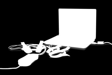
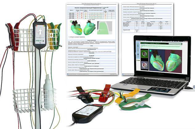
3 Kardi is a revolutionizing non-invasive diagnostic tool used for testing ischaemic heart disease It s technology generates high quality 3D visuals projected from traditional ECG plus Dispersion Mapping. The actual test only lasts 30 seconds.
 but those of the QRS and R-wave complex as")
4 By analyzing low amplitude oscillations of conventional ECG signals, we can generate a stable signal of ECG micro fluctuations, by reflecting not just T-wave alternans (noise and artifact) but those of the QRS and R-wave complex as well

5 The 3-dimensional image projected by the system allows for physicians to observe the condition of the heart muscle and the intensity of ischemic heart disease.
6 The Composition

7 Four electrodes are applied in accordance to standard ECG arrangement of ECG limb leads ECG data acquisitioned in 30 seconds An image of the heart is formed on screen together with quantitative and qualitative analysis of cardiac electrical activity.

8 Results Kardi results are shown in the form of a numeric dispersive characteristics range and a disperson mapping The dispersive mapping is a colour-coded image of the heart in which green represented healthy and red represented pathological changes
9 Results Dispersive characteristics are expressed by 9 analyzed groups of deviations. In these groups, the characteristics were analyzed reflecting electrophysiological abnormalities in the: depolarization (generation of an electrical impulse that causes an action potential or a short lasting contraction of the heart muscles) and; repolarization (heart muscle returns to its original state) of the myocardium.

10 Dispersive Indices (as seen online)

11 Dispersive Indices (as printed/ ed)

12 ECG Tracing
13 The system therefore reacts to changes that exist or operate, below the threshold of normal ECG, that violate the synchrony of ventricular electrical excitation, as well as microscopic changes in the ionic balance of myocytes (muscle cell or muscle fiber) and other changes that are due to small quantities that do not appear in the morphology of the traditional ECG signals.
14 Why say YES to Kardi?
15 Feature: ECG dispersion mapping by capturing low amplitude waveforms 3D portrait of the heart highlights impaired areas of the myocardium Benefit: Real time assessment for ischemic heart disease quicker diadnosis quicker prescription of treatment
16 Feature: Uses ECG wave alternans (noise) for information Benefit: Ability to use information that is regarded as insignificant or unusable by the traditional ECG systems, giving more information to doctors about heart health
17 Feature: Uses four limb lead wires with patients sitting in an upright position and fully clothed Benefit: More comfortability during the procedure without undressing or limited to lying down
18 Feature: Specificity and sensitivity to ischemic heart disease increased by 80% Benefit: Detection of extremely small deviations during cardiac cycles which are invisible to regular ECG systems
19 Feature: Color-coded 3D portrait of the heart Benefit: Reduces the need for 2D echocardiography or other visualization tests cutting expenses used for diagnostics alone

20 Used for assessing if modes of treatment prescribed work

21 Visuals help patients to be more conscious of lifestyle choices before and 1 day after taking Max One

22 easy detection of patterns of heart anomalies
23 For an easier, faster, accurate heart diagnosis, Kardi paves the way for a more effective practice for doctors and more proactive health management of patients.
Interpreting Electrocardiograms (ECG) Physiology Name: Per:
 Physiology Name: Per:") Interpreting Electrocardiograms (ECG) Physiology Name: Per: Introduction The heart has its own system in place to create nerve impulses and does not actually require the brain to make it beat. This electrical
Interpreting Electrocardiograms (ECG) Physiology Name: Per: Introduction The heart has its own system in place to create nerve impulses and does not actually require the brain to make it beat. This electrical
3/26/15 HTEC 91. EKG Sign-in Book. The Cardiac Cycle. Parts of the ECG. Waves. Waves. Review of protocol Review of placement of chest leads (V1, V2)
") EKG Sign-in Book HTEC 91 Review of protocol Review of placement of chest leads (V1, V2) Medical Office Diagnostic Tests Week 2 http://www.cvphysiology.com/arrhythmias/a013c.htm The Cardiac Cycle Represents
EKG Sign-in Book HTEC 91 Review of protocol Review of placement of chest leads (V1, V2) Medical Office Diagnostic Tests Week 2 http://www.cvphysiology.com/arrhythmias/a013c.htm The Cardiac Cycle Represents
Chapter 12: Cardiovascular Physiology System Overview
 Chapter 12: Cardiovascular Physiology System Overview Components of the cardiovascular system: Heart Vascular system Blood Figure 12-1 Plasma includes water, ions, proteins, nutrients, hormones, wastes,
INTRODUCTION TO ECG. Dr. Tamara Alqudah
 INTRODUCTION TO ECG Dr. Tamara Alqudah Excitatory & conductive system of the heart + - The ECG The electrocardiogram, or ECG, is a simple & noninvasive diagnostic test which records the electrical
Cardiac Telemetry Self Study: Part One Cardiovascular Review 2017 THINGS TO REMEMBER
 Please review the above anatomy of the heart. THINGS TO REMEMBER There are 3 electrolytes that affect cardiac function o Sodium, Potassium, and Calcium When any of these electrolytes are out of the normal
RN-BC, MS, CCRN, FAHA
 Presented By: Barbara Furry, RN-BC, MS, CCRN, FAHA Director The Center of Excellence in Education Director of HERO Follow me on Twitter! CEE Med Updates@BarbaraFurryRN Like me on Facebook! 1 A. Atropine
Testing the Accuracy of ECG Captured by Cronovo through Comparison of ECG Recording to a Standard 12-Lead ECG Recording Device
 Testing the Accuracy of ECG Captured by through Comparison of ECG Recording to a Standard 12-Lead ECG Recording Device Data Analysis a) R-wave Comparison: The mean and standard deviation of R-wave amplitudes
Enhanced Cellular Automation Model For Detecting Heart Fibrillation
 Enhanced Cellular Automation Model For Detecting Heart Fibrillation R. Touqan, A. Nasser & N. Hamdy Arab Academy for Science and Technology (AAST), email< rtouqan@hotmail.com Abstract: A new technique
Lab Activity 24 EKG. Portland Community College BI 232
 Lab Activity 24 EKG Reference: Dubin, Dale. Rapid Interpretation of EKG s. 6 th edition. Tampa: Cover Publishing Company, 2000. Portland Community College BI 232 Graph Paper 1 second equals 25 little boxes
The Electrocardiogram
 The Electrocardiogram Chapters 11 and 13 AUTUMN WEDAN AND NATASHA MCDOUGAL The Normal Electrocardiogram P-wave Generated when the atria depolarizes QRS-Complex Ventricles depolarizing before a contraction
for Heart-Health Scanning
 Magnetocardiography for Heart-Health Scanning CardioMag Imaging, Inc. 1 Basic Principles of Magnetocardiography (MCG) The cardiac electric activity that produces a voltage difference on the body surface
Presented By: Barbara Furry, RN-BC, MS, CCRN, FAHA Director The Center of Excellence in Education Director of HERO
 Presented By: Barbara Furry, RN-BC, MS, CCRN, FAHA Director The Center of Excellence in Education Director of HERO Follow me on Twitter! CEE Med Updates@BarbaraFurryRN Like me on Facebook! What is a
: Biomedical Signal Processing
 : Biomedical Signal Processing 0. Introduction: Biomedical signal processing refers to the applications of signal processing methods, such as Fourier transform, spectral estimation and wavelet transform,
Understanding basics of EKG
 Understanding basics of EKG By Alula A.(R III) www.le.ac.uk Topic for discussion Understanding of cellular electrophysiology Basics Rate Rhythm Axis Intervals P wave QRS ST/T wave Abnormal EKGs Understanding
Birmingham Regional Emergency Medical Services System
 Birmingham Regional Emergency Medical Services System 2018 ALCTE Summer Conference EKG Basics Brian Gober, MAT, ATC, NRP, CSCS Education Services Manager ECC Training Center Coordinator Birmingham Regional
Cardiac Cycle. Each heartbeat is called a cardiac cycle. First the two atria contract at the same time.
 The Heartbeat Cardiac Cycle Each heartbeat is called a cardiac cycle. First the two atria contract at the same time. Next the two ventricles contract at the same time. Then all the chambers relax. http://www.youtube.com/watch?v=frd3k6lkhws
Lab 4: Introduction to Physiological Measurements - Cardiovascular
 Lab 4: Introduction to Physiological Measurements - Cardiovascular INTRODUCTION: This lab will demonstrate cardiovascular measurements by creating an ECG with instruments used in previous labs. Students
BIO 360: Vertebrate Physiology Performing and analyzing an EKG Lab 11: Performing and analyzing an EKG Lab report due April 17 th
 BIO 60: Vertebrate Physiology Lab : Lab report due April 7 th All muscles produce an electrical current when they contract. The heart is no exception. An electrocardiogram (ECG or EKG) is a graphical recording
12 Lead ECG Interpretation: Color Coding for MI s
 12 Lead ECG Interpretation: Color Coding for MI s Anna E. Story, RN, MS Director, Continuing Professional Education Critical Care Nurse Online Instructional Designer 2004 Anna Story 1 Objectives review
Analyzing the Heart with EKG
 Analyzing the Heart with EKG LabQuest An electrocardiogram (ECG or EKG) is a graphical recording of the electrical events occurring within the heart. In a healthy heart there is a natural pacemaker in
Electrocardiography for Healthcare Professionals
 Electrocardiography for Healthcare Professionals Kathryn A. Booth Thomas O Brien Chapter 10: Pacemaker Rhythms and Bundle Branch Block Learning Outcomes 10.1 Describe the various pacemaker rhythms. 10.2
Mathematical modeling of ischemia and infarction
 Mathematical modeling of ischemia and infarction Mostly based on Cimponeriu, Starmer and Bezerianos: A theoretical analysis of acute ischemia and infarction using ECG reconstruction on a 2-D model of myocardium
Electrocardiography for Healthcare Professionals
 Electrocardiography for Healthcare Professionals Chapter 9: Ventricular Dysrhythmias 2012 The Companies, Inc. All rights reserved. Learning Outcomes 9.1 Describe the various ventricular dysrhythmias 9.2
Figure 1 muscle tissue to its resting state. By looking at several beats you can also calculate the rate for each component.
 ANALYZING THE HEART WITH EKG WITH LABQUEST LAB From Human Physiology with Vernier Westminster College INTRODUCTION An electrocardiogram (ECG or EKG) is a graphical recording of the electrical events occurring
Figure 2. Normal ECG tracing. Table 1.
 Figure 2. Normal ECG tracing that navigates through the left ventricle. Following these bundle branches the impulse finally passes to the terminal points called Purkinje fibers. These Purkinje fibers are
- why the T wave is deflected upwards although it's a repolarization wave?
 Cardiac Electrograph: - why the T wave is deflected upwards although it's a repolarization wave? After depolarization the ventricle contracts but since the heart is a volume conductor (3D not 2D), when
I PART I. Timing Cycles and Troubleshooting Review COPYRIGHTED MATERIAL
 I PART I Timing Cycles and Troubleshooting Review COPYRIGHTED MATERIAL 1 SECTION 1 Calculating Rates and Intervals Figure 1.1 Few clinicians can escape their training without having to learn to read a
Cardiac physiology. b. myocardium -- cardiac muscle and fibrous skeleton of heart
 I. Heart anatomy -- general gross. A. Size/orientation - base/apex B. Coverings D. Chambers 1. parietal pericardium 2. visceral pericardium 3. Layers of heart wall a. epicardium Cardiac physiology b. myocardium
5- The normal electrocardiogram (ECG)
") 5- The (ECG) Introduction Electrocardiography is a process of recording electrical activities of heart muscle at skin surface. The electrical current spreads into the tissues surrounding the heart, a small
5- The (ECG) Introduction Electrocardiography is a process of recording electrical activities of heart muscle at skin surface. The electrical current spreads into the tissues surrounding the heart, a small
HST-582J/6.555J/16.456J-Biomedical Signal and Image Processing-Spring Laboratory Project 1 The Electrocardiogram
 HST-582J/6.555J/16.456J-Biomedical Signal and Image Processing-Spring 2007 DUE: 3/8/07 Laboratory Project 1 The Electrocardiogram 1 Introduction The electrocardiogram (ECG) is a recording of body surface
Family Medicine for English language students of Medical University of Lodz ECG. Jakub Dorożyński
 Family Medicine for English language students of Medical University of Lodz ECG Jakub Dorożyński Parts of an ECG The standard ECG has 12 leads: six of them are considered limb leads because they are placed
Introduction to ECG Gary Martin, M.D.
 Brief review of basic concepts Introduction to ECG Gary Martin, M.D. The electrical activity of the heart is caused by a sequence of rapid ionic movements across cell membranes resulting first in depolarization
Electrocardiography Normal 5. Faisal I. Mohammed, MD, PhD
 Electrocardiography Normal 5 Faisal I. Mohammed, MD, PhD 1 Objectives 2 1. Describe the different waves in a normal electrocardiogram. 2. Recall the normal P-R and Q-T interval time of the QRS wave. 3.
EEG, ECG, EMG. Mitesh Shrestha
 EEG, ECG, EMG Mitesh Shrestha What is Signal? A signal is defined as a fluctuating quantity or impulse whose variations represent information. The amplitude or frequency of voltage, current, electric field
Full file at
 MULTIPLE CHOICE. Choose the one alternative that best completes the statement or answers the question. 1) What electrical event must occur for atrial kick to occur? 1) A) Atrial repolarization B) Ventricular
BASIC CONCEPT OF ECG
 BASIC CONCEPT OF ECG Electrocardiogram The electrocardiogram (ECG) is a recording of cardiac electrical activity. The electrical activity is readily detected by electrodes attached to the skin. After the
Practice Exercises for the Cardiovascular System
 Practice Exercises for the Cardiovascular System On the diagram below, color the oxygen-rich blood red and the oxygen-poor blood blue. Label the parts: Continued on the next page... Label the parts on
ECG Interpretation Cat Williams, DVM DACVIM (Cardiology)
") ECG Interpretation Cat Williams, DVM DACVIM (Cardiology) Providing the best quality care and service for the patient, the client, and the referring veterinarian. GOAL: Reduce Anxiety about ECGs Back to
ECG Interpretation Cat Williams, DVM DACVIM (Cardiology) Providing the best quality care and service for the patient, the client, and the referring veterinarian. GOAL: Reduce Anxiety about ECGs Back to
PART I. Disorders of the Heart Rhythm: Basic Principles
 PART I Disorders of the Heart Rhythm: Basic Principles FET01.indd 1 1/11/06 9:53:05 AM FET01.indd 2 1/11/06 9:53:06 AM CHAPTER 1 The Cardiac Electrical System The heart spontaneously generates electrical
CORONARY ARTERIES HEART
 CARDIAC/ECG MODULE THE HEART CORONARY ARTERIES FIBRILLATING HEART CORONARY ARTERIES HEART PRACTICE RHYTHMS PRACTICE RHYTHMS ELECTRICAL CONDUCTION SA Node (60 100) Primary pacemaker AV Node (40 60) ***Creates
Shock-induced termination of cardiac arrhythmias
 Shock-induced termination of cardiac arrhythmias Group members: Baltazar Chavez-Diaz, Chen Jiang, Sarah Schwenck, Weide Wang, and Jinglei Zhang Cardiac arrhythmias, also known as irregular heartbeat, occur
Biology 13A Lab #10: Cardiovascular System II ECG & Heart Disease
 Biology 13A Lab #10: Cardiovascular System II ECG & Heart Disease Lab #10 Table of Contents: Expected Learning Outcomes...... 83 Introduction....... 84 Activity 1: Collecting ECG Data..... 85 Activity
Sheet 5 physiology Electrocardiography-
 *questions asked by some students Sheet 5 physiology Electrocardiography- -why the ventricles lacking parasympathetic supply? if you cut both sympathetic and parasympathetic supply of the heart the heart
Long Term Monitoring of the Intrinsic Ventricular Monophasic Action Potential with an Implantable DDD Pacemaker
 May 1998 79 Long Term Monitoring of the Intrinsic Ventricular Monophasic Action Potential with an Implantable DDD Pacemaker T. LAWO, J. BARMEYER Abteilung für Kardiologie, Universitätsklinik Bergmannsheil,
ELECTROCARDIOGRAPHY (ECG)
") ELECTROCARDIOGRAPHY (ECG) The heart is a muscular organ, which pumps blood through the blood vessels of the circulatory system. Blood provides the body with oxygen and nutrients, as well as assists in
ELECTROCARDIOGRAPHY (ECG) The heart is a muscular organ, which pumps blood through the blood vessels of the circulatory system. Blood provides the body with oxygen and nutrients, as well as assists in
ECG UPDATE COURSE DESCRIPTION
 ECG UPDATE COURSE DESCRIPTION This CE course discusses techniques to improve ECG quality, interpretation of normal and abnormal ECG tracings, and troubleshooting ECG tracings. Note: An internet connection
Lab #3: Electrocardiogram (ECG / EKG)
") Lab #3: Electrocardiogram (ECG / EKG) An introduction to the recording and analysis of cardiac activity Introduction The beating of the heart is triggered by an electrical signal from the pacemaker. The
Lab #3: Electrocardiogram (ECG / EKG) An introduction to the recording and analysis of cardiac activity Introduction The beating of the heart is triggered by an electrical signal from the pacemaker. The
By the end of this lecture, you will be able to: Understand the 12 lead ECG in relation to the coronary circulation and myocardium Perform an ECG
 By the end of this lecture, you will be able to: Understand the 12 lead ECG in relation to the coronary circulation and myocardium Perform an ECG recording Identify the ECG changes that occur in the presence
CAMOSUN COLLEGE BIOLOGY 144 (2010) LABS
 LABS") LAB 8: CARDIOVASCULAR PHYSIOLOGY PART 1. HEART SOUNDS AND PULSE DETERMINATIONS Introduction Two distinct sounds can be heard during each cardiac cycle. These sounds are commonly described as lub and dup
LAB 8: CARDIOVASCULAR PHYSIOLOGY PART 1. HEART SOUNDS AND PULSE DETERMINATIONS Introduction Two distinct sounds can be heard during each cardiac cycle. These sounds are commonly described as lub and dup
iworx Sample Lab Experiment HH-4: The Six-Lead Electrocardiogram
 Experiment HH-4: The Six-Lead Electrocardiogram Background The cardiac cycle involves a sequential contraction of the atria and the ventricles. These contractions are triggered by the coordinated electrical
Electrocardiography I Laboratory
 Introduction The body relies on the heart to circulate blood throughout the body. The heart is responsible for pumping oxygenated blood from the lungs out to the body through the arteries and also circulating
CASE 10. What would the ST segment of this ECG look like? On which leads would you see this ST segment change? What does the T wave represent?
 CASE 10 A 57-year-old man presents to the emergency center with complaints of chest pain with radiation to the left arm and jaw. He reports feeling anxious, diaphoretic, and short of breath. His past history
Speed - Accuracy - Exploration. Pathfinder SL
 Speed - Accuracy - Exploration Pathfinder SL 98000 Speed. Accuracy. Exploration. Pathfinder SL represents the evolution of over 40 years of technology, design, algorithm development and experience in the
Electrocardiogram sensor (ECG/EKG sensor)
") [KDS-1040] Electrocardiogram sensor (ECG/EKG sensor) User's Manual Note This product is designed for educational use only. Not recommended for industrial, medical, commercial use. What is Electrocardiogram?
[KDS-1040] Electrocardiogram sensor (ECG/EKG sensor) User's Manual Note This product is designed for educational use only. Not recommended for industrial, medical, commercial use. What is Electrocardiogram?
Atlantic Health System
 Atlantic Health System Morristown Medical Center Newton Medical Center Overlook Medical Center Basic Dysrhythmia Course Day 1 1 2 Chapter 1 Anatomy and Physiology Learning Objectives 1) Identify electrophysiology
Objectives of the Heart
 Objectives of the Heart Electrical activity of the heart Action potential EKG Cardiac cycle Heart sounds Heart Rate The heart s beat separated into 2 phases Relaxed phase diastole (filling of the chambers)
DETECTION OF HEART ABNORMALITIES USING LABVIEW
 IASET: International Journal of Electronics and Communication Engineering (IJECE) ISSN (P): 2278-9901; ISSN (E): 2278-991X Vol. 5, Issue 4, Jun Jul 2016; 15-22 IASET DETECTION OF HEART ABNORMALITIES USING
CS 100. Diagnostic Manual
 CS 100 Frequency Cardiograph (FCG R ) Analysis Systems Diagnostic Manual Version 3.1 Trigon Medical Inc. ALL COPYRIGHT RESERED 2013 1 General Information: Device Name: CS-100 (Cardio Scan-100) Software
ECG and Cardiac Electrophysiology
 ECG and Cardiac Electrophysiology Simon Some very basic electrophysiology Intracellular fluid: 10 mm Na, 140 mm K, etc. K Na-K ATPase Extracellular fluid: 140mM Na, 4mM K, etc. Na Ion gradient plus selective
Electrocardiography Abnormalities (Arrhythmias) 7. Faisal I. Mohammed, MD, PhD
 7. Faisal I. Mohammed, MD, PhD") Electrocardiography Abnormalities (Arrhythmias) 7 Faisal I. Mohammed, MD, PhD 1 Causes of Cardiac Arrythmias Abnormal rhythmicity of the pacemaker Shift of pacemaker from sinus node Blocks at different
Electrocardiography Abnormalities (Arrhythmias) 7 Faisal I. Mohammed, MD, PhD 1 Causes of Cardiac Arrythmias Abnormal rhythmicity of the pacemaker Shift of pacemaker from sinus node Blocks at different
WHAT S THAT RHYTHM I AM HEARING? GUIDE TO AUSCULTATION OF ARRHYTHMIAS IN HORSES
 WHAT S THAT RHYTHM I AM HEARING? GUIDE TO AUSCULTATION OF ARRHYTHMIAS IN HORSES Michelle Henry Barton DVM, PhD, DACVIM University of Georgia, Athens, GA INTRODUCTION The purpose of this talk is to review
12-Lead ECG Interpretation. Kathy Kuznar, RN, ANP
 12-Lead ECG Interpretation Kathy Kuznar, RN, ANP The 12-Lead ECG Objectives Identify the normal morphology and features of the 12- lead ECG. Perform systematic analysis of the 12-lead ECG. Recognize abnormalities
Panorama. Arrhythmia Analysis Frequently Asked Questions
 Panorama Arrhythmia Analysis Frequently Asked Questions What ECG vectors are used for Beat Detection? 3-wire lead set 5-wire lead set and 12 lead What ECG vectors are used for Beat Typing? 3-wire lead
What the ECG is about
 What to expect from the ECG 00 The electricity of the heart 00 The shape of the ECG 00 The ECG electrical pictures 00 The shape of the QRS complex 00 Making a recording practical points 00 How to report
Lect.6 Electrical axis and cardiac vector Cardiac vector: net result Vector that occurs during depolarization of the ventricles Figure:
 Lect.6 Electrical axis and cardiac vector Objectives: 1. State the relationship between the direction of cardiac vector with the direction (-ve, +ve) and amplitude of an ECG waves. 2. Draw diagram indicting
DEFIBRILLATORS. Prof. Yasser Mostafa Kadah
 DEFIBRILLATORS Prof. Yasser Mostafa Kadah Basics Defibrillation is definitive treatment for life-threatening cardiac arrhythmias such as ventricular fibrillation Defibrillation consists of delivering therapeutic
Anatomy Review: The Heart Graphics are used with permission of A.D.A.M. Software, Inc. and Benjamin/Cummings Publishing Co.
 Anatomy Review: The Heart Graphics are used with permission of A.D.A.M. Software, Inc. and Benjamin/Cummings Publishing Co. Anatomy Views Label the diagrams of the heart below: Interactive Physiology Study
Electrocardiogram and Heart Sounds
 Electrocardiogram and Heart Sounds Five physiologic properties of cardiac muscle Automaticity: SA node is the primary pacemaker of the heart, but any cells in the conduction system can initiate their
ECG. Prepared by: Dr.Fatima Daoud Reference: Guyton and Hall Textbook of Medical Physiology,12 th edition Chapters: 11,12,13
 ECG Prepared by: Dr.Fatima Daoud Reference: Guyton and Hall Textbook of Medical Physiology,12 th edition Chapters: 11,12,13 The Concept When the cardiac impulse passes through the heart, electrical current
The Normal Electrocardiogram
 C H A P T E R 1 1 The Normal Electrocardiogram When the cardiac impulse passes through the heart, electrical current also spreads from the heart into the adjacent tissues surrounding the heart. A small
Lesson 4 ECG 2 Electrocardiography
 Physiology Lessons for use with the Biopac Science Lab MP40 Lesson 4 ECG 2 Electrocardiography PC running Windows XP or Mac OS X 10.3-10.4 Lesson Revision 1.20.2006 BIOPAC Systems, Inc. 42 Aero Camino,
8/20/ Identify the functions of common ECG machines. 3.3 Explain how each ECG machine control is used. 3.4 Recognize common electrodes.
 1 2 Electrocardiography for Healthcare Professionals Chapter 3: The Electrocardiograph Learning Outcomes 3.1 Identify three types of leads, and explain how each is recorded. 3.2 Identify the functions
CRITICAL THINKING QUESTIONS AND ANSWERS AND CYCLE 2 LAB EXAM TEMPLATE. There are two main mechanisms that work in conjunction to return the blood
 CRITICAL THINKING QUESTIONS AND ANSWERS AND CYCLE 2 LAB EXAM TEMPLATE There are two main mechanisms that work in conjunction to return the blood THE CARDIAC PUMP 1) The forward pull(vis a fronte) This
Systolic and Diastolic Currents of Injury
 Systolic and Diastolic Currents of Injury Figure 1 Action Potentials of Normal and Ischemic Tissue In Figure 1 above, the action potential of normal myocardium is represented by the solid lines and the
Ventricular Parasystole
 Ventricular Parasystole 1 WHAT IS IT? In addition to the sinus node, there are many accessory pacemakers throughout the conducting system of the atria, junction and ventricles that are ready to assume
A Cellular Automata Model For Dynamics and Control of Cardiac Arrhythmias DANNY GALLENBERGER (CARROLL UNIVERSITY) XIAOPENG ZHAO (UTK) KWAI WONG (UTK)
 XIAOPENG ZHAO (UTK) KWAI WONG (UTK)") A Cellular Automata Model For Dynamics and Control of Cardiac Arrhythmias DANNY GALLENBERGER (CARROLL UNIVERSITY) XIAOPENG ZHAO (UTK) KWAI WONG (UTK) Introduction Sudden cardiac arrest is responsible for
A Cellular Automata Model For Dynamics and Control of Cardiac Arrhythmias DANNY GALLENBERGER (CARROLL UNIVERSITY) XIAOPENG ZHAO (UTK) KWAI WONG (UTK) Introduction Sudden cardiac arrest is responsible for
ABCs of ECGs. Shelby L. Durler
 ABCs of ECGs Shelby L. Durler Objectives Review the A&P of the cardiac conduction system Placement and obtaining 4-lead and 12-lead ECGs Overview of the basics of ECG rhythm interpretation Intrinsic
Chapter 08. Health Screening and Risk Classification
 Chapter 08 Health Screening and Risk Classification Preliminary Health Screening and Risk Classification Protocol: 1) Conduct a Preliminary Health Evaluation 2) Determine Health /Disease Risks 3) Determine
This presentation will deal with the basics of ECG description as well as the physiological basics of
 Snímka 1 Electrocardiography basics This presentation will deal with the basics of ECG description as well as the physiological basics of Snímka 2 Lecture overview 1. Cardiac conduction system functional
DR QAZI IMTIAZ RASOOL OBJECTIVES
 PRACTICAL ELECTROCARDIOGRAPHY DR QAZI IMTIAZ RASOOL OBJECTIVES Recording of electrical events in heart Established electrode pattern results in specific tracing pattern Health of heart i. e. Anatomical
Warning: Avoid putting the tip of the syringe needle too deeply into the hole to avoid damage to the heart tissue.
 Experiment AM-12: Crayfish Electrocardiogram (ECG) Exercise 1: The Crayfish ECG and Heart Rate Aim: To record the electrical trace (ECG) produced by the contraction of a resting heart, and to determine
BME 365 Website. Project Directions
 Lecture 17 EKG BME 365 Website Project Directions Heart rate Factors Affecting CO Parasympathetic activity decreases HR Sympathetic activity increases HR Stroke volume Depends on force generated by cardiac
PROBLEM SET 3. Assigned: February 19, 2004 Due: February 26, 2004
 Harvard-MIT Division of Health Sciences and Technology HST.542J: Quantitative Physiology: Organ Transport Systems Instructors: Roger Mark and Jose Venegas MASSACHUSETTS INSTITUTE OF TECHNOLOGY Departments
CIS 632 / EEE 687 Mobile Computing
 CIS 632 / EEE 687 Mobile Computing MC Platform #2: BioRadio Chansu Yu Body Signals Temperature, Skin humidity (dehydration) Lung sounds (LS), heart sounds (HS), and bowel sounds (BS) Blood pressure, Blood
Lab 2. The Intrinsic Cardiac Conduction System. 1/23/2016 MDufilho 1
 Lab 2 he Intrinsic Cardiac Conduction System 1/23/2016 MDufilho 1 Figure 18.13 Intrinsic cardiac conduction system and action potential succession during one heartbeat. Superior vena cava ight atrium 1
Chapter 9, Part 2. Cardiocirculatory Adjustments to Exercise
 Chapter 9, Part 2 Cardiocirculatory Adjustments to Exercise Electrical Activity of the Heart Contraction of the heart depends on electrical stimulation of the myocardium Impulse is initiated in the right
Assessment of Reliability of Hamilton-Tompkins Algorithm to ECG Parameter Detection
 Proceedings of the 2012 International Conference on Industrial Engineering and Operations Management Istanbul, Turkey, July 3 6, 2012 Assessment of Reliability of Hamilton-Tompkins Algorithm to ECG Parameter
Cardiovascular Physiology
 Cardiovascular Physiology The mammalian heart is a pump that pushes blood around the body and is made of four chambers: right and left atria and right and left ventricles. The two atria act as collecting
Cardiology. Objectives. Chapter
 1:44 M age 1121 Chapter Cardiology Objectives art 1: Cardiovascular natomy and hysiology, ECG Monitoring, and Dysrhythmia nalysis (begins on p. 1127) fter reading art 1 of this chapter, you should be able
Cardiovascular System Notes: Heart Disease & Disorders
 Cardiovascular System Notes: Heart Disease & Disorders Interesting Heart Facts The Electrocardiograph (ECG) was invented in 1902 by Willem Einthoven Dutch Physiologist. This test is still used to evaluate
Shock-induced termination of cardiac arrhythmias
 Shock-induced termination of cardiac arrhythmias Group members: Baltazar Chavez-Diaz, Chen Jiang, Sarah Schwenck, Weide Wang, and Jinglei Zhang Abstract: Cardiac arrhythmias occur when blood flow to the
Sample. Analyzing the Heart with EKG. Computer
 Analyzing the Heart with EKG Computer An electrocardiogram (ECG or EKG) is a graphical recording of the electrical events occurring within the heart. In a healthy heart there is a natural pacemaker in
MARS Ambulatory ECG Analysis The power to assess and predict
 GE Healthcare MARS Ambulatory ECG Analysis The power to assess and predict Connecting hearts and minds Prevention starts with knowledge Around the world, heart disease is one of our fastest-growing health
Medical Electronics Dr. Neil Townsend Michaelmas Term 2001 (www.robots.ox.ac.uk/~neil/teaching/lectures/med_elec) The story so far
 The story so far") Medical Electronics Dr. Neil Townsend Michaelmas Term 2001 (www.robots.ox.ac.uk/~neil/teaching/lectures/med_elec) The story so far The heart pumps blood around the body. It has four chambers which contact
Medical Electronics Dr. Neil Townsend Michaelmas Term 2001 (www.robots.ox.ac.uk/~neil/teaching/lectures/med_elec) The story so far The heart pumps blood around the body. It has four chambers which contact
11/18/13 ECG SIGNAL ACQUISITION HARDWARE DESIGN. Origin of Bioelectric Signals
 ECG SIGNAL ACQUISITION HARDWARE DESIGN Origin of Bioelectric Signals 1 Cell membrane, channel proteins Electrical and chemical gradients at the semi-permeable cell membrane As a result, we get a membrane
Electrocardiographic Markers Associated with Sotalol-induced Torsades de Pointes
 Electrocardiographic Markers Associated with Sotalol-induced Torsades de Pointes Based on Data from the Telemetric and Holter ECG Warehouse (THEW) Initiative Jean-PhiIippe Couderc, PhD Center for Quantitative
Electrocardiography for Healthcare Professionals
 Electrocardiography for Healthcare Professionals Kathryn A. Booth Thomas O Brien Chapter 5: Rhythm Strip Interpretation and Sinus Rhythms Learning Outcomes 5.1 Explain the process of evaluating ECG tracings
Simulation of T-Wave Alternans and its Relation to the Duration of Ventricular Action Potentials Disturbance
 The Open Pacing, Electrophysiology & Therapy Journal, 2010, 3, 21-27 21 Open Access Simulation of T-Wave Alternans and its Relation to the Duration of Ventricular Action Potentials Disturbance Dariusz
UNDERSTANDING YOUR ECG: A REVIEW
 UNDERSTANDING YOUR ECG: A REVIEW Health professionals use the electrocardiograph (ECG) rhythm strip to systematically analyse the cardiac rhythm. Before the systematic process of ECG analysis is described
Identification of Ischemic Lesions Based on Difference Integral Maps, Comparison of Several ECG Intervals
 1.2478/v148-9-21-7 MEASUREMENT SCIENCE REVIEW, Volume 9, No. 5, 29 Identification of Ischemic Lesions Based on Difference Integral Maps, Comparison of Several ECG Intervals J. Švehlíková, M. Kania 1, M.