QUIZ/TEST REVIEW NOTES SECTION 1 CARDIAC MYOCYTE PHYSIOLOGY [CARDIOLOGY]
|
|
|
- Clifton Shields
- 5 years ago
- Views:
Transcription
![QUIZ/TEST REVIEW NOTES SECTION 1 CARDIAC MYOCYTE PHYSIOLOGY [CARDIOLOGY] Learning Objectives: Describe the ionic basis of action potentials in cardiac contractile and autorhythmic cells Explain the](/docs-images/96/126998162/images/1-0.jpg "relationship between refractory periods and the absence of summation in cardiac muscle contraction I. TWO TYPES OF ELECTRICAL SIGNALS a.")

1 QUIZ/TEST REVIEW NOTES SECTION 1 CARDIAC MYOCYTE PHYSIOLOGY [CARDIOLOGY] Learning Objectives: Describe the ionic basis of action potentials in cardiac contractile and autorhythmic cells Explain the relationship between refractory periods and the absence of summation in cardiac muscle contraction I. TWO TYPES OF ELECTRICAL SIGNALS a. Structure of cardiac myocytes Appearance under microscope: Fiber arrangement: Fiber proteins: Control: Nervous control: Hormonal influence: Location: Morphology: Internal structure: Contraction speed: Contraction force of single fiber twitch: Initiation of contraction: Striated Sarcomeres Actin, myosin: troponin/tropomyosin Involuntary/Ca2+ and troponin/fibers electrically linked via gap junctions Autonomic neurons Epinephrine Heart muscle Uninucleate; branch fibers T-tubule and Sarcoplasmic reticulum Intermediate Graded Autorhythmic MAJOR POINTS INSERT FIGURE CARDIAC MUSCLE - Single nucleus per fiber - Individual cardiac cells branch and join neighboring cells end-to-end creating complex of networks formed through cell junctions known as intercalated disks Intercalated Disks components: (1) Desmosomes: tie cell together allowing contractile force to be transfer cell to cell (2) Gap Junctions: Electrically connect cardiac cells together - T-tubules branch inside the myocardial cells - Sarcoplasmic reticulum is smaller because Extracellular Ca2+ is needed to initiate contraction; not a adjacent protein - Mitochondria occupy about 1/3 the cell volume of a contractile fiber TWO FORMS OF MYOCYTES - Contractile Cardiac Muscle Cells (myocardium) [Organized sarcomeres/99% of heart] - Autorhythmic Myocytes (pacemakers) [Unorganized sarcomeres/1% of heart/generate A.P. spontaneously] 1

2 II. MYOCARDIAL CONTRACTION a. Depolarization - A.P. enters contractile cell moving across the sarcolemma into the t-tubes - A.P. then opens voltage-gated Ca2+ channels in the t-tubules, allowing Ca2+ to open up ryanodine receptor channels (RyR) in the Sarcoplasmic reticulum [RyR operated by Ca2+ binding unlike mechanical linkage of RyR found in skeletal muscle] - Ca2+ Induced Ca2+ Release: RyR opening caused by calcium, which leads to the flow of stored calcium out into the cytosol to meet with troponin and initiate the cycle of cross-bridge formation and movement REUPTAKE: Cytoplasmic Ca2+ concentrations decrease, Ca2+ unbinds from troponin, myosin releases actin, and contractile filaments slide back to relaxed position (1) Ca2+ transported back to Sarcoplasmic reticulum with Ca2+-ATPase (2) Ca2+ removed via Na+-Ca2+ antiport protein [3 Na+ in for 1 Ca2+ out] [Na is later removed by Na+-K+-ATPase] GRADED CONTRACTIONS: Force generated by cardiac muscle is proportional to the number of cross-bridges that are active; number of active cross-bridges is determined by how much Ca2+ is bound to troponin b. Action Potential of contractile Myocytes 1. Membrane Potential Changes - Resting potential -90mV [skeletal muscle -70mV] - Depolarization: Na+ influx - Repolarization: K+ efflux - Longer action potential caused by Ca2+ entry 2. Role of Calcium - Ca2+ influx at the plateau lengthens total duration of A.P, slowing down repolarization; causing the cell to stay positive for longer period of time 2


3 3. Increased length of refractory period - Longer A.P. helps prevent tetanus or sustained contraction which is important because muscles must relax between contractions so ventricles can fill with blood - Tetanus cannot occur in cardiac muscle because the longer A.P. means the refractory period and the contraction end almost simultaneously Phase 4: Resting Membrane Potential [resting potential of -90mV] Phase 0: Depolarization [Depolarization spreads through gap junctions, membrane becomes more positive. Voltage gated Na+ channels open allowing Na+ to enter cell and rapidly depolarize; potential reaches about +20mV then double-gated Na+ channels close] Phase 1: Initial Repolarization: Na+ channels close; cell begins to repolarize by efflux of K+ through K+ channels Phase 2: The Plateau: Decrease in K+ permeability and increase in Ca2+ permeability; Ca2+ channels slowly opening since phases 0 and 1; When Ca2+ channels open some K+ channels close, a influx of Ca2+ and decreased efflux of Na+ causes A.P. to flatten out into plateau [extending refractory period/action potential] Phase 3: Rapid Repolarization: Plateau ends with Ca2+ channels close and K+ permeability increase; K+ channels are activated during depolarization but are slow to open; Once K+ channels open K+ efflux occurs rapidly returning cell to resting potential III. AUTORHYTHMIC CELLS [pacemakers] a. Characteristics - Fulfill 1% of cardiac myocytes - Unstable membrane potential - No organized sarcomeres - Generated spontaneous action potentials 3
4 - Pacemaker cells found toward apex of the heart are faster then pacemaker cells found toward the base of the heart (slow) - Speed with which pacemaker cells depolarizes determines the rate at which heart contracts (bpm) b. Instability - If Channels: Permeable to both K+ and Na+ > Allow current (I) to flow > Subscript (f) stands for funny current because scientists did not understand them - If Channel opens at negative membrane potential: > Na+ influx exceeds K+ efflux > Net influx of positive charge depolarizes autorhythmic cell > As membrane becomes more, If channels close and Ca2+ channels open > Ca2+ channels close at peak of A.P, slow K+ channels now open > Repolarization phase autorhythmic action due to efflux K+ c. Pacemaker Autonomic Cardiac myocytes - SA node [Sinoatrial] - Internodal pathway - AV node [atrioventricular] - AV bundle - AV branch - Perkinje fibers Populations of cells and not neurons d. Action Potentials of autorhythmic cells (1) Unstable Membrane Potential > Membrane potential never rests at a constant value and is thus called a pacemaker potential rather then a resting membrane potential (2) Depolarization to threshold Unstable pacemaker potential usually starts at -60mV (3) Depolarization Na+ influx through If channels and Ca2+ influx NO REFRACTORY PERIOD (4) Repolarization K+ efflux e. Autonomic Neurotransmitters Modulate Heart Rate - Sympathetic stimulation of pacemaker cells increase heart rate > The catecholamines Norepinephrine (from sympathetic neurons) and epinephrine (from adrenal medulla) increase ion flow through both If and Ca2+ channels > Catecholamines bind to B1-adrenergic receptors on autorhythmic cells 4


5 - Parasympathetic stimulation of pacemaker cells decrease heart rate > Acetylcholine (ACh) activates Muscarinic cholinergic receptors that influence K+ and Ca2+ channels in pacemaker cell > K+ permeability increases, hyperpolarizing cell so pacemaker potential begins at more negative value > Ca2+ permeability of pacemaker decreases, slowing the rate at which the pacemaker potential depolarizes IV. CARDIAC CYCLE a. Anatomy (1) General Information - Heart is found at the center of thoracic cavity - Pointed apex of heart angles down to left side of the body - The base lies just behind the sternum - Incased with a tough membranous sac pericardium 5

![being pushed back into atrium; prolapse) > Chordae tendineae connect to papillary muscles in ventricular chamber to provide stability [Neither Chordae or Papillary muscles open/close the AV valves]](/docs-images/96/126998162/images/6-1.jpg "Tricuspid Valve (Right AV) - Three flaps - Separates right atrium and right ventricle Mitral Valve (Left AV; Bicuspid) - Two flaps - Separates left atrium and left ventricle c.")
6 (2) Chambers (3) Valves a. General Information > b. Atrioventricular Valves > Opening between each atrium and its ventricles > Formed by thin flaps that are anchored to ventricular side to collagenous tendons, chordae tendineae (prevent valve from being pushed back into atrium; prolapse) > Chordae tendineae connect to papillary muscles in ventricular chamber to provide stability [Neither Chordae or Papillary muscles open/close the AV valves] Tricuspid Valve (Right AV) - Three flaps - Separates right atrium and right ventricle Mitral Valve (Left AV; Bicuspid) - Two flaps - Separates left atrium and left ventricle c. Semilunar Valves > Between the ventricles and the arteries > Prevent backward flow of blood Pulmonary Semilunar Valve - Lies between right ventricle and the pulmonary trunk Aortic Semilunar Valve - Lies between left ventricle and the aorta 6


7 (4) Blood flow Right Atrium Tricuspid Valve Right Ventricle Pulmonary Semilunar Valve Pulmonary Trunk / Pulmonary Arties Lungs Left Atrium Bicuspid/Left AV/Mitral Valve Left Ventricle Aortic Semilunar Valve Aorta Systematic Circuit 7
![(5) Contractions Diastole: Time during cardiac muscles relaxes Systole: Time during which muscle is contracting [Contractions of Atria and ventricles do not contract and relax at same time] a.](/docs-images/96/126998162/images/8-0.jpg "Atrial Diastole Atrial Relaxation (both Atrium/Ventricle chambers fill passively) b. Atrial Systole Atrial contraction forces a small amount of additional blood into ventricles; c.")
8 (5) Contractions Diastole: Time during cardiac muscles relaxes Systole: Time during which muscle is contracting [Contractions of Atria and ventricles do not contract and relax at same time] a. Atrial Diastole Atrial Relaxation (both Atrium/Ventricle chambers fill passively) b. Atrial Systole Atrial contraction forces a small amount of additional blood into ventricles; c. Ventricular Systole (Part 1 Isovumic) Contraction closes AV valves but not enough pressure to open semilunar valves (Part 2 Ejection) Pressure rises, opening semilunar valves to eject blood d. Ventricular Diastole Ventricles relax, pressure falls, blood flows back into cups of semilunar valves and snaps them closed (6) Intrinsic Conducting System > Electrical communication in heart begins with A.P. in autorhythmic cell; depolarization spreads through gap junctions in intercalated disks; depolarization wave followed by wave of contraction that passes across atria, then moves into the ventricles a. Sinoatrial SA node > Autorhythmic cells in the right atrium > Serve as pacemaker to heart > Sets the pace for the entire heart > P-Waves in ECG [Internodal Pathway: Link between SA and AV nodes] 8
 > Found in superior atrioventricular septum > Link between atria and ventricles d. Bilateral Bundle branches > Convey impulse down right and left ventricles to the hearts apex e.")
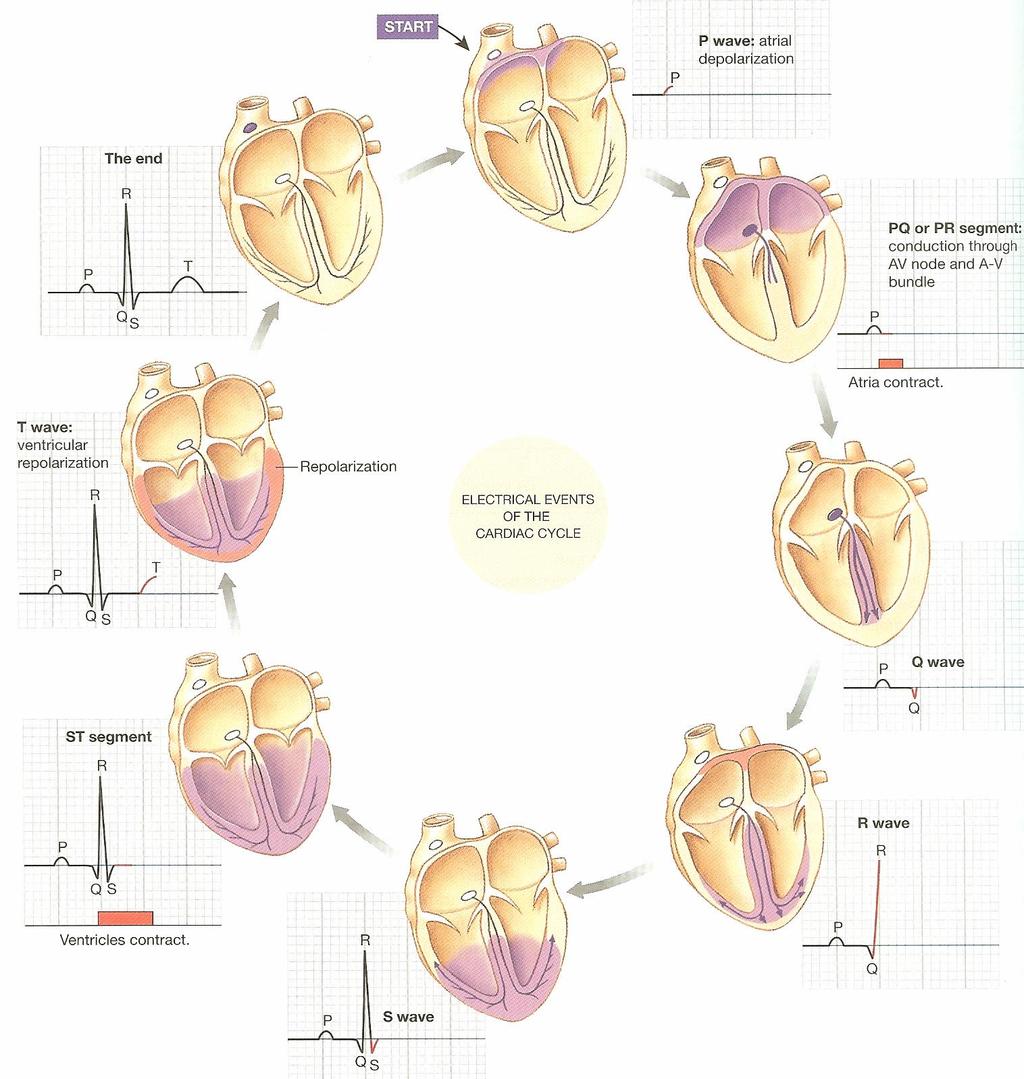
9 b. Atrioventircular AV node > Near the floor of the Atrium > Delay occurs at AV node while atria contract c. AV Bundle (of His) > Found in superior atrioventricular septum > Link between atria and ventricles d. Bilateral Bundle branches > Convey impulse down right and left ventricles to the hearts apex e. Purkinje fibers >Convey impulse throughout the ventricular walls [Ejection of blood aided by spiral arrangement of muscles/during contractions muscle pull the apex and base close together squeezing blood out the openings at the top of the ventricles] V. ELECTROCARDIOGRAM (ECG) 1. Background - ECG tracing shows the summed electrical potentials generated by all cells of the heart - ECG reflects depolarization and repolarizations of atria and ventricles - Contraction Cycle: Single contraction-relaxation cycle 9

10 - Heart rate is normally timed from the beginning of one P wave to the beginning of the next P wave or from peak of one R wave to peak of next R wave Waves: Deflections above or below the baseline Segments: Sections of baseline between two waves Intervals: Combinations of waves and segments NOTE: Cannot tell if ECG recording represents depolarization or repolarization by looking at the shape of wave relative to the baseline (example: P represents depolarization and T represents repolarization; even though both are above the baseline) a. Atrial Depolarization > P Wave b. Atrial systole > PR interval c. Atrial repolarization/relaxtion > Not represented by special wave but incorporated into QRS complex d. Atrial diastole > Later drop of QRS complex (S) to prefix of P wave e. Ventricular depolarization > QRS complex f. Ventricular systole > QT interval g. Ventricular repolarization > T wave h. Ventricular diastole > End of T wave to the first drop of QRS complex (Q) i. U-wave 10
11 11

12 b. Arrhythmias a. Sinus Rhythm > NORMAL > bpm adults > bpm older children > bpm small children > bpm infants b. Bradycardia > Slower then normal heart rate c. Trachycardia > Faster then normal heart rate d. Fluter > Coordinated fast heart rate > In Atrium > Little blood flow > Usually above 200 bpm > Light headed/dizziness/pass-out e. Fibrillation > Non-coordinated fast heart fate > Circus Rhythms > No blood flow [Fibulator repolarizes (resets) intrinsic conducting system to base] f. AV Heart Block > Slow conduction through AV node > Independence of atrial and ventricular rhythms 12
13 13
Electrical Conduction
 Sinoatrial (SA) node Electrical Conduction Sets the pace of the heartbeat at 70 bpm AV node (50 bpm) and Purkinje fibers (25 40 bpm) can act as pacemakers under some conditions Internodal pathway from
THE CARDIOVASCULAR SYSTEM. Heart 2
 THE CARDIOVASCULAR SYSTEM Heart 2 PROPERTIES OF CARDIAC MUSCLE Cardiac muscle Striated Short Wide Branched Interconnected Skeletal muscle Striated Long Narrow Cylindrical PROPERTIES OF CARDIAC MUSCLE Intercalated
Cardiovascular System
 Cardiovascular System The Heart Cardiovascular System The Heart Overview What does the heart do? By timed muscular contractions creates pressure gradients blood moves then from high pressure to low pressure
Chapter 20: Cardiovascular System: The Heart
 Chapter 20: Cardiovascular System: The Heart I. Functions of the Heart A. List and describe the four functions of the heart: 1. 2. 3. 4. II. Size, Shape, and Location of the Heart A. Size and Shape 1.
Introduction. Circulation
 Introduction Circulation 1- Systemic (general) circulation 2- Pulmonary circulation carries oxygenated blood to all parts of the body carries deoxygenated blood to the lungs From Lt. ventricle aorta From
CARDIOVASCULAR SYSTEM
 CARDIOVASCULAR SYSTEM Overview Heart and Vessels 2 Major Divisions Pulmonary Circuit Systemic Circuit Closed and Continuous Loop Location Aorta Superior vena cava Right lung Pulmonary trunk Base of heart
Cardiac physiology. b. myocardium -- cardiac muscle and fibrous skeleton of heart
 I. Heart anatomy -- general gross. A. Size/orientation - base/apex B. Coverings D. Chambers 1. parietal pericardium 2. visceral pericardium 3. Layers of heart wall a. epicardium Cardiac physiology b. myocardium
Collin County Community College. ! BIOL Anatomy & Physiology! WEEK 5. The Heart
 Collin County Community College! BIOL. 2402 Anatomy & Physiology! WEEK 5 The Heart 1 (1578-1657) A groundbreaking work in the history of medicine, English physician William Harvey s Anatomical Essay on
Chapter 18 - Heart. I. Heart Anatomy: size of your fist; located in mediastinum (medial cavity)
") Chapter 18 - Heart I. Heart Anatomy: size of your fist; located in mediastinum (medial cavity) A. Coverings: heart enclosed in double walled sac called the pericardium 1. Fibrous pericardium: dense connective
Chapter 18 - Heart I. Heart Anatomy: size of your fist; located in mediastinum (medial cavity) A. Coverings: heart enclosed in double walled sac called the pericardium 1. Fibrous pericardium: dense connective
Chapter 20b Cardiac Physiology
 Chapter 20b Cardiac Physiology Heart Valve Mechanics The heart valve openand close because of pressure gradients. When pressure on one side is greater than the other, it pushes the valve open. For example,
Chapter 13 The Cardiovascular System: Cardiac Function
 Chapter 13 The Cardiovascular System: Cardiac Function Overview of the Cardiovascular System The Path of Blood Flow through the Heart and Vasculature Anatomy of the Heart Electrical Activity of the Heart
Cardiovascular system
 BIO 301 Human Physiology Cardiovascular system The Cardiovascular System: consists of the heart plus all the blood vessels transports blood to all parts of the body in two 'circulations': pulmonary (lungs)
Department of medical physiology 7 th week and 8 th week
 Department of medical physiology 7 th week and 8 th week Semester: winter Study program: Dental medicine Lecture: RNDr. Soňa Grešová, PhD. Department of medical physiology Faculty of Medicine PJŠU Cardiovascular
The Heart. Size, Form, and Location of the Heart. 1. Blunt, rounded point; most inferior part of the heart.
 12 The Heart FOCUS: The heart is composed of cardiac muscle cells, which are elongated, branching cells that appear striated. Cardiac muscle cells behave as a single electrical unit, and the highly coordinated
10/23/2017. Muscular pump Two atria Two ventricles. In mediastinum of thoracic cavity 2/3 of heart's mass lies left of midline of sternum
 It beats over 100,000 times a day to pump over 1,800 gallons of blood per day through over 60,000 miles of blood vessels. During the average lifetime, the heart pumps nearly 3 billion times, delivering
AnS SI 214 Practice Exam 2 Nervous, Muscle, Cardiovascular
 AnS SI 214 Practice Exam 2 Nervous, Muscle, Cardiovascular Select the best answer choice in the questions below. 1) On the electrocardiogram, repolarization of the atria is represented by the: A) P wave
The Cardiovascular System
 Chapter 18 Part A The Cardiovascular System 1/19/16 1 Annie Leibovitz/Contact Press Images Similarities of Cardiac and Skeletal Muscle RMP Ion concentration Deploarization Action Potential Repolarization
4. The two inferior chambers of the heart are known as the atria. the superior and inferior vena cava, which empty into the left atrium.
 Answer each statement true or false. If the statement is false, change the underlined word to make it true. 1. The heart is located approximately between the second and fifth ribs and posterior to the
*Generating blood pressure *Routing blood: separates. *Ensuring one-way blood. *Regulating blood supply *Changes in contraction
 *Generating blood pressure *Routing blood: separates pulmonary and systemic circulations *Ensuring one-way blood flow: valves *Regulating blood supply *Changes in contraction rate and force match blood
Practice Exercises for the Cardiovascular System
 Practice Exercises for the Cardiovascular System On the diagram below, color the oxygen-rich blood red and the oxygen-poor blood blue. Label the parts: Continued on the next page... Label the parts on
Cardiovascular System: The Heart
 Cardiovascular System: The Heart I. Anatomy of the Heart (See lab handout for terms list) A. Describe the size, shape and location of the heart B. Describe the structure and function of the pericardium
The HEART. What is it???? Pericardium. Heart Facts. This muscle never stops working It works when you are asleep
 This muscle never stops working It works when you are asleep The HEART It works when you eat It really works when you exercise. What is it???? Located between the lungs in the mid thoracic region Apex
11/10/2014. Muscular pump Two atria Two ventricles. In mediastinum of thoracic cavity 2/3 of heart's mass lies left of midline of sternum
 It beats over 100,000 times a day to pump over 1,800 gallons of blood per day through over 60,000 miles of blood vessels. During the average lifetime, the heart pumps nearly 3 billion times, delivering
Collin County Community College
 Collin County Community College BIOL. 2402 Anatomy & Physiology WEEK 5 The Heart 1 The Heart Beat and the EKG 2 1 The Heart Beat and the EKG P-wave = Atrial depolarization QRS-wave = Ventricular depolarization
Approximately the size of your fist Location. Pericardial physiology
 Heart Anatomy Approximately the size of your fist Location Superior surface of diaphragm Left of the midline Anterior to the vertebral column, posterior to the sternum Wednesday, March 28, 2012 Muscle
The Heart. The Heart A muscular double pump. The Pulmonary and Systemic Circuits
 C H A P T E R 19 The Heart The Heart A muscular double pump circuit takes blood to and from the lungs Systemic circuit vessels transport blood to and from body tissues Atria receive blood from the pulmonary
Cardiac Properties MCQ
 Cardiac Properties MCQ Abdel Moniem Ibrahim Ahmed, MD Professor of Cardiovascular Physiology Cairo University 2007 1- Cardiac Valves: a- Prevent backflow of blood from the ventricles to the atria during
The cardiovascular system is composed of the heart and blood vessels that carry blood to and from the body s organs. There are 2 major circuits:
 1 The cardiovascular system is composed of the heart and blood vessels that carry blood to and from the body s organs. There are 2 major circuits: pulmonary and systemic. The pulmonary goes out to the
Chapter 20 (2) The Heart
 The Heart") Chapter 20 (2) The Heart ----------------------------------------------------------------------------------------------------------------------------------------- Describe the component and function of
Chapter 20 (2) The Heart ----------------------------------------------------------------------------------------------------------------------------------------- Describe the component and function of
The Heart. C h a p t e r. PowerPoint Lecture Slides prepared by Jason LaPres Lone Star College - North Harris
 C h a p t e r 20 The Heart PowerPoint Lecture Slides prepared by Jason LaPres Lone Star College - North Harris Copyright 2009 Pearson Education, Inc., publishing as Pearson Benjamin Cummings Introduction
Circulatory System Function Move circulatory fluid (blood) around body Gas Transport Nutrient Transport Excretory Product Transport
 around body Gas Transport Nutrient Transport Excretory Product Transport") Lecture 37 Introduction to Circulation BY DR QAZI IMTIAZ RASOOL OBJECTIVES Functions of the Heart Generating blood pressure Routing blood: separates pulmonary and systemic circulations Ensuring one-way
Lecture 37 Introduction to Circulation BY DR QAZI IMTIAZ RASOOL OBJECTIVES Functions of the Heart Generating blood pressure Routing blood: separates pulmonary and systemic circulations Ensuring one-way
Conduction System of the Heart 4. Faisal I. Mohammed, MD, PhD
 Conduction System of the Heart 4 Faisal I. Mohammed, MD, PhD 1 Objectives List the parts that comprise the conduction system Explain the mechanism of slow response action potential (pacemaker potential)
Ch 19: Cardiovascular System - The Heart -
 Ch 19: Cardiovascular System - The Heart - Give a detailed description of the superficial and internal anatomy of the heart, including the pericardium, the myocardium, and the cardiac muscle. Trace the
The Cardiovascular System: The Heart
 PowerPoint Lecture Slides prepared by Meg Flemming Austin Community College C H A P T E R 12 The Cardiovascular System: The Heart Chapter 12 Learning Outcomes 12-1 12-2 Describe the anatomy of the heart,
THE HEART. A. The Pericardium - a double sac of serous membrane surrounding the heart
 THE HEART I. Size and Location: A. Fist-size weighing less than a pound (250 to 350 grams). B. Located in the mediastinum between the 2 nd rib and the 5 th intercostal space. 1. Tipped to the left, resting
Lab 2. The Intrinsic Cardiac Conduction System. 1/23/2016 MDufilho 1
 Lab 2 he Intrinsic Cardiac Conduction System 1/23/2016 MDufilho 1 Figure 18.13 Intrinsic cardiac conduction system and action potential succession during one heartbeat. Superior vena cava ight atrium 1
The Heart. Happy Friday! #takeoutyournotes #testnotgradedyet
 The Heart Happy Friday! #takeoutyournotes #testnotgradedyet Introduction Cardiovascular system distributes blood Pump (heart) Distribution areas (capillaries) Heart has 4 compartments 2 receive blood (atria)
2. right heart = pulmonary pump takes blood to lungs to pick up oxygen and get rid of carbon dioxide
 A. location in thorax, in inferior mediastinum posterior to sternum medial to lungs superior to diaphragm anterior to vertebrae orientation - oblique apex points down and to the left 2/3 of mass on left
Cardiovascular System Notes: Physiology of the Heart
 Cardiovascular System Notes: Physiology of the Heart Interesting Heart Fact Capillaries are so small it takes ten of them to equal the thickness of a human hair. Review What are the 3 parts of the cardiovascular
The conduction system
 The conduction system In today s lecture we will discuss the conducting system of the heart. If we placed the heart in a special solution that contains Ca+ it will keep on contracting, keep in mind that
PART I. Disorders of the Heart Rhythm: Basic Principles
 PART I Disorders of the Heart Rhythm: Basic Principles FET01.indd 1 1/11/06 9:53:05 AM FET01.indd 2 1/11/06 9:53:06 AM CHAPTER 1 The Cardiac Electrical System The heart spontaneously generates electrical
Human Anatomy, First Edition
 Human Anatomy, First Edition McKinley & O'Loughlin Chapter 22 : Heart 1 Functions of the Heart Center of the cardiovascular system, the heart. Connects to blood vessels that transport blood between the
Functions of the Heart
 Cardiovascular System The Heart What is the Cardiovascular System? Blood circulated in Arteries, veins, and capillaries by the Pumping action of the heart Functions of the Heart Generating blood pressure
Cardiac Cycle. Each heartbeat is called a cardiac cycle. First the two atria contract at the same time.
 The Heartbeat Cardiac Cycle Each heartbeat is called a cardiac cycle. First the two atria contract at the same time. Next the two ventricles contract at the same time. Then all the chambers relax. http://www.youtube.com/watch?v=frd3k6lkhws
- what other structures, besides the heart, does the mediastinum contain?
 Basic A & P II Dr. L. Bacha Chapter Outline (Martini & Nath 2010) An Introduction to the Cardiovascular System - read the paragraphs under this heading on page 580 The Heart is a Four Chambered Organ describe
Conduction System of the Heart. Faisal I. Mohammed, MD, PhD
 Conduction System of the Heart Faisal I. Mohammed, MD, PhD 1 Objectives l List the parts that comprise the conduction system l Explain the mechanism of slow response action potential (pacemaker potential)
Circulatory system. Terminology. Ventricles and resistance. Pressure gradients move blood through the heart and vessels.
 Circulatory system Pressure gradients move blood through the heart and vessels. Pulmonary circulation vs. systemic circulation (to pulmonary circuit) liver head and arms heart aorta diaphragm (from pulmonary
Where are the normal pacemaker and the backup pacemakers of the heart located?
 CASE 9 A 68-year-old woman presents to the emergency center with shortness of breath, light-headedness, and chest pain described as being like an elephant sitting on her chest. She is diagnosed with a
The Cardiovascular System
 Essentials of Human Anatomy & Physiology Elaine N. Marieb Seventh Edition Chapter 11 The Cardiovascular System Slides 11.1 11.19 Lecture Slides in PowerPoint by Jerry L. Cook The Cardiovascular System
IP: Regulation of Cardiac Output
 ANP 1105D Winter 2013 Assignment 9: The Heart, part 2: Chap... Assignment 9: The Heart, part 2: Chapter 18 Signed in as Alex Sokolowski Help Close Resources Due: 11:59pm on Monday, March 25, 2013 Note:
Heart. Heart 2-Tunica media: middle layer (media ='middle') muscle fibers (smooth or cardiac).
 muscle fibers (smooth or cardiac).") t. innermost lumenal General Circulatory system heart and blood vessels walls have 3 layers (inside to outside) 1-Tunica interna: aka tunica intima layer--lumenal layer epithelium--endothelium simple squamous
t. innermost lumenal General Circulatory system heart and blood vessels walls have 3 layers (inside to outside) 1-Tunica interna: aka tunica intima layer--lumenal layer epithelium--endothelium simple squamous
BIPN100 F15 Human Physiology I (Kristan) Problem set #5 p. 1
 Problem set #5 p. 1") BIPN100 F15 Human Physiology I (Kristan) Problem set #5 p. 1 1. Dantrolene has the same effect on smooth muscles as it has on skeletal muscle: it relaxes them by blocking the release of Ca ++ from the
BIPN100 F15 Human Physiology I (Kristan) Problem set #5 p. 1 1. Dantrolene has the same effect on smooth muscles as it has on skeletal muscle: it relaxes them by blocking the release of Ca ++ from the
(D) (E) (F) 6. The extrasystolic beat would produce (A) increased pulse pressure because contractility. is increased. increased
 (E) (F) 6. The extrasystolic beat would produce (A) increased pulse pressure because contractility. is increased. increased") Review Test 1. A 53-year-old woman is found, by arteriography, to have 5% narrowing of her left renal artery. What is the expected change in blood flow through the stenotic artery? Decrease to 1 2 Decrease
Review Test 1. A 53-year-old woman is found, by arteriography, to have 5% narrowing of her left renal artery. What is the expected change in blood flow through the stenotic artery? Decrease to 1 2 Decrease
Principles of Biomedical Systems & Devices. Lecture 8: Cardiovascular Dynamics Dr. Maria Tahamont
 Principles of Biomedical Systems & Devices Lecture 8: Cardiovascular Dynamics Dr. Maria Tahamont Review of Cardiac Anatomy Four chambers Two atria-receive blood from the vena cave and pulmonary veins Two
Anatomy Review: The Heart Graphics are used with permission of A.D.A.M. Software, Inc. and Benjamin/Cummings Publishing Co.
 Anatomy Review: The Heart Graphics are used with permission of A.D.A.M. Software, Inc. and Benjamin/Cummings Publishing Co. Anatomy Views Label the diagrams of the heart below: Interactive Physiology Study
Marah karablieh. Osama khader. Muhammad khatatbeh. 0 P a g e
 15 Marah karablieh Osama khader 0 P a g e Muhammad khatatbeh Cardiac Muscle Physiology Introduction The heart has two ventricles and two atriums. The heart wall is composed primarily of spirally arranged
CARDIAC PHYSIOLOGY. Amelyn U. Ramos-Rafael,M.D. Functional Anatomy of the Heart
 CARDIAC PHYSIOLOGY Amelyn U. Ramos-Rafael,M.D. Functional Anatomy of the Heart 1 Functional Anatomy of The Heart The Atria relatively thin walled The Ventricles ventricular walls thicker than atrial walls
Full file at
 MULTIPLE CHOICE. Choose the one alternative that best completes the statement or answers the question. 1) What electrical event must occur for atrial kick to occur? 1) A) Atrial repolarization B) Ventricular
Chapter 12: Cardiovascular Physiology System Overview
 Chapter 12: Cardiovascular Physiology System Overview Components of the cardiovascular system: Heart Vascular system Blood Figure 12-1 Plasma includes water, ions, proteins, nutrients, hormones, wastes,
The Cardiovascular System
 11 PART A The Cardiovascular System PowerPoint Lecture Slide Presentation by Jerry L. Cook, Sam Houston University ESSENTIALS OF HUMAN ANATOMY & PHYSIOLOGY EIGHTH EDITION ELAINE N. MARIEB The Cardiovascular
Lab Activity 23. Cardiac Anatomy. Portland Community College BI 232
 Lab Activity 23 Cardiac Anatomy Portland Community College BI 232 Cardiac Muscle Histology Branching cells Intercalated disc: contains many gap junctions connecting the adjacent cell cytoplasm, creates
flow of blood May carry blood
 Heart 1 CARDIOVASCULAR SYSTEM: The Heart I. THE HEART & BLOOD FLOW Functional Anatomy off the heart - CARDIOVASCULAR SYSTEM - heart + blood vessels. - HEART = muscular pump; keeps blood CONSTANTLY moving
The Circulatory System. The Heart, Blood Vessels, Blood Types
 The Circulatory System The Heart, Blood Vessels, Blood Types The Closed Circulatory System Humans have a closed circulatory system, typical of all vertebrates, in which blood is confined to vessels and
EXAM II Animal Physiology ZOO 428 Fall 2006
 V Eq EXAM II Animal Physiology ZOO 428 Fall 2006 = RT X o. ln( [ zf [ X ) RT p K[K o pna[na o pcl[cl i V = m ln i F pk[k i pna[na i pcl[cl o I = g(v m V eq. ) Q = C m V m Q Driving Force = V m V eq. 10
current, and acting like
 Heart 10 IV. HEART PHYSIOLOGY - How the heart beats. How the heart depolarizes the myocardium, which leads to a contraction. A) INTRINSIC CONTROL - Heart controls its own rhythm. HOW? The presence of gap
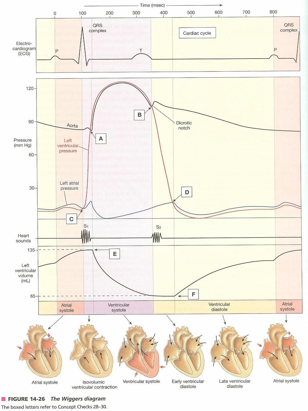
CARDIAC CYCLE CONTENTS. Divisions of cardiac cycle 11/13/13. Definition. Badri Paudel GMC
 CARDIAC CYCLE Badri Paudel GMC CONTENTS Ø DEFINATION Ø DIVISION OF CARDIAC CYCLE Ø SUB DIVISION AND DURATION OF CARDIAC CYCLE Ø SYSTOLE Ø DIASTOLE Ø DESCRIPTION OF EVENTS OF CARDIAC CYCLE Ø SUMMARY Ø ELECTROCARDIOGRAPHY
The Cardiovascular System
 The Cardiovascular System The Cardiovascular System A closed system of the heart and blood vessels The heart pumps blood Blood vessels allow blood to circulate to all parts of the body The function of
The Cardiovascular System
 Essentials of Human Anatomy & Physiology Elaine N. Marieb Slides 11.1 11.19 Seventh Edition Chapter 11 The Cardiovascular System Functions of the Cardiovascular system Function of the heart: to pump blood
Using Figure 14.1, match the following: 1) Myelin sheath. 1) 2) Cell body of ANS preganglionic neuron. 2)
 Myelin sheath. 1) 2) Cell body of ANS preganglionic neuron. 2)") Practice Exam 1 AP 2 chapters 14 and 18 Name MATCHING: Match labeled areas with the appropriate terminology from the list below. Figure 14.1 Using Figure 14.1, match the following: 1) Myelin sheath. 1)
Practice Exam 1 AP 2 chapters 14 and 18 Name MATCHING: Match labeled areas with the appropriate terminology from the list below. Figure 14.1 Using Figure 14.1, match the following: 1) Myelin sheath. 1)
Physiology sheet #2. The heart composed of 3 layers that line its lumen and cover it from out side, these layers are :
 Physiology sheet #2 * We will talk in this lecture about cardiac muscle physiology, the mechanism and the energy sources of their contraction and intracellular calcium homeostasis. # Slide 4 : The heart
The Heart and Heart Disease
 The Heart and Heart Disease Illustration of the heart by Leonardo DaVinci heart-surgeon.com/ history.html 2/14/2010 1 I. Location, Size and Position of the Heart A. Triangular organ located 1. of mass
Heart: Cardiac Function & ECGs. The Heart and Circulatory System
 Heart: Cardiac Function & ECGs Adapted From: Textbook Of Medical Physiology, 11 th Ed. Arthur C. Guyton, John E. Hall Chapters 9, 10, 11, 12, & 13 John P. Fisher The Heart and Circulatory System Introduction
Atlantic Health System
 Atlantic Health System Morristown Medical Center Newton Medical Center Overlook Medical Center Basic Dysrhythmia Course Day 1 1 2 Chapter 1 Anatomy and Physiology Learning Objectives 1) Identify electrophysiology
1. Which of the following conditions will result in a dilated, flaccid heart?
 1. Which of the following conditions will result in a dilated, flaccid heart? A) Excess calcium ions in the blood B) Excess potassium ions in the blood C) Excess sodium ions in the blood D) Increased sympathetic
37 1 The Circulatory System
 H T H E E A R T 37 1 The Circulatory System The circulatory system and respiratory system work together to supply cells with the nutrients and oxygen they need to stay alive. a) The respiratory system:
The Cardiovascular System: The Heart: Part A
 PowerPoint Lecture Slides prepared by Janice Meeking, Mount Royal College CHAPTER 18 The Cardiovascular System: The Heart: Part A Heart Anatomy Approximately the size of a fist Location In the mediastinum
10. Thick deposits of lipids on the walls of blood vessels, called, can lead to serious circulatory issues. A. aneurysm B. atherosclerosis C.
 Heart Student: 1. carry blood away from the heart. A. Arteries B. Veins C. Capillaries 2. What is the leading cause of heart attack and stroke in North America? A. alcohol B. smoking C. arteriosclerosis
Cardiovascular System Notes: Heart Disease & Disorders
 Cardiovascular System Notes: Heart Disease & Disorders Interesting Heart Facts The Electrocardiograph (ECG) was invented in 1902 by Willem Einthoven Dutch Physiologist. This test is still used to evaluate
Human Anatomy and Physiology II Laboratory Cardiovascular Physiology
 Human Anatomy and Physiology II Laboratory Cardiovascular Physiology 1 This lab involves two exercises: 1) Conduction System of the Heart and Electrocardiography and 2) Human Cardiovascular Physiology:
CV Anatomy Quiz. Dr Ella Kim Dr Pip Green
 CV Anatomy Quiz Dr Ella Kim Dr Pip Green Q1 The location of the heart is correctly described as A) lateral to the lungs. B) medial to the sternum. C) superior to the diaphragm. D) posterior to the spinal
Cardiovascular health & Health Promotion HH2602 & HH5607
 Cardiovascular health & Health Promotion HH2602 & HH5607 Lecture 2: Microscopic Structure and Function of the Heart 2pm 28-02-17 ESGW Teaching Aims To introduce you to the microstructure of heart muscle.
BIOL 4350 Cardiovascular Physiology Dr. Hamilton. Using the figure above, match the following: 1. Purkinje fibers. 2. SA node. 3. AV node.
 BIOL 4350 Cardiovascular Physiology Dr. Hamilton Using the figure above, match the following: 1. Purkinje fibers. 2. SA node. 3. AV node. 1 Using the figure above, match the following: 4. Atrial depolarization.
CIRCULATION & GAS EXCHANGE
 AP BIOLOGY ACTIVITY2.13 Text:Campbell,v.8,chapter42 NAME DATE HOUR CIRCULATION & GAS EXCHANGE 1. In general, what is the function of transport systems? 2. What method/structure do most invertebrates use
آالء العجرمي أسامة الخضر. Faisal Muhammad
 16 آالء العجرمي أسامة الخضر Faisal Muhammad 1. Summary for what taken : *changes in permeability of ions: 1. During phase 0: changes happen due to the influx of Na+, the permeability of Na ions increase
the Cardiovascular System I
 the Cardiovascular System I By: Dr. Nabil A Khouri MD, MsC, Ph.D MEDIASTINUM 1. Superior Mediastinum 2. inferior Mediastinum Anterior mediastinum. Middle mediastinum. Posterior mediastinum Anatomy of
Effects of Temperature, Stretch, and Various Drug Treatments on the
 Nicole Rodi Bio 235: Animal Physiology Heart Muscle Lab Report 10/24/2014 Effects of Temperature, Stretch, and Various Drug Treatments on the Cardiac Muscle Activity of Rana pipiens Abstract Mechanical
Circulation. Circulation = is a process used for the transport of oxygen, carbon! dioxide, nutrients and wastes through-out the body
 Circulation Circulation = is a process used for the transport of oxygen, carbon! dioxide, nutrients and wastes through-out the body Heart = muscular organ about the size of your fist which pumps blood.
BME 5742 Bio-Systems Modeling and Control. Lecture 41 Heart & Blood Circulation Heart Function Basics
 BME 5742 Bio-Systems Modeling and Control Lecture 41 Heart & Blood Circulation Heart Function Basics Dr. Zvi Roth (FAU) 1 Pumps A pump is a device that accepts fluid at a low pressure P 1 and outputs the
Cardiovascular System
 Cardiovascular System Purpose Transport oxygen and nutrients Take waste products away from tissues & organs Things we learned Blood pressure: the force of blood pushing against the walls of blood vessels
Heart. Structure Physiology of blood pressure and heartbeat
 Heart Structure Physiology of blood pressure and heartbeat Location and Anatomy Location and Anatomy Pericardial cavity: surrounds, isolates, and anchors heart Parietal pericardium lined with serous membrane
The Cardiovascular System (Heart)
") The Cardiovascular System The Cardiovascular System (Heart) A closed system of the heart and blood vessels The heart pumps blood Blood vessels allow blood to circulate to all parts of the body The function
The Cardiovascular System The Cardiovascular System (Heart) A closed system of the heart and blood vessels The heart pumps blood Blood vessels allow blood to circulate to all parts of the body The function
Circulatory system of mammals
 Circulatory system of mammals Explain the cardiac cycle and its initiation Discuss the internal factors that control heart action Blood flows through the heart as a result of pressure differences Blood
THE HEART Dr. Ali Ebneshahidi
 THE HEART Dr. Ali Ebneshahidi Functions is of the heart & blood vessels 1. The heart is an essential pumping organ in the cardiovascular system where the right heart pumps deoxygenated blood (returned
About This Chapter. Skeletal muscle Mechanics of body movement Smooth muscle Cardiac muscle Pearson Education, Inc.
 About This Chapter Skeletal muscle Mechanics of body movement Smooth muscle Cardiac muscle Skeletal Muscle Usually attached to bones by tendons Origin: closest to the trunk or to more stationary bone Insertion:
Chapter 20! The Heart!
 Chapter 20! The Heart! SECTION 20-1! The heart is a four-chambered organ, supplied by the coronary circulation, that pumps oxygen-poor blood to the lungs and oxygen-rich blood to the rest of the body!
Objectives of the Heart
 Objectives of the Heart Electrical activity of the heart Action potential EKG Cardiac cycle Heart sounds Heart Rate The heart s beat separated into 2 phases Relaxed phase diastole (filling of the chambers)
Conduction system of the heart
 Conduction system of the heart -For skeletal muscle to contract, it has to be innervated by spinal nerves (there must be a neuromuscular junction). *The heart is innervated by autonomic nervous system
Cardiac muscle is different from other types of muscle in that cardiac muscle
 6 E X E R C I S E Cardiovascular Physiology O B J E C T I V E S 1. To define autorhythmicity, sinoatrial node, pacemaker cells, and vagus nerves 2. To understand the effects of the sympathetic and parasympathetic
Derived copy of Cardiac Muscle and Electrical Activity *
 OpenStax-CNX module: m47785 1 Derived copy of Cardiac Muscle and Electrical Activity * Steven Telleen Based on Cardiac Muscle and Electrical Activity by OpenStax This work is produced by OpenStax-CNX and
Biology November 2009 Exam Three FORM W KEY
 Biology 251 3 November 2009 Exam Three FORM W KEY PRINT YOUR NAME AND ID NUMBER in the space that is provided on the answer sheet, and then blacken the letter boxes below the corresponding letters of your