CT angiography techniques. Boot camp
|
|
|
- Jonah Ambrose McLaughlin
- 6 years ago
- Views:
Transcription
1 CT angiography techniques Boot camp

2 Overview Basic concepts Contrast administration arterial opacification Time scan acquisition during the arterial phase Protocol examples Helical non-gated CTA Pulmonary embolism Abdominal aorta Aortogram with run-off DIEP flap ECG-synchronized CTA Thoracic aorta Thoraco-abdominal aorta
3 Contrast administration basic concepts

 Amount of")
 Fleischmann, D.")
4 Achieve arterial enhancement CT angiography Need intravenous contrast to achieve arterial enhancement Proportional to the iodine administration rate Increasing iodine concentration of contrast medium Increasing Injection flow rate (ml/s) Amount of iodinated contrast delivered per unit time Longer injection duration (larger volume of contrast) Fleischmann, D. Radiol Clin N Am, 2010; 48: 237
5 Achieve arterial enhancement CT angiography Need intravenous contrast to achieve arterial enhancement Proportional to the iodine administration rate Increasing iodine concentration of contrast medium Increasing Injection flow rate (ml/s) Amount of iodinated contrast delivered per unit time Longer injection duration (larger volume of contrast) Fleischmann, D. Radiol Clin N Am, 2010; 48: 237
6 Higher concentration of Iodine 0.1 mg/ml 1.0 mg/ml 10 mg/ml 100 mg/ml 14 HU 24 HU 305 HU 2679 HU Higher Iodine concentration increased arterial enhancement Bae KT. Radiology 2010;256:32 Simulated contrast enhancement curves of the abdominal aorta 125 ml of contrast at 4 ml/s Three CM concentrations
7 Flow rate Higher rate Enhancement increases Duration decreases Routine injections rates 4-5 ml/sec Needle sizes Vein size Flow rates > 8 ml/s Don t result in greater enhancement Pooling in central venous system, reflux into IVC Bae KT. Radiology 2010;256:32 Simulated contrast enhancement curves of the abdominal aorta 125 ml of 350 mg/ml contrast Injected at three different rates Higher flow rate of CM increased arterial enhancement
8 Flow rate Higher rate Enhancement increases Duration decreases Routine injections rates 4-5 ml/sec Needle sizes Vein size Flow rates > 8 ml/s Don t result in greater enhancement Pooling in central venous system, reflux into IVC Bae KT. Radiology 2010;256:32 Simulated contrast enhancement curves of the abdominal aorta 125 ml of 350 mg/ml contrast Injected at three different rates Higher flow rate of CM increased arterial enhancement
9 Flow rate Higher rate Enhancement increases Duration decreases Routine injections rates 4-5 ml/sec catheter size Vein size Flow rates > 8 ml/s Don t result in greater enhancement Pooling in central venous system, reflux into IVC Bae KT. Radiology 2010;256:32 Simulated contrast enhancement curves of the abdominal aorta 125 ml of 350 mg/ml contrast Injected at three different rates Higher flow rate of CM increased arterial enhancement
10 Aortic attenuation (HU) Injection duration = contrast volume Simulated aortic enhancement curves (adult male, 70kg, 170cm). Varying injection durations of 350 mg/ml contrast at 3 cc/s. 5 sec = 15 cc 20 sec = 60 cc 40 sec = 120 cc 60 sec = 180 cc Time in seconds Longer injection duration increased peak arterial enhancement Bae KT. Radiology 2010;256:32
11 Contrast administration summary Iodinated contrast needed for CTA to enhance arterial vasculature greater arterial opacification with higher iodine flux at the time of the scan Higher iodine concentration contrast media Higher flow rates Longer injection durations


12 Saline chaser Pushes contrast in tubing and peripheral veins into central veins cc Allows reduction in contrast volume Increases peak attenuation Reduced streak artifacts from veins and right heart Simpler to implement with dual head injectors
13 noise (HU) Attenuation (HU) kvp Measured attenuation and image noise of a 2% iodine solution at different tube potentials attenuation increases with lower kvp kvp noise increases with lower kvp kvp McCollough CM. Radiol Clin N Am, 2009;47:27

14 Timing scan acquisition to the arterial phase
15 Enhancement (HU) Arterial phase of contrast bolus After contrast injection Time-to-peak enhancement differs for different target arteries (PA coronary aorta foot) Distance from venous access site individual cardiac output Differ due to local vascular pathology Stenosis aneurysm time Bae KT. Radiology 2010;256:32
16 Enhancement (HU) Arterial phase of contrast bolus After contrast injection Time-to-peak enhancement differs for different target arteries (PA coronary aorta foot) Distance from venous access site individual cardiac output Differ due to local vascular pathology Stenosis aneurysm time Bae KT. Radiology 2010;256:32
17 Enhancement (HU) Arterial phase of contrast bolus After contrast injection Time-to-peak enhancement differs for different target vessels (PA coronary aorta foot) Distance from venous access site individual cardiac output Differ due to local vascular pathology Stenosis aneurysm time Bae KT. Radiology 2010;256:32
 Distance from venous access site individual cardiac output Differ due to local vascular pathology Stenosis aneurysm time Bae KT.")
18 Enhancement (HU) Arterial phase of contrast bolus After contrast injection Time-to-peak enhancement differs for different target vessels (PA coronary aorta foot) Distance from venous access site individual cardiac output Differ due to local vascular pathology Stenosis aneurysm time Bae KT. Radiology 2010;256:32
 Distance from venous access site individual cardiac output Differ due to local vascular pathology Stenosis aneurysm time Bae KT.")
19 Enhancement (HU) Arterial phase of contrast bolus After contrast injection Time-to-peak enhancement differs for different target vessels (PA coronary aorta foot) Distance from venous access site individual cardiac output Differ due to local vascular pathology Stenosis aneurysm time Bae KT. Radiology 2010;256:32

20 Enhancement (HU) Arterial phase of contrast bolus Contrast media arrival time (t arr ) Time for the bolus to reach target vessel Can be determined for each individual and desired target vessel Timing bolus Bolus tracking t arr time Bae KT. Radiology 2010;256:32


21 Scan timing methods Timing bolus Select target location from scout topogram Inject small test-bolus ml contrast Acquire low-dose dynamic scan at specified location during injection ROI in target structure Measure time-attenuation curve Contrast material arrival time in aortic root


22 Scan timing methods Bolus triggering Select trigger location Acquire reference image Place ROI in vascular structure of interest Inject contrast bolus Acquire low-dose dynamic scans Monitor attenuation in ROI Start scan when desired threshold reached Fleischmann, D. Radiol Clin N Am, 2010; 48: 237
23 Scan timing methods Timing bolus Advantages Test adequacy of contrast path Multiple ROIs (art and veins) replace if error Avoid artifacts Test patient response Heart rate Disadvantages Two contrast injections time Bolus triggering Advantages Time efficient less contrast Disadvantages Different scan delay times Single shot Unable to trouble shoot Adjust to problems Streak artifacts, misplaced ROI, occluded vein, connector leak
24 Scan timing methods Timing bolus Advantages Test adequacy of contrast path Multiple ROIs (art and veins) replace if error Avoid artifacts Test patient response Heart rate Disadvantages Two contrast injections time Bolus triggering Advantages Time efficient less contrast Disadvantages Different scan delay times Single shot Unable to trouble shoot Adjust to problems Streak artifacts, misplaced ROI, occluded vein, connector leak

25 Aortic attenuation (HU) Enhancement (HU) CT angiography: basic strategy Use a bolus of iodinated contrast to produce arterial enhancement determine the contrast arrival time Timing bolus Bolus tracking time Perform diagnostic scan near peak enhancement achieved with the contrast bolus in the target vessel scan Time in seconds
26 Aortic attenuation (HU) Basic strategy with timing bolus Select bolus to achieve sufficient vascular attenuation Vol: 150 cc (350 mgi/ml) Rate: 5 cc/s t arr t d Determine contrast media arrival time (t arr ) Timing bolus 15 5 cc/s + saline flush scan Specify diagnostic delay (t d ) Account for larger volume of primary bolus Achieve greater enhancement Start scan = t arr + t d Scan delay = t arr + 8 sec Time in seconds Fleischmann, D. Radiol Clin N Am, 2010; 48: 237
27 Aortic attenuation (HU) Basic strategy with timing bolus Select bolus to achieve sufficient vascular attenuation Vol: 150 cc (350 mgi/ml) Rate: 5 cc/s t arr t d Determine contrast media arrival time (t arr ) Timing bolus 15 5 cc/s + saline flush scan Shorter diagnostic delay (t d ) Lower arterial enhancement 150 vs 200 HU Time in seconds Start scan = t arr + t d Scan delay = t arr + 4 sec Fleischmann, D. Radiol Clin N Am, 2010; 48: 237
28 Aortic attenuation (HU) Basic strategy with bolus tracking Select bolus to achieve sufficient vascular attenuation t arr t d Determine contrast media arrival time (t arr ) Inject primary bolus Bolus tracking 100 HU threshold (50 HU) Specify diagnostic delay (t d ) Scan delay = t arr + t d t arr + 8 sec scan Time in seconds Fleischmann, D. Radiol Clin N Am, 2010; 48: 237

29 CTA summary points Higher contrast concentrations Higher arterial enhancement for the same volume of contrast Flow rate 4 5 ml/s Timing bolus or bolus tracking Location Size of target vessel Expected complexity Saline chaser Lower kvp when possible
30 CTA protocols examples Putting it all together

 + 15 cc")
31 Imaging protocol: pulmonary embolism Timing bolus AP and lateral scouts Timing bolus below carina. ROI in PA. helical acquisition at timing bolus peak + 5 sec contrast Omnipaque 350 caudal-cranial scan direction from diaphragm to lung apices Timing bolus: 15 cc contrast (5 cc/s) + 15 cc saline (5cc/s) Primary bolus: 85 cc contrast (5 cc/s) + 30 cc saline (5 cc/s)

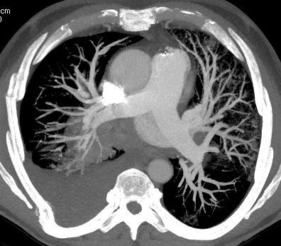
32 CTA pulmonary embolism

 + flush 85-94 kg: 145 cc contrast (4.")
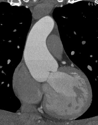
33 Imaging protocol: CTA abdominal aorta Bolus tracking AP and lateral scouts Bolus tracking at L1. ROI in abdominal aorta. Helical acquisition at threshold of 70 HU Contrast bolus Omnipaque 350 Cranial-caudal scan direction from diaphragm to lesser trochanter < 55 kg: 120 cc contrast (4 cc/s) + 30 cc saline (4 cc/s) kg: 125 cc contrast (4 cc/s) + flush kg: 130 cc contrast (4.5 cc/s) + flush kg: 145 cc contrast (4.5 cc/s) + flush > 95 kg: 150 cc contrast (5 cc/s) + flush

34 CTA abdominal aorta

 + 30 cc saline (3.")
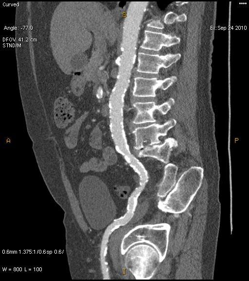
35 Imaging protocol: CTA aortogram with run-off Long scan duration AP and lateral scouts Bolus tracking at L1. ROI in abdominal aorta. Helical acquisition at threshold of 100 HU Contrast bolus Omnipaque 350 Scan delay = 40 sec scan duration Biphasic rate injection Cranial-caudal scan direction from diaphragm through toes Small patient: 20 cc contrast (4 cc/s) + 95 cc contrast (3.2 cc/s) + 30 cc saline (3.2 cc/s) Medium patient: 25 cc (5 cc/s) cc (4 cc/s) + 30 cc saline (4 cc/s) Large patient: 30 cc (6 cc/s) cc (4.8 cc/s) + 30 cc saline (4.8 cc/s)

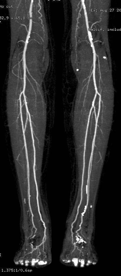
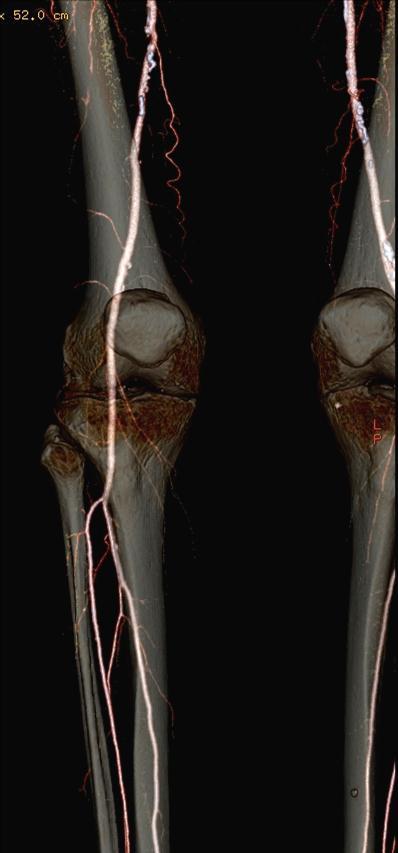
36 Aortogram with peripheral run-off
37 enhancement (HU) enhancement (HU) Biphasic injection: prolong plateau Uniphasic injection Biphasic rate injection Time (sec) 50 2 ml/s Continuously upsloping curve Time (sec) 25 2 ml/s ml/s More prolonged enhancement curve with two peaks Bae KT. Radiology, 2000; 216:872


38 CTA for DIEP flap breast reconstruction Alternate delay for veins AP and lateral scouts Timing bolus at level of acetabulum. ROI in right external iliac artery. Helical acquisition at timing bolus peak + 10 sec Caudal-cranial scan direction from lesser femoral trochanters to 4 cm above umbilicus Primary bolus: 100 cc contrast (5 cc/s) + 30 cc saline (5 cc/s)


39 Example 3d Images Volume rendered skin view for Location map of where perforator exits fascia Sagittal and axial oblique views of point where perforator exits fascia and IM course


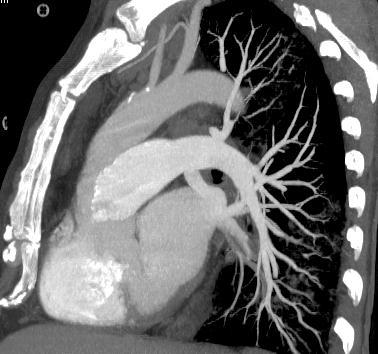
40 Imaging protocol: prospective ECG-triggered thoracic CT aortogram Gated scan, timing bolus AP and lateral scouts Right arm IV placement Avoid streak artifacts across arch vessels Timing bolus below carina. ROI in ascending thoracic aorta. PT sequential axial acquisition at timing bolus peak + 6 sec Contrast Omnipaque 350 Biphasic blended Cranial-caudal scan direction from clavicles to L1 70 cc contrast (5 cc/s) + 50 cc (70/30 blend, 5 cc/s) + 50 cc saline (5 cc/s)
41 Prospective ECG triggered CTA thoracic aorta


 + 30 cc saline (3.")
42 Imaging protocol: prospective ECG-triggered CT aortogram ECG gated, prolonged scan duration AP and lateral scouts Right arm IV placement Avoid streak artifacts across arch vessels Timing bolus below carina. ROI in descending thoracic aorta. PT sequential axial acquisition at timing bolus peak + 4 sec Contrast Biphasic size based bolus Cranial-caudal scan direction from clavicles to lesser femoral trochanter Small patient: 20 cc contrast (4 cc/s) + 95 cc contrast (3.2 cc/s) + 30 cc saline (3.2 cc/s) Medium patient: 25 cc contrast (5 cc/s) cc (4 cc/s) + 30 cc saline (4 cc/s) Large patient: 30 cc contrast (6 cc/s) cc (4.8 cc/s) + 30 cc saline (4.8 cc/s)


, and thin MIP images of the")
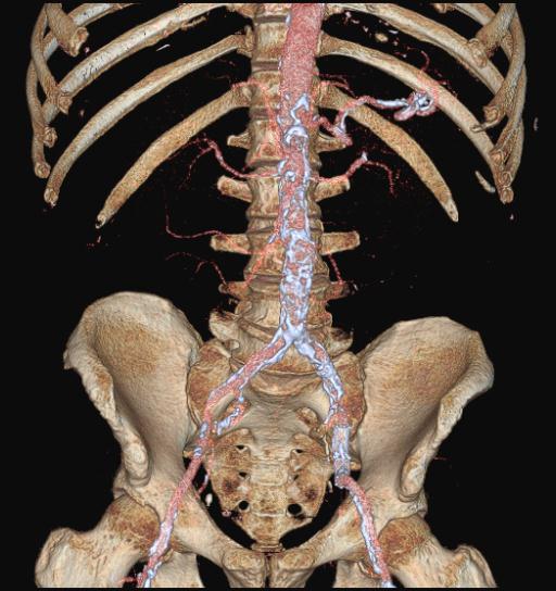
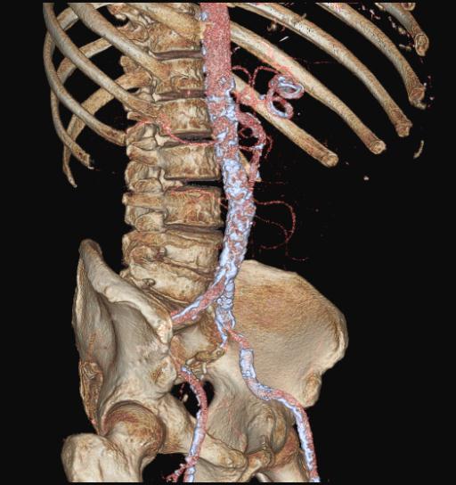
43 PT - CT aortogram: multifocal aneurysms Sagittal oblique MPR of the arch and descending thoracic aorta Coronal oblique thin MIP of the abdominal aorta 76 yo female with multifocal disease aneurysms in the aortic arch, descending thoracic and abdominal aorta. Volume rendered (left), multiplanar reformatted (MPR), and thin MIP images of the thoracic and abdominal aorta.


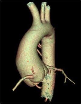
44 Imaging protocol: upper extremity CTA Distal timing bolus AP and lateral scouts contralateral arm IV placement Timing bolus near area of concern. ROI in target artery. helical acquisition at timing bolus peak + 4 sec scan through extremity from hand to chest. Decrease venous contamination Bolus > 20 sec, 100 cc (5 cc/s) Anderson S, Radiology 2008;249:1064

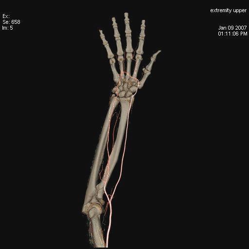
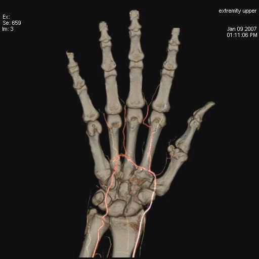
45 upper extremity

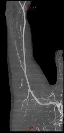
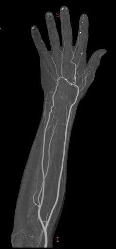
46 upper extremity


47 In closing Basic concepts of CT angiography Contrast administration and bolus shaping Scan timing Protocol examples Modifications based on the anatomy and pathology imaged
Vascular CT Protocols
 Vascular CT Protocols V 1D: Chest and abdominal CT angiogram (aortic dissection protocol) V 1T: Chest CT angiogram (aortic trauma protocol) V 2: Abdominal and pelvis CT angiogram (aortic aneurysm protocol)
Customizing Contrast Injection for Body MDCT: Algorithmic Approach
 Customizing Contrast Injection for Body MDCT: Algorithmic Approach Lincoln L. Berland, M.D., F.A.C.R. University of Alabama at Birmingham Before Contrast Prep and Hydration Hydration single most important
CT: Common Protocols. Michael Steigner, M.D. Director, Vascular CT/MR Assistant Professor of Radiology
 CT: Common Protocols BRIGHAM AND WOMEN S HOSPITAL Heart & Vascular Center Harvard Medical School Teaching Hospital Michael Steigner, M.D. Director, Vascular CT/MR Assistant Professor of Radiology msteigner@bwh.harvard.edu
(Non-EKG Gated) CTA Thoracic Aorta = CTA Chest
 CTA Thoracic Aorta = CTA Chest") (Non-EKG Gated) CTA Thoracic Aorta = CTA Chest Reviewed By: Dan Verdini, MD, Rachael Edwards, MD Last Reviewed: January 2019 Contact: (866) 761-4200, Option 1 In accordance with the ALARA principle, TRA
(Non-EKG Gated) CTA Thoracic Aorta = CTA Chest Reviewed By: Dan Verdini, MD, Rachael Edwards, MD Last Reviewed: January 2019 Contact: (866) 761-4200, Option 1 In accordance with the ALARA principle, TRA
CTA Chest Pulmonary Embolism & Routine CT Abdomen + Pelvis W CTA Chest W (arterial) & CT Abdomen + Pelvis W (venous)
 & CT Abdomen + Pelvis W (venous)") CTA Chest Pulmonary Embolism & Routine CT Abdomen + Pelvis W CTA Chest W (arterial) & CT Abdomen + Pelvis W (venous) Reviewed By: Anna Ellermeier, MD; Brett Mollard, MD Last Reviewed: August 2018 Contact:
CTA Chest Pulmonary Embolism & Routine CT Abdomen + Pelvis W CTA Chest W (arterial) & CT Abdomen + Pelvis W (venous) Reviewed By: Anna Ellermeier, MD; Brett Mollard, MD Last Reviewed: August 2018 Contact:
Scientific Exhibit. Authors: D. Takenaka, Y. Ohno, Y. Onishi, K. Matsumoto, T.
 The feasibility of biphasic contrast-media-injection-protocol for chest imaging on 320-slice volume MDCT: Direct comparison of biphasic and bolus contrast-media injection protocols on 320-slice volume
Technologist Error Patient Dynamics Anomalies Leaks & Computer Errors
 Daisha Marsh RT (R)(CT) Technologist Error Patient Dynamics Anomalies Leaks & Computer Errors Poor ROI placement Example: PE Placed in the wrong anatomy -We placed the ROI in the incorrect anatomy -We
Fundamentals, Techniques, Pitfalls, and Limitations of MDCT Interpretation and Measurement
 Fundamentals, Techniques, Pitfalls, and Limitations of MDCT Interpretation and Measurement 3 rd Annual Imaging & Physiology Summit November 20-21, 21, 2009 Seoul, Korea Wm. Guy Weigold, MD, FACC Cardiovascular
Bottom up cardiac CT for CABG assessment to resolve breathing artefact
 Bottom up cardiac CT for CABG assessment to resolve breathing artefact Poster No.: C-0589 Congress: ECR 2010 Type: Educational Exhibit Topic: Cardiac Authors: P. Glass, P. Donnelly, P. Hanley, D. Higginson,
Handzettel 1. CT Contrast Media. Agenda. Contrast Media Definition. Agenda. Why we need contrast media? Agenda
 Agenda CT Contrast Media Weena Swatdiswanee Factorinvolvein contrast enchancement Senior Application Specialist, CT Regional Headquarter Asia Australia weena.swat@siemens.com Page 1 Page 2 Agenda Contrast
Protocols in Cardiac CT Dr. Bruce Precious Dalhousie University Friday, April 15, 2016
 Protocols in Cardiac CT Dr. Bruce Precious Dalhousie University Friday, April 15, 2016 Disclosure Statement: No Conflict of Interest I do not have an affiliation, financial or otherwise, with a pharmaceutical
CTA Pulmonary Embolism CTA Chest W (arterial)
") CTA Pulmonary Embolism CTA Chest W (arterial) Reviewed By: Rachael Edwards, MD; Anna Ellermeier, MD; Brett Mollard, MD Last Reviewed: January 2019 Contact: (866) 761-4200, Option 1 In accordance with the
CTA Pulmonary Embolism CTA Chest W (arterial) Reviewed By: Rachael Edwards, MD; Anna Ellermeier, MD; Brett Mollard, MD Last Reviewed: January 2019 Contact: (866) 761-4200, Option 1 In accordance with the
Ultrasound. Computed tomography. Case studies. Utility of IQon Spectral CT in. cardiac imaging
 Ultrasound Computed tomography Case studies Utility of IQon Spectral CT in cardiac imaging Cardiac imaging is a challenging procedure where it is necessary to image a motion-free heart. This requires a
CT Renal 3 Phase + Pelvis CT Abdomen Pelvis WO W - NC.A.V, Pelvis during V
 CT Renal 3 Phase + Pelvis CT Abdomen Pelvis WO W - NC.A.V, Pelvis during V Reviewed By: Anna Ellermeier, MD; Brett Mollard, MD Last Reviewed: August 2018 Contact: (866) 761-4200, Option 1 In accordance
Case 9799 Stanford type A aortic dissection: US and CT findings
 Case 9799 Stanford type A aortic dissection: US and CT findings Accogli S, Aringhieri G, Scalise P, Angelini G, Pancrazi F, Bemi P, Bartolozzi C Department of Diagnostic and Interventional Radiology, University
CT Pancreas 3 Phase CT Abdomen WO W - NC.A.V
 CT Pancreas 3 Phase CT Abdomen WO W - NC.A.V Reviewed By: Rachael Edwards, MD; Anna Ellermeier, MD; Brett Mollard, MD Last Reviewed: January 2019 Contact: (866) 761-4200, Option 1 In accordance with the
ECG Gated CT Aorta in Transcatheter Aortic Valve Implantation
 ECG Gated CT Aorta in Transcatheter Aortic Valve Implantation Poster No.: C-2014 Congress: ECR 2014 Type: Educational Exhibit Authors: M. A. Ottesen; Oslo/NO Keywords: Cardiac, Arteries / Aorta, CT, CT-Angiography,
Liver 4 Phase CT Abdomen WO W - NC.A.V.D
 Liver 4 Phase CT Abdomen WO W - NC.A.V.D Reviewed By: Rachael Edwards, MD; Anna Ellermeier, MD; Brett Mollard, MD Last Reviewed: January 2019 Contact: (866) 761-4200, Option 1 In accordance with the ALARA
Non Contrast MRA. Mayil Krishnam. Director, Cardiovascular and Thoracic Imaging University of California, Irvine
 Non Contrast MRA Mayil Krishnam Director, Cardiovascular and Thoracic Imaging University of California, Irvine No disclosures Non contrast MRA-Why? Limitations of CTA Radiation exposure Iodinated contrast
Lung Perfusion Analysis New Pathways in Lung Imaging. Case Study Brochure PLA 309 Hospital
 Lung Perfusion Analysis New Pathways in Lung Imaging Case Study Brochure PLA 309 Hospital http://www.toshibamedicalsystems.com Toshiba Medical Systems Corporation 2012 all rights reserved. Design and specifications
Aorta ct w runoff The Borg System is 100 % Retrievable & Reusable Aorta ct w runoff
 Aorta ct w runoff The Borg System is 100 % Aorta ct w runoff Sep 5, 2016. CT images demonstrated extensive atherosclerotic soft mural and calcified plaques, involving the abdominal aorta, the major lower
, David Stultz, MD. Cardiac CT. David Stultz, MD Cardiology Fellow, PGY 6 March 28, 2006
 Cardiac CT David Stultz, MD Cardiology Fellow, PGY 6 March 28, 2006 Courtesy Tom Kracus Courtesy Kettering Tom Medical Kracus Cente Kettering Medical Center 2003-2006, David Stultz, MD Courtesy Tom Kracus
Acute Aortic Syndromes
 Acute Aortic Syndromes Carole J. Dennie, MD Acute Thoracic Aortic Syndromes Background Non-Traumatic Acute Thoracic Aortic Syndromes Carole Dennie MD FRCPC Associate Professor of Radiology and Cardiology
Triple Rule-out using 320-row-detector volume MDCT: A comparison of the wide volume and helical modes
 Triple Rule-out using 320-row-detector volume MDCT: A comparison of the wide volume and helical modes Poster No.: C-0488 Congress: ECR 2012 Type: Authors: Keywords: DOI: Scientific Exhibit E.-J. Kang,
Case Report 1. CTA head. (c) Tele3D Advantage, LLC
 Tele3D Advantage, LLC") Case Report 1 CTA head 1 History 82 YEAR OLD woman with signs and symptoms of increased intra cranial pressure in setting of SAH. CT Brain was performed followed by CT Angiography of head. 2 CT brain Extensive
Case Report 1 CTA head 1 History 82 YEAR OLD woman with signs and symptoms of increased intra cranial pressure in setting of SAH. CT Brain was performed followed by CT Angiography of head. 2 CT brain Extensive
Pediatric CT Protocols (18 years old or less)
") Pediatric CT Protocols (18 years old or less) Ped1: Head CT Ped2: Cervical spine CT Ped3: Sinus CT Ped4: Neck CT Ped5: Chest CT Ped6: Abdomen and pelvis CT Ped7: Thoracic or lumbar spine CT Ped8: Extremity
Pediatric CT Protocols (18 years old or less) Ped1: Head CT Ped2: Cervical spine CT Ped3: Sinus CT Ped4: Neck CT Ped5: Chest CT Ped6: Abdomen and pelvis CT Ped7: Thoracic or lumbar spine CT Ped8: Extremity
Assignable revenue codes: Explanation of services:
 computed tomography Chest/Cardiac Assignable revenue codes: Explanation of services: 0350 CT Scan General Classification 0351 CT Scan Head Scan 0352 CT Scan Body Scan 0359 CT Scan Other CT Scans Known
Cardiac CTA Prospective Gating Broad Beam
 Cardiac CTA Prospective Gating Broad Beam ACQUISITION- Broad Beam Gating: Prospective Non Contrast Scan-Calcium Score Patient Position Supine Feet First into Gantry Heart Isocenter Scanogram AP and Lateral
To Shield or Not to Shield? Lincoln L. Berland, M.D.
 To Shield or Not to Shield? Lincoln L. Berland, M.D. Disclosures Consultant to: Nuance, Inc. Page 2 Breast Radiation on CT Use of chest CT has increased in women vulnerable to cancer induction by radiation.
Cardiac CT - Coronary Calcium Basics Workshop II (Basic)
") Cardiac CT - Coronary Calcium Basics Workshop II (Basic) J. Jeffrey Carr, MD, MSCE Dept. of Radiology & Public Health Sciences Wake Forest University School of Medicine Winston-Salem, NC USA No significant
Cardiac CT - Coronary Calcium Basics Workshop II (Basic) J. Jeffrey Carr, MD, MSCE Dept. of Radiology & Public Health Sciences Wake Forest University School of Medicine Winston-Salem, NC USA No significant
Cardiac CT Techniques in Neonates (and infants)
") Cardiac CT Techniques in Neonates (and infants) Siddharth P. Jadhav, MD Director, Body CT and MRI Edward B. Singleton Department of Pediatric Radiology Texas Children s Hospital Disclosures None Objectives
Cardiac CT Techniques in Neonates (and infants) Siddharth P. Jadhav, MD Director, Body CT and MRI Edward B. Singleton Department of Pediatric Radiology Texas Children s Hospital Disclosures None Objectives
Anterior Spinal Artery and Artery of Adamkiewicz Detected by Using Multi-Detector Row CT
 AJNR Am J Neuroradiol 24:13 17, January 2003 Anterior Spinal Artery and Artery of Adamkiewicz Detected by Using Multi-Detector Row CT Kohsuke Kudo, Satoshi Terae, Takeshi Asano, Masaki Oka, Kenshi Kaneko,
Update on Acute Aortic Syndrome
 SUNDAY Update on Acute Aortic Syndrome Diana Litmanovich, MD Learning objectives To be familiar with the definition, natural history, and imaging findings of acute aortic syndrome, including: I. Aortic
Cardiac Computed Tomography
 Cardiac Computed Tomography Authored and approved by Koen Nieman Stephan Achenbach Francesca Pugliese Bernard Cosyns Patrizio Lancellotti Anastasia Kitsiou Contents CARDIAC COMPUTED TOMOGRAPHY Page 1.
The Impact of Warmed Intravenous Contrast Material on the Bolus Geometry of Coronary CT Angiography Applications
 The Impact of Warmed Intravenous Contrast Material on the Bolus Geometry of Coronary CT Angiography Applications Tuncay Hazirolan, MD 1 Baris Turkbey, MD 1 Erhan Akpinar, MD 1 Murat Canyigit, MD 1 Musturay
Pulmonary Embolism. Thoracic radiologist Helena Lauri
 Pulmonary Embolism Thoracic radiologist Helena Lauri 8.5.2017 Statistics 1-2 out of 1000 adults annually are diagnosed with deep vein thrombosis (DVT) and/or pulmonary embolism (PE) About half of patients
North American Society of Cardiovascular Imaging Annual Meeting, Baltimore MD, October 15-18, Tips and Tricks in Vascular Imaging
 North American Society of Cardiovascular Imaging Annual Meeting, Baltimore MD, October 15-18, 2016 Tips and Tricks in Vascular Imaging Lower Extremity CTA Dominik Fleischmann, Richard Hallett Division
Patient preparation and coronary CTA techniques. Gregory Kicska, M.D. Ph.D. University of Washington, Thoracic Imaging
 Patient preparation and coronary CTA techniques Gregory Kicska, M.D. Ph.D. University of Washington, Thoracic Imaging Overview 1. Patient preparation 2. Scanning techniques Patient preparation Preparation
Feasibility of contrast agent volume reduction on 640-slice CT coronary angiography in patients with low heart rate
 Feasibility of contrast agent volume reduction on 640-slice CT coronary angiography in patients with low heart rate Poster No.: B-0742 Congress: ECR 2013 Type: Authors: Keywords: DOI: Scientific Paper
The comparison of bolus tracking and test bolus techniques for computed tomography thoracic angiography in healthy beagles.
 The comparison of bolus tracking and test bolus techniques for computed tomography thoracic angiography in healthy beagles by Nicolette Lindsay Submitted to the Faculty of Veterinary Science, University
CT of Acute Thoracic Aortic Syndromes Stuart S. Sagel, M.D.
 CT of Acute Thoracic Aortic Syndromes Stuart S. Sagel, M.D. Thoracic Aortic Aneurysms Atherosclerotic Dissection Penetrating ulcer Mycotic Inflammatory (vasculitis) Traumatic Aortic Imaging Options Catheter
HI-Res Extremity Sensation 16
 Page 1 Routine Extremity - (2/14/2013) CTDI: ~20 mgy per acquisition Used for evaluation of: Humerus Forearm Femur Knee Tib/Fib Billing: 1. CT Upper/Lower Extremity of concern without contrast, with contrast,
Whole Body CT Protocol Update 2018
 Whole Body CT Protocol Update 2018 10 th Nordic Course in Trauma Radiology Gothenburg, Sweden K.SHANMUGANATHAN M.D. Disclosure of Commercial Interest Neither I nor my immediate family members have a financial
Cardiac Radiography. Jared D. Christensen, M.D.
 Cardiac Radiography Jared D. Christensen, M.D. Cardiac radiography Jared D. Christensen, M.D. Overview Basic Concepts Technique Normal anatomy Cases Technique 3 Standard Views Posterior-Anterior (PA) Anterior-Posterior
Hi RES Extremity - (04/18/2011) CTDI: ~13 mgy per acquisition Used for evaluation of: Ankle Elbow Hand Wrist Foot /Calcaneous Toes Fingers
 CTDI: ~13 mgy per acquisition Used for evaluation of: Ankle Elbow Hand Wrist Foot /Calcaneous Toes Fingers") P a g e 1 Hi RES Extremity - (04/18/2011) CTDI: ~13 mgy per acquisition Used for evaluation of: Ankle Elbow Hand Wrist Foot /Calcaneous Toes Fingers Billing: 1. CT Upper/Lower Extremity of concern without
P a g e 1 Hi RES Extremity - (04/18/2011) CTDI: ~13 mgy per acquisition Used for evaluation of: Ankle Elbow Hand Wrist Foot /Calcaneous Toes Fingers Billing: 1. CT Upper/Lower Extremity of concern without
Imaging ischemic heart disease: Role of CCTA
 Plenary Session II Ischemic Heart Disease Imaging ischemic heart disease: Role of CCTA Florian Wolf Medical University of Vienna Department of Biomedical Imaging and Image Guided Therapy Division of Cardi0vascular
Richard L. Hallett, MD
 SCCT 2015 LAS VEGAS, NV 18 JULY 2015 Richard L. Hallett, MD Chief, Cardiovascular Imaging Northwest Radiology Network Indianapolis St. Vincent Heart Center of Indiana Adjunct Assistant Professor of Radiology
MR Advance Techniques. Vascular Imaging. Class II
 MR Advance Techniques Vascular Imaging Class II 1 Vascular Imaging There are several methods that can be used to evaluate the cardiovascular systems with the use of MRI. MRI will aloud to evaluate morphology
CT of the Head, Spine, and Cerebral Vessels
 CT of the Head, Spine, and Cerebral Vessels Objectives Determine specific imaging plane used to acquire or reformat CT scan, i.e. sagittal, coronal, transverse, and offaxis or oblique. Assess and evaluate
Improvement of Image Quality with ß-Blocker Premedication on ECG-Gated 16-MDCT Coronary Angiography
 16-MDCT Coronary Angiography Shim et al. 16-MDCT Coronary Angiography Sung Shine Shim 1 Yookyung Kim Soo Mee Lim Received December 1, 2003; accepted after revision June 1, 2004. 1 All authors: Department
Low-dose prospective ECG-triggering dual-source CT angiography in infants and children with complex congenital heart disease: first experience
 Low-dose prospective ECG-triggering dual-source CT angiography in infants and children with complex congenital heart disease: first experience Ximing Wang, M.D., Zhaoping Cheng, M.D., Dawei Wu, M.D., Lebin
Index. radiologic.theclinics.com. Note: Page numbers of article titles are in boldface type.
 Index Note: Page numbers of article titles are in boldface type. A ALCAPA. See Anomalous left coronary artery from the pulmonary artery. Angiosarcoma computed tomographic assessment of, 809 811 Anomalous
Utility of Variable Helical Pitch CT Scanning Technique for CT Angiography of Aortoiliac and Lower Extremity Arteries
 Utility of Variable Helical Pitch CT Scanning Technique for CT Angiography of Aortoiliac and Lower Extremity Arteries Poster No.: C-0863 Congress: ECR 2015 Type: Scientific Exhibit Authors: A. Nakamoto,
Evaluation of Contrast Injection Site Effectiveness: Thoracic CT Angiography in Children With Hand Injection of IV Contrast Material
 Pediatric Imaging Original Research Schooler et al. Injection Sites for CTA in Children Pediatric Imaging Original Research Gary R. Schooler 1 David Zurakowski 2 Edward Y. Lee 1 Schooler GR, Zurakowski
High Opacification of Hilar Pulmonary Vessels with a Small Amount of Nonionic Contrast Medium for General Thoracic CT: A Prospective Study
 Pierre Loubeyre 1 Isabelle Debard 2 Chantal Nemoz 3 Van André Tran Minh 2 Received April 5, 2001; accepted after revision December 28, 2001. 1 Service de Radiologie, l Hôpital Cantonal Universitaire de
Combined Anatomical and Functional Imaging with Revolution * CT
 GE Healthcare Case studies Combined Anatomical and Functional Imaging with Revolution * CT Jean-Louis Sablayrolles, M.D. Centre Cardiologique du Nord, Saint-Denis, France Case 1 Whole Brain Perfusion and
CHEST CT PROTOCOL FOR MULTIPLE DETECTOR ROW SCANNERS
 1. Standard ed 2. Standard ed & Abdomen 3. Standard ed, Abdomen, & Pelvis 4. Aortic Dissection arch dome thru adrenals apex arch + 2 arch dome thru abdomen apex arch + 2 arch dome to crests apex arch +
Medical Radiology Contrast Medium Injection Technique
 Medical Radiology Contrast Medium Injection Technique --Manuscript Draft-- Manuscript Number: Article Type: Corresponding Author: Multidetector-Row CT of the Thorax D. Fleischmann, MD UNITED STATES First
128-slice dual-source CT coronary angiography using highpitch scan protocols in 102 patients
 128-slice dual-source CT coronary angiography using highpitch scan protocols in 102 patients Poster No.: C-0634 Congress: ECR 2010 Type: Scientific Exhibit Topic: Cardiac Authors: Y. H. Choe, J. W. Lee,
Computed tomographic pulmonary angiography procedures: Contrast media dilution from the venous to the systemic circulation
 Computed tomographic pulmonary angiography procedures: Contrast media dilution from the venous to the systemic circulation Petter Bugge Askeland Project thesis at the Faculty of Medicine UNIVERSITETET
Study of aortic ulcer by using MDCTA
 Study of aortic ulcer by using MDCTA Poster No.: C-3085 Congress: ECR 2010 Type: Topic: Educational Exhibit Vascular Authors: L. Saba, R. Sanfilippo, M. Atzeni, D. Ribuffo, R. Montisci, G. Mallarini; Cagliari/IT
General Imaging. Imaging modalities. Incremental CT. Multislice CT Multislice CT [ MDCT ]
![General Imaging. Imaging modalities. Incremental CT. Multislice CT Multislice CT [ MDCT ]](/thumbs/76/74079340.jpg "General Imaging. Imaging modalities. Incremental CT. Multislice CT Multislice CT [ MDCT ]") General Imaging Imaging modalities Conventional X-rays Ultrasonography [ US ] Computed tomography [ CT ] Radionuclide imaging Magnetic resonance imaging [ MRI ] Angiography conventional, CT,MRI Interventional
General Imaging Imaging modalities Conventional X-rays Ultrasonography [ US ] Computed tomography [ CT ] Radionuclide imaging Magnetic resonance imaging [ MRI ] Angiography conventional, CT,MRI Interventional
Original Article Application of flash dual-source CT at low radiation dose and low contrast medium dose in triple-rule-out (tro) examination
 examination") Int J Clin Exp Med 2015;8(11):21898-21905 www.ijcem.com /ISSN:1940-5901/IJCEM0015005 Original Article Application of flash dual-source CT at low radiation dose and low contrast medium dose in triple-rule-out
Int J Clin Exp Med 2015;8(11):21898-21905 www.ijcem.com /ISSN:1940-5901/IJCEM0015005 Original Article Application of flash dual-source CT at low radiation dose and low contrast medium dose in triple-rule-out
Three-dimensional CT angiography of the canine hepatic vasculature
 J. Vet. Sci. (2008), 9(4), 407 413 JOURNAL OF Veterinary Science Three-dimensional CT angiography of the canine hepatic vasculature Yucheol Jeong, Changyun Lim, Sunkyoung Oh, Joohyun Jung, Jinhwa Chang,
Dr Winnie Sze-Wun Chan. Cardiac Team Deputy Team Head Department of Radiology and Imaging Queen Elizabeth Hospital Hong Kong
 Dr Winnie Sze-Wun Chan Cardiac Team Deputy Team Head Department of Radiology and Imaging Queen Elizabeth Hospital Hong Kong Why? Is CT reliable? How to perform the CT study? How to interpret the CT study?
Partial Anomalous Pulmonary Venous Connection in Adults: Evaluation with MDCT
 Partial Anomalous Pulmonary Venous Connection in Adults: Evaluation with MDCT e-poster: 349 Congress: 2WCTI 2009 Type: Educational poster Topic: Pulmonary circulation Authors: MeSH: Bhatti W, Maldjian
Pre-procedural CT angiography for Transcatheter Aortic Valve Implantation: What a Radiologist Needs to Know?
 Pre-procedural CT angiography for Transcatheter Aortic Valve Implantation: What a Radiologist Needs to Know? E O Dwyer, C O Brien, I Murphy, C Shortt, O Buckley Department of Radiology, AMNCH, Dublin,
PERIPHERAL CTA. Richard L. Hallett, MD
 RC 812B Lakeside E351 1 December 2017 0830 1000 Richard L. Hallett, MD Chief, Cardiovascular Imaging Northwest Radiology Network Indianapolis, IN Adjunct Assistant Professor Radiology Cardiovascular Imaging
Assignable revenue codes: Explanation of services:
 COMPUTED TOMOGRAPHY Chest/Cardiac Assignable revenue codes: 0350 CT Scan General Classification 0351 CT Scan Head Scan 0352 CT Scan Body Scan 0359 CT Scan Other CT Scans Explanation of services: Known
Cardiac Imaging Tests
 Cardiac Imaging Tests http://www.medpagetoday.com/upload/2010/11/15/23347.jpg Standard imaging tests include echocardiography, chest x-ray, CT, MRI, and various radionuclide techniques. Standard CT and
Cardiac CTA without and with IV Contrast
 Cardiac CTA without and with IV Contrast IMG 12122 Cardiac Prospective 12123 Cardiac Retrospective 12119 Cardiac ED Chest Pain 12150 Calcium Score A B A B Toshiba AquilionONE Toshiba AquilionPRIME Calcium
Case 47 Clinical Presentation
 93 Case 47 C Clinical Presentation 45-year-old man presents with chest pain and new onset of a murmur. Echocardiography shows severe aortic insufficiency. 94 RadCases Cardiac Imaging Imaging Findings C
Banding and Step-Stair Artifacts on the Cardiac-CT Caused By Pseudo-Ectopic Beats
 Banding and Step-Stair Artifacts on the Cardiac-CT Caused By Pseudo-Ectopic Beats Amolak Singh 1*, Yash Sethi 1, Sonya Watkins 1, Angela Youtsey 1, Angie Thomas 1 1. Department of Radiology, University
Abdomen and Pelvis CT (1) By the end of the lecture students should be able to:
 By the end of the lecture students should be able to:") RAD 451 Abdomen and Pelvis CT (1) By the end of the lecture students should be able to: State the common indications for Abdomen and pelvis CT exams Identify possible contra indications for Abdomen and
RAD 451 Abdomen and Pelvis CT (1) By the end of the lecture students should be able to: State the common indications for Abdomen and pelvis CT exams Identify possible contra indications for Abdomen and
Aortic Coarctation: Evaluation with Computed Tomography Angiography in Pediatric Patients
 Med. J. Cairo Univ., Vol. 83, No. 2, June: 63-70, 2015 www.medicaljournalofcairouniversity.net Aortic Coarctation: Evaluation with Computed Tomography Angiography in Pediatric Patients MOHAMED ZAKI, M.D.
A rare case: Coronary sinus thrombosis
 A rare case: Coronary sinus thrombosis Poster No.: P-0085 Congress: ESTI 2014 Type: Educational Poster Authors: B. Özkul, N. Inan, Ö. Özkul, H. T. Sarisoy, G. Akansel, A. Akça, #. Çam; Kocaeli/TR Keywords:
EVAR and TEVAR: Extending Their Use for Rupture and Traumatic Injury. Conflict of Interest. Hypotensive shock 5/5/2014. none
 EVAR and TEVAR: Extending Their Use for Rupture and Traumatic Injury Bruce H. Gray, DO MSVM FSCAI Professor of Surgery/Vascular Medicine USC SOM-Greenville Greenville, South Carolina none Conflict of Interest
Renal vascular evaluation with 64 Multislice Computerized Tomography Daniela Stoisa, Fabrizzio E. Galiano, Andrés Quaranta, Roberto L.
 Renal vascular evaluation with 64 Multislice Computerized Tomography Daniela Stoisa, Fabrizzio E. Galiano, Andrés Quaranta, Roberto L. Villavicencio Footnote Diagnóstico Médico Oroño. Bv. Oroño 1515. 2000.
Percutaneous Transapical Access for Thoracic Endovascular Repair
 Percutaneous Transapical Access for Thoracic Endovascular Repair Atman P. Shah MD FACC FSCAI Co-Director, Hans Hecht Cardiac Catheterization Laboratory Clinical Director, Section of Cardiology Associate
Imminent Cardiac Collapse: The Catastrophe You Cannot Afford To Miss
 Imminent Cardiac Collapse: The Catastrophe You Cannot Afford To Miss Presenting Authors Ameya J Baxi, MD (baxi@uthscsa.edu) Carlos Restrepo, MD Disclaimer: We do not have any conflict of interest or financial
1Pulse sequences for non CE MRA
 MRI: Principles and Applications, Friday, 8.30 9.20 am Pulse sequences for non CE MRA S. I. Gonçalves, PhD Radiology Department University Hospital Coimbra Autumn Semester, 2011 1 Magnetic resonance angiography
University of Groningen. Quantitative CT myocardial perfusion Pelgrim, Gert
 University of Groningen Quantitative CT myocardial perfusion Pelgrim, Gert IMPORTANT NOTE: You are advised to consult the publisher's version (publisher's PDF) if you wish to cite from it. Please check
Dual Energy CT Aortography: Can We Reduce Iodine Dose??
 Dual Energy CT Aortography: Can We Reduce Iodine Dose?? William P. Shuman MD, FACR FSCBTMR Department of Radiology University of Washington SCBTMR Annual Course Boston, October 10, 2012 Conflict of Interest
The Computed Tomography Examination
 CONTENT SPECIFICATIONS The Computed Tomography Examination The purpose of The American Registry of Radiologic Technologists (ARRT ) Computed Tomography Examination is to assess the knowledge and cognitive
Imaging of Thoracic Trauma: Tips and Traps. Arun C. Nachiappan, MD Associate Professor of Clinical Radiology University of Pennsylvania
 Imaging of Thoracic Trauma: Tips and Traps Arun C. Nachiappan, MD Associate Professor of Clinical Radiology University of Pennsylvania None Disclosures Objectives Describe blunt and penetrating traumatic
Radiation Exposure in Pregnancy. John R. Mayo UNIVERSITY OF BRITISH COLUMBIA
 Radiation Exposure in Pregnancy John R. Mayo UNIVERSITY OF BRITISH COLUMBIA Illustrative Clinical Scenario 32 year old female 34 weeks pregnant with recent onset shortness of breath and central chest pain
A new method for radiation dose reduction at cardiac CT with multi-phase data-averaging and non-rigid image registration: preliminary clinical trial
 A new method for radiation dose reduction at cardiac CT with multi-phase data-averaging and non-rigid image registration: preliminary clinical trial Poster No.: C-0595 Congress: ECR 2013 Type: Authors:
Typical PET Image. Elevated uptake of FDG (related to metabolism) Lung cancer example: But where exactly is it located?
 Lung cancer example: But where exactly is it located?") Typical PET Image Elevated uptake of FDG (related to metabolism) Lung cancer example: But where exactly is it located? PET/CT Oncology Imaging Anatometabolic fusion images are useful in the management
Typical PET Image Elevated uptake of FDG (related to metabolism) Lung cancer example: But where exactly is it located? PET/CT Oncology Imaging Anatometabolic fusion images are useful in the management
CARDIAC AND CORONARY ARTERY ANATOMY NO DISCLOSURES. Axial Anatomy of Heart. Axial Anatomy of Heart. Axial Anatomy of Heart
 CARDIAC AND CORONARY ARTERY ANATOMY NO DISCLOSURES NASCI MEETING, ORLANDO FLORIDA 2009 KOSTAKI G. BIS, MD, FACR DEPARTMENT OF RADIOLOGY WILLIAM BEAUMONT HOSPITAL Royal Oak, Michigan OBJECTIVES CARDIAC
Utility of CT angiography for pre-operative evaluation of robotic-assisted minimally invasive mitral valve surgery.
 Utility of CT angiography for pre-operative evaluation of robotic-assisted minimally invasive mitral valve surgery. Poster No.: C-2214 Congress: ECR 2014 Type: Educational Exhibit Authors: M. Muthuvelu,
B-Flow, Power Doppler and Color Doppler Ultrasound in the Assessment of Carotid Stenosis: Comparison with 64-MD-CT Angiography
 Med. J. Cairo Univ., Vol. 85, No. 2, March: 805-809, 2017 www.medicaljournalofcairouniversity.net B-Flow, Power Doppler and Color Doppler Ultrasound in the Assessment of Carotid Stenosis: Comparison with
PULMONARY EMBOLISM ANGIOCT (CTA) ASSESSMENT OF VASCULAR OCCLUSION EXTENT AND LOCALIZATION OF EMBOLI 1. BACKGROUND
 ASSESSMENT OF VASCULAR OCCLUSION EXTENT AND LOCALIZATION OF EMBOLI 1. BACKGROUND") JOURNAL OF MEDICAL INFORMATICS & TECHNOLOGIES Vol. 11/2007, ISSN 1642-6037 Damian PTAK * pulmonary embolism, AngioCT, postprocessing techniques, Mastora score PULMONARY EMBOLISM ANGIOCT (CTA) ASSESSMENT
JOURNAL OF MEDICAL INFORMATICS & TECHNOLOGIES Vol. 11/2007, ISSN 1642-6037 Damian PTAK * pulmonary embolism, AngioCT, postprocessing techniques, Mastora score PULMONARY EMBOLISM ANGIOCT (CTA) ASSESSMENT
Multislice CT. - fast scanning - submilimeter slices
 CT angiography Multislice CT - fast scanning - submilimeter slices CT angiography - Minimal invasivity - High resolution (similar to DSA, higher than MRI) - Cannot assess hemodynamics (contrary to DSA)
STRUCTURED EDUCATION REQUIREMENTS EFFECTIVE: JANUARY 1, 2016
 Computed Tomography The purpose of structured education is to provide the opportunity for individuals to develop mastery of discipline-specific knowledge that, when coupled with selected clinical experiences,
Computed Tomography of the Coronary Arteries
 Cardiology Update DAVOS 2011 Computed Tomography of the Coronary Arteries Anders Persson M.D., Ph.D Director, Assoc. Professor Center for Medical Image Science and Visualization Linköping University SWEDEN
CRITICAL THINKING QUESTIONS AND ANSWERS AND CYCLE 2 LAB EXAM TEMPLATE. There are two main mechanisms that work in conjunction to return the blood
 CRITICAL THINKING QUESTIONS AND ANSWERS AND CYCLE 2 LAB EXAM TEMPLATE There are two main mechanisms that work in conjunction to return the blood THE CARDIAC PUMP 1) The forward pull(vis a fronte) This
cardiac imaging planes planning basic cardiac & aortic views for MR
 cardiac imaging planes planning basic cardiac & aortic views for MR Dianna M. E. Bardo, M. D. Assistant Professor of Radiology & Cardiovascular Medicine Director of Cardiac Imaging cardiac imaging planes
RC 612B 3 December Richard L. Hallett, MD
 RC 612B 3 December 2015 0830 1000 Richard L. Hallett, MD Chief, Cardiovascular Imaging Northwest Radiology Network Indianapolis, IN Adjunct Assistant Professor Radiology Stanford University Stanford, CA