Advances in Ablation Therapy for Ventricular Tachycardia
|
|
|
- Joel Knight
- 6 years ago
- Views:
Transcription
1 Advances in Ablation Therapy for Ventricular Tachycardia Nitish Badhwar, MD, FACC, FHRS Director, Cardiac Electrophysiology Training Program University of California, San Francisco For those of you who watch what you eat, here's the final word on nutrition and health. It's a relief to know the truth after all those conflicting nutritional studies. 1. The Japanese eat very little fat And suffer fewer heart attacks than Americans. 2. The Mexicans eat a lot of fat and suffer fewer heart attacks than Americans. 3. The Chinese drink very little red wine and suffer fewer heart attacks than Americans. 4. The Italians drink a lot of red wine and suffer fewer heart attacks than Americans. 5. The Germans drink a lot of beers and eat lots of sausages and fats and suffer fewer heart attacks than Americans. 1
 3 or more consecutive QRS complexes of ventricular origin at rate of more than 100 bpm Sustained VT Lasts more than")
 Syncope, near syncope")
2 CONCLUSION: Eat and drink what you like. Speaking English is apparently what kills you. Non-sustained ventricular tachycardia (NSVT) 3 or more consecutive QRS complexes of ventricular origin at rate of more than 100 bpm Sustained VT Lasts more than 30 seconds, usually requires intervention for termination Monomorphic VT Uniform QRS configuration Polymorphic VT Beat to beat variation in QRS configuration Electrical storm > 3 VT/VF episodes in 24 hours Definitions Clinical Manifestations Sudden cardiac arrest (ventricular fibrillation) Syncope, near syncope Palpitations with wide complex tachycardia (hemodynamically tolerated) or frequent PVCs 2


3 Wide complex Tachycardia 1. VT 2. Pacemaker mediated tachycardia 3.Artifact 4. WPW with preexcited tachycardia 5. SVT with aberrancy Bigeminal PVC 3


4 Holter monitor PVC induced Cardiomyopathy Associated with high burden ( >24% Holter burden...bogun et al) Monomorphic PVCs Cardiomyopathy reverses with ablation of PVC Mechanism not well defined?interpolation?dyssnchrony?rate related Idiopathic VT VT associated with structural heart disease 4


5 Polymorphic VT /VF Monomorphic VT Outflow tract VT- RVOT VT, LVOT VT,cusp VT Fascicular VT- LAF, LPF, Septal Annular VT- Mitral, Tricuspid VT from crux of heart I II III avr avl avf V1 V2 V3 V4 V5 V6 5


6 Pulmonary valve Ablation site Tricuspid valve Idiopathic LBBB VT may originate above the Pulmonary Valve Outflow Tract VT 6

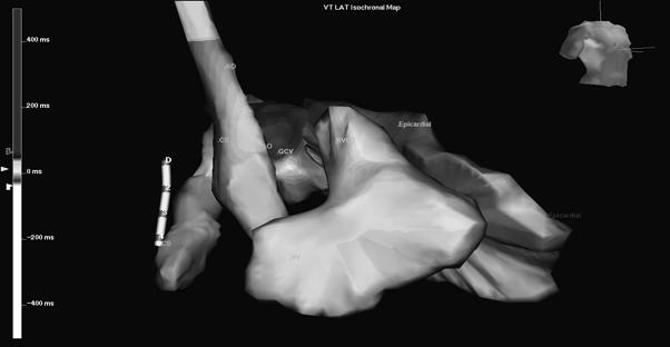

7 ESI Anatomy (RAO) Epicardial and CS catheter Epi Epi CS CS 7


8 Left Aortic Cusp VT Left Aortic Cusp VT Left main coronary artery Ablation catheter Intracardiac ultrasound showing ablator in L coronary cusp 8


 Left Posterior")
9 Aortic Cusp VT ECG Criteria Early transition in precordial leads (V1, V2) Tall R waves in inferior leads Notch in V5, lack of S in V5, V6 Broad R wave in V1, V2 Larger R/S amplitude in V1, V2 Notch in V1 in L cusp VT (transeptal conduction) Lead I negative in L cusp, positive in R cusp Phase analysis (as measured from earliest surface onset) Local onset in V2 7 ms Initial peak / nadir in III 120 ms Initial peak / nadir in V2 78 ms LVOT VT (Aortomitral continuity) Left Posterior Fascicular VT 9

 RBBB, RAD (Ohe,")

10 Features Induction with atrial pacing RBBB, LAD No structural heart disease (Zipes, 1979) Verapamil sensitive (Belhassen, 1981) RBBB, RAD (Ohe, 1988) Upper septal (Shimoike, 2000) Left Anterior Fascicular VT Fascicular VT 10


11 Mitral Annular VT a. Anterolateral b. Posterior c. Posteroseptal PVCs arising from Posterolateral Tricuspid Annulus PVC from the Crux of the Heart 11


12 VT arising from Crux of the Heart VT arising from Crux of the Heart LAO RAO 12


13 Idiopathic VT VT associated with structural heart disease LV Septal VT LV Septal Fibroma 13
 Infiltrative cardiomyopathy- amyloidosis, sarcoidosis Chagas disease Congenital heart disease Tetrology")

14 Real Time Intracardiac Ultrasound Coronary artery disease Idiopathic dilated cardiomyopathy Hypertrophic cardiomyopathy (HOCM) Arrhythmogenic right ventricular cardiomyopathy (ARVC) Infiltrative cardiomyopathy- amyloidosis, sarcoidosis Chagas disease Congenital heart disease Tetrology of Fallot, aortic stenosis Valvular heart disease Pre surgery Post surgical repair Mitral valve prolapse Right sided AV bundle, fibrotic scars in septum, degenerative changes in conduction system Myotonic dystrophy AV block more common Familial VT Genetic abnormality of conduction system 14


15 Ischemic VT Ablation in the Left Ventricle 15
 limited by hemodynamic stability Catheter ablation in sinus")
")
16 Ablation approaches Catheter ablation during VT (critical area of slow conduction) limited by hemodynamic stability Catheter ablation in sinus rhythm (scar mapping) Channels Late potentials Pace mapping Long term results 50-60% Arrhythmogenic Right Ventricular Cardiomyopathy (ARVC) 47 16


17 ARVC: Ventricular Tachycardia arising from Right Ventricle 49 Role of imaging modalities in the diagnosis of ARVC Holter Carto RV angio MRI Non ischemic Dilated Cardiomyopathy Annular scar VT arising from the conduction system (bundle branch reentry, fascicular, interfascicular Epicardial scar 17


18 Bundle Branch Reentry VT Bundle Branch Reentry Reentry circuit is confined to the left and right bundle branches Usually LBBB, during sinus rhythm Presents with: Syncope Palpitations Sudden cardiac death Treatment: RF ablation of right bundle Catheter Ablation of Right Bundle Branch I V 1 II RA Current Voltage 18
 Epicardial and endocardial mapping Hybrid procedures (OR) Remote Navigation")
19 Epicardial VT in Patient with Dilated Cardiomyopathy Failed VT Ablation Redo endocardial mapping with remote navigation Mapping with hemodynamic support (IABP, echmo, LV assist devices (Impella, Tandem heart) Epicardial and endocardial mapping Hybrid procedures (OR) Remote Navigation (Stereotaxis) 19


20 Impella device and VT Ablation Subxyphoid Access to Pericardial Space Epicardial Mapping 20

 Conclusion Ventricular tachycardia is an important cause of sudden cardiac arrest ECG characteristics can localize the site and origin")

21 Epicardial VT: EKG criteria QRS duration > 200 ms Pseudo delta wave > 34 ms Intrinsicoid deflection > 85 ms Shortest RS > 121 ms Precordial MDI > 0.55 ms Non ischemic cardiomyopathy: lack of q wave in inferior leads, positive q wave in lead I Epicardial VT Chagas 30-40% Non ischemic cardiomyopathy 25-50% Ischemic cardiomyopathy 10-15% ARVC 5-10% LV aneurysm, Sarcoid, Non compaction Idiopathic VT 10% (mainly around epicardial arteries) Conclusion Ventricular tachycardia is an important cause of sudden cardiac arrest ECG characteristics can localize the site and origin of VT Idiopathic VT Monomorphic VT / Frequent PVCs curable with catheter ablation Polymorphic VT treated with ICD and drugs VT associated with structural heart disease ICD and antiarrhythmic drugs Catheter ablation is mainly palliative, improved efficacy with epicardial mapping, impella/echmo/iabp 21

22 22
Ablative Therapy for Ventricular Tachycardia
 Ablative Therapy for Ventricular Tachycardia Nitish Badhwar, MD, FACC, FHRS 2 nd Annual UC Davis Heart and Vascular Center Cardiovascular Nurse / Technologist Symposium May 5, 2012 Disclosures Research
Catheter Ablation of VT Without Structural Heart Disease 성균관의대 온영근
 Catheter Ablation of VT Without Structural Heart Disease 성균관의대 온영근 Idiopathic Monomorphic Ventricular Tachycardia Adenosine-sensitive Verapamil-sensitive Propranolol-sensitive Mech (Triggered activity)
Technique of Epicardial VT Ablation
 CARTO Club Jan 2014 Technique of Epicardial VT Ablation Amir AbdelWahab, MD Electrophysiology and Pacing Service Department of Cardiovascular Medicine, Cairo University Need for Epicardial VT ablation
Mapping and Ablation of Challenging Outflow Tract VTs: Pulmonary Artery, LVOT, Epicardial
 Mapping and Ablation of Challenging Outflow Tract VTs: Pulmonary Artery, LVOT, Epicardial Samuel J. Asirvatham, MD Mayo Clinic Rochester California Heart Rhythm Symposium San Francisco, CA September 8,
VENTRICULAR TACHYCARDIA IN THE ABSENCE OF STRUCTURAL HEART DISEASE
 VENTRICULAR TACHYCARDIA IN THE ABSENCE OF STRUCTURAL HEART DISEASE Dimosthenis Avramidis, MD. Consultant Mitera Children s Hospital Athens Greece Scientific Associate 1st Cardiology Dpt Evangelismos Hospital
Arrhythmias (II) Ventricular Arrhythmias. Disclosures
 Ventricular Arrhythmias. Disclosures") Arrhythmias (II) Ventricular Arrhythmias Amy Leigh Miller, MD, PhD Cardiovascular Electrophysiology, Brigham & Women s Hospital Disclosures None Rhythms and Mortality Implantable loop recorder post-mi
Arrhythmias (II) Ventricular Arrhythmias Amy Leigh Miller, MD, PhD Cardiovascular Electrophysiology, Brigham & Women s Hospital Disclosures None Rhythms and Mortality Implantable loop recorder post-mi
NAAMA s 24 th International Medical Convention Medicine in the Next Decade: Challenges and Opportunities Beirut, Lebanon June 26 July 2, 2010
 NAAMA s 24 th International Medical Convention Medicine in the Next Decade: Challenges and Opportunities Beirut, Lebanon June 26 July 2, 2010 I have a financial interest/arrangement or affiliation with
Idiopathic Ventricular Tachycardia Need for an Update in EHRA/HRS Consensus?
 Idiopathic Ventricular Tachycardia Need for an Update in EHRA/HRS Consensus? Arash Arya, M.D. Department of Interventional Electrophysiology Heart Center University of Leipzig Disclosures: NONE Idiopathic
ECG Cases and Questions. Ashish Sadhu, MD, FHRS, FACC Electrophysiology/Cardiology
 ECG Cases and Questions Ashish Sadhu, MD, FHRS, FACC Electrophysiology/Cardiology 32 yo female Life Insurance Physical 56 yo male with chest pain Terminology Injury ST elevation Ischemia T wave inversion
December 2018 Tracings
 Tracings Tracing 1 Tracing 4 Tracing 1 Answer Tracing 4 Answer Tracing 2 Tracing 5 Tracing 2 Answer Tracing 5 Answer Tracing 3 Tracing 6 Tracing 3 Answer Tracing 6 Answer Questions? Contact Dr. Nelson
Sudden cardiac death: Primary and secondary prevention
 Sudden cardiac death: Primary and secondary prevention By Kai Chi Chan Penultimate Year Medical Student St George s University of London at UNic Sheba Medical Centre Definition Sudden cardiac arrest (SCA)
Urgent VT Ablation in a Patient with Presumed ARVC
 Urgent VT Ablation in a Patient with Presumed ARVC Mr Alex Cambridge, Chief Cardiac Physiologist, St. Barts Hospital, London, UK The patient, a 52 year-old male, attended the ICD clinic without an appointment
Epicardial VT Ablation The Cleveland Clinic Experience
 Epicardial VT Ablation The Cleveland Clinic Experience Walid Saliba, MD, FHRS Director, EP Lab Cardiac Electrophysiology Heart and Vascular Institute Epicardial Access in the EP Lab Why Epicardial Special
Acute Coronary Syndromes Unstable Angina Non ST segment Elevation MI (NSTEMI) ST segment Elevation MI (STEMI)
 ST segment Elevation MI (STEMI)") Leanna R. Miller, RN, MN, CCRN-CSC, PCCN-CMC, CEN, CNRN, CMSRN, NP Education Specialist LRM Consulting Nashville, TN Objectives Evaluate common abnormalities that mimic myocardial infarction. Identify
Leanna R. Miller, RN, MN, CCRN-CSC, PCCN-CMC, CEN, CNRN, CMSRN, NP Education Specialist LRM Consulting Nashville, TN Objectives Evaluate common abnormalities that mimic myocardial infarction. Identify
Medicine. Dynamic Changes of QRS Morphology of Premature Ventricular Contractions During Ablation in the Right Ventricular Outflow Tract
 Medicine CLINICAL CASE REPORT Dynamic Changes of QRS Morphology of Premature Ventricular Contractions During Ablation in the Right Ventricular Outflow Tract A Case Report Li Yue-Chun, MD, Lin Jia-Feng,
12-Lead ECG Interpretation. Kathy Kuznar, RN, ANP
 12-Lead ECG Interpretation Kathy Kuznar, RN, ANP The 12-Lead ECG Objectives Identify the normal morphology and features of the 12- lead ECG. Perform systematic analysis of the 12-lead ECG. Recognize abnormalities
REtrive. REpeat. RElearn Design by. Test-Enhanced Learning based ECG practice E-book
 Test-Enhanced Learning Test-Enhanced Learning Test-Enhanced Learning Test-Enhanced Learning based ECG practice E-book REtrive REpeat RElearn Design by S I T T I N U N T H A N G J U I P E E R I Y A W A
General Introduction to ECG. Reading Assignment (p2-16 in PDF Outline )
") General Introduction to ECG Reading Assignment (p2-16 in PDF Outline ) Objectives 1. Practice the 5-step Method 2. Differential Diagnosis: R & L axis deviation 3. Differential Diagnosis: Poor R-wave progression
General Introduction to ECG Reading Assignment (p2-16 in PDF Outline ) Objectives 1. Practice the 5-step Method 2. Differential Diagnosis: R & L axis deviation 3. Differential Diagnosis: Poor R-wave progression
Section V. Objectives
 Section V Landscape of an MI Objectives At the conclusion of this presentation the participant will be able to Outline a systematic approach to 12 lead ECG interpretation Demonstrate the process for determining
Ventricular Tachycardia in Normal Heart: Approach and Management
 ndian Journal of Cardiology SSN-0972-1622 20 ] 2012 by the ndian Society of Cardiology Vol. 15, (3-4), 20-26 Review Article Ventricular Tachycardia in Normal Heart: Approach and Management S.K. Chutani
Ventricular Tachycardia Ablation. Saverio Iacopino, MD, FACC, FESC
 Ventricular Tachycardia Ablation Saverio Iacopino, MD, FACC, FESC ü Ventricular arrhythmias, both symptomatic and asymptomatic, are common, but syncope and SCD are infrequent initial manifestations of
INTRODUCTION. left ventricular non-compaction is a sporadic or familial cardiomyopathy characterized by
 A Rare Case of Arrhythmogenic Right Ventricular Cardiomyopathy Co-existing with Isolated Left Ventricular Non-compaction NS Yelgeç, AT Alper, Aİ Tekkeşin, C Türkkan INTRODUCTION Arrhythmogenic right ventricular
Conduction Problems / Arrhythmias. Conduction
 Conduction Problems / Arrhythmias Conduction Wolf-Parkinson White Syndrome (WPW) and Lown-Ganong-Levine (LGL): Atrial impulses bypass the AV node through an accessory pathway or bypass tract (bundle of
Ablation of Ventricular Tachycardia in Non-Ischemic Cardiomyopathy
 Ablation of Ventricular Tachycardia in Non-Ischemic Cardiomyopathy Fermin C Garcia, MD University of Pennsylvania Cardiac Electrophysiology Philadelphia, PA Nothing to disclose No conflict of interest
Clinical Cardiac Electrophysiology
 Clinical Cardiac Electrophysiology Certification Examination Blueprint Purpose of the exam The exam is designed to evaluate the knowledge, diagnostic reasoning, and clinical judgment skills expected of
Premature ventricular complexes or contractions
 CLINICAL STUDY Analysis of Morphological Characteristics and Origins of Idiopathic Premature Ventricular Contractions Under a 12-Lead Electrocardiogram in Children with Structurally Normal Hearts Jianbin
Map-Guided Ablation of Non-ischemic VT. Takashi Nitta Cardiovascular Surgery, Nippon Medical School Tokyo, JAPAN
 Map-Guided Ablation of Non-ischemic VT Takashi Nitta Cardiovascular Surgery, Nippon Medical School Tokyo, JAPAN nothing Declaration of Interest Catheter Ablation of Non-ischemic VT Sarcoidosis, 13, 6%
ECG Interpretation Made Easy
 ECG Interpretation Made Easy Dr. A Tageldien Abdellah, MSc MD EBSC Lecturer of Cardiology- Hull University Hull York Medical School 2007-2008 ECG Interpretation Made Easy Synopsis Benefits Objectives Process
Office ECG Interpretation
 Office ECG Interpretation Jason Evanchan, DO Assistant Professor of Medicine Division of Cardiovascular Medicine The Ohio State University Wexner Medical Center Outline of topics High risk ischemia T wave
Cardiology Flash Cards
 Cardiology Flash Cards EKG in a nut shell www.brain101.info Conduction System www.brain101.info 2 Analyzing EKG Step by step Steps in Analyzing ECG'S 1. Rhythm: - Regular _ Sinus, Junctional or Ventricular.
TACHYARRHYTHMIAs. Pawel Balsam, MD, PhD
 TACHYARRHYTHMIAs Pawel Balsam, MD, PhD SupraVentricular Tachycardia Atrial Extra Systole Sinus Tachycardia Focal A. Tachycardia AVRT AVNRT Atrial Flutter Atrial Fibrillation Ventricular Tachycardia Ventricular
Managing Hypertrophic Cardiomyopathy with Imaging. Gisela C. Mueller University of Michigan Department of Radiology
 Managing Hypertrophic Cardiomyopathy with Imaging Gisela C. Mueller University of Michigan Department of Radiology Disclosures Gadolinium contrast material for cardiac MRI Acronyms Afib CAD Atrial fibrillation
12 Lead ECG Interpretation
 12 Lead ECG Interpretation Julie Zimmerman, MSN, RN, CNS, CCRN Significant increase in mortality for every 15 minutes of delay! N Engl J Med 2007;357:1631-1638 Who should get a 12-lead ECG? Also include
EHRA Accreditation Exam - Sample MCQs Invasive cardiac electrophysiology
 EHRA Accreditation Exam - Sample MCQs Invasive cardiac electrophysiology Dear EHRA Member, Dear Colleague, As you know, the EHRA Accreditation Process is becoming increasingly recognised as an important
ECG Basics Sonia Samtani 7/2017 UCI Resident Lecture Series
 ECG Basics Sonia Samtani 7/2017 UCI Resident Lecture Series Agenda I. Introduction II.The Conduction System III.ECG Basics IV.Cardiac Emergencies V.Summary The Conduction System Lead Placement avf Precordial
Basic electrocardiography reading. R3 lee wei-chieh
 Basic electrocardiography reading R3 lee wei-chieh The Normal Conduction System Lead Placement avf Limb Leads Precordial Leads Interpretation Rate Rhythm Interval Axis Chamber abnormality QRST change What
Cardiovascular Nursing Practice: A Comprehensive Resource Manual and Study Guide for Clinical Nurses 2 nd Edition
 Cardiovascular Nursing Practice: A Comprehensive Resource Manual and Study Guide for Clinical Nurses 2 nd Edition Table of Contents Volume 1 Chapter 1: Cardiovascular Anatomy and Physiology Basic Cardiac
The Efficient and Smart Methods for Diagnosis of SVT 대구파티마병원순환기내과정병천
 The Efficient and Smart Methods for Diagnosis of SVT 대구파티마병원순환기내과정병천 Differentiation Supraventricular Origin from Ventricular Origin on ECG. QRS-Complex Width. 1. Narrow QRS-Complex Tachycardia (
Paroxysmal Supraventricular Tachycardia PSVT.
 Atrial Tachycardia; is the name for an arrhythmia caused by a disorder of the impulse generation in the atrium or the AV node. An area in the atrium sends out rapid signals, which are faster than those
Reentrant Ventricular Tachycardia Originating in the Right Ventricular Outflow Tract
 Circ J 2008; 72: 855 860 Reentrant Ventricular Tachycardia Originating in the Right Ventricular Outflow Tract Slow Conduction Identified by Right Coronary Artery Ostium Pacing Emi Nakano, MD; Tomoo Harada,
Tachycardias II. Štěpán Havránek
 Tachycardias II Štěpán Havránek Summary 1) Supraventricular (supraventricular rhythms) Atrial fibrillation and flutter Atrial ectopic tachycardia / extrabeats AV nodal reentrant a AV reentrant tachycardia
Ventricular arrhythmias
 Ventricular arrhythmias Assoc.Prof. Lucie Riedlbauchová, MD, PhD Department of Cardiology University HospitalMotol and2nd FacultyofMedicine, Charles University in Prague Definition and classification Ventricular
Myocardial Infarction. Reading Assignment (p66-78 in Outline )
") Myocardial Infarction Reading Assignment (p66-78 in Outline ) Objectives 1. Why do ST segments go up or down in ischemia? 2. STEMI locations and culprit vessels 3. Why 15-lead ECGs? 4. What s up with avr?
Myocardial Infarction Reading Assignment (p66-78 in Outline ) Objectives 1. Why do ST segments go up or down in ischemia? 2. STEMI locations and culprit vessels 3. Why 15-lead ECGs? 4. What s up with avr?
Ventricular Tachycardia in Structurally Normal Hearts (Idiopathic VT) Patient Information
 Patient Information") Melbourne Heart Rhythm Ventricular Tachycardia in Structurally Normal Hearts (Idiopathic VT) Patient Information What is Ventricular Tachycardia? Ventricular tachycardia (VT) is an abnormal rapid heart
Melbourne Heart Rhythm Ventricular Tachycardia in Structurally Normal Hearts (Idiopathic VT) Patient Information What is Ventricular Tachycardia? Ventricular tachycardia (VT) is an abnormal rapid heart
Supraventricular Arrhythmias. Reading Assignment. Chapter 5 (p17-30)
") Supraventricular Arrhythmias Reading Assignment Chapter 5 (p17-30) The Supraventricular Rhythms In Our Lives Site of Origin Single Events Slow Rates Intermediate Rates Fast Rates (>100 bpm) Sinus Sinus
Supraventricular Arrhythmias Reading Assignment Chapter 5 (p17-30) The Supraventricular Rhythms In Our Lives Site of Origin Single Events Slow Rates Intermediate Rates Fast Rates (>100 bpm) Sinus Sinus
ΔΠΔΜΒΑΣΙΚΗ ΘΔΡΑΠΔΙΑ ΚΟΙΛΙΑΚΩΝ ΑΡΡΤΘΜΙΩΝ
 ΔΠΔΜΒΑΣΙΚΗ ΘΔΡΑΠΔΙΑ ΚΟΙΛΙΑΚΩΝ ΑΡΡΤΘΜΙΩΝ ΣΔΛΙΟ ΠΑΡΑΚΔΤΑÏΓΗ ΓΙΔΤΘΤΝΣΗ ΔΤ Α Καρδιολογική Κλινική ΑΠΘ, Νοζοκομείο ΑΧΕΠΑ, Θεζζαλονίκη NO CONFLICT OF INTEREST INTRODUCTION Sustained VT is an important cause
Treatment of VT of Purkinje fiber origin: ablation targets and outcome
 Treatment of VT of Purkinje fiber origin: ablation targets and outcome Ch. Piorkowski University Leipzig - Heart Center - Dept. of Electrophysiology Leipzig, Germany Presenter Disclosure Information Gerhard
CLINICAL CARDIAC ELECTROPHYSIOLOGY Maintenance of Certification (MOC) Examination Blueprint
 Examination Blueprint") CLINICAL CARDIAC ELECTROPHYSIOLOGY Maintenance of Certification (MOC) Examination Blueprint ABIM invites diplomates to help develop the Clinical Cardiac Electrophysiology MOC exam blueprint Based on feedback
CLINICAL CARDIAC ELECTROPHYSIOLOGY Maintenance of Certification (MOC) Examination Blueprint ABIM invites diplomates to help develop the Clinical Cardiac Electrophysiology MOC exam blueprint Based on feedback
Miscellaneous Stuff Keep reading the Outline
 Miscellaneous Stuff Keep reading the Outline Welcome to the 5-Step Method ECG #: Mearurements: Rhythm (s): Conduction: Waveform: Interpretation: A= V= PR= QRS= QT= Axis= 1. Compute the 5 basic measurements:
PAEDIATRIC ECG Dimosthenis Avramidis, MD.
 PAEDIATRIC ECG Dimosthenis Avramidis, MD. Consultant Mitera Children s Hospital Athens Greece S. Associate 1st Cardiology Dpt Evangelismos Hospital Athens Greece 5 y/o with sinus tach Background ECG changes
THE ELECTROCARDIOGRAM A UBIQUITOUS AND COST-EFFECTIVE DIAGNOSTIC TOOL FOR THE FAMILY MEDICINE REFRESHER COURSE MARCH 8, 2019
 THE ELECTROCARDIOGRAM A UBIQUITOUS AND COST-EFFECTIVE DIAGNOSTIC TOOL FOR THE FAMILY MEDICINE REFRESHER COURSE MARCH 8, 2019 Major Clinical Disorders Pulmonary Embolism 69 y/o woman with dyspnea and an
What s New in IV Conduction? (Quadrafascicular, not Trifascicular)
") What s New in IV Conduction? (Quadrafascicular, not Trifascicular) Frank Yanowitz, MD Professor, University of Utah School of Medicine Medical Director, IHC ECG Services (Urban Central Region) http://ecg.utah.edu
What s New in IV Conduction? (Quadrafascicular, not Trifascicular) Frank Yanowitz, MD Professor, University of Utah School of Medicine Medical Director, IHC ECG Services (Urban Central Region) http://ecg.utah.edu
Electrocardiogram A valuable diagnostic tool. Jean Vorster Netcare Unitas Hospital STEMI Early Reperfusion Initiative 2015
 Electrocardiogram A valuable diagnostic tool Jean Vorster Netcare Unitas Hospital STEMI Early Reperfusion Initiative 2015 Overview 1. Arrhythmias 2. Structural heart disease 3. Ischaemia Arrhythmias Sinus
ECG ABNORMALITIES D R. T AM A R A AL Q U D AH
 ECG ABNORMALITIES D R. T AM A R A AL Q U D AH When we interpret an ECG we compare it instantaneously with the normal ECG and normal variants stored in our memory; these memories are stored visually in
Bundle Branch & Fascicular Blocks. Reading Assignment (p53-58 in Outline )
") Bundle Branch & Fascicular Blocks Reading Assignment (p53-58 in Outline ) Objectives 1. QRS analysis of Right and Left BBB 2. Uncomplicated vs complicated BBB 3. Diagnosis of RBBB with LAFB and LPFB 4.
Bundle Branch & Fascicular Blocks Reading Assignment (p53-58 in Outline ) Objectives 1. QRS analysis of Right and Left BBB 2. Uncomplicated vs complicated BBB 3. Diagnosis of RBBB with LAFB and LPFB 4.
12 Lead ECG Skills: Building Confidence for Clinical Practice. Presented By: Cynthia Webner, BSN, RN, CCRN-CMC. Karen Marzlin, BSN, RN,CCRN-CMC
 12 Lead ECG Skills: Building Confidence for Clinical Practice NTI 2009 Preconference Session 803 Presented By: Karen Marzlin, BSN, RN,CCRN-CMC 1 12 Lead ECG Fundamentals: The Starting Place for Linking
Index. cardiacep.theclinics.com. Note: Page numbers of article titles are in boldface type.
 Note: Page numbers of article titles are in boldface type. A AEDs. See Automated external defibrillators (AEDs) AF. See Atrial fibrillation (AF) Age as factor in SD in marathon runners, 45 Antiarrhythmic
Theroleofcatheterablationinthemanagement of ventricular tachycardia
 European Heart Journal Advance Access published August 31, 2015 European Heart Journal doi:10.1093/eurheartj/ehv421 REVIEW Novel therapeutic concepts Theroleofcatheterinthemanagement of ventricular tachycardia
Outflow Tract Ventricular Tachycardia Always Benign?
 Outflow Tract Ventricular Tachycardia Always Benign? Arash Arya, M.D. Department of Interventional Electrophysiology Heart Center University of Leipzig Disclosures: NONE Outflow Ventricular Tachycardia
Return to Basics. ECG Rate and Rhythm. Management of the Hospitalized Patient September 25, 2009
 Management of the Hospitalized Patient September 25, 2009 ECG Refresher and Update 2009 Return to Basics Determine rate and rhythm Determine intervals and axes Define morphology of P-QRS-T-U Compare with
Conventional Mapping. Introduction
 Conventional Mapping Haitham Badran Ain Shams University it Introduction The mapping approach used to guide ablation depends on the type of arrhythmia being assessed. Simple fluoroscopic anatomy is essential
The Electrocardiogram part II. Dr. Adelina Vlad, MD PhD
 The Electrocardiogram part II Dr. Adelina Vlad, MD PhD Basic Interpretation of the ECG 1) Evaluate calibration 2) Calculate rate 3) Determine rhythm 4) Determine QRS axis 5) Measure intervals 6) Analyze
LONG RP TACHYCARDIA MAPPING AND RF ABLATION
 LONG RP TACHYCARDIA MAPPING AND RF ABLATION Dr. Hayam Eldamanhoury Ain shams univeristy Arrhythmia is a too broad topic SVT is broadly defined as narrow complex ( unless aberrant conduction ) Requires
Title. CitationJournal of Electrocardiology, 43(5): Issue Date Doc URL. Type. File Information.
: Issue Date Doc URL. Type. File Information.") Title Pleomorphic ventricular tachycardia originating from Author(s)Yokoshiki, Hisashi; Mitsuyama, Hirofumi; Watanabe, M CitationJournal of Electrocardiology, 43(5): 452-458 Issue Date 2010-09 Doc URL
Title Pleomorphic ventricular tachycardia originating from Author(s)Yokoshiki, Hisashi; Mitsuyama, Hirofumi; Watanabe, M CitationJournal of Electrocardiology, 43(5): 452-458 Issue Date 2010-09 Doc URL
Catheter ablation of monomorphic ventricular tachycardia. Department of Cardiology, IKEM, Prague, Czech Republic
 Catheter ablation of monomorphic ventricular tachycardia Department of Cardiology, IKEM, Prague, Czech Republic DECLARATION OF CONFLICT OF INTEREST None Ventricular tachycardia ablation in IKEM, Prague
ACCESSORY PATHWAYS AND SVT. Neil Grubb Royal Infirmary of Edinburgh
 ACCESSORY PATHWAYS AND SVT Neil Grubb Royal Infirmary of Edinburgh Bypass tracts - properties accessory AV connections usually endocardial may exhibit unidirectional conduction conduction properties similar
ECG Workshop. Nezar Amir
 ECG Workshop Nezar Amir Myocardial Ischemia ECG Infarct ECG in STEMI is dynamic & evolving Common causes of ST shift Infarct Localisation Left main artery occlusion: o diffuse ST-depression with ST elevation
Chapter 2 Practical Approach
 Chapter 2 Practical Approach There are beginners in electrocardiogram (ECG) analysis who are fascinated by a special pattern (e.g., a bundle-branch block or a striking Q wave) and thereby overlook other
Adult Echocardiography Examination Content Outline
 Adult Echocardiography Examination Content Outline (Outline Summary) # Domain Subdomain Percentage 1 2 3 4 5 Anatomy and Physiology Pathology Clinical Care and Safety Measurement Techniques, Maneuvers,
Basic Electrophysiology Protocols
 Indian Journal of Cardiology ISSN-0972-1622 2012 by the Indian Society of Cardiology Vol. 15, (3-4), 27-37 [ 27 Review Article Shomu Bohora Assistant Professor, Deptt. of Cardiology, U.N. Mehta Institute
SIMPLY ECGs. Dr William Dooley
 SIMPLY ECGs Dr William Dooley Content Basic ECG interpretation pattern Some common (examined) abnormalities Presenting ECGs in context Setting up an ECG Setting up an ECG 1 V1-4 th Right intercostal space
Ekg pra pr c a tice D.HAMMOUDI.MD
 Ekg practice D.HAMMOUDI.MD Anatomy Revisited RCA (Right Coronary Artery) Right ventricle Inferior wall of LV Posterior wall of LV (75%) SA Node (60%) AV Node (>80%) LCA (Left Coronary Artery) Septal wall
Biventricular Enlargement/ Hypertrophy
 Biventricular Enlargement/ Hypertrophy Keywords congenital heart disease left ventricular hypertrophy right ventricular hypertrophy SR MITTAL Abstract Electrocardiographic diagnosis of early biventricular
Tachy. Induction tachycardia lead ECG during Tachy /25/2009. Sinus Rhythm Single His
 12-lead ECG during Tachy 10.30.31 Sinus Rhythm Single His 11.20.02 Induction tachycardia 11.23.23 Tachy 11.25.23 1 I This finding excludes: (a) AVNRT (either typical or atypical) Tachy: Alternating cycle
Ventricular tachycardia Ventricular fibrillation and ICD
 EKG Conference Ventricular tachycardia Ventricular fibrillation and ICD Samsung Medical Center CCU D.I. Hur Ji Won 2006.05.20 Ventricular tachyarrhythmia ventricular tachycardia ventricular fibrillation
ΔΙΑΤΑΡΑΧΕΣ ΕΝΔΟΚΟΙΛΙΑΚΗΣ ΑΓΩΓΙΜΟΤΗΤΑΣ ΔΗΜΗΤΡΙΟΣ Δ. ΜΑΝΩΛΑΤΟΣ Β ΚΑΡΔΙΟΛΟΓΙΚΗ ΚΛΙΝΙΚΗ ΕΡΓΑΣΤΗΡΙΟ ΗΛΕΚΤΡΟΦΥΣΙΟΛΟΓΙΑΣ Γ.Ν.Α.
 ΔΙΑΤΑΡΑΧΕΣ ΕΝΔΟΚΟΙΛΙΑΚΗΣ ΑΓΩΓΙΜΟΤΗΤΑΣ ΔΗΜΗΤΡΙΟΣ Δ. ΜΑΝΩΛΑΤΟΣ Β ΚΑΡΔΙΟΛΟΓΙΚΗ ΚΛΙΝΙΚΗ ΕΡΓΑΣΤΗΡΙΟ ΗΛΕΚΤΡΟΦΥΣΙΟΛΟΓΙΑΣ Γ.Ν.Α. «ΕΥΑΓΓΕΛΙΣΜΟΣ» Intraventricular conduction delay and Blocks Right Bundle Branch
Return to Basics. Normal Intervals & Axes. ECG Rate and Rhythm
 Return to Basics Management of the Hospitalized Patient October 15, 2010 ECG Refresher and Update 2010 Determine rate and rhythm Determine intervals and axes Define morphology of P-QRS-T-U Compare with
Fast & Slow Tachy & Brady Arrhythmias DAVID STULTZ, MD, FACC KPN HEART & VASCULAR AUGUST 7, 2017
 Fast & Slow Tachy & Brady Arrhythmias DAVID STULTZ, MD, FACC KPN HEART & VASCULAR AUGUST 7, 2017 Normal EKG EKG boxes Heart Rate 1 big box = 200ms 1 small box = 40ms Big Boxes Between QRS complexes Heart
SIMPLY ECGs. Dr William Dooley
 SIMPLY ECGs Dr William Dooley 1 No anatomy just interpretation 2 Setting up an ECG 3 Setting up an ECG 1 V1-4 th Right intercostal space at sternal border 2 V2-4 th Left intercostal space at sternal border
Arrhythmia Management Joshua M. Cooper, MD, FHRS, FACC
 Arrhythmia Management Joshua M. Cooper, MD, FHRS, FACC Professor of Medicine Director of Cardiac Electrophysiology Temple University Health System Plumbing Electrical System Bradyarrhythmias Sinus Node
Intermediate ECG Course - Part 4. Joe M. Moody, Jr, MD UTHSCSA and STVAHCS
 Intermediate ECG Course - Part 4 Joe M. Moody, Jr, MD UTHSCSA and STVAHCS Topics in Intermediate ECG Consolidation of prior information with additional details Not advanced, but feel free to ask advanced
Case Report Coexistence of Atrioventricular Nodal Reentrant Tachycardia and Idiopathic Left Ventricular Outflow-Tract Tachycardia
 www.ipej.org 149 Case Report Coexistence of Atrioventricular Nodal Reentrant Tachycardia and Idiopathic Left Ventricular Outflow-Tract Tachycardia Majid Haghjoo, M.D, Arash Arya, M.D, Mohammadreza Dehghani,
When to implant an ICD in systemic right ventricle?
 When to implant an ICD in systemic right ventricle? Département de rythmologie et de stimulation cardiaque Nicolas Combes n.combes@clinique-pasteur.com Pôle de cardiologie pédiatrique et congénitale Risk
Study methodology for screening candidates to athletes risk
 1. Periodical Evaluations: each 2 years. Study methodology for screening candidates to athletes risk 2. Personal history: Personal history of murmur in childhood; dizziness, syncope, palpitations, intolerance
Electroanatomic Substrate and Outcome of Catheter Ablative Therapy for Ventricular Tachycardia in Setting of Right Ventricular Cardiomyopathy
 Electroanatomic Substrate and Outcome of Catheter Ablative Therapy for Ventricular Tachycardia in Setting of Right Ventricular Cardiomyopathy Francis E. Marchlinski, MD; Erica Zado, PA-C; Sanjay Dixit,
EHRA EUROPACE How to perform epicardial ventricular tachycardia mapping and ablation
 EHRA EUROPACE 2011 How to perform epicardial ventricular tachycardia mapping and ablation Jacob Atié Director Arrhythmias Department Federal University of Rio de janeiro jacobatie1@gmail.com Presenter
Please check your answers with correct statements in answer pages after the ECG cases.
 ECG Cases ECG Case 1 Springer International Publishing AG, part of Springer Nature 2018 S. Okutucu, A. Oto, Interpreting ECGs in Clinical Practice, In Clinical Practice, https://doi.org/10.1007/978-3-319-90557-0
ELECTROCARDIOGRAPH. General. Heart Rate. Starship Children s Health Clinical Guideline
 General Heart Rate QRS Axis T Wave Axis PR Interval according to Heart Rate & Age P Wave Duration and Amplitude QRS Duration according to Age QT Interval R & S voltages according to Lead & Age R/S ratio
Ventricular Tachycardia Substrate. For the ablationist. Stanley Tung, MD FRCPC Arrhythmia Service/St Paul Hospital University of British Columbia
 Ventricular Tachycardia Substrate For the ablationist Stanley Tung, MD FRCPC Arrhythmia Service/St Paul Hospital University of British Columbia Two Attitudes of Ventricular Tachycardia Ablation 1 2C:\Documents
Ben Taylor, PhD, PA-C
 Ben Taylor, PhD, PA-C The patient is a 23-year-old white male with a history of polysubstance abuse who was found unresponsive, last seen the day before. Classic signs of systemic hypothermia with prominent
ECG Workshop. Carolyn Shepherd And Anya Horne UWE Principles of Cardiac Care
 ECG Workshop Carolyn Shepherd And Anya Horne UWE Principles of Cardiac Care ECG workshop case study1 44 Year old male. Reports SOB, Lethargy, tiredness. PMH: Hypertension, nil else. What tests? What treatment?
CATHETER ABLATION FOR TACHYCARDIAS
 190 CATHETER ABLATION FOR TACHYCARDIAS MASOOD AKHTAR, M.D. T ACHY ARRHYTHMIAS constitute a major cause of mortality and morbidity. The most serious manifestation of cardiac arrhythmia is sudden cardiac
, David Stultz, MD.
 http://www.dilbert.com EKG Rounds Handouts available at http://www.drstultz.com January 5, 2004 David Stultz, MD Cardiology Fellow, PGY 4 Overview of Topics How to read an EKG Normal EKG Determination
Implantable Cardioverter Defibrillator (ICD)
") Medical Coverage Policy Effective Date... 3/15/2018 Next Review Date... 3/15/2019 Coverage Policy Number... 0181 Implantable Cardioverter Defibrillator (ICD) Table of Contents Related Coverage Resources
Medical Coverage Policy Effective Date... 3/15/2018 Next Review Date... 3/15/2019 Coverage Policy Number... 0181 Implantable Cardioverter Defibrillator (ICD) Table of Contents Related Coverage Resources
15 16 September Seminar W10O. ECG for General Practice
 15 16 September 2012 Seminar W10O ECG for General Practice Speaker: Ms Natasha Eaton ECG for General Practice Speaker: Natasha Eaton Cardiac CNC Executive Representative Electrocardiography The graphic
EKG Competency for Agency
 EKG Competency for Agency Name: Date: Agency: 1. The upper chambers of the heart are known as the: a. Atria b. Ventricles c. Mitral Valve d. Aortic Valve 2. The lower chambers of the heart are known as
ECGs on the acute admission ward. - Cardiology Update -
 ECGs on the acute admission ward - Cardiology Update - Dr Simon Fynn Consultant Cardiologist Papworth Hospital, Cambridge RCP London Oct 2017 ECG 1 1. AF with BBB 2. Pre-excited AF 3. SVT with BBB 4.
All About STEMIs. Presented By: Brittney Urvand, RN, BSN, CCCC. Essentia Health Fargo Cardiovascular Program Manager.
 All About STEMIs Presented By: Brittney Urvand, RN, BSN, CCCC Essentia Health Fargo Cardiovascular Program Manager Updated 10/2/2018 None Disclosures Objectives Identify signs and symptoms of a heart attack
Purkinje-related Arrhythmias
 J Arrhythmia Vol 27 No 1 2011 Review Article Purkinje-related Arrhythmias Akihiko Nogami MD Department of Heart Rhythm Management, Yokohama Rosai Hospital, Yokohama, Japan The Purkinje system has been
Supraventricular Tachycardia (SVT)
") Supraventricular Tachycardia (SVT) Bruce Stambler, MD Piedmont Heart Atlanta, GA Supraventricular Tachycardia Objectives Types and mechanisms AV nodal reentrant tachycardia (AVNRT) AV reciprocating tachycardia
Supraventricular Tachycardia (SVT) Bruce Stambler, MD Piedmont Heart Atlanta, GA Supraventricular Tachycardia Objectives Types and mechanisms AV nodal reentrant tachycardia (AVNRT) AV reciprocating tachycardia