Multimodality Imaging of Anomalous Left Coronary Artery from the Pulmonary
|
|
|
- Octavia Martina Norman
- 6 years ago
- Views:
Transcription
1 1 IMAGES IN CARDIOVASCULAR ULTRASOUND Multimodality Imaging of Anomalous Left Coronary Artery from the Pulmonary Artery Byung Gyu Kim, MD 1, Sung Woo Cho, MD 1, Dae Hyun Hwang, MD 2 and Jong Chun Nah, MD Division of Cardiology, Department of Internal Medicine, Inje University College of Medicine, Seoul Paik Hospital, Seoul, Korea Department of Radiology, Inje University College of Medicine, Seoul Paik Hospital, Seoul, Korea 12 Running title: Multimodality Imaging of ALCAPA Address for Correspondence: Jong Chun Nah, Division of Cardiology, Department of Internal Medicine, Inje University College of Medicine, Seoul Paik Hospital, 9 Mareunnae-ro, Jung-gu, Seoul 04551, Korea Tel: , Fax: , drnah1970@daum.net 18 * Byung Gyu Kim and Sung Woo Cho contributed equally to this work. 19
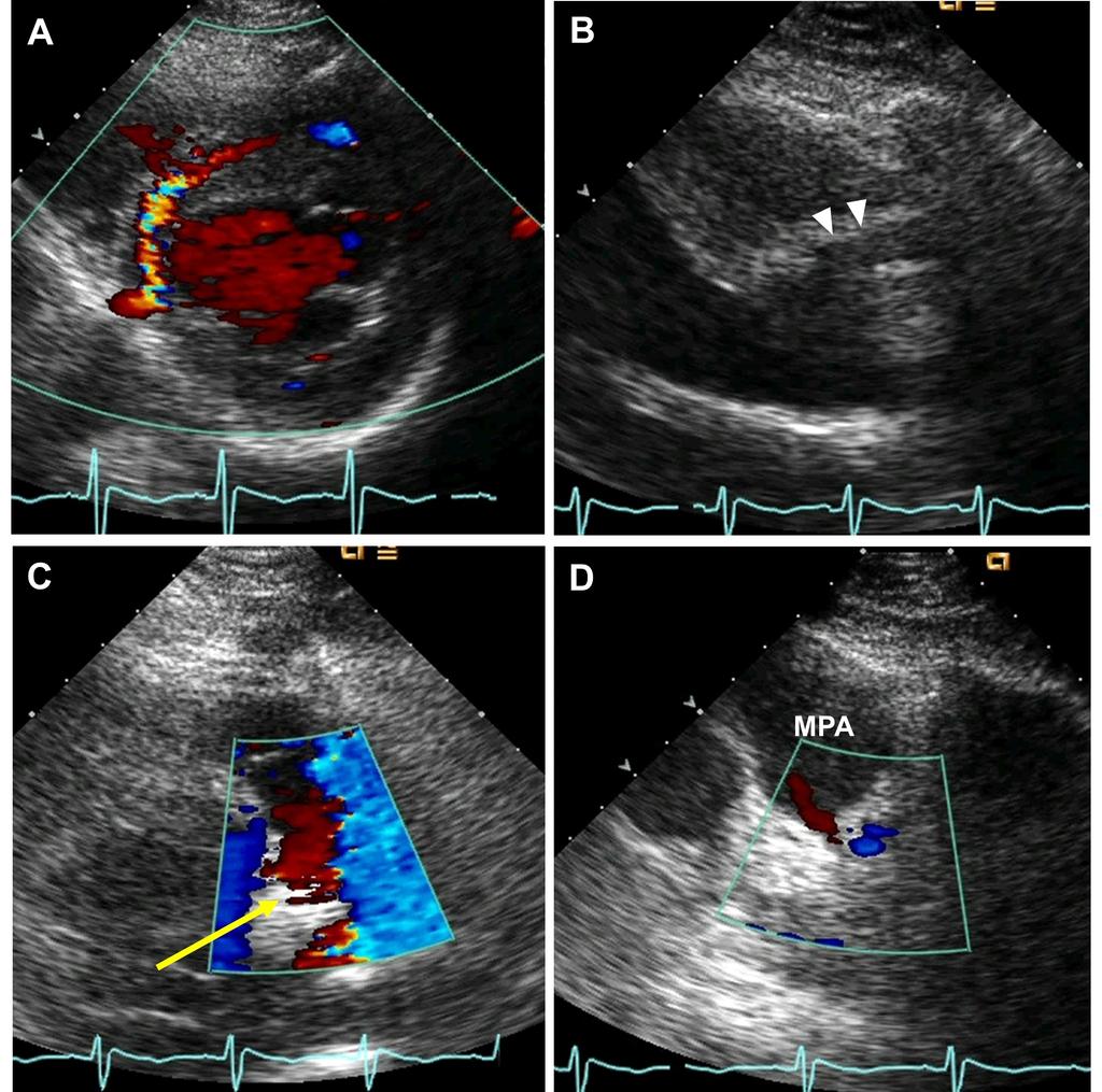
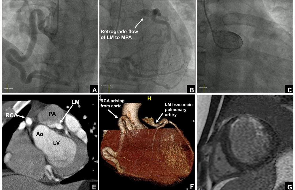
2 A 22-year-old man was hospitalized for right femur fracture due to a motorcycle accident. Although he had no known cardiac or family history, he felt intermittent chest tightness during moderate intensity of exercise. His electrocardiography showed patterns of left ventricular strain. The echocardiography showed left ventricular hypertrophy, mild eccentric mitral regurgitation, and regional wall motion abnormality and thinning of left anterior descending (LAD) coronary artery territory with lower normal left ventricular systolic function, in which ejection fraction was 50%. Diastolic flow showing peak velocity of 2.5 cm/sec was observed at interventricular septum, which was suspicious of excessive collateral flow at parasternal short axis view (Fig. 1A). Dilated right coronary artery (RCA) ostium of 10 mm was observed (Fig. 1B) on parasternal long axis view, whereas left main coronary artery was not detected in typical situs. Notably, an abnormal retrograde shunt flow was detected (Fig. 1C, Supplementary Movie 1) and a drainage site of abnormal shunt flow was observed at the main pulmonary artery level of parasternal short axis view (Fig. 1D). Thus, we suspected a congenital anomaly of the coronary arteries. Coronary angiography revealed an enlarged and tortuous RCA with abundant septal collateral flows toward the left coronary artery. An unusual location of the left main coronary artery opening with an abnormal retrograde shunt flow was observed in the left superior part of aorta, most likely pulmonary artery (Fig. 2A and 2B, Supplementary Movie 2). However, left coronary artery was not shown in the left coronary cusp (Fig. 2C). To specify the location of the left main coronary artery opening, cardiac multidetector computed tomography (CT) was performed and the anomalous origin of left coronary artery from pulmonary artery (ALCAPA) was finally confirmed (Fig. 2D and 2E). To determine myocardial viability, cardiac magnetic resonance imaging (MRI) was performed and the thinning and subendocardial delayed enhancement of anterior wall from base to middle left ventricle was observed, which suggested chronic subendocardial infarction of LAD territory (Fig. 2F). The patient received beta-blocker and
3 angiotensin converting enzyme inhibitor, and completed femur fracture surgery without any cardiovascular events. He was discharged and is scheduled for an open cardiac surgery for the correction of ALCAPA. ALCAPA is a very rare congenital heart disease with reported incidence rate of 1 in 300,000 children, or 0.5% of those with congenital heart disease. 1) If untreated, up to 90% of patients with ALCAPA die during the first year of life due to severe myocardial ischemia. Only 10%- 15% of ALCAPA patients reach adulthood depending on the development of sufficient intercoronary communications. 2) Despite abundant collaterals, blood supply to left coronary artery territory can be inadequate, especially to the subendocardial region according to the proportion of coronary steal from coronary artery to pulmonary artery. For a precise diagnosis and risk stratification of ALCAPA, multimodality imaging studies are needed. Echocardiography is essential and provides important clues for diagnosis of ALCAPA, as demonstrated in our case. RCA dilatation, collateral coronary artery flow, left coronary artery flow reversal, mitral regurgitation, and left ventricular dysfunction are known as important echocardiographic findings of ALCAPA. 3) Cardiac CT is useful for visualization of coronary arteries, but is limited in evaluating coronary flow, valvular function, and myocardial viability. Cardiac MRI provides more detailed information on myocardial perfusion, fibrosis, and viability for deciding treatment plan. Coronary angiography is definitive diagnostic modality for providing anatomic information of anomalous coronary arteries and their flow. There are no current guidelines for the optimal treatment of ALCAPA. According to previous literatures, surgical correction has good prognosis and is recommended regardless of age or symptoms in order to prevent adverse outcomes including myocardial infarction or sudden cardiac death. 4) We present a very rare case of an adult type ALCAPA accompanied by chronic subendocardial infarction, which was initially suspected by echocardiography and confirmed
4 70 71 by multimodality imaging studies. The present case also emphasizes the essential role of echocardiography for providing important clues of ALCAPA. 72
5 REFERENCES 1. Keith JD. The anomalous origin of the left coronary artery from the pulmonary artery. Br Heart J 1959;21: Moodie DS, Fyfe D, Gill CC, Cook SA, Lytle BW, Taylor PC, Fitzgerald R, Sheldon WC. Anomalous origin of the left coronary artery from the pulmonary artery (Bland-White- Garland syndrome) in adult patients: long-term follow-up after surgery. Am Heart J 1983;106: Patel SG, Frommelt MA, Frommelt PC, Kutty S, Cramer JW. Echocardiographic diagnosis, surgical treatment, and outcomes of anomalous left coronary artery from the pulmonary artery. J Am Soc Echocardiogr. Epub 2017 Jun Kottayil BP, Jayakumar K, Dharan BS, Pillai VV, Ajitkumar V, Menon S, Sanjay G. Anomalous origin of left coronary artery from pulmonary artery in older children and adults: direct aortic implantation. Ann Thorac Surg 2011;91:
6 87 Figure Legends Fig. 1. (A) An accelerated diastolic color flow within the interventricular septum indicating a large septal collateral flow with frontal direction from right coronary artery to left anterior descending artery on parasternal short axis view. (B) Markedly dilated right coronary artery ostium on parasternal long axis view (arrow heads). (C) Retrograde diastolic shunt flow toward pulmonary valve at MPA (arrow). (D) Drainage site of abnormal shunt flow at MPA with diastolic reversal flow. MPA: main pulmonary artery Fig. 2. (A) Coronary angiography showing an enlarged and tortuous RCA from aorta. (B) Retrograde filling of LCA through abundant collaterals from RCA and abnormal shunt flow from left main stem to main PA. (C) No visualization of LCA in left coronary cusp. (D, F) Sagittal section and 3D reconstruction images of computed tomography of the LCA originating from the PA with retrograde contrast flow from LCA to PA. (G) Thinning and subendocardial delayed enhancement of anterior LV wall on cardiac magnetic resonance imaging. Ao: aorta, LM: left main artery, LCA: left coronary artery, LV: left ventricle, MPA: main pulmonary artery, PA: pulmonary artery, RCA: right coronary artery. 103
7 104 Supplementary Movie Legends 105 Movie 1. Echocardiography: parasternal short axis view at the level of pulmonary trunk. 106 Movie 2. Coronary angiography. 107
8 Figure 1
9 Figure 2
Coronary Artery Anomalies from Birth to Adulthood; the Role of CT Coronary Angiography in Sudden Cardiac Death Screening
 Coronary Artery Anomalies from Birth to Adulthood; the Role of CT Coronary Angiography in Sudden Cardiac Death Screening E O Dwyer 1, C O Brien 1, B Loo 1, A Snow Hogan 1, O Buckley1 2, B 1. Department
Department of Medicine, New Jersey Medical School, Newark, New Jersey Department of Radiology, New Jersey Medical School, Newark, New Jersey
 Journal compilation C 2010, Wiley Periodicals, Inc. DOI: 10.1111/j.1540-8175.2009.01040.x C 2010, the Authors An Unusual Combination of an Anomalous Origin of the Left Coronary Artery from the Pulmonary
Cardiac MRI in ACHD What We. ACHD Patients
 Cardiac MRI in ACHD What We Have Learned to Apply to ACHD Patients Faris Al Mousily, MBChB, FAAC, FACC Consultant, Pediatric Cardiology, KFSH&RC/Jeddah Adjunct Faculty, Division of Pediatric Cardiology
Case Report Preoperative Assessment of Anomalous Right Coronary Artery Arising from the Main Pulmonary Artery
 Case Reports in Medicine Volume 2011, Article ID 642126, 4 pages doi:10.1155/2011/642126 Case Report Preoperative Assessment of Anomalous Right Coronary Artery Arising from the Main Pulmonary Artery Marshall
Isolated congenital coronary anomalies: Evaluation by multislice-ct or MRI
 Isolated congenital coronary anomalies: Evaluation by multislice-ct or MRI B.K. Velthuis, Dept. of Radiology UMC Utrecht, the Netherlands ESC 2010 Coronary artery anomalies CAA Uncommon 0.3-5% normal population
Anomalous Origin of Left Coronary Artery from Main Pulmonary Artery (ALCAPA) Who Underwent Two Coronary System Repair with a Novel Technique
 Who Underwent Two Coronary System Repair with a Novel Technique") Open Journal of Clinical Diagnostics, 2014, 4, 182-191 Published Online September 2014 in SciRes. http://www.scirp.org/journal/ojcd http://dx.doi.org/10.4236/ojcd.2014.43027 Anomalous Origin of Left Coronary
Open Journal of Clinical Diagnostics, 2014, 4, 182-191 Published Online September 2014 in SciRes. http://www.scirp.org/journal/ojcd http://dx.doi.org/10.4236/ojcd.2014.43027 Anomalous Origin of Left Coronary
CARDIAC AND CORONARY ARTERY ANATOMY NO DISCLOSURES. Axial Anatomy of Heart. Axial Anatomy of Heart. Axial Anatomy of Heart
 CARDIAC AND CORONARY ARTERY ANATOMY NO DISCLOSURES NASCI MEETING, ORLANDO FLORIDA 2009 KOSTAKI G. BIS, MD, FACR DEPARTMENT OF RADIOLOGY WILLIAM BEAUMONT HOSPITAL Royal Oak, Michigan OBJECTIVES CARDIAC
Bland - White - Garland Syndrome confirmed by dual source computed tomography angiography
 ISPUB.COM The Internet Journal of Cardiology Volume 7 Number 1 Bland - White - Garland Syndrome confirmed by dual source computed tomography angiography V Mendoza-Rodríguez, L Llerena, E Olivares-Aquiles,
Late presentation of anomalous origin of the left coronary artery from the pulmonary artery (ALCAPA) was confused with coronary artery fistula.
 was confused with coronary artery fistula.") Case Report http://www.alliedacademies.org/annals-of-cardiovascular-and-thoracic-surgery/ Late presentation of anomalous origin of the left coronary artery from the pulmonary artery (ALCAPA) was confused
Case Report http://www.alliedacademies.org/annals-of-cardiovascular-and-thoracic-surgery/ Late presentation of anomalous origin of the left coronary artery from the pulmonary artery (ALCAPA) was confused
Congenital Coronary Anomalies
 Chapter 50 Congenital Coronary Anomalies S. Adil Husain, Brett C. Sheridan, and Michael R. Mill Congenital coronary anomalies may have a significant impact on myocardial perfusion and secondary ischemia,
Preserved Left Ventricular Function in Two Infants with Anomalous Left Coronary Artery from the Pulmonary Artery
 Open Journal of Clinical & Medical Case Reports Volume 1 (2015) Issue 8 Abstract ISSN 2379-1039 Preserved Left Ventricular Function in Two Infants with Anomalous Left Coronary Artery from the Pulmonary
CORONARY ANOMALIES. Clinical Significance. Disclosures. Definitions. Learning Objectives. Prevalence. Consultant for M2S, Inc.
 Disclosures CORONARY ANOMALIES Consultant for M2S, Inc. Julianna M. Czum, MD Director, Division of Cardiothoracic Imaging Department of Radiology Dartmouth Hitchcock Medical Center Assistant Professor
Index. Note: Page numbers of article titles are in boldface type.
 Index Note: Page numbers of article titles are in boldface type. A Acute coronary syndrome(s), anticoagulant therapy in, 706, 707 antiplatelet therapy in, 702 ß-blockers in, 703 cardiac biomarkers in,
ADVANCED CARDIOVASCULAR IMAGING. Medical Knowledge. Goals and Objectives PF EF MF LF Aspirational
 Medical Knowledge Goals and Objectives PF EF MF LF Aspirational Know the basic principles of magnetic resonance imaging (MRI) including the role of the magnetic fields and gradient coil systems, generation
Adult Echocardiography Examination Content Outline
 Adult Echocardiography Examination Content Outline (Outline Summary) # Domain Subdomain Percentage 1 2 3 4 5 Anatomy and Physiology Pathology Clinical Care and Safety Measurement Techniques, Maneuvers,
GoodPrognosisofALCAPAAnomalousOriginoftheLeftCoronaryArteryfromthePulmonaryArterySyndromewithearlyDiagnosisandSurgicalTreatment
 Global Journal of Medical Research: I Surgeries and Cardiovascular System Volume 18 Issue 3 Version 1.0 Type: Double Blind Peer Reviewed International Research Journal Publisher: Global Journals Online
Left atrial function. Aliakbar Arvandi MD
 In the clinic Left atrial function Abstract The left atrium (LA) is a left posterior cardiac chamber which is located adjacent to the esophagus. It is separated from the right atrium by the inter-atrial
University Journal of Medicine and Medical Sciences
 ISSN 2455-2852 Volume 2 issue 1 2016 Infantile Heart Failure, Exotic Diagnosis but Dismal Prognosis LIJO VARGHESE PUTHENMADOM Department of Cardiology, CHRISTIAN MEDICAL COLLEGE Abstract : Infantile heart
cardiac imaging planes planning basic cardiac & aortic views for MR
 cardiac imaging planes planning basic cardiac & aortic views for MR Dianna M. E. Bardo, M. D. Assistant Professor of Radiology & Cardiovascular Medicine Director of Cardiac Imaging cardiac imaging planes
Case Report Anomalous Origin of Left Coronary Artery from the Pulmonary Trunk in a Mildly Symptomatic Adult Female
 Case Reports in Surgery Volume 2013, Article ID 840741, 4 pages http://dx.doi.org/10.1155/2013/840741 Case Report Anomalous Origin of Left Coronary Artery from the Pulmonary Trunk in a Mildly Symptomatic
Anomalous left coronary artery from the pulmonary artery: a 15 year sample*
 Br Heart J 1987;58:378-84 Anomalous left coronary artery from the pulmonary artery: a 15 year sample* SAMUEL MENAHEM, ALEXANDER W VENABLES From the Department of Cardiology, Royal Children's Hospital,
2019 Qualified Clinical Data Registry (QCDR) Performance Measures
 Performance Measures") 2019 Qualified Clinical Data Registry (QCDR) Performance Measures Description: This document contains the 18 performance measures approved by CMS for inclusion in the 2019 Qualified Clinical Data Registry
2019 Qualified Clinical Data Registry (QCDR) Performance Measures Description: This document contains the 18 performance measures approved by CMS for inclusion in the 2019 Qualified Clinical Data Registry
Cardiovascular Images
 Cardiovascular Images Pulmonary Embolism Diagnosed From Right Heart Changes Seen After Exercise Stress Echocardiography Brian C. Case, MD; Micheas Zemedkun, MD; Amarin Sangkharat, MD; Allen J. Taylor,
PROSTHETIC VALVE BOARD REVIEW
 PROSTHETIC VALVE BOARD REVIEW The correct answer D This two chamber view shows a porcine mitral prosthesis with the typical appearance of the struts although the leaflets are not well seen. The valve
Coronary Anomalies & Hemodynamic Identification
 Coronary Anomalies & Hemodynamic Identification David Stultz, MD Cardiology Fellow, PGY 6 May 2, 2006 Anomaly #1 Anomaly #2 Anomaly #3 Figure 18-27 Anomalous origin of the left circumflex artery.
Scholars Journal of Medical Case Reports
 Scholars Journal of Medical Case Reports Sch J Med Case Rep 2016; 4(5):338-342 Scholars Academic and Scientific Publishers (SAS Publishers) (An International Publisher for Academic and Scientific Resources)
INTRODUCTION CASE REPORT
 Yonsei Med J 50(1):164-168, 2009 DOI 10.3349/ymj.2009.50.1.164 A Case of Acute Myocardial Infarction with the Anomalous Origin of the Right Coronary Artery from the Ascending Aorta above the Left Sinus
Index. radiologic.theclinics.com. Note: Page numbers of article titles are in boldface type.
 Index Note: Page numbers of article titles are in boldface type. A ALCAPA. See Anomalous left coronary artery from the pulmonary artery. Angiosarcoma computed tomographic assessment of, 809 811 Anomalous
Jae Hoon Lim, M.D., Song Choi, M.D. 2, Yang Jun Kang, M.D. 2, Hyun Ju Seon, M.D., Yun Hyeon Kim, M.D.
 J Korean Soc Radiol 2010;62:113-117 The Noninvasive Diagnosis and Postoperative Evaluation of nomalous Right Coronary rtery from the Pulmonary rtery (RCP) using Coronary MDCT: Case Report 1 Jae Hoon Lim,
Budi Yuli Setianto, Anggoro Budi Hartopo, Putrika Prastuti Ratna Gharini, and Nahar Taufiq. 1. Introduction. 2. Case Report
 Case Reports in Cardiology Volume 2016, Article ID 7652869, 4 pages http://dx.doi.org/10.1155/2016/7652869 Case Report Anomalous Origination of Right Coronary Artery from Left Sinus in Asymptomatic Young
Sports cardiology: Pre-competition screening
 Sports cardiology: Pre-competition screening Dr. med Andreas E. Brauchlin Division of cardiology, University Hospital, Zurich andreas.brauchlin@usz.ch Content Interactive case presentation Background and
Doppler-echocardiographic findings in a patient with persisting right ventricular sinusoids
 Zurich Open Repository and Archive University of Zurich Main Library Strickhofstrasse 39 CH-8057 Zurich www.zora.uzh.ch Year: 1990 Doppler-echocardiographic findings in a patient with persisting right
General Cardiovascular Magnetic Resonance Imaging
 2 General Cardiovascular Magnetic Resonance Imaging 19 Peter G. Danias, Cardiovascular MRI: 150 Multiple-Choice Questions and Answers Humana Press 2008 20 Cardiovascular MRI: 150 Multiple-Choice Questions
CT for Myocardial Characterization of Cardiomyopathy. Byoung Wook Choi, Yonsei University Severance Hospital, Seoul, Korea
 CT for Myocardial Characterization of Cardiomyopathy Byoung Wook Choi, Yonsei University Severance Hospital, Seoul, Korea Cardiomyopathy Elliott P et al. Eur Heart J 2008;29:270-276 The European Society
Cardiac ultrasound protocols
 Cardiac ultrasound protocols IDEXX Telemedicine Consultants Two-dimensional and M-mode imaging planes Right parasternal long axis four chamber Obtained from the right side Displays the relative proportions
Cigna - Prior Authorization Procedure List: Radiology & Cardiology
 Cigna - Prior Authorization Procedure List: Radiology & Cardiology Product Category CPT Code CPT Code Description Radiology MR 70336 MRI Temporomandibular Joint(s), (TMJ) Radiology CT 70450 CT Head or
Detection and Assessment of MI: Use of Imaging Methods. Robert O. Bonow, M.D.
 Detection and Assessment of MI: Use of Imaging Methods Robert O. Bonow, M.D. Detection and Assessment of MI: Use of Imaging Methods Robert O. Bonow, M.D. No Relationships to Disclose Expert Consensus Document
The Heart & Pericardium Dr. Rakesh Kumar Verma Assistant Professor Department of Anatomy KGMU UP Lucknow
 The Heart & Pericardium Dr. Rakesh Kumar Verma Assistant Professor Department of Anatomy KGMU UP Lucknow Fibrous skeleton Dense fibrous connective tissue forms a structural foundation around AV & arterial
Appendix II: ECHOCARDIOGRAPHY ANALYSIS
 Appendix II: ECHOCARDIOGRAPHY ANALYSIS Two-Dimensional (2D) imaging was performed using the Vivid 7 Advantage cardiovascular ultrasound system (GE Medical Systems, Milwaukee) with a frame rate of 400 frames
Case Report Sinus Venosus Atrial Septal Defect as a Cause of Palpitations and Dyspnea in an Adult: A Diagnostic Imaging Challenge
 Case Reports in Medicine Volume 2015, Article ID 128462, 4 pages http://dx.doi.org/10.1155/2015/128462 Case Report Sinus Venosus Atrial Septal Defect as a Cause of Palpitations and Dyspnea in an Adult:
What s New in Cardiac MRI
 What s New in Cardiac MRI Katie M. Hawthorne, MD Director, Cardiac MRI Main Line Health Philadelphia Cardiovascular Summit November 18, 2017 Cardiac MRI: Disclosure 2 Disclosures No financial disclosures
Specific Basic Standards for Osteopathic Fellowship Training in Cardiology
 Specific Basic Standards for Osteopathic Fellowship Training in Cardiology American Osteopathic Association and American College of Osteopathic Internists BOT 07/2006 Rev. BOT 03/2009 Rev. BOT 07/2011
Radiology of the respiratory/cardiac diseases (part 2)
") Cardiology Cycle - Lecture 6 436 Teams Radiology of the respiratory/cardiac diseases (part 2) Objectives Done By Team Leaders: Khalid Alshehri Hanin Bashaikh Team Members: Leena Alwakeel Aroob Alhuthail
Cardiology Cycle - Lecture 6 436 Teams Radiology of the respiratory/cardiac diseases (part 2) Objectives Done By Team Leaders: Khalid Alshehri Hanin Bashaikh Team Members: Leena Alwakeel Aroob Alhuthail
For Personal Use. Copyright HMP 2013
 12-00415 Case Report J INVASIVE CARDIOL 2013;25(4):E69-E71 A Concert in the Heart. Bilateral Melody Valve Implantation in the Branch Pulmonary Arteries Nicola Maschietto, MD, PhD and Ornella Milanesi,
SPECT-CT: Τι πρέπει να γνωρίζει ο Καρδιολόγος
 SPECT-CT: Τι πρέπει να γνωρίζει ο Καρδιολόγος Δρ Αναστασία Κίτσιου Διευθύντρια, Καρδιολογική Κλινική, Σισμανόγλειο ΓΝΑ Chair, Education Committee, Section on Nuclear Cardiology & Cardiac CT, EACVI, ESC
Case Report Anomalous Left Main Coronary Artery: Case Series of Different Courses and Literature Review
 Case Reports in Vascular Medicine Volume 2013, Article ID 380952, 5 pages http://dx.doi.org/10.1155/2013/380952 Case Report Anomalous Left Main Coronary Artery: Case Series of Different Courses and Literature
Research Presentation June 23, Nimish Muni Resident Internal Medicine
 Research Presentation June 23, 2009 Nimish Muni Resident Internal Medicine Research Question In adult patients with repaired Tetralogy of Fallot, how does Echocardiography compare to MRI in evaluating
Blood supply of the Heart & Conduction System. Dr. Nabil Khouri
 Blood supply of the Heart & Conduction System Dr. Nabil Khouri Arterial supply of Heart Right coronary artery Left coronary artery 3 Introduction: Coronary arteries - VASAVASORUM arising from aortic sinuses
The Value of Stress MRI in Evaluation of Myocardial Ischemia
 The Value of Stress MRI in Evaluation of Myocardial Ischemia Dr. Saeed Al Sayari, MBBS, EBCR, MBA Department of Radiology and Nuclear Medicine Mafraq Hospital, Abu Dhabi United Arab Emirates Introduction
MRI (AND CT) FOR REPAIRED TETRALOGY OF FALLOT
 FOR REPAIRED TETRALOGY OF FALLOT") MRI (AND CT) FOR REPAIRED TETRALOGY OF FALLOT Linda B Haramati MD, MS Departments of Radiology and Medicine Bronx, New York OUTLINE Pathogenesis Variants Initial surgical treatments Basic MR protocols
MRI (AND CT) FOR REPAIRED TETRALOGY OF FALLOT Linda B Haramati MD, MS Departments of Radiology and Medicine Bronx, New York OUTLINE Pathogenesis Variants Initial surgical treatments Basic MR protocols
Normal and Abnormal Coronary Artery Anatomy: Is it significant?
 Normal and Abnormal Coronary Artery Anatomy: Is it significant? Poster No.: C-2112 Congress: ECR 2012 Type: Educational Exhibit Authors: M.-Y. Ng, S. Kumar, C. K. Liew, R. W. Bury; Blackpool/UK Keywords:
S. Bruce Greenberg, MD FNASCI and President, NASCI Professor of Radiology and Pediatrics University of Arkansas for Medical Sciences
 S. Bruce Greenberg, MD FNASCI and President, NASCI Professor of Radiology and Pediatrics University of Arkansas for Medical Sciences No financial disclosures Aorta Congenital aortic stenosis/insufficiency
Anomalous Origin of Left Coronary Artery From Pulmonary Artery in Older Children and Adults: Direct Aortic Implantation
 Anomalous Origin of Left Coronary Artery From Pulmonary Artery in Older Children and Adults: Direct Aortic Implantation Brijesh P. Kottayil, MS, Karunakaran Jayakumar, MCh, Baiju S. Dharan, MCh, Vivek
Case 47 Clinical Presentation
 93 Case 47 C Clinical Presentation 45-year-old man presents with chest pain and new onset of a murmur. Echocardiography shows severe aortic insufficiency. 94 RadCases Cardiac Imaging Imaging Findings C
가천의대길병원소아심장과최덕영 PA C IVS THE EVALUATION AND PRINCIPLES OF TREATMENT STRATEGY
 가천의대길병원소아심장과최덕영 PA C IVS THE EVALUATION AND PRINCIPLES OF TREATMENT STRATEGY PA c IVS (not only pulmonary valve disease) Edwards JE. Pathologic Alteration of the right heart. In: Konstam MA, Isner M, eds.
Evaluation of the Right Ventricle and Risk Stratification for Sudden Cardiac Death
 Evaluation of the Right Ventricle and Risk Stratification for Sudden Cardiac Death Presenters: Sabrina Phillips, MD FACC FASE Director, Adult Congenital Heart Disease Services The University of Oklahoma
Pediatric Echocardiography Examination Content Outline
 Pediatric Echocardiography Examination Content Outline (Outline Summary) # Domain Subdomain Percentage 1 Anatomy and Physiology Normal Anatomy and Physiology 10% 2 Abnormal Pathology and Pathophysiology
An Unreported Type of Coronary Artery Anomaly in Congenitally Corrected Transposition of Great Arteries 선천성수정대혈관전위환자에서새롭게보고된관상동맥변이
 Case Report pissn 1738-2637 / eissn 2288-2928 J Korean Soc Radiol 2016;75(1):62-67 http://dx.doi.org/10.3348/jksr.2016.75.1.62 An Unreported Type of Coronary Artery Anomaly in Congenitally Corrected Transposition
Objectives. Diastology: What the Radiologist Needs to Know. LV Diastolic Function: Introduction. LV Diastolic Function: Introduction
 Objectives Diastology: What the Radiologist Needs to Know. Jacobo Kirsch, MD Cardiopulmonary Imaging, Section Head Division of Radiology Cleveland Clinic Florida Weston, FL To review the physiology and
Rotation: Echocardiography: Transthoracic Echocardiography (TTE)
") Rotation: Echocardiography: Transthoracic Echocardiography (TTE) Rotation Format and Responsibilities: Fellows rotate in the echocardiography laboratory in each clinical year. Rotations during the first
Rotation: Echocardiography: Transthoracic Echocardiography (TTE) Rotation Format and Responsibilities: Fellows rotate in the echocardiography laboratory in each clinical year. Rotations during the first
Managing Hypertrophic Cardiomyopathy with Imaging. Gisela C. Mueller University of Michigan Department of Radiology
 Managing Hypertrophic Cardiomyopathy with Imaging Gisela C. Mueller University of Michigan Department of Radiology Disclosures Gadolinium contrast material for cardiac MRI Acronyms Afib CAD Atrial fibrillation
Debate in Management of native COA; Balloon Versus Surgery
 Debate in Management of native COA; Balloon Versus Surgery Dr. Amira Esmat, El Tantawy, MD Professor of Pediatrics Consultant Pediatric Cardiac Interventionist Faculty of Medicine Cairo University 23/2/2017
Little is known about the degree and time course of
 Differential Changes in Regional Right Ventricular Function Before and After a Bilateral Lung Transplantation: An Ultrasonic Strain and Strain Rate Study Virginija Dambrauskaite, MD, Lieven Herbots, MD,
Form 4: Coronary Evaluation
 Form : Coronary Evaluation Print this Form t Started Date of Coronary Evaluation Coronary Evaluation Indication for Coronary Evaluation Check only one. Angio NOT DONE: n invasive test performed Followup
Isolated right ventricular ballooning syndrome: a new variant of transient cardiomyopathy
 Zurich Open Repository and Archive University of Zurich Main Library Strickhofstrasse 39 CH-8057 Zurich www.zora.uzh.ch Year: 2011 Isolated right ventricular ballooning syndrome: a new variant of transient
Cigna - Prior Authorization Procedure List: Radiology & Cardiology
 Cigna - Prior Authorization Procedure List: Radiology & Cardiology Category CPT Code CPT Code Description 93451 Right heart catheterization 93452 Left heart catheterization 93453 Combined right and left
Cardiac Conditions in Sport & Exercise. Cardiac Conditions in Sport. USA - Sudden Cardiac Death (SCD) Dr Anita Green. Sudden Cardiac Death
 Dr Anita Green. Sudden Cardiac Death") Cardiac Conditions in Sport & Exercise Dr Anita Green Cardiac Conditions in Sport Sudden Cardiac Death USA - Sudden Cardiac Death (SCD)
Cardiac Conditions in Sport & Exercise Dr Anita Green Cardiac Conditions in Sport Sudden Cardiac Death USA - Sudden Cardiac Death (SCD)
Uncommon Doppler Echocardiographic Findings of Severe Pulmonic Insufficiency
 Uncommon Doppler Echocardiographic Findings of Severe Pulmonic Insufficiency Rahul R. Jhaveri, MD, Muhamed Saric, MD, PhD, FASE, and Itzhak Kronzon, MD, FASE, New York, New York Background: Two-dimensional
Looking Outside the Box: Incidental Extracardiac Finding in Echo
 Looking Outside the Box: Incidental Extracardiac Finding in Echo Dr. Aijaz Shah Head of Division, Adult Echocardiography Laboratory Prince Sultan Cardiac Centre Riyadh Case 1 17 year old boy presented
Screening of Children and Adolescents at Risk of Sudden Cardiac Arrest: What Is the Utility of Non-Invasive Imaging?
 Screening of Children and Adolescents at Risk of Sudden Cardiac Arrest: What Is the Utility of Non-Invasive Imaging? Beth F. Printz, M.D., Ph.D. Medical Director, Non-Invasive Imaging Rady Children s Hospital,
Apical Hypertrophic Cardiomyopathy With Hemodynamically Unstable Ventricular Arrhythmia Atypical Presentation
 Cronicon OPEN ACCESS Hemant Chaturvedi* Department of Cardiology, Non-Invasive Cardiology, Eternal Heart Care Center & research Institute, Rajasthan, India Received: September 15, 2015; Published: October
Policy #: 222 Latest Review Date: March 2009
 Name of Policy: MRI Phase-Contrast Flow Measurement Policy #: 222 Latest Review Date: March 2009 Category: Radiology Policy Grade: Active Policy but no longer scheduled for regular literature reviews and
Cardiac Imaging Tests
 Cardiac Imaging Tests http://www.medpagetoday.com/upload/2010/11/15/23347.jpg Standard imaging tests include echocardiography, chest x-ray, CT, MRI, and various radionuclide techniques. Standard CT and
Myocardial Infarction
 Myocardial Infarction MI = heart attack Defined as necrosis of heart muscle resulting from ischemia. A very significant cause of death worldwide. of these deaths, 33% -50% die before they can reach the
A structured report for assessment of Tetralogy of Fallot by Cardiac MRI according to recent guidelines
 A structured report for assessment of Tetralogy of Fallot by Cardiac MRI according to recent guidelines Poster No.: C-0125 Congress: ECR 2016 Type: Educational Exhibit Authors: N. Stagnaro, G. Trocchio,
LV FUNCTION ASSESSMENT: WHAT IS BEYOND EJECTION FRACTION
 LV FUNCTION ASSESSMENT: WHAT IS BEYOND EJECTION FRACTION Jamilah S AlRahimi Assistant Professor, KSU-HS Consultant Noninvasive Cardiology KFCC, MNGHA-WR Introduction LV function assessment in Heart Failure:
Gated blood pool ventriculography: Is there still a role in myocardial viability?
 Gated blood pool ventriculography: Is there still a role in myocardial viability? Oliver C. Alix, MD Adult Clinical and Nuclear Cardiology St. Luke s Medical Centre - Global City Case Presentation A 62-year-old
Case 1. Case 2. Case 3
 Case 1 The correct answer is D. Occasionally, the Brugada syndrome can present similar morphologies to A and also change depending on the lead position but in the Brugada pattern the r is wider and ST
J. Schwitter, MD, FESC Section of Cardiology
 J. Schwitter, MD, FESC Section of Cardiology CMR Center of the CHUV University Hospital Lausanne - CHUV Switzerland Centre de RM Cardiaque J. Schwitter, MD, FESC Section of Cardiology CMR Center of the
Cardiology for the Practitioner Advanced Cardiac Imaging: Worth the pretty pictures?
 Keenan Research Centre Li Ka Shing Knowledge Institute Cardiology for the Practitioner Advanced Cardiac Imaging: Worth the pretty pictures? Howard Leong-Poi, MD, FRCPC Associate Professor of Medicine St.
Abnormal, Autoquant Adenosine Myocardial Perfusion Heart Imaging. ID: GOLD Date: Age: 46 Sex: M John Doe Phone (310)
") Background: Reason: preoperative assessment of CAD, Shortness of Breath Symptom: atypical chest pain Risk factors: hypertension Under influence: a beta blocker Medications: digoxin Height: 66 in. Weight:
Background: Reason: preoperative assessment of CAD, Shortness of Breath Symptom: atypical chest pain Risk factors: hypertension Under influence: a beta blocker Medications: digoxin Height: 66 in. Weight:
CMS Limitations Guide - Radiology Services
 CMS Limitations Guide - Radiology Services Starting October 1, 2015, CMS will update their existing medical necessity limitations on tests and procedures to correspond to ICD-10 codes. This limitations
Review of Cardiac Imaging Modalities in the Renal Patient. George Youssef
 Review of Cardiac Imaging Modalities in the Renal Patient George Youssef ECHO Left ventricular hypertrophy (LVH) assessment Diastolic dysfunction Stress ECHO Cardiac CT angiography Echocardiography - positives
Form 4: Coronary Evaluation
 Patient Details Hidden Show Show/Hide Annotations Form : Coronary Evaluation Print this Form t Started Date of Coronary Evaluation Coronary Evaluation Indication for Coronary Evaluation Check only one.
Index of subjects. effect on ventricular tachycardia 30 treatment with 101, 116 boosterpump 80 Brockenbrough phenomenon 55, 125
 145 Index of subjects A accessory pathways 3 amiodarone 4, 5, 6, 23, 30, 97, 102 angina pectoris 4, 24, 1l0, 137, 139, 140 angulation, of cavity 73, 74 aorta aortic flow velocity 2 aortic insufficiency
AP2 Lab 3 Coronary Vessels, Valves, Sounds, and Dissection
 AP2 Lab 3 Coronary Vessels, Valves, Sounds, and Dissection Project 1 - BLOOD Supply to the Myocardium (Figs. 18.5 &18.10) The myocardium is not nourished by the blood while it is being pumped through the
Myocardial contusion injury (MCI) may occur as a rare
 may occur as a rare") Cardiovascular Images Myocardial Contusion in an 8-Year-Old Boy A Kick to the Heart Danielle M. Moyé, MD; Adrian K. Dyer, MD; Poonam P. Thankavel, MD Myocardial contusion injury (MCI) may occur as a rare
Cardiovascular Images Myocardial Contusion in an 8-Year-Old Boy A Kick to the Heart Danielle M. Moyé, MD; Adrian K. Dyer, MD; Poonam P. Thankavel, MD Myocardial contusion injury (MCI) may occur as a rare
Aortic Regurgitation and Aortic Aneurysm - Epidemiology and Guidelines -
 Reconstruction of the Aortic Valve and Root - A Practical Approach - Aortic Regurgitation and Aortic Aneurysm Wednesday 14 th September - 9.45 Practice must always be founded on sound theory. Leonardo
Cardiology Fellowship Manual. Goals & Objectives -Cardiac Imaging- 1 P a g e
 Cardiology Fellowship Manual Goals & Objectives -Cardiac Imaging- 1 P a g e UNIV. OF NEBRASKA CHILDREN S HOSPITAL & MEDICAL CENTER DIVISION OF CARDIOLOGY FELLOWSHIP PROGRAM CARDIAC IMAGING ROTATION GOALS
We present the case of an asymptomatic, 75-year-old
 Images in Cardiovascular Medicine Asymptomatic Rupture of the Left Ventricle Lech Paluszkiewicz, MD; Stefan Ożegowski, MD; Mohammad Amin Parsa, MD; Jan Gummert, PhD, MD We present the case of an asymptomatic,
Septal Myectomy, Papillary Muscle Resection, and Mitral Valve Replacement for Hypertrophic Obstructive Cardiomyopathy: A Case Report
 Case Report Septal Myectomy, Papillary Muscle Resection, and Mitral Valve Replacement for Hypertrophic Obstructive Cardiomyopathy: A Case Report Junichiro Takahashi, MD, 1 Yutaka Wakamatsu, MD, 1 Jun Okude,
ARIC HEART FAILURE HOSPITAL RECORD ABSTRACTION FORM. General Instructions: ID NUMBER: FORM NAME: H F A DATE: 10/13/2017 VERSION: CONTACT YEAR NUMBER:
 ARIC HEART FAILURE HOSPITAL RECORD ABSTRACTION FORM General Instructions: The Heart Failure Hospital Record Abstraction Form is completed for all heart failure-eligible cohort hospitalizations. Refer to
Atrial Septal Defects
 Supplementary ACHD Echo Acquisition Protocol for Atrial Septal Defects The following protocol for echo in adult patients with atrial septal defects (ASDs) is a guide for performing a comprehensive assessment
Νεότερα ςτην Υπερηχοκαρδιογραφία. Βαςίλειοσ Καμπερίδησ Clinical research fellow in Cardiology
 Νεότερα ςτην Υπερηχοκαρδιογραφία Βαςίλειοσ Καμπερίδησ Clinical research fellow in Cardiology Disclosures ESC training grant EACVI research grant HCS training grant ELIKAR research grant Evolution of Echocardiography
Multiple Gated Acquisition (MUGA) Scanning
 Scanning") Multiple Gated Acquisition (MUGA) Scanning Dmitry Beyder MPA, CNMT Nuclear Medicine, Radiology Barnes-Jewish Hospital / Washington University St. Louis, MO Disclaimers/Relationships Standard of care research
Multiple Gated Acquisition (MUGA) Scanning Dmitry Beyder MPA, CNMT Nuclear Medicine, Radiology Barnes-Jewish Hospital / Washington University St. Louis, MO Disclaimers/Relationships Standard of care research
Congenital heart disease involving the coronary artery
 Anomalous Coronary Artery With Aortic Origin and Course Between the Great Arteries: Improved Diagnosis, Anatomic Findings, and Surgical Treatment Eldad Erez, MD, Vincent K. H. Tam, MD, Nancy A. Doublin,
Anomalous origin of the right subclavian artery from main pulmonary artery
 Anomalous origin of the right subclavian artery from main pulmonary artery Award: AOSR Best Exhibit Prize - Bronze Poster No.: R-0178 Congress: RANZCR-AOCR 2012 Type: Educational Exhibit Authors: U. Chaumrattanakul,
Current Indications for Cardiac MRI: What You See is What You Get?
 Current Indications for Cardiac MRI: What You See is What You Get? Javier Ganame, MD, PhD, FASE No disclosures Cardiology Update, Niagara, Sept 24th, 2016 The Ideal Diagnostic Technique Easy to apply Accurate
Case based learning: CMR in Heart Failure
 Case based learning: CMR in Heart Failure Milind Y Desai, MD FACC FAHA FESC Associate Professor of Medicine Heart and Vascular Institute, Cleveland Clinic Cleveland, OH Disclosures: none Use of Gadolinium
COMPLEX CONGENITAL HEART DISEASE: WHEN IS IT TOO LATE TO INTERVENE?
 COMPLEX CONGENITAL HEART DISEASE: WHEN IS IT TOO LATE TO INTERVENE? Aurora S. Gamponia, MD, FPPS, FPCC, FPSE OBJECTIVES Identify complex congenital heart disease at high risk or too late for intervention