3D-stress echocardiography Bernard Cosyns, MD, PhD
|
|
|
- Gordon James
- 6 years ago
- Views:
Transcription

1 3D-stress echocardiography Bernard Cosyns, MD, PhD No Disclosure

2 The Pro-Technology bias Sicari et al. Cardiovascular Ultrasound 2006, 4:11
3 Overview 2D stress echocardiography: main limitations 3D echocardiography: potential incremental value Accuracy of 3D stress echocardiography Which modalities of 3D echo during stress Which modalities of stress during 3D - echo Do we have to throw the baby out with the bath water?
4 Overview 2D stress echocardiography: main limitations 3D echocardiography: potential incremental value Accuracy of 3D stress echocardiography Which modalities of 3D echo during stress Which modalities of stress during 3D - echo Do we have to throw the baby out with the bath water?
5 Main limitations of 2D-stress echo Image quality during transthoracic scanning with insufficient visualization of left ventricle (LV) walls Probe positioning difficulties resulting in inadequate image planes Time-consuming serial acquisition of different image planes which has to be performed in a narrow time window during peak stress while wall motion abnormalities exist Data analysis: subjectivity of image interpretation still is the major problem, which leads to poor inter-observer agreement and causes a relevant examiner-dependency
6 Overview 2D stress echocardiography: main limitations 3D echocardiography: potential incremental value Accuracy of 3D stress echocardiography Which modalities of 3D echo during stress Which modalities of stress during 3D - echo Do we have to throw the baby out with the bath water?
7 3D echocardiography: incremental value Avoid foreshortening More accurate evaluation of LV volumes and LVEF Time saving for acquisition LV dyssynchrony 3D stress map contraction

8 3DE to avoid foreshortening: Reproducibility of the scan planes between stages Mor-Avi et al JACC 2008 and Circulation 2009
9 3D echocardiography: incremental value Avoid foreshortening More accurate evaluation of LV volumes and LVEF Time saving for acquisition LV dyssynchrony 3D stress map contraction


10 Evaluation of LV volumes and LVEF Changes in ejection fraction and ventricular volume = stress-induced regional wall motion abnormalities for diagnosis of the extent of coronary disease and predictive of outcome Arruda et al Am J Cardiol 2001;87: Chuah et al Circulation 1998;97:
11 Need for accuracy and reproducibility Agreement of 2D vs RT3D compared to MRI Jacobs Eur Heart J 2006; 27:
12 RT3D improves reproducibility 2D 3D Jenkins JACC 2004
13 Reproducibility in serial fup (1y) Jenkins AJC 2007
14 3D echocardiography: incremental value Avoid foreshortening More accurate evaluation of LV volumes and LVEF Time saving for acquisition LV dyssynchrony 3D stress map contraction
15 Stress 3DE : Time saving No need to change the transducer position during apical scanning once the echo window is found Acquisition easier and faster for both the beginner and the expert echocardiographer The narrow time window at peak stress (especially in exercise stress echo) can be used much more effectively when acquiring two or even three image planes simultaneously
16 Stress 3DE : Acquisition Time Saving A shorter time needed for scanning at peak stress More complete monitoring (more segments can be observed on-line during stress testing) Also reduces the potential risk of prolonged myocardial ischaemia for the individual patient. Eroglu et al Eur Heart J 2006: 27,
17 3D echocardiography: incremental value Avoid foreshortening More accurate evaluation of LV volumes and LVEF Time saving for acquisition LV dyssynchrony 3D stress map contraction

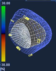
18 LV 3D - stress dynamic contraction map Accurately localize and estimate the severity of stress-induced ischemia by identifying areas of delayed contraction
19 Overview 2D stress echocardiography: main limitations 3D echocardiography: potential incremental value Accuracy of 3D stress echocardiography Which modalities of 3D echo during stress Which modalities of stress during 3D - echo Do we have to throw the baby out with the bath water?

20 Accuracy of 3D stress echocardiography 2D 3D Aggeli et al Heart 2007;93:672-75


21 Accuracy of Dipyridamole-3DE Badano et al J Am Soc Echocardiogr 2010;23:628-35

22 Improved apical visualization Lang et al, EJCI in press
23 Overview 2D stress echocardiography: main limitations 3D echocardiography: potential incremental value Accuracy of 3D stress echocardiography Which modalities of 3D echo during stress Which modalities of stress during 3D - echo Do we have to throw the baby out with the bath water?

24 Modalities of 3D echo during stress Lang et al, EJCI in press

25 Tri-plane 3D stress echo

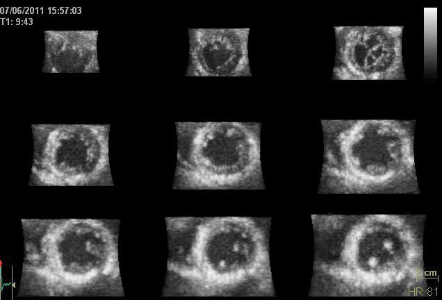
26 Full 3D stress echo

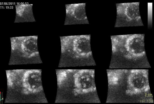
27 Contrast 3D stress echo
 No flash destruction of bubbles Only adenosine (lower heart rates) Courtesy of I.")
28 Contrast perfusion 3D stress echo Is feasible Low spatial resolution Lower frame rate (volume rate) No flash destruction of bubbles Only adenosine (lower heart rates) Courtesy of I. Felekos
 12, 648")
29 Perfusion Contrast 3D stress echo SVD MVD Aggeli et al European Journal of Echocardiography (2011) 12,

30 3D stress STI


 False motion / deformation")
31 Rationale For 3D Speckle Myocardial deformation is 3D Better geometric description of LV with 3D Avoid out-of-plane motion artifacts Speckle de-correlation (measurement artifacts) False motion / deformation Courtesy of J. Crosby


32 Challenges for 3D speckle tracking Increased FOV at the cost of both spatial and temporal resolution Speckle pattern has less details and de-correlation between subsequent volumes is high 3D strain normal values depending on the method used (block matching vs elastic registration) Same information 2D in less time J. Crosby et al., Ultrasound in Medicine & Biology 2008 K. Saito et al. Journal Am. Soc.Echocardiography 2009
33 3D stress STI vs Sonomicrometry X = dobutamine infusion Seo et al Circ Cardiovasc Imaging. 2009;2:
34 Overview 2D stress echocardiography: main limitations 3D echocardiography: potential incremental value Accuracy of 3D stress echocardiography Which modalities of 3D echo during stress Which modalities of stress during 3D - echo Do we have to throw the baby out with the bath water?

35 Which Stress? Pratali et al Cardiovascular Ultrasound 2010, 8:10
36 Stress 3D- Echocardiography Advantages 1) better visualization of the LV apex, which is frequently foreshortened on standard 2DE apical images 2) rapid acquisition of peak stress images before the heart rate declines in recovery 3) evaluation of multiple segments from different planes from a single dataset. Disadvantages 1) lower spatial resolution 2) lower frame rates (tardokinesia can be missed) 3) Learning curve required 4) No side-by-side display of rest and stress images for comparison * *Recently overcome
37 Overview 2D stress echocardiography: main limitations 3D echocardiography: potential incremental value Accuracy of 3D stress echocardiography Which modalities of 3D echo during stress Which modalities of stress during 3D - echo Do we have to throw the baby out with the bath water?

38 Do we have to throw the baby out with the bath water? Sicari et al. European Journal of Echocardiography 2008

39 Feasibility of 3D stress echo Badano et al J Am Soc Echocardiogr 2010;23: Pratali et al Cardiovascular Ultrasound 2010, 8:10
40 Prolonged Analysis Time? 2D ECHO 3D ECHO P Acquisition time 65 +/ /- 3 P< Analysis time 176 +/ /- 5 P< Varnero Cardiovascular ultrasound 2008 Badano et al J Am Soc Echocardiogr 2010;23:628-35
41 Additional value of 3D > 2D-stress echo Image quality during transthoracic scanning with insufficient visualization of left ventricle (LV) walls Probe positioning difficulties resulting in inadequate image planes Time-consuming serial acquisition of different image planes which has to be performed in a narrow time window during peak stress while wall motion abnormalities exist Data analysis: subjectivity of image interpretation still is the major problem, which leads to poor inter-observer agreement and causes a relevant examiner-dependency
 D. Barbosa et al. JASE 2011 (Submitted)")
42 Fully automated segmentation Real-time segmentation Real-time volume measurements! D. Barbosa et al. IEEE-TIP 2011 (In press) D. Barbosa et al. JASE 2011 (Submitted)
43 Limitations still to overcome 3D stress echo is still work in progress Further quality image improvement Higher Frame Rates, less stitching (tachycardia, arrhythmias) More well validated automated algorithms Side by side analysis and synchronization (different stages) Implementation and validation of other echo- modalities (STI) Validation in various indications and non selected patients
Assessment of cardiac function with 3D echocardiography. Đánh giá chức năng tim bằng siêu âm tim 3D
 Assessment of cardiac function with 3D echocardiography Đánh giá chức năng tim bằng siêu âm tim 3D TS. BS. Nguyễn Thị Thu Hoài Viện Tim Mạch Quốc Gia Việt Nam TỪ SIÊU ÂM M-mode ĐẾN SIÊU ÂM 3D TỪ SIÊU ÂM
Conflict of Interests
 The Left Ventricle: How Should We Quantify Its Size and Function; Is It Time for 3D in Everyone? Roberto M Lang, MD Conflict of Interests Philips Medical Imaging Research Grants Speakers bureau Advisory
10/7/2013. Systolic Function How to Measure, How Accurate is Echo, Role of Contrast. Thanks to our Course Director: Neil J.
 Systolic Function How to Measure, How Accurate is Echo, Role of Contrast Neil J. Weissman, MD MedStar Health Research Institute & Professor of Medicine Georgetown University Washington, D.C. No Disclosures
Quantifying LV function how good are we?
 Quantifying LV function how good are we? Professor Alan G Fraser Wales Heart Research Institute Cardiff University, U.K. Support for research from Hitachi Aloka, & GE Ultrasound Visual assessment of synchronicity
DECLARATION OF CONFLICT OF INTEREST. None
 DECLARATION OF CONFLICT OF INTEREST None Hot Topics in Echocardiography: The position of the EAE EAE / ASE recommendation about Echo Assessment of Cardiac Mechanics Jens-Uwe Voigt Dpt. of Cardiovascular
DECLARATION OF CONFLICT OF INTEREST
 DECLARATION OF CONFLICT OF INTEREST Clinica Cardiologica Università degli Studi di Padova Direttore: Prof. Sabino Iliceto BENEFITS OF REAL-TIME 3D STRESS ECHOCARDIOGRAPHY Luigi P. Badano**, MD, FESC **Dr.
Beginner s Guide to Strain: What should be in your lab in Disclosures
 Beginner s Guide to Strain: What should be in your lab in 2018 Bonita Anderson DMU (Cardiac), MApplSc (Med Ultrasound), ACS, AMS, FASE None Disclosures Calculation of Strain Strain can be Positive Strain
Strain and Strain Rate Imaging How, Why and When?
 Strain and Strain Rate Imaging How, Why and When? João L. Cavalcante, MD Advanced Cardiac Imaging Fellow Cleveland Clinic Foundation Disclosures: No conflicts of interest Movement vs Deformation Movement
Velocity, strain and strain rate: Doppler and Non-Doppler methods. Thoraxcentre, Erasmus MC,Rotterdam
 Velocity, strain and strain rate: Doppler and Non-Doppler methods J Roelandt J. Roelandt Thoraxcentre, Erasmus MC,Rotterdam Basics of tissue Doppler imaging Instantaneous annular velocity profiles IVCT
Chamber Quantitation Guidelines: What is New?
 Chamber Quantitation Guidelines: What is New? Roberto M Lang, MD J AM Soc Echocardiogr 2005; 18:1440-1463 1 Approximately 10,000 citations iase in itune Cardiac Chamber Quantification: What is New? Database
Cardiac Chamber Quantification by Echocardiography
 Cardiac Chamber Quantification by Echocardiography Maryam Bokhamseen, RCS, RCDS, EACVI Echotechnologist ǁ, Non invasive Cardiac Laboratory King Abdulaziz Cardiac Center. Outline: Introduction. Background
2/2/2011. Strain and Strain Rate Imaging How, Why and When? Movement vs Deformation. Doppler Myocardial Velocities. Movement. Deformation.
 Strain and Strain Rate Imaging How, Why and When? João L. Cavalcante, MD Advanced Cardiac Imaging Fellow Cleveland Clinic Foundation Disclosures: No conflicts of interest Movement vs Deformation Movement
Cardiology for the Practitioner Advanced Cardiac Imaging: Worth the pretty pictures?
 Keenan Research Centre Li Ka Shing Knowledge Institute Cardiology for the Practitioner Advanced Cardiac Imaging: Worth the pretty pictures? Howard Leong-Poi, MD, FRCPC Associate Professor of Medicine St.
Tissue Doppler Imaging in Congenital Heart Disease
 Tissue Doppler Imaging in Congenital Heart Disease L. Youngmin Eun, M.D. Department of Pediatrics, Division of Pediatric Cardiology, Kwandong University College of Medicine The potential advantage of ultrasound
Echocardiographic Assessment of the Left Ventricle
 Echocardiographic Assessment of the Left Ventricle Theodora Zaglavara, MD, PhD, BSCI/BSCCT Department of Cardiovascular Imaging INTERBALKAN EUROPEAN MEDICAL CENTER 2015 The quantification of cardiac chamber
Automated Volumetric Cardiac Ultrasound Analysis
 Whitepaper Automated Volumetric Cardiac Ultrasound Analysis ACUSON SC2000 Volume Imaging Ultrasound System Bogdan Georgescu, Ph.D. Siemens Corporate Research Princeton, New Jersey USA Answers for life.
Conflict of Interests
 New Approaches to Systolic Function: 4D Roberto M Lang, MD Conflict of Interests Philips Medical Imaging Research Grants Speakers bureau Advisory bureau Tomtec Research Grants Epsilon Research Grants 1
Three-dimensional Wall Motion Tracking:
 Three-dimensional Wall Motion Tracking: A Novel Echocardiographic Method for the Assessment of Ventricular Volumes, Strain and Dyssynchrony Jeffrey C. Hill, BS, RDCS, FASE Jennifer L. Kane, RCS Gerard
LV FUNCTION ASSESSMENT: WHAT IS BEYOND EJECTION FRACTION
 LV FUNCTION ASSESSMENT: WHAT IS BEYOND EJECTION FRACTION Jamilah S AlRahimi Assistant Professor, KSU-HS Consultant Noninvasive Cardiology KFCC, MNGHA-WR Introduction LV function assessment in Heart Failure:
How to Assess Dyssynchrony
 How to Assess Dyssynchrony Otto A. Smiseth, Professor, MD, PhD Oslo University Hospital None Conflicts of interest Cardiac resynchronization therapy effect on mortality Cleland JG et al, N Engl J Med
EDITOR S PICK CURRENT STATUS OF FULLY AUTOMATED SOFTWARE WITH THREE-DIMENSIONAL ECHOCARDIOGRAPHY FOR THE QUANTIFICATION OF LEFT VENTRICULAR FUNCTION
 ITOR S PICK This paper, courtesy of Yang and Takeuchi, provides a timely and well-considered update on the current status of fully-automated software with three-dimensional echocardiography for quantifying
Strain/Untwisting/Diastolic Suction
 What Is Diastole and How to Assess It? Strain/Untwisting/Diastolic Suction James D. Thomas, M.D., F.A.C.C. Cardiovascular Imaging Center Department of Cardiology Cleveland Clinic Foundation Cleveland,
Alicia Armour, MA, BS, RDCS
 Alicia Armour, MA, BS, RDCS No disclosures Review 2D Speckle Strain (briefly) Discuss some various patient populations & disease pathways where Strain can be helpful Discuss how to acquire images for Strain
Tissue Doppler and Strain Imaging. Steven J. Lester MD, FRCP(C), FACC, FASE
, FACC, FASE") Tissue Doppler and Strain Imaging Steven J. Lester MD, FRCP(C), FACC, FASE Relevant Financial Relationship(s) None Off Label Usage None a. Turn the wall filters on and turn down the receiver gain. b. Turn
Tissue Doppler and Strain Imaging Steven J. Lester MD, FRCP(C), FACC, FASE Relevant Financial Relationship(s) None Off Label Usage None a. Turn the wall filters on and turn down the receiver gain. b. Turn
Tissue Doppler and Strain Imaging
 Tissue Doppler and Strain Imaging Steven J. Lester MD, FRCP(C), FACC, FASE Relevant Financial Relationship(s) None Off Label Usage None 1 Objective way with which to quantify the minor amplitude and temporal
On the feasibility of speckle reduction in echocardiography using strain compounding
 Title On the feasibility of speckle reduction in echocardiography using strain compounding Author(s) Guo, Y; Lee, W Citation The 2014 IEEE International Ultrasonics Symposium (IUS 2014), Chicago, IL.,
Lingyun Kong, Chao Yu, Jihong Guo and Tiangang Zhu. Abstract. Introduction. Department of Cardiology, Peking University People s Hospital.
 24 Original Article Comparison of Left Ventricular Global Longitudinal Strain Measured with Real Time Triplane and 2-Dimensional Echocardiography in Patients with Atrial Fibrillation Lingyun Kong, Chao
Three-dimensional speckle tracking echocardiography for the evaluation of segmental myocardial deformation
 ORIGINAL RESEARCH Three-dimensional speckle tracking echocardiography for the evaluation of segmental myocardial deformation Janine Baum, Florian Beeres, Silke Van Hall, Yang Chul Boering, Eva Susanne
Tissue Doppler and Strain Imaging
 Tissue Doppler and Strain Imaging Steven J. Lester MD, FRCP(C), FACC, FASE Relevant Financial Relationship(s) None Off Label Usage None 1 Objective way with which to quantify the minor amplitude and temporal
When Does 3D Echo Make A Difference?
 When Does 3D Echo Make A Difference? Wendy Tsang, MD, SM Assistant Professor, University of Toronto Toronto General Hospital, University Health Network 1 Practical Applications of 3D Echocardiography Recommended
Quantitation of right ventricular dimensions and function
 SCCS Basics of cardiac assessment Quantitation of right ventricular dimensions and function Tomasz Kukulski, MD PhD Dept of Cardiology, Congenital Heart Disease and Electrotherapy Silesian Medical University
Conflict of interest: none declared
 The value of left ventricular global longitudinal strain assessed by three-dimensional strain imaging in the early detection of anthracycline-mediated cardiotoxicity C. Mornoş, A. Ionac, D. Cozma, S. Pescariu,
Three-dimensional echocardiography in the clinical world
 Three-dimensional echocardiography in the clinical world Dr. JL Zamorano Director CV Institute University Clinic SC, Madrid Advantages of 3D. Spatial manipulation. Optimal alineation of structures. Views
Assessment of left ventricular systolic function by deformation imaging derived from speckle tracking: a comparison between 2D and 3D echo modalities
 European Heart Journal Cardiovascular Imaging (2014) 15, 316 323 doi:10.1093/ehjci/jet103 Assessment of left ventricular systolic function by deformation imaging derived from speckle tracking: a comparison
Guideline-Driven Care in Cardio- Oncology: Utilizing Recommendations Across Disciplines
 Guideline-Driven Care in Cardio- Oncology: Utilizing Recommendations Across Disciplines Jennifer Liu, MD FACC FASE Director of CV Laboratories Associate Professor of Clinical Medicine Memorial Sloan Kettering
Stress echo workshop STRESSORS
 Stress echo workshop STRESSORS Adham Ahmed, MD Lecturer of Cardiology, Ain Shams Indications of Stress Echo CAD Diagnosis Prognosticat ion 1 Physiologic Basis 1930s: Tennant and Wiggers Relationship between
Incorporating the New Echo Guidelines Into Everyday Practice
 Incorporating the New Echo Guidelines Into Everyday Practice Clinical Case RIGHT VENTRICULAR FAILURE Gustavo Restrepo MD President Elect Interamerican Society of Cardiology Director Fellowship Training
Νεότερα ςτην Υπερηχοκαρδιογραφία. Βαςίλειοσ Καμπερίδησ Clinical research fellow in Cardiology
 Νεότερα ςτην Υπερηχοκαρδιογραφία Βαςίλειοσ Καμπερίδησ Clinical research fellow in Cardiology Disclosures ESC training grant EACVI research grant HCS training grant ELIKAR research grant Evolution of Echocardiography
Strain imaging in children: from Tissue Doppler to 3 D
 Strain imaging in children: from Tissue Doppler to 3 D Mark kk. Friedberg Fi Outline Deformation in the fetus and neonate Deformation in pediatric cardiomyopathy y (briefly!) Deformation in Congenital
DISCLOSURE. Myocardial Mechanics. Relevant Financial Relationship(s) Off Label Usage
 Off Label Usage") 7th Annual Team Echocardiography: The Heart of Cardiovascular Medicine Tissue Doppler, Strain, Speckle: What? How? Christopher J Kramer RDCS Aurora Medical Group Advanced Cardiovascular Services, Aurora
7th Annual Team Echocardiography: The Heart of Cardiovascular Medicine Tissue Doppler, Strain, Speckle: What? How? Christopher J Kramer RDCS Aurora Medical Group Advanced Cardiovascular Services, Aurora
Left atrial function. Aliakbar Arvandi MD
 In the clinic Left atrial function Abstract The left atrium (LA) is a left posterior cardiac chamber which is located adjacent to the esophagus. It is separated from the right atrium by the inter-atrial
Advanced imaging of the left atrium - strain, CT, 3D, MRI -
 Advanced imaging of the left atrium - strain, CT, 3D, MRI - Monica Rosca, MD Carol Davila University of Medicine and Pharmacy, Bucharest, Romania Declaration of interest: I have nothing to declare Case
RIGHT VENTRICULAR SIZE AND FUNCTION
 RIGHT VENTRICULAR SIZE AND FUNCTION Edwin S. Tucay, MD, FPCC, FPCC, FPSE Philippine Society of Echocardiography Quezon City, Philippines Echo Mission, BRTTH, Legaspi City, July 1-2, 2016 NO DISCLOSURE
Advanced Multi-Layer Speckle Strain Permits Transmural Myocardial Function Analysis in Health and Disease:
 Advanced Multi-Layer Speckle Strain Permits Transmural Myocardial Function Analysis in Health and Disease: Clinical Case Examples Jeffrey C. Hill, BS, RDCS Echocardiography Laboratory, University of Massachusetts
Appropriateness of Stress Echocardiography and Nuclear Stress Thallium/Sesta Mibi Testing Methods
 Appropriateness of Stress Echocardiography and Nuclear Stress Thallium/Sesta Mibi Testing Methods Review by Michael Devenport, MS, RDCS Objectives: A basic description and review of appropriate use of
Value of echocardiography in chronic dyspnea
 Value of echocardiography in chronic dyspnea Jahrestagung Schweizerische Gesellschaft für /Schweizerische Gesellschaft für Pneumologie B. Kaufmann 16.06.2016 Chronic dyspnea Shortness of breath lasting
Novel echocardiographic modalities: 3D echo, speckle tracking and strain rate imaging. Potential roles in sports cardiology. Stefano Caselli, MD, PhD
 Novel echocardiographic modalities: 3D echo, speckle tracking and strain rate imaging. Potential roles in sports cardiology. Stefano Caselli, MD, PhD Ospedale San Pietro Fatebenefratelli Rome, Italy Differential
Long Axis Strain by MRI and Echocardiography in a Postmyocardial Infarct Population
 CME JOURNAL OF MAGNETIC RESONANCE IMAGING 40:1247 1251 (2014) Clinical Note Long Axis Strain by MRI and Echocardiography in a Postmyocardial Infarct Population Ola Gjesdal, MD, PhD, 1,2 Andre L.C. Almeida,
HYPERTROPHY: Behind the curtain. V. Yotova St. Radboud Medical University Center, Nijmegen
 HYPERTROPHY: Behind the curtain V. Yotova St. Radboud Medical University Center, Nijmegen Disclosure of interest: none Relative wall thickness (cm) M 0.22 0.42 0.43 0.47 0.48 0.52 0.53 F 0.24 0.42 0.43
Measuring cardiac tissue motion and strain
 Ultrasound Measuring cardiac tissue motion and strain Automated Cardiac Motion Quantification A.I. (acmq A.I. ) David Prater, MS, Clinical Scientist, Philips Jane Vogel, MD, Senior Product Manager, Philips
JACC: CARDIOVASCULAR IMAGING VOL. 8, NO. 2, 2015 ª 2015 BY THE AMERICAN COLLEGE OF CARDIOLOGY FOUNDATION ISSN X/$36.00
 JACC: CARDIOVASCULAR IMAGING VOL. 8, NO. 2, 15 ª 15 BY THE AMERICAN COLLEGE OF CARDIOLOGY FOUNDATION ISSN 1936-878X/$36. PUBLISHED BY ELSEVIER INC. http://dx.doi.org/1.116/j.jcmg.14.1.1 How to Define End-Diastole
Strain Imaging: Myocardial Mechanics Simplified and Applied
 9/28/217 Strain Imaging: Myocardial Mechanics Simplified and Applied John Gorcsan III, MD Professor of Medicine Director of Clinical Research Division of Cardiology VECTORS OF CONTRACTION Shortening Thickening
Mechanisms of False Positive Exercise Electrocardiography: Is False Positive Test Truly False?
 Mechanisms of False Positive Exercise Electrocardiography: Is False Positive Test Truly False? Masaki Izumo a, Kengo Suzuki b, Hidekazu Kikuchi b, Seisyo Kou b, Keisuke Kida b, Yu Eguchi b, Nobuyuki Azuma
Challenge on Endocardial Three-dimensional Ultrasound Segmentation (CETUS)
") Challenge on Endocardial Three-dimensional Ultrasound Segmentation (CETUS) Olivier Bernard 1,BrechtHeyde 2, Martino Alessandrini 2, Daniel Barbosa 3, Sorina Camarasu-Pop 1, Frederic Cervenansky 1, Sebastien
Challenge on Endocardial Three-dimensional Ultrasound Segmentation (CETUS) Olivier Bernard 1,BrechtHeyde 2, Martino Alessandrini 2, Daniel Barbosa 3, Sorina Camarasu-Pop 1, Frederic Cervenansky 1, Sebastien
Altered left ventricular geometry and torsional mechanics in high altitude-induced pulmonary hypertension:
 Altered left ventricular geometry and torsional mechanics in high altitude-induced pulmonary hypertension: a 3-D echocardiographic study B.W. De Boeck,* S. Kiencke, C. Dehnert, K. Auinger, # M. Maggiorini,
Multiple Gated Acquisition (MUGA) Scanning
 Scanning") Multiple Gated Acquisition (MUGA) Scanning Dmitry Beyder MPA, CNMT Nuclear Medicine, Radiology Barnes-Jewish Hospital / Washington University St. Louis, MO Disclaimers/Relationships Standard of care research
Multiple Gated Acquisition (MUGA) Scanning Dmitry Beyder MPA, CNMT Nuclear Medicine, Radiology Barnes-Jewish Hospital / Washington University St. Louis, MO Disclaimers/Relationships Standard of care research
The Value of 2D Strain Imaging during Stress Testing
 The Value of 2D Strain Imaging during Stress Testing Marie Moonen, M.D., Patrizio Lancellotti, M.D., Ph.D., Dimitrios Zacharakis, M.D., and Luc Pierard, M.D., Ph.D., F.E.S.C., F.A.C.C. CHU Sart Tilman,
Prof. JL Zamorano Hospital Universitario Ramón y Cajal
 Prof. JL Zamorano Hospital Universitario Ramón y Cajal Fully Automated Quantification Software Adaptive analytical algorithm consists in knowledge-based identification of global shape and specific adaptation
Reproducibility and Accuracy of Echocardiographic Measurements of Left Ventricular Parameters Using Real-Time Three-Dimensional Echocardiography
 Journal of the American College of Cardiology Vol. 44, No. 4, 2004 2004 by the American College of Cardiology Foundation ISSN 0735-1097/04/$30.00 Published by Elsevier Inc. doi:10.1016/j.jacc.2004.05.050
The road to successful CRT implantation: The role of echo
 The road to successful CRT implantation: The role of echo Tae-Ho Park Dong-A University Hospital, Busan, Korea Terminology Cardiac Resynchronization Therapy (CRT) = Biventricular pacing (BiV) = Left ventricular
Assessment of right ventricular contraction by speckle tracking echocardiography in pulmonary hypertension patients.
 Biomedical Research 2017; 28 (1): 173-177 ISSN 0970-938X www.biomedres.info Assessment of right ventricular contraction by speckle tracking echocardiography in pulmonary hypertension patients. Yudong Peng,
MRI Assessment of the Right Ventricle and Pulmonary Blood Flow, Perfusion and Ventilation
 MRI Assessment of the Right Ventricle and Pulmonary Blood Flow, Perfusion and Ventilation Dr. Richard Thompson Department of Biomedical Engineering University of Alberta Heart and Lung Imaging Many Constantly
Mechanisms and role of contrast echocardiography
 Mechanisms and role of contrast echocardiography Seol Sang-Hoon Inje University College of Medicine, Haeundae Paik Hospital, Busan, Korea Physical Principles of Contrast Ultrasound Contrast echocardiography
Qualitative and Quantitative Assessment of Perfusion
 APCDE 2011 Qualitative and Quantitative Assessment of Perfusion Hyun Ju Yoon Chonnam National University Hospital Gwangju, Korea ISCHEMIC CASCADE Blood flow mismatch Perfusion defects on nuclear imaging,
Basic Assessment of Left Ventricular Systolic Function
 WINFOCUS BASIC ECHO (WBE) Basic Assessment of Left Ventricular Systolic Function Ritesh Dhar, MD Director, Echocardiography Lab and Staff Cardiologist Intermountain Medical Center Murray, Utah Outline
Assessment of Ischemia and Viability
 EAE Teaching Course Bucharest, 2010 Assessment of Ischemia and Viability Jens-Uwe Voigt Dpt. of Cardiology University Leuven Belgium Assessment of Ischemia & Viability resting wall motion Stress Testing
Echocardiographic assessment of the right ventricle in paediatric pulmonary hypertension.
 Echocardiographic assessment of the right ventricle in paediatric pulmonary hypertension. Mark K. Friedberg, MD No disclosures Outline RV response to increased afterload Echo assessment of RV function
How To Perform Strain Imaging; Step By Step Approach. Maryam Bo Khamseen Echotechnoligist II EACVI, ARDMS, RCS King Abdulaziz Cardiac Center- Riyadh
 How To Perform Strain Imaging; Step By Step Approach Maryam Bo Khamseen Echotechnoligist II EACVI, ARDMS, RCS King Abdulaziz Cardiac Center- Riyadh Outlines: Introduction Describe the basic of myocardium
Introduction. Cardiac Imaging Modalities MRI. Overview. MRI (Continued) MRI (Continued) Arnaud Bistoquet 12/19/03
 MRI (Continued) Arnaud Bistoquet 12/19/03") Introduction Cardiac Imaging Modalities Arnaud Bistoquet 12/19/03 Coronary heart disease: the vessels that supply oxygen-carrying blood to the heart, become narrowed and unable to carry a normal amount
Introduction Cardiac Imaging Modalities Arnaud Bistoquet 12/19/03 Coronary heart disease: the vessels that supply oxygen-carrying blood to the heart, become narrowed and unable to carry a normal amount
When is strain assessment mandatory?
 When is strain assessment mandatory? Geneviève Derumeaux University of Lyon France Presenter Disclosure Information Geneviève Derumeaux When is strain assessment mandatory? DISCLOSURE INFORMATION: None
PRESENTER DISCLOSURE INFORMATION. There are no potential conflicts of interest regarding current presentation
 PRESENTER DISCLOSURE INFORMATION There are no potential conflicts of interest regarding current presentation Better synchrony and diastolic function for septal versus apical right ventricular permanent
Cardiovascular Imaging Endpoints in Oncology Clinical Trials
 Cardiovascular Imaging Endpoints in Oncology Clinical Trials Bonnie Ky, MD, MSCE Assistant Professor of Medicine and Epidemiology Director, Penn Cardio-Oncology Center of Excellence Director, Penn Center
좌심실수축기능평가 Cardiac Function
 Basic Echo Review Course 좌심실수축기능평가 Cardiac Function Seonghoon Choi Cardiology Hallym university LV systolic function Systolic function 좌심실수축기능 - 심근의수축으로심실에서혈액을대동맥으로박출하는기능 실제임상에서 LV function 의의미 1Diagnosis
Myocardial Strain Imaging in Cardiac Diseases and Cardiomyopathies.
 Myocardial Strain Imaging in Cardiac Diseases and Cardiomyopathies. Session: Cardiomyopathy Tarun Pandey MD, FRCR. Associate Professor University of Arkansas for Medical Sciences Disclosures No relevant
Cardiovascular Imaging Stress Echo
 Cardiovascular Imaging Stress Echo Theodora A Zaglavara, MD, PhD Cardiac Imaging Department INTERBALKAN MEDICAL CENTER Thessaloniki GREECE Evolution of Stress Echo: From Innovation to a Widely Established
21st Annual Contemporary Therapeutic Issues in Cardiovascular Disease
 21st Annual Contemporary Therapeutic Issues in Cardiovascular Disease Noninvasive Evaluation of Coronary Artery Disease: Anatomical, Functional, Clinical May 5, 2018 Mark Hansen MD FRCPC Cardiologist,
IRM cardiaque en cancérologie: le rôle du radiologue
 IRM cardiaque en cancérologie: le rôle du radiologue Laurent MACRON Centre Cardiologique du Nord (CCN) Saint Denis Centre Cardiologique du Nord - Saint Denis - France CMR in oncology Characterisation of
DECLARATION OF CONFLICT OF INTEREST
 DECLARATION OF CONFLICT OF INTEREST The additive prognostic value of myocardial perfusion defects, coronary flow reserve and wall motion abnormalities during dipyridamole contrast stress-echo: a prospective
A Practical Approach to Using Strain Echocardiography to Evaluate the Left Ventricle
 Circulation Journal Official Journal of the Japanese Circulation Society http://www.j-circ.or.jp REVIEW A Practical Approach to Using Strain Echocardiography to Evaluate the Left Ventricle Harvey Feigenbaum,
Cover Page. The handle holds various files of this Leiden University dissertation.
 Cover Page The handle http://hdl.handle.net/1887/38522 holds various files of this Leiden University dissertation. Author: Ewe, See Hooi Title: Aortic valve disease : novel imaging insights from diagnosis
Cardiac magnetic resonance imaging in rheumatoid arthritis: promising or misleading? Sophie Mavrogeni MD FESC
 Cardiac magnetic resonance imaging in rheumatoid arthritis: promising or misleading? Sophie Mavrogeni MD FESC Onassis Cardiac Surgery Center Athens Greece Nothing to disclose Financial disclosure Cardiac
GE Healthcare. Vivid i Cardiovascular ultrasound system
 GE Healthcare Vivid i Cardiovascular ultrasound system Excellent raw data image quality. Advanced quantification. Increased diagnostic confidence. Streamlined workflow. In any clinical environment. With
Cardial MRI; Approaching the Level of Gold Standard for Viability Assessment
 Cardial MRI; Approaching the Level of Gold Standard for Viability Assessment 용환석 고려대학교구로병원영상의학과 Viability Hibernating myocardium a state of myocardial hypocontractility during chronic hypoperfusion, in
CHRONIC CAD DIAGNOSIS
 CHRONIC CAD DIAGNOSIS Chest Pain Evaluation 1. Approach to diagnosis of CAD 2. Classification of chest pain 3. Pre-test likelihood CAD 4. Algorithm for chest pain evaluation in women 5. Indications for
Impaired Regional Myocardial Function Detection Using the Standard Inter-Segmental Integration SINE Wave Curve On Magnetic Resonance Imaging
 Original Article Impaired Regional Myocardial Function Detection Using the Standard Inter-Segmental Integration Ngam-Maung B, RT email : chaothawee@yahoo.com Busakol Ngam-Maung, RT 1 Lertlak Chaothawee,
4D Auto LAQ (Left Atrial Quantification)
") 4D Auto LAQ (Left Atrial Quantification) Introduction There has been an increased interest in quantification of the left atrium (LA) for various types of diseases; e.g. heart failure, heart valve diseases,
4D Auto LAQ (Left Atrial Quantification) Introduction There has been an increased interest in quantification of the left atrium (LA) for various types of diseases; e.g. heart failure, heart valve diseases,
DOWNLOAD PDF MYOCARDIAL CONTRAST TWO DIMENSIONAL ECHOCARDIOGRAPHY (DEVELOPMENTS IN CARDIOVASCULAR MEDICINE)
") Chapter 1 : Imaging Cardiovascular Medicine Stanford Medicine contrast two-dimensional echocardiography (MC-2DE), a new and exciting diagnostic methodology for assessment of myocardial perfusion, which
Chapter 1 : Imaging Cardiovascular Medicine Stanford Medicine contrast two-dimensional echocardiography (MC-2DE), a new and exciting diagnostic methodology for assessment of myocardial perfusion, which
Evaluation of Left Ventricular Diastolic Dysfunction by Doppler and 2D Speckle-tracking Imaging in Patients with Primary Pulmonary Hypertension
 ESC Congress 2011.No 85975 Evaluation of Left Ventricular Diastolic Dysfunction by Doppler and 2D Speckle-tracking Imaging in Patients with Primary Pulmonary Hypertension Second Department of Internal
The best from Euro-Echo Ischemic heart disease. Fausto Rigo,FESC Department of Cardiology Mestre-Venezia Hospital,Italy
 The best from Euro-Echo 2011 Ischemic heart disease Fausto Rigo,FESC Department of Cardiology Mestre-Venezia Hospital,Italy faustorigo@alice.it DECLARATION OF CONFLICT OF INTEREST No conflict of interest
Η ηχωκαρδιολογία στην διάγνωση κα πρόγνωση της καρδιακής ανεπάρκειας µε µειωµένο και φυσιολογικό κλάσµα εξώθησης
 Η ηχωκαρδιολογία στην διάγνωση κα πρόγνωση της καρδιακής ανεπάρκειας µε µειωµένο και φυσιολογικό κλάσµα εξώθησης Βασίλειος Σαχπεκίδης Επιµελητής Β Καρδιολογίας Γ.Ν. Παπαγεωργίου Θεσσαλονίκη ESC Guidelines
Left atrial Function In Echocardiography
 Left atrial Function In Echocardiography Pr Erwan DONAL erwan.donal@chu-rennes.fr Inserm Diastole LA Pressure (mmhg) -t/ 17 LA function 15 A-Loop 13 V-Loop 11 9 7 5 1 2 3 4 20 30 40 50 60 70 LA Volume
Coronary artery disease (CAD) risk factors
 risk factors") Background Coronary artery disease (CAD) risk factors CAD Risk factors Hypertension Insulin resistance /diabetes Dyslipidemia Smoking /Obesity Male gender/ Old age Atherosclerosis Arterial stiffness precedes
Background Coronary artery disease (CAD) risk factors CAD Risk factors Hypertension Insulin resistance /diabetes Dyslipidemia Smoking /Obesity Male gender/ Old age Atherosclerosis Arterial stiffness precedes
2019 Qualified Clinical Data Registry (QCDR) Performance Measures
 Performance Measures") 2019 Qualified Clinical Data Registry (QCDR) Performance Measures Description: This document contains the 18 performance measures approved by CMS for inclusion in the 2019 Qualified Clinical Data Registry
2019 Qualified Clinical Data Registry (QCDR) Performance Measures Description: This document contains the 18 performance measures approved by CMS for inclusion in the 2019 Qualified Clinical Data Registry
Sung A Chang Department of Internal Medicine, Division of Cardiology, Sungkyunkwan University School of Medicine, Samsung Medical Center
 CMR Perfusion and Viability A STICH Out of Time? Sung A Chang Department of Internal Medicine, Division of Cardiology, Sungkyunkwan University School of Medicine, Samsung Medical Center Can Imaging Improve
Multimodality Imaging in Cardiac Stem Cell Research
 Multimodality Imaging in Cardiac Stem Cell Research IL SUK SOHN, MD, PhD Department of Cardiology Kyung Hee University Hospital at Gangdong Kyung Hee University School of Medicine, Seoul, Korea Stem Cell
Case Report. Case Report. Ana Lúcia Martins Arruda, Altamiro Ozório, Eloisa Mattos, José Lázaro de Andrade, Thomas Porter, Wilson Mathias Jr
 Case Report Hypoperfusion of the Left Ventricle in the Absence of Changes in Segmental Contractility as Observed through Echocardiography by Using Microbubbles During Dobutamine Infusion Ana Lúcia Martins
Right ventricular adaptation in endurance athletes. António Freitas. No conflict of interest
 The role of echocardiography in sports cardiology Right ventricular adaptation in endurance athletes. António Freitas Cardiology Department - Fernando Fonseca Hospital Lisbon Sports Medicine Centre - Lisbon
Role of echocardiography in the assessment of ischemic heart disease 분당서울대학교병원윤연이
 Role of echocardiography in the assessment of ischemic heart disease 분당서울대학교병원윤연이 Outline Evaluation of Chest pain Evaluation of MI complications Prediction of Outcomes Evaluation of Chest pain Evaluation
I have no financial disclosures
 Manpreet Singh MD I have no financial disclosures Exercise Treadmill Bicycle Functional capacity assessment Well validated prognostic value Ischemic assessment ECG changes ST segments Arrhythmias Hemodynamic
22 nd Annual Conference of the Saudi Heart Association Riyadh, Saudi Arabia
 22 nd Annual Conference of the Saudi Heart Association Riyadh, Saudi Arabia New Echocardiographic Modalities to Evaluate Ventricular Function in Congenital Heart Disease: Tissue Doppler & Strain Rate Imaging