HYPERTROPHY: Behind the curtain. V. Yotova St. Radboud Medical University Center, Nijmegen
|
|
|
- Eustace Bennett
- 6 years ago
- Views:
Transcription

1 HYPERTROPHY: Behind the curtain V. Yotova St. Radboud Medical University Center, Nijmegen
2 Disclosure of interest: none


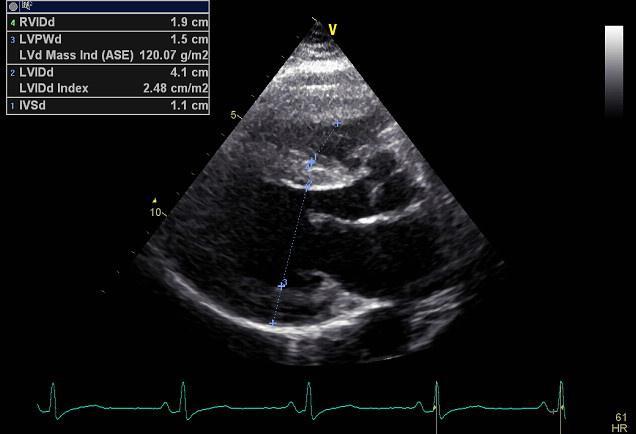
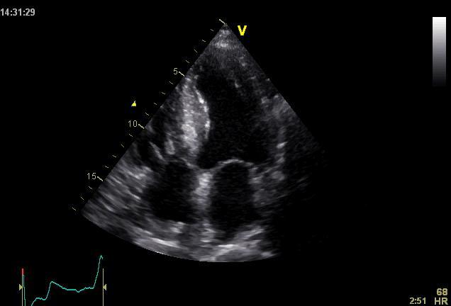
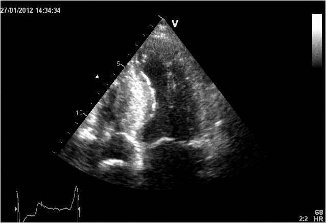
3 Relative wall thickness (cm) M F Left ventricular hypertrophy is defined as an increase in the mass of the left ventricle, which can be secondary to an increase in wall thickness, an increase in cavity size, or both.

/regional Diastolic")
4 Too simple to be enough! Hypertension Hypertrophic cardiomyopathy Aortic valve stenosis Amyloidosis Genetic diseases: Fabry, Friedreich Non-compaction Systemic approach The type of LV remodeling Systolic function: global (EF)/regional Diastolic function: global/regional Special features of specific diseases
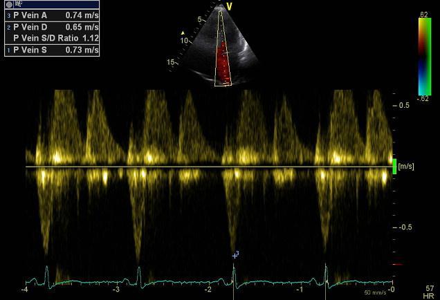
5 Role of echocardiography Echocardiography is a reliable method to visualize the specific patterns of LV hypertrophy, and quantify the systolic and diastolic function. New techniques in echocardiography have provided insight into regional myocardial motion and deformation. Tissue Doppler imaging, as well as grayscale 2D speckle tracking, provides more sensitive markers of early myocardial dysfunction compared with standard echocardiography.
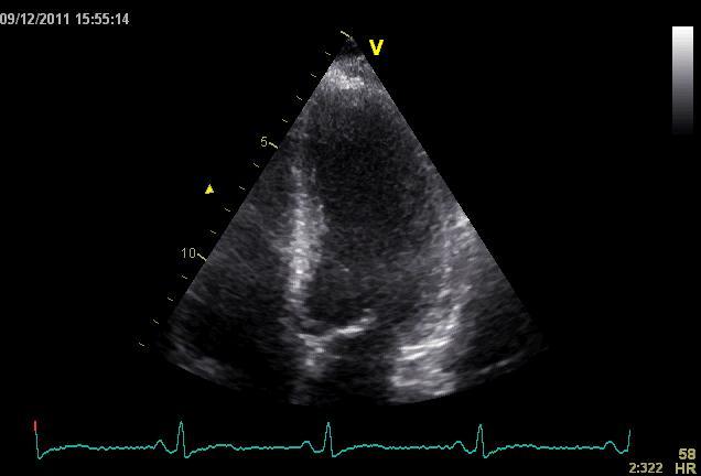
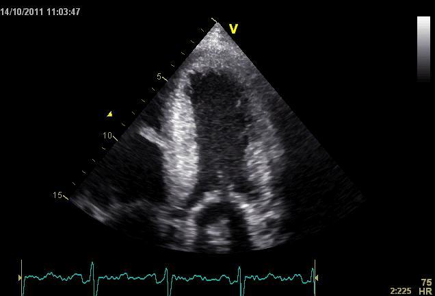
6 Arterial Hypertension


7 Arterial Hypertension

8 Aortic stenosis


9 Aortic stenosis

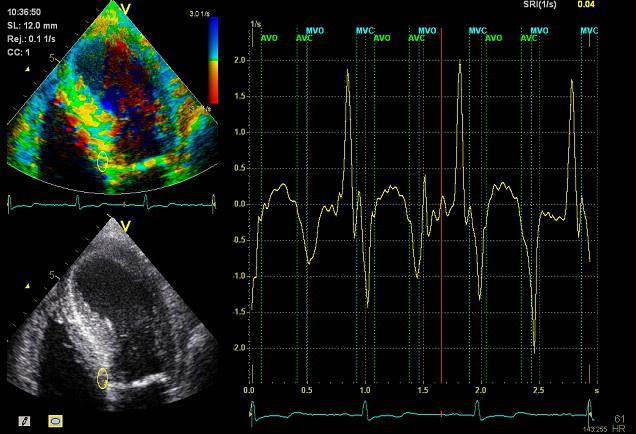
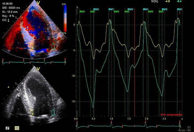
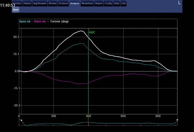
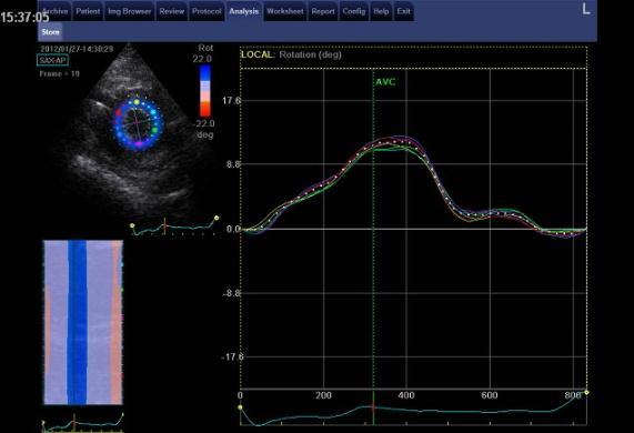
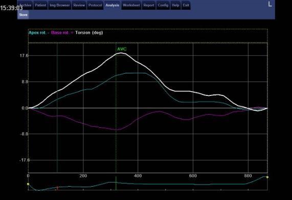
10 Rotational mechanics are clearly increased in all pressure overload pathologies. This observation results from the changing interaction between the endocardial and epicardial fibers, which induces LV torsion. Endocardial function is partially lost, therefore, and epicardial torsion becomes even more dominant, resulting in increased overall torsion.
11 The deformation indices are strongly related to the amount of fibrosis Weidemann F et al. Eur Heart J 2007;28:


12 HCM & HOCM An unexplained enddiastolic LV wall thickness > 15 mm in any myocardial segment accompanied by a normal wall thickness (<12 mm) in other segments and a nondilated left ventricle is suspicious for the diagnosis of idiopathic HCM.

13 Hypertension vs NHCM The combination of the IVST/PWT ratio > 1.3 and systolic strain of % was able to discriminate HCM from H-LVH with a predictive accuracy of 96.1%. Kato et al., Circulation 2004;110:3808

14 Apical CMP
15 Apical CMP


16 Athlete s heart No replacement fibrosis No fiber disarray

17 Physiologic vs Pathologic Hypertrophy Mean ( SD) systolic velocities. Cut-off 9 cm/sec. p <0.01 HCM or systemic hypertension versus athletes or normal subjects. Vinereanu et al.am J Cardiol 2001;88:53 58 Floresku et al.mædica A Journal of Clinical Medicine, Vol1 No
18 Fabri CMP


19 Fabri CMP Concentric hypertrophy up to 16 mm No LVOT obstruction Prominence of the lateral papillary muscle


20 MRI of 3 typical Fabry patients at baseline. An additional typical feature in Fabry cardiomyopathy is the development of replacement fibrosis in the basal posterolateral segments. Weidemann F et al. Circulation 2009;119:
21 Myocardial segments demonstrating late enhancement showed no functional improvement during enzyme-replacement therapy, which suggests that early disease detection and initiation of treatment is crucial. Weidemann Circulation 2009;119:524
22


23 Concentric hypertrophy up to 15 mm. Never LVOT obstruction Pericardial effusions IAS, RV Stiff Heart
 Mean LV basal strain is an independent predictor of both cardiac and overall deaths.")
24 Cardiac amyloidosis is the disease in which longitudinal function is most homogeneously reduced and long. strain is usually <10% Wiedemann et al. J Am Soc Echocardiogr 2010;23: ) Mean LV basal strain is an independent predictor of both cardiac and overall deaths. Obama J, JACC Cardiovasc Imaging 2010;3:
25 Noncompaction CMP
26 Noncompaction CMP The most important echo criterion is the typical 2-layered structure of the myocardium, with a thin, compacted epicardial band and a much thicker, noncompacted mid and endocardial band with deep endocardial recesses.

27 Typical echocardiographic features of the left ventricle in different hypertrophic hearts Maja Cikes et al.
28 Message For the clinic: Stay informed: Doppler myocardial imaging and speckle tracking are moresensitive imaging modalities than conventional echocardiography. This enable the detection of hypertrophic myopathies at an earlier stage. For the Echolab: In some patients, the correct diagnosis can be achieved only by obtaining additional history information or laboratory tests: Age of clinical expression Mode of inheritance (genealogical tree) Rate of progression Non cardiac features (phenotypic red flags): Skin-hair-eyes-facies Pectus, back, skeletal muscles) Neurological/mental status

29 Thank you Aelbert Cuyp, View from Dordrecht, 1660
How NOT to miss Hypertrophic Cardiomyopathy? Adaya Weissler-Snir, MD University Health Network, University of Toronto
 How NOT to miss Hypertrophic Cardiomyopathy? Adaya Weissler-Snir, MD University Health Network, University of Toronto Introduction Hypertrophic cardiomyopathy is the most common genetic cardiomyopathy,
Utility of Echocardiography
 Hypertrophic Cardiomyopathy and Beyond- Echo Hawaii 2018 Lawrence Rudski MD FRCPC FACC FASE Professor of Medicine Director, Division of Cardiology and Azrieli Heart Center Jewish General Hospital, McGill
Echocardiographic Evaluation of the Cardiomyopathies. Stephanie Coulter, MD, FACC, FASE April, 2016
 Echocardiographic Evaluation of the Cardiomyopathies Stephanie Coulter, MD, FACC, FASE April, 2016 Cardiomyopathies (CMP) primary disease intrinsic to cardiac muscle Dilated CMP Hypertrophic CMP Infiltrative
Novel echocardiographic modalities: 3D echo, speckle tracking and strain rate imaging. Potential roles in sports cardiology. Stefano Caselli, MD, PhD
 Novel echocardiographic modalities: 3D echo, speckle tracking and strain rate imaging. Potential roles in sports cardiology. Stefano Caselli, MD, PhD Ospedale San Pietro Fatebenefratelli Rome, Italy Differential
Nancy Goldman Cutler, MD Beaumont Children s Hospital Royal Oak, Mi
 Nancy Goldman Cutler, MD Beaumont Children s Hospital Royal Oak, Mi Identify increased LV wall thickness (WT) Understand increased WT in athletes Understand hypertrophic cardiomyopathy (HCM) Enhance understanding
Restrictive Cardiomyopathy
 ESC Congress 2011, Paris Imaging Unusual Causes of Cardiomyopathy Restrictive Cardiomyopathy Kazuaki Tanabe, MD, PhD Professor of Medicine Chair, Division of Cardiology Izumo, Japan I Have No Disclosures
What s New in Cardiac MRI
 What s New in Cardiac MRI Katie M. Hawthorne, MD Director, Cardiac MRI Main Line Health Philadelphia Cardiovascular Summit November 18, 2017 Cardiac MRI: Disclosure 2 Disclosures No financial disclosures
Review of Cardiac Imaging Modalities in the Renal Patient. George Youssef
 Review of Cardiac Imaging Modalities in the Renal Patient George Youssef ECHO Left ventricular hypertrophy (LVH) assessment Diastolic dysfunction Stress ECHO Cardiac CT angiography Echocardiography - positives
Managing Hypertrophic Cardiomyopathy with Imaging. Gisela C. Mueller University of Michigan Department of Radiology
 Managing Hypertrophic Cardiomyopathy with Imaging Gisela C. Mueller University of Michigan Department of Radiology Disclosures Gadolinium contrast material for cardiac MRI Acronyms Afib CAD Atrial fibrillation
Highlights from EuroEcho 2009 Echo in cardiomyopathies
 Highlights from EuroEcho 2009 Echo in cardiomyopathies Bogdan A. Popescu University of Medicine and Pharmacy, Bucharest, Romania ESC Congress 2010 Hypertrophic cardiomyopathy To determine the differences
How To Perform Strain Imaging; Step By Step Approach. Maryam Bo Khamseen Echotechnoligist II EACVI, ARDMS, RCS King Abdulaziz Cardiac Center- Riyadh
 How To Perform Strain Imaging; Step By Step Approach Maryam Bo Khamseen Echotechnoligist II EACVI, ARDMS, RCS King Abdulaziz Cardiac Center- Riyadh Outlines: Introduction Describe the basic of myocardium
Myocardial Strain Imaging in Cardiac Diseases and Cardiomyopathies.
 Myocardial Strain Imaging in Cardiac Diseases and Cardiomyopathies. Session: Cardiomyopathy Tarun Pandey MD, FRCR. Associate Professor University of Arkansas for Medical Sciences Disclosures No relevant
Quantitation of right ventricular dimensions and function
 SCCS Basics of cardiac assessment Quantitation of right ventricular dimensions and function Tomasz Kukulski, MD PhD Dept of Cardiology, Congenital Heart Disease and Electrotherapy Silesian Medical University
The Athlete s Heart. Role of Echo. Neil J. Weissman, MD MedStar Health Research Institute & Professor of Medicine Georgetown University
 The Athlete s Heart Role of Echo Neil J. Weissman, MD MedStar Health Research Institute & Professor of Medicine Georgetown University Washington, D.C. Disclosures Grant support (to institution) for Core
LV FUNCTION ASSESSMENT: WHAT IS BEYOND EJECTION FRACTION
 LV FUNCTION ASSESSMENT: WHAT IS BEYOND EJECTION FRACTION Jamilah S AlRahimi Assistant Professor, KSU-HS Consultant Noninvasive Cardiology KFCC, MNGHA-WR Introduction LV function assessment in Heart Failure:
Aortic valve Stenosis: Insights in the evaluation of LV function. Erwan DONAL Cardiologie CHU Rennes
 Aortic valve Stenosis: Insights in the evaluation of LV function Erwan DONAL Cardiologie CHU Rennes erwan.donal@chu-rennes.fr Preload Afterload Myocardial Fiber Shortening Circumferential Longitudinal
Hypertrophic Cardiomyopathy
 Hypertrophic Cardiomyopathy From Genetics to ECHO Alexandra A Frogoudaki Second Cardiology Department ATTIKON University Hospital Athens University Athens, Greece EUROECHO 2010, Copenhagen, 11/12/2010
The Athlete s Heart. Critical Role of Echo. Neil J. Weissman, MD MedStar Health Research Institute & Professor of Medicine Georgetown University
 The Athlete s Heart Critical Role of Echo Neil J. Weissman, MD MedStar Health Research Institute & Professor of Medicine Georgetown University Washington, D.C. Disclosures Grant support (to institution)
Tissue Doppler and Strain Imaging
 Tissue Doppler and Strain Imaging Steven J. Lester MD, FRCP(C), FACC, FASE Relevant Financial Relationship(s) None Off Label Usage None 1 Objective way with which to quantify the minor amplitude and temporal
Tissue Doppler and Strain Imaging. Steven J. Lester MD, FRCP(C), FACC, FASE
, FACC, FASE") Tissue Doppler and Strain Imaging Steven J. Lester MD, FRCP(C), FACC, FASE Relevant Financial Relationship(s) None Off Label Usage None a. Turn the wall filters on and turn down the receiver gain. b. Turn
Tissue Doppler and Strain Imaging Steven J. Lester MD, FRCP(C), FACC, FASE Relevant Financial Relationship(s) None Off Label Usage None a. Turn the wall filters on and turn down the receiver gain. b. Turn
Cardiac MRI: Cardiomyopathy
 Cardiac MRI: Cardiomyopathy Laura E. Heyneman, MD I do not have any relevant financial relationships with any commercial interests Cardiac MRI: Cardiomyopathy Laura E. Heyneman, MD Duke University Medical
VECTORS OF CONTRACTION
 1/3/216 Strain, Strain Rate, and Torsion: Myocardial Mechanics Simplified and Applied VECTORS OF CONTRACTION John Gorcsan, MD University of Pittsburgh, Pittsburgh, PA Shortening Thickening Twisting No
Rotation: Echocardiography: Transthoracic Echocardiography (TTE)
") Rotation: Echocardiography: Transthoracic Echocardiography (TTE) Rotation Format and Responsibilities: Fellows rotate in the echocardiography laboratory in each clinical year. Rotations during the first
Rotation: Echocardiography: Transthoracic Echocardiography (TTE) Rotation Format and Responsibilities: Fellows rotate in the echocardiography laboratory in each clinical year. Rotations during the first
Case based learning: CMR in Heart Failure
 Case based learning: CMR in Heart Failure Milind Y Desai, MD FACC FAHA FESC Associate Professor of Medicine Heart and Vascular Institute, Cleveland Clinic Cleveland, OH Disclosures: none Use of Gadolinium
10/7/2013. Systolic Function How to Measure, How Accurate is Echo, Role of Contrast. Thanks to our Course Director: Neil J.
 Systolic Function How to Measure, How Accurate is Echo, Role of Contrast Neil J. Weissman, MD MedStar Health Research Institute & Professor of Medicine Georgetown University Washington, D.C. No Disclosures
Strain and Strain Rate Imaging How, Why and When?
 Strain and Strain Rate Imaging How, Why and When? João L. Cavalcante, MD Advanced Cardiac Imaging Fellow Cleveland Clinic Foundation Disclosures: No conflicts of interest Movement vs Deformation Movement
Velocity, strain and strain rate: Doppler and Non-Doppler methods. Thoraxcentre, Erasmus MC,Rotterdam
 Velocity, strain and strain rate: Doppler and Non-Doppler methods J Roelandt J. Roelandt Thoraxcentre, Erasmus MC,Rotterdam Basics of tissue Doppler imaging Instantaneous annular velocity profiles IVCT
Tissue Doppler and Strain Imaging
 Tissue Doppler and Strain Imaging Steven J. Lester MD, FRCP(C), FACC, FASE Relevant Financial Relationship(s) None Off Label Usage None 1 Objective way with which to quantify the minor amplitude and temporal
Value of echocardiography in chronic dyspnea
 Value of echocardiography in chronic dyspnea Jahrestagung Schweizerische Gesellschaft für /Schweizerische Gesellschaft für Pneumologie B. Kaufmann 16.06.2016 Chronic dyspnea Shortness of breath lasting
Strain imaging in children: from Tissue Doppler to 3 D
 Strain imaging in children: from Tissue Doppler to 3 D Mark kk. Friedberg Fi Outline Deformation in the fetus and neonate Deformation in pediatric cardiomyopathy y (briefly!) Deformation in Congenital
Athlete s Heart vs. Cardiomyopathy
 Athlete s Heart vs. Cardiomyopathy Linda D. Gillam, MD, MPH, FASE Chair, Department of Cardiovascular Medicine Medical Director, Cardiovascular Service Line Former Team Cardiologist to the New York Jets
Dr. Dermot Phelan MB BCh BAO PhD European Society of Cardiology 2012
 Relative Apical Sparing of Longitudinal Strain Using 2- Dimensional Speckle-Tracking Echocardiography is Both Sensitive and Specific for the Diagnosis of Cardiac Amyloidosis. Dr. Dermot Phelan MB BCh BAO
Genotype Positive/ Phenotype Negative: Is It a Disease?
 Genotype Positive/ Phenotype Negative: Is It a Disease? Michelle Michels MD, PhD Center of Inherited Cardiovascular Diseases Erasmus MC, Rotterdam, the Netherlands No disclosures What is phenotype negative
Heart Failure in Women: Dr Goh Ping Ping Cardiologist Asian Heart & Vascular Centre
 Heart Failure in Women: More than EF? Dr Goh Ping Ping Cardiologist Asian Heart & Vascular Centre Overview Review pathophysiology as it relates to diagnosis and management Rational approach to workup:
Left atrial function. Aliakbar Arvandi MD
 In the clinic Left atrial function Abstract The left atrium (LA) is a left posterior cardiac chamber which is located adjacent to the esophagus. It is separated from the right atrium by the inter-atrial
RIGHT VENTRICULAR SIZE AND FUNCTION
 RIGHT VENTRICULAR SIZE AND FUNCTION Edwin S. Tucay, MD, FPCC, FPCC, FPSE Philippine Society of Echocardiography Quezon City, Philippines Echo Mission, BRTTH, Legaspi City, July 1-2, 2016 NO DISCLOSURE
Alicia Armour, MA, BS, RDCS
 Alicia Armour, MA, BS, RDCS No disclosures Review 2D Speckle Strain (briefly) Discuss some various patient populations & disease pathways where Strain can be helpful Discuss how to acquire images for Strain
Advanced Multi-Layer Speckle Strain Permits Transmural Myocardial Function Analysis in Health and Disease:
 Advanced Multi-Layer Speckle Strain Permits Transmural Myocardial Function Analysis in Health and Disease: Clinical Case Examples Jeffrey C. Hill, BS, RDCS Echocardiography Laboratory, University of Massachusetts
Evalua&on)of)Le-)Ventricular)Diastolic) Dysfunc&on)by)Echocardiography:) Role)of)Ejec&on)Frac&on)
of)Le-)Ventricular)Diastolic) Dysfunc&on)by)Echocardiography:) Role)of)Ejec&on)Frac&on)") Evalua&on)of)Le-)Ventricular)Diastolic) Dysfunc&on)by)Echocardiography:) Role)of)Ejec&on)Frac&on) N.Koutsogiannis) Department)of)Cardiology) University)Hospital)of)Patras)! I have no conflicts of interest
Evalua&on)of)Le-)Ventricular)Diastolic) Dysfunc&on)by)Echocardiography:) Role)of)Ejec&on)Frac&on) N.Koutsogiannis) Department)of)Cardiology) University)Hospital)of)Patras)! I have no conflicts of interest
Ιπποκράτειες μέρες καρδιολογίας Θεσσαλονίκη, 9-10 Μαρτίου Φωτεινή Α. Λαζαρίδου Επιμελήτρια Α Γενικό Νοσοκομείο Αγιος Παύλος, Θεσσαλονίκη
 Ιπποκράτειες μέρες καρδιολογίας Θεσσαλονίκη, 9-10 Μαρτίου 2018 Φωτεινή Α. Λαζαρίδου Επιμελήτρια Α Γενικό Νοσοκομείο Αγιος Παύλος, Θεσσαλονίκη RV shape Triangular shape in frontal plane crescent shape in
27-year-old professionnal rugby player: asymptomatic
 27-year-old professionnal rugby player: asymptomatic Benefits and limits of cardiac MRI in the young athlete with a suspected heart disease. Philippe PAULE Service de Cardiologie, HIA Clermont Tonnerre,
Cardiac hypertrophy : differentiating disease from athlete
 Cardiac hypertrophy : differentiating disease from athlete Ario Soeryo Kuncoro, MD, Cardiologist Echocardiography Division, National Cardiovascular Centre Harapan Kita-Jakarta Departement of Cardiology
Chamber Quantitation Guidelines: What is New?
 Chamber Quantitation Guidelines: What is New? Roberto M Lang, MD J AM Soc Echocardiogr 2005; 18:1440-1463 1 Approximately 10,000 citations iase in itune Cardiac Chamber Quantification: What is New? Database
Global left ventricular circumferential strain is a marker for both systolic and diastolic myocardial function
 Global left ventricular circumferential strain is a marker for both systolic and diastolic myocardial function Toshinari Onishi 1, Samir K. Saha 2, Daniel Ludwig 1, Erik B. Schelbert 1, David Schwartzman
Left ventricular non-compaction: the New Cardiomyopathy on the Block
 Left ventricular non-compaction: the New Cardiomyopathy on the Block Aamir Jeewa MB BCh, FAAP, FRCPC Section Head, Cardiomyopathy & Heart Function Program The Hospital for Sick Children Assistant Professor
Evaluation of Left Ventricular Diastolic Dysfunction by Doppler and 2D Speckle-tracking Imaging in Patients with Primary Pulmonary Hypertension
 ESC Congress 2011.No 85975 Evaluation of Left Ventricular Diastolic Dysfunction by Doppler and 2D Speckle-tracking Imaging in Patients with Primary Pulmonary Hypertension Second Department of Internal
Cardiology for the Practitioner Advanced Cardiac Imaging: Worth the pretty pictures?
 Keenan Research Centre Li Ka Shing Knowledge Institute Cardiology for the Practitioner Advanced Cardiac Imaging: Worth the pretty pictures? Howard Leong-Poi, MD, FRCPC Associate Professor of Medicine St.
9/23/2011. Cardiac MRI Evaluation of Cardiomyopathy and Myocarditis. Primary Hypertrophic Cardiomyopathy. Cardiomyopathy.
 Cardiomyopathy Cardiac MRI Evaluation of Cardiomyopathy and Myocarditis Laureen Sena Children s Hospital Boston, MA NASCI 2011 Baltimore, Maryland Primary Hypertrophic ARVD Dilated Restrictive Unclassified
Athlete s Heart vs. Cardiomyopathy
 Athlete s Heart vs. Cardiomyopathy Linda D. Gillam, MD, MPH, FASE Chair, Department of Cardiovascular Medicine Medical Director, Cardiovascular Service Line Former Team Cardiologist to the New York Jets
Left atrial Function In Echocardiography
 Left atrial Function In Echocardiography Pr Erwan DONAL erwan.donal@chu-rennes.fr Inserm Diastole LA Pressure (mmhg) -t/ 17 LA function 15 A-Loop 13 V-Loop 11 9 7 5 1 2 3 4 20 30 40 50 60 70 LA Volume
Νεότερα ςτην Υπερηχοκαρδιογραφία. Βαςίλειοσ Καμπερίδησ Clinical research fellow in Cardiology
 Νεότερα ςτην Υπερηχοκαρδιογραφία Βαςίλειοσ Καμπερίδησ Clinical research fellow in Cardiology Disclosures ESC training grant EACVI research grant HCS training grant ELIKAR research grant Evolution of Echocardiography
Martin G. Keane, MD, FASE Temple University School of Medicine
 Martin G. Keane, MD, FASE Temple University School of Medicine Measurement of end-diastolic LV internal diameter (LVIDd) made by properly-oriented M-Mode techniques in the Parasternal Long Axis View (PLAX):
Cardiomyopathy in Fabry s disease
 Cardiomyopathy in Fabry s disease Herzinsuffizienzlunch Basel, 11.09.2018 Christiane Gruner Kardiologie, UniversitätsSpital Zürich Content Background / epidemiology Differential diagnosis Clinical presentations
Two Cardiology Zebras ERIC MARTIN MD
 Two Cardiology Zebras ERIC MARTIN MD Disclosures Bayer Gilead Sciences NIH Vascular Dynamics, In. Employer Iowa Heart Center/Mercy Des Moines Zebra # 1 History CC: 52-year-old man seen in consultation
Outline. EuroScore II. Society of Thoracic Surgeons Score. EuroScore II
 SURGICAL RISK IN VALVULAR HEART DISEASE: WHAT 2D AND 3D ECHO CAN TELL YOU AND WHAT THEY CAN'T Ernesto E Salcedo, MD Professor of Medicine University of Colorado School of Medicine Director of Echocardiography
Is normal ejection fraction equivalent to normal systolic function?
 Is normal ejection fraction equivalent to normal systolic function? D. Vinereanu University of Medicine, Bucharest, Romania EAE course, Bucharest No 2 nd criterion (out of 3) for the diagnosis of HFNEF:
Echo assessment of the failing heart
 Echo assessment of the failing heart Mark K. Friedberg, MD The Labatt Family Heart Center The Hospital for Sick Children Toronto, Ontario, Canada Cardiac function- definitions Cardiovascular function:
Role of CMR in heart failure and cardiomyopathy
 Role of CMR in heart failure and cardiomyopathy Hajime Sakuma Department of Radiology, Mie University Late gadolinium enhancement (LGE) LGE MRI can demonstrate site of necrosis, fibrosis or deposition
Athlete s Heart: Clinical Relevance. Athlete's Heart vs. Cardiac Pathology Clinical Approaches. Echo Florida Conflicts of Interest: None
 Athlete's Heart vs. Cardiac Pathology Clinical Approaches Echo Florida 2012 Aaron L. Baggish MD Cardiovascular Performance Program Cardiac Ultrasound Laboratory Division of Cardiology Massachusetts General
Beginner s Guide to Strain: What should be in your lab in Disclosures
 Beginner s Guide to Strain: What should be in your lab in 2018 Bonita Anderson DMU (Cardiac), MApplSc (Med Ultrasound), ACS, AMS, FASE None Disclosures Calculation of Strain Strain can be Positive Strain
Questions on Chamber Quantitation
 Questions on Chamber Quantitation @RobertoMLang Which of the following statements is true? 1. The aortic annulus should be measured in midsystole. 2. The aortic annulus should be measured in enddiastole.
Strain/Untwisting/Diastolic Suction
 What Is Diastole and How to Assess It? Strain/Untwisting/Diastolic Suction James D. Thomas, M.D., F.A.C.C. Cardiovascular Imaging Center Department of Cardiology Cleveland Clinic Foundation Cleveland,
Velocity Vector Imaging as a new approach for cardiac magnetic resonance: Comparison with echocardiography
 Velocity Vector Imaging as a new approach for cardiac magnetic resonance: Comparison with echocardiography Toshinari Onishi 1, Samir K. Saha 2, Daniel Ludwig 1, Erik B. Schelbert 1, David Schwartzman 1,
DELAYED ENHANCEMENT IMAGING IN CHILDREN
 NASCI 38 TH ANNUAL MEENG, SEATLE October 3-5, 21 1. DELAYED ENHANCEMENT IN CHILDREN Shi-Joon Yoo, MD Lars Grosse-Wortmann, MD University of Toronto Canada -1. 1. 1. Magnitude image Magnitude images -1.
Stress, strain and contrast. UK available agents. Safety 13/06/2018. Which enhancing agent do you use? Ultrasound enhancing agents.
 Stress, strain and contrast Stephen Glen Ultrasound enhancing agents Safety Effectiveness during stress Perfusion / myocardial contrast UK available agents Which enhancing agent do you use? Name Bubble
ECHO HAWAII. Role of Stress Echo in Valvular Heart Disease. Not only ischemia! Cardiomyopathy. Prosthetic Valve. Diastolic Dysfunction
 Role of Stress Echo in Valvular Heart Disease ECHO HAWAII January 15 19, 2018 Kenya Kusunose, MD, PhD, FASE Tokushima University Hospital Japan Not only ischemia! Cardiomyopathy Prosthetic Valve Diastolic
CT for Myocardial Characterization of Cardiomyopathy. Byoung Wook Choi, Yonsei University Severance Hospital, Seoul, Korea
 CT for Myocardial Characterization of Cardiomyopathy Byoung Wook Choi, Yonsei University Severance Hospital, Seoul, Korea Cardiomyopathy Elliott P et al. Eur Heart J 2008;29:270-276 The European Society
Right ventricular adaptation in endurance athletes. António Freitas. No conflict of interest
 The role of echocardiography in sports cardiology Right ventricular adaptation in endurance athletes. António Freitas Cardiology Department - Fernando Fonseca Hospital Lisbon Sports Medicine Centre - Lisbon
Hypertrophic Cardiomyopathy: beyond gradient and wall thickness
 Hypertrophic Cardiomyopathy: beyond gradient and wall thickness Michael H. Picard, M.D. Massachusetts General Hospital Harvard Medical School no disclosures special thanks to A. Baggish 1 Hypertrophic
L ecocardiografia nello Scompenso Cardiaco Acuto e cronico: vecchi dogmi e nuovi trends.
 V SESSIONE SCOMPENSO CARDIACO 2015 Genova, 13-14 Novembre 2015 L ecocardiografia nello Scompenso Cardiaco Acuto e cronico: vecchi dogmi e nuovi trends. Gian Paolo Bezante, MD, FACC UOC Clinica di Malattie
Imaging in Heart Failure: A Multimodality Approach. Thomas Ryan, MD
 Imaging in Heart Failure: A Multimodality Approach Thomas Ryan, MD Heart Failure HFrEF HFpEF EF50% Lifetime risk 20% Prevalence 6M Americans Societal costs - $30B 50% 5-year survival 1 Systolic
Pulmonary Hypertension: Echocardiographic Evaluation of Pulmonary Hypertension and Right Ventricular Function. Irmina Gradus-Pizlo, MD
 Pulmonary Hypertension: Echocardiographic Evaluation of Pulmonary Hypertension and Right Ventricular Function Irmina Gradus-Pizlo, MD Disclosures: Nothing to disclose Overview Is pulmonary hypertension
Failing right ventricle
 Failing right ventricle U. Herberg 1, U. Gembruch 2 1 Pediatric Cardiology, 2 Prenatal Diagnostics and Fetal Therapy, University of Bonn, Germany Prenatal Physiology Right ventricle dominant ventricle
Top 10 Facts in Contrast Echocardiography. Pamela R. Burgess, BS, RDCS, RDMS, RVT, FASE
 Top 10 Facts in Contrast Echocardiography Pamela R. Burgess, BS, RDCS, RDMS, RVT, FASE Presenter Disclosure The following relationship exist related to this presentation: Pamela R. Burgess, BS, RDCS, RDMS,
Cardiomyopathy. ACOI IM Board Review 2018 Martin C. Burke DO, FACOI
 Cardiomyopathy ACOI IM Board Review 2018 Martin C. Burke DO, FACOI No Disclosures Cardiomyopathies Definition: diseases of heart muscle 1980 WHO: unknown causes Not clinically relevant 1995 WHO: diseases
Echocardiographic Assessment of the Left Ventricle
 Echocardiographic Assessment of the Left Ventricle Theodora Zaglavara, MD, PhD, BSCI/BSCCT Department of Cardiovascular Imaging INTERBALKAN EUROPEAN MEDICAL CENTER 2015 The quantification of cardiac chamber
Tissue Doppler Imaging in Congenital Heart Disease
 Tissue Doppler Imaging in Congenital Heart Disease L. Youngmin Eun, M.D. Department of Pediatrics, Division of Pediatric Cardiology, Kwandong University College of Medicine The potential advantage of ultrasound
The Athletic Heart Takes Shape: Overview of Cardiac Remodeling Rory B. Weiner, MD
 The Athletic Heart Takes Shape: Overview of Cardiac Remodeling Rory B. Weiner, MD Cardiovascular Performance Program, MGH Assistant Professor of Medicine, Harvard Medical School Outline Athlete s Heart
Left ventricular rotational and strain analysis by three-dimensional speckle tracking echocardiography in cardiomyopathies.
 1 2nd Department of Medicine and Cardiology Center, Medical Faculty, Albert Szent-Györgyi Clinical Center, University of Szeged Left ventricular rotational and strain analysis by three-dimensional speckle
Feasibility and limitations of 2D speckle tracking echocardiography
 ORIGINAL ARTICLE 204 A prospective study in daily clinical practice Feasibility and limitations of 2D speckle tracking echocardiography Lina Melzer, Anja Faeh-Gunz, Barbara Naegeli, Burkhardt Seifert*,
Acute impairment of basal left ventricular rotation but not twist and untwist are involved in the pathogenesis of acute hypertensive pulmonary oedema
 Acute impairment of basal left ventricular rotation but not twist and untwist are involved in the pathogenesis of acute hypertensive pulmonary oedema A.D. Margulescu 1,2, R.C. Sisu 1,2, M. Florescu 2,
Alcohol Septal Ablation for Hypertrophic Obstructive Cardiomyopathy. CardioVascular Research Foundation
 Alcohol Septal Ablation for Hypertrophic Obstructive Cardiomyopathy Alcohol Septal Ablation (ASA) Nonsurgical technique for septal myocardial reduction Dramatic hemodynamic improvement Technically easy
22 nd Annual Conference of the Saudi Heart Association Riyadh, Saudi Arabia
 22 nd Annual Conference of the Saudi Heart Association Riyadh, Saudi Arabia New Echocardiographic Modalities to Evaluate Ventricular Function in Congenital Heart Disease: Tissue Doppler & Strain Rate Imaging
Risk Stratification in Non-Compaction Cardiomyopathy. Who Should Get an ICD or CRT-D? Prof. Néstor Galizio
 Risk Stratification in Non-Compaction Cardiomyopathy. Who Should Get an ICD or CRT-D? (COMPAS Registry) Prof. Néstor Galizio Director of the Electrophysiology Division University Hospital - Favaloro Foundation
Adel Hasanin Ahmed 1 CARDIOMYOPATHIES
 Adel Hasanin Ahmed 1 CARDIOMYOPATHIES 1. DILATED CARDIOMYOPATHY DCM is characterized by dilatation and systolic impairment of the LV, usually accompanied by dilatation of the RV and the atria. Diagnostic
Strain Imaging: Myocardial Mechanics Simplified and Applied
 9/28/217 Strain Imaging: Myocardial Mechanics Simplified and Applied John Gorcsan III, MD Professor of Medicine Director of Clinical Research Division of Cardiology VECTORS OF CONTRACTION Shortening Thickening
A Study of Regional and Global Myocardial Morphology and Function in Various Substrates of Cardiac Remodelling
 UNIVERSITY OF ZAGREB SCHOOL OF MEDICINE Maja Čikeš A Study of Regional and Global Myocardial Morphology and Function in Various Substrates of Cardiac Remodelling PhD THESIS Zagreb, 2009. The scientific
Pericardial Diseases. Smonporn Boonyaratavej, MD. Division of Cardiology, Department of Medicine Chulalongkorn University
 Pericardial Diseases Smonporn Boonyaratavej, MD Division of Cardiology, Department of Medicine Chulalongkorn University Cardiac Center, King Chulalongkorn Memorial Hospital 21 AUGUST 2016 Pericardial
Little is known about the degree and time course of
 Differential Changes in Regional Right Ventricular Function Before and After a Bilateral Lung Transplantation: An Ultrasonic Strain and Strain Rate Study Virginija Dambrauskaite, MD, Lieven Herbots, MD,
The importance of left atrium in LV diastolic function
 II Baltic Heart Failure Meeting and Congress of Latvian Society of Cardiology The importance of left atrium in LV diastolic function Dr. Artem Kalinin Eastern Clinical University Hospital Riga 30.09.2010.
Right Ventricular Strain in Normal Healthy Adult Filipinos: A Retrospective, Cross- Sectional Pilot Study
 Right Ventricular Strain in Normal Healthy Adult Filipinos: A Retrospective, Cross- Sectional Pilot Study By Julius Caesar D. de Vera, MD Jonnah Fatima B. Pelat, MD Introduction Right ventricle contributes
Alcohol Septal Abla-on: Is This Now First Line Treatment for Hypertrophic Obstruc-ve Cardiomyopathy (HOCM)?
?") Alcohol Septal Abla-on: Is This Now First Line Treatment for Hypertrophic Obstruc-ve Cardiomyopathy (HOCM)? Sarang Mangalmur+, MD Bryn Mawr Hospital, PA NCVH New Jersey 2015 Disclosures No relevant disclosures
Alcohol Septal Abla-on: Is This Now First Line Treatment for Hypertrophic Obstruc-ve Cardiomyopathy (HOCM)? Sarang Mangalmur+, MD Bryn Mawr Hospital, PA NCVH New Jersey 2015 Disclosures No relevant disclosures
How does the heart pump? From sarcomere to ejection volume
 How does the heart pump? From sarcomere to ejection volume Piet Claus Cardiovascular Imaging and Dynamics Department of Cardiovascular Diseases University Leuven, Leuven, Belgium Course on deformation
ESC Guidelines on Hypertrophic Cardiomyopathy
 2014 version ES Guidelines on Hypertrophic ardiomyopathy Pr Michel KOMAJDA Dept of ardiology HU PTE SALPETRERE University Pierre et Marie urie PARS FRANE European Heart Journal (2014):doi:10.1093/eurheartj/ehu284
Cardiac MRI: Clinical Application to Disease
 Cardiac MRI: Clinical Application to Disease Jessi Smith, MD Cardiothoracic imaging, Indiana University Slides courtesy of Stacy Rissing, MD Outline Imaging planes Disease findings Pulse sequences used
Echocardiography for the Electrophysiologist: Day-to-day practice. Emmanuel Fares, MD
 Echocardiography for the Electrophysiologist: Day-to-day practice Emmanuel Fares, MD EP and pacing service, Department of Cardiovascular Medicine, Cairo University Agenda Role of echo in arrhythmia management:
Case based learning: CMR in Heart Failure
 Case based learning: CMR in Heart Failure Milind Y Desai, MD FACC FAHA FESC Associate Professor of Medicine Heart and Vascular Institute, Cleveland Clinic Cleveland, OH Disclosures: none Use of Gadolinium
ECHOCARDIOGRAPHY. Patient Care. Goals and Objectives PF EF MF LF Aspirational
 Patient Care Be able to: Perform and interpret basic TTE and X cardiac Doppler examinations Perform and interpret a comprehensive X TTE and cardiac Doppler examination Perform and interpret a comprehensive
Etiology, Classification & Management. Sheba Medical Center Cardiology Department Matthew Wright St. George s University of London
 Etiology, Classification & Management Sheba Medical Center Cardiology Department Matthew Wright St. George s University of London Introduction World Health Organization (1995): Diseases of myocardium (heart
Cardiac Chamber Quantification by Echocardiography
 Cardiac Chamber Quantification by Echocardiography Maryam Bokhamseen, RCS, RCDS, EACVI Echotechnologist ǁ, Non invasive Cardiac Laboratory King Abdulaziz Cardiac Center. Outline: Introduction. Background