Heart and Soul Evaluation of the Fetal Heart
|
|
|
- Nora Webster
- 6 years ago
- Views:
Transcription


1 Heart and Soul Evaluation of the Fetal Heart Ivana M. Vettraino, M.D., M.B.A. Clinical Associate Professor, Michigan State University College of Human Medicine
2 Objectives Review the embryology of the formation of the heart Compare and contrast fetal versus post-natal circulation List indications for detailed assessment of the fetal heart Review abnormal fetal cardiac anatomy. Discuss the role of the 3 vessel and 3 vessel trachea view
3 Introduction Congenital anomalies are the leading cause of infant death Congenital heart disease (CHD) accounts for 30 to 50 percent of these deaths CHD is the most common congenital disorder in newborns Affect 40,000 infants born in the USA each year Prevalence 6 to 13 per 1000 live births 8 per 1000 live births the average quoted
4 Introduction March of Dimes Data
5 Introduction Critical CHD is present in approximately 25 to 50 percent Most are ductal dependent lesions Coarctation of the aorta, interrupted aortic arch, aortic stenosis, pulmonary stenosis, hypoplastic left heart syndrome, transpositions of the great vessels, tetralogy of Fallot Non ductal dependent lesions Total anomalous pulmonary return, truncus arteriosus Risk of morbidity and mortality increases in this group with delayed diagnosis A missed diagnosis is thought to occur in approximatley 1 in 15,000 births
6 EMBRYOLOGY OF THE HEART

7 Congenital Heart Disease Stage of Development Primitive heart tube Looping Wedging/ventricular development Atrial septation Systemic and pulmonary veins Atrioventricular valves Aortic and Pulmonary outflow tracts Aortic arch Associated Anomaly Lethal defects Dextrocardia, Situs inversus totalis, Heterotaxy, Corrected TGA VSDs, hypoplastic ventricles, Double outlet right ventricle, double inlet left ventricle Common atrium, atrial septal defects Bilateral SVCs, Interrupted IVC/azygous vein, Total anomalous pulmonary return Epstein's, atresia of the AV valve Truncus arteriosus, double outlet RV, double inlet LV, TGA, absent DA, DiGeorge syndrome, CATCH 22 Catch 22, interrupted aortic arch, right aortic arch, tetralogy of Fallot, aberrant subclavian veins
8 FETAL AND NEONATAL CIRCULATION

9 The fetal circulation. Fetal Circulation
10 Summary
11 THE FETAL HEART EXAM
12 2007

13 2013

14 General Considerations

15 Changes Since 2007 Color Doppler sonography was optional now required M-Mode optional now required Pulsed Doppler required AV valves Semilunar valves Ductus venosus Cardiac rhythm disturbance
16 Essentially Unchanged Since 2007 Cardiac Biometry Optional But Should Be Considered for Suspected Structural or Functional Anomalies Cardiac Function Assessment Optional But Should Be Considered for Suspected Structural or Functional Cardiac Anomalies Complementary Imaging Strategies Optional 3- and 4-dimensional sonography
17 Indications for Fetal Echocardiogram Maternal Indications Autoimmune antibodies anti-ro (SSA)/anti-La (SSB) Familial inherited disorders 22q11.2 deletion In vitro fertilization Metabolic disease Diabetes mellitus Teratogen exposure Lithium Fetal Indications Abnormal cardiac screen First-degree relative of fetus with congenital heart disease Abnormal heart rate or rhythm Fetal chromosomal anomaly Extracardiac anomaly Hydrops Increased nuchal translucency/fold Monochorionic twins
18 The Basics Determine situs Do not assume that the situs is correct if the stomach and heart are on the same side Evaluate size Fetal heart occupies one third of the area of the fetal chest with an axis ~ 45 degrees to the left Cardiac circumference to chest circumference greater than 0.5 consistent with an enlarged heart
19 The Basics Visualization of fetal heart possible in the first trimester Optimal time to perform cardiac screening is between 18 and 22 weeks gestation Apical four-chamber is main screening view Evaluation of situs Evaluation of size Position Anatomy Function


20 Situs
21 The Basics The position Levocardia Heart located in left chest with apex pointing to the left Dextrocardia Heart located in right chest with apex pointing to the right Mesocardia Heart centrally located with apex pointing anteriorly Abnormalities of position can be associated with other cardiac anomalies
22 Cardiac Axis Obstet Gynecol 1987;70:255.

23

24

25 Cardiac Size
26 The Apical Four Chamber View
27 The Basics Four-chamber view Detect 43% to 96% of fetuses with CHD As a screening tool in general population expected to detect 40% to 50% of cases of CHD Ability to image the fetal heart influenced by gestational age, fetal position, amniotic fluid volume, and maternal body habitus
28 Structures Seen in the 4 Chamber View Atrial and ventricular size Atrial and ventricular septae Atrioventricular size and function Coronary sinus Ventricular function in long axis Semilunar valve function Pulmonary veins
29 Four Chamber Screening View Abnormalities easily missed on four chamber Ventricular septal defects Atrial septal defects Coarctation Tetralogy of Fallot Transposition of the great arteries Double-outlet right ventricle Truncus arteriosus Total anomalous pulmonary venous return
30 The Outflow Tracts Increases detection rate of CHD to 70 to 90% Cardiac anomalies associated with outflow tracts detected in only 6.7 percent of cases Left ventricular outflow tract (LVOT) 45 tilt of transducer from the four chamber view perpendicular to the septum to an oblique view from the fetal left upper quadrant of the abdomen to the right fetal shoulder Right ventricular outflow tract (RVOT) Further rotation in the same direction as noted above and rocking the transducer from the LVOT

31 Four Chamber and Outflows

32 Scanning Planes
33 Fetal Cardiac Scanning Short-axis view Obtained by scanning perpendicular to long axis of the heart Long-axis view Aligned with the left ventricular outflow tract Caval long-axis view Obtained with the imaging plane parallel to the caval connections to the right atrium Ductal view Obtained when the imaging plane is aligned with the right ventricular outflow tract and main pulmonary artery Aortic arch Obtained with the beam aligned from anterior right of fetal chest to posterior left of fetal chest

34 The Outflow Tracts Left Ventricular Outflow Tract (LVOT)


35 The Outflow Tracts Right Ventricular Outflow Tract (RVOT)
36 The Outflow Tracts

37 EXAMPLES


38 The Apical Four Chamber View

39 The Short Axis View RV mpa lpa aao rpa SVC

40 The Aortic Arch

41 Aortic Arch and Inferior Vena Cava

42 The Vena Cavas

43 The Ductal Arch

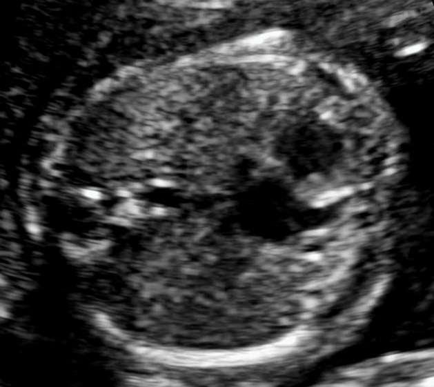
44 Three Vessel View

45 Four Chamber View

46

47

48


49 Ventricular Septal Defect
50 Ventricular Septal Defect


51 Ventricular Septal Defect


52 Ventricular Septal Defect


53 Atrioventricular Septal Defect

54 The AVSD


55 Ebstein s Anomaly

56 Ebstein s Anomaly


57 Tricuspid Regurgitation

58 Hypoplastic Left Heart

59 Hypoplastic Left Heart

60 HLHS
61 Transposition of the Great Vessels (TGA)
62 TGA Three Vessel View
63 Tetralogy of Fallot Large ventricular septal defect Over-riding aorta Pulmonary stenosis Right ventricular hypertrophy

64 Case

65 Case Liver Bowel
66 Pentalogy of Cantrell Rare form of abdominal wall defect Five Associated Anomalies Midline epigastric abdominal wall defect Defect of the lower sternum Deficiency of the anterior diaphragm Defect in the diaphragmatic pericardium Intra cardiac defects


67 Case 2

68 Case 2

69 Case 2


70 The Cardiac Mass


71 The Cardiac Mass

72 The Cardiac Mass

73 The Cardiac Mass
74 FETAL CARDIAC DYSRHYTHMIAS
75 Cardiac Dysrhythmia Occur in 1 to 3 percent of all pregnancies Detected by auscultation Most are benign Most are ectopic premature atrial contractions 10% of pregnancies complicated by fetal arrhythmias, have a potentially life threatening arrhythmia Tachyarrhythmias (heart rate in excess of 180 beats/min) Supraventricular tachycardia (SVT) Atrial flutter (AFL) Bradyarrhythmias (heart rate less than 100 beats/min) Second-degree atrioventricular (AV) block Complete AV block (CAVB)

76 Premature Atrial Contraction

77 PACs


78 PACs

79 PACs

80 Supraventricular Tachycardia

81 SVT

82 Pericardial Effusion


83 The Sail Sign

84 Mechanical PR Interval

85 Mechanical PR Interval
86 THE FUTURE

87 The Three Vessel View

88 The Three Vessel View


89 The Three Vessel Trachea View

90 The Three Vessel Trachea View
 anterior of the Aorta (Ao) Continuous PA and Ao Flow toward the spine in PA")
91 The Three Vessel Trachea View The Normal View Three vessels All vessels to the left of the trachea Vessels similar in size Pulmonary artery (PA) anterior of the Aorta (Ao) Continuous PA and Ao Flow toward the spine in PA and Ao


92 The Three Vessel View Blood flow toward the spine in PA and Ao
93 The Three Vessel Trachea View Examples of abnormalities Too Many Vessels PA and AO Not Continuous Vessel to Right of Trachea Ao Anterior to PA Small PA anterograde Flow Small PA Retrograde Flow Small Ao Left superior vena cava, azygous vein Interupted aortic arch Right sided aortic arch Transposition of the great vessels Tetralogy of Fallot Pulmonary Atresia Hypoplastic left heart

94 Summary Planes


95


96

97


98
ULTRASOUND OF THE FETAL HEART
 ULTRASOUND OF THE FETAL HEART Cameron A. Manbeian, MD Disclosure Statement Today s faculty: Cameron Manbeian, MD does not have any relevant financial relationships with commercial interests or affiliations
Basic Fetal Cardiac Evaluation
 Basic Fetal Cardiac Evaluation Mert Ozan Bahtiyar, MD Director, Fetal Care Center Division of Maternal Fetal Medicine Department of Obstetrics, Gynecology and Reproductive Sciences S L I D E 1 Background
Systematic approach to Fetal Echocardiography. Objectives. Introduction 11/2/2015
 Systematic approach to Fetal Echocardiography. Pediatric Echocardiography Conference, JCMCH November 7, 2015 Rajani Anand Objectives Fetal cardiology pre-test Introduction Embryology and Physiology of
Heart and Lungs. LUNG Coronal section demonstrates relationship of pulmonary parenchyma to heart and chest wall.
 Heart and Lungs Normal Sonographic Anatomy THORAX Axial and coronal sections demonstrate integrity of thorax, fetal breathing movements, and overall size and shape. LUNG Coronal section demonstrates relationship
ISUOG Basic Training. Obtaining & Interpreting Heart Views Correctly Alfred Abuhamad, USA. Basic training. Editable text here
 ISUOG Basic Training Obtaining & Interpreting Heart Views Correctly Alfred Abuhamad, USA Learning Objectives 6, 7 & 8 At the end of the lecture you will be able to: describe how to assess cardiac situs
The Fetal Cardiology Program
 The Fetal Cardiology Program at Texas Children s Fetal Center About the program Since the 1980s, Texas Children s Fetal Cardiology Program has provided comprehensive fetal cardiac care to expecting families
COMPREHENSIVE EVALUATION OF FETAL HEART R. GOWDAMARAJAN MD
 COMPREHENSIVE EVALUATION OF FETAL HEART R. GOWDAMARAJAN MD Disclosure No Relevant Financial Relationships with Commercial Interests Fetal Echo: How to do it? Timing of Study -optimally between 22-24 weeks
Fetal Echocardiography and the Routine Obstetric Sonogram
 JDMS 23:143 149 May/June 2007 143 Fetal Echocardiography and the Routine Obstetric Sonogram SHELLY ZIMBELMAN, RT(R)(CT), RDMS, RDCS ASAD SHEIKH, MD, RDCS Congenital heart disease (CHD) is the most common
PRACTICAL GUIDE TO FETAL ECHOCARDIOGRAPHY IC Huggon and LD Allan
 PRACTICAL GUIDE TO FETAL ECHOCARDIOGRAPHY IC Huggon and LD Allan Fetal Cardiology Unit, Harris Birthright Research Centre for Fetal Medicine, King's College Hospital, London, UK IMPORTANCE OF PRENATAL
Making Sense of Cardiac Views and Imaging Characteristics for 13 Congenital Heart Defects (CHDs)
") Making Sense of Cardiac Views and Imaging Characteristics for 13 Congenital Heart Defects (CHDs) Manny Gaziano, MD, FACOG obimages.net obimages.net@gmail.com Acknowledgements: Krista Wald, RDMS, sonographer,
Making Sense of Cardiac Views and Imaging Characteristics for 13 Congenital Heart Defects (CHDs) Manny Gaziano, MD, FACOG obimages.net obimages.net@gmail.com Acknowledgements: Krista Wald, RDMS, sonographer,
Congenital Heart Defects
 Normal Heart Congenital Heart Defects 1. Patent Ductus Arteriosus The ductus arteriosus connects the main pulmonary artery to the aorta. In utero, it allows the blood leaving the right ventricle to bypass
ECHOCARDIOGRAPHIC APPROACH TO CONGENITAL HEART DISEASE: THE UNOPERATED ADULT
 ECHOCARDIOGRAPHIC APPROACH TO CONGENITAL HEART DISEASE: THE UNOPERATED ADULT Karen Stout, MD, FACC Divisions of Cardiology University of Washington Medical Center Seattle Children s Hospital NO DISCLOSURES
Before we are Born: Fetal Diagnosis of Congenital Heart Disease
 Before we are Born: Fetal Diagnosis of Congenital Heart Disease Mohamed Sulaiman, MD Pediatric cardiologist Kidsheart: American Fetal & Children's Heart Center Dubai Healthcare City, Dubai-UAE First Pediatric
Cardiac Catheterization Cases Primary Cardiac Diagnoses Facility 12 month period from to PRIMARY DIAGNOSES (one per patient)
") PRIMARY DIAGNOSES (one per patient) Septal Defects ASD (Atrial Septal Defect) PFO (Patent Foramen Ovale) ASD, Secundum ASD, Sinus venosus ASD, Coronary sinus ASD, Common atrium (single atrium) VSD (Ventricular
PRIMARY DIAGNOSES (one per patient) Septal Defects ASD (Atrial Septal Defect) PFO (Patent Foramen Ovale) ASD, Secundum ASD, Sinus venosus ASD, Coronary sinus ASD, Common atrium (single atrium) VSD (Ventricular
Disclosures. Outline. Learning Objectives. Introduction. Introduction. Sonographic Screening Examination of the Fetal Heart
 Sonographic Screening Examination of the Fetal Heart Lami Yeo, MD Director of Fetal Cardiology Perinatology Research Branch of NICHD / NIH / DHHS Bethesda, MD and Detroit, Michigan, USA Professor, Division
Pediatric Echocardiography Examination Content Outline
 Pediatric Echocardiography Examination Content Outline (Outline Summary) # Domain Subdomain Percentage 1 Anatomy and Physiology Normal Anatomy and Physiology 10% 2 Abnormal Pathology and Pathophysiology
Summary. HVRA s Cardio Vascular Genetic Detailed L2 Obstetrical Ultrasound. CPT 76811, 76825, _ 90% CHD detection. _ 90% DS detection.
 What is the role of fetal echocardiography (2D 76825, cardiovascular color flow mapping 93325) as performed in conjunction with detailed fetal anatomy scan (CPT 76811) now that AIUM requires limited outflow
UPDATE FETAL ECHO REVIEW
 UPDATE 1 FETAL ECHO REVIEW Study Alert for RDCS Candidates D A V I E S P U B L I S H I N G I N C. Fetal Echo Review Study Alert U P D A T E D A U G U S T 1, 2 0 1 2 Nikki Stahl, RT(R)(M)(CT), RDMS, RVT
Screening for Critical Congenital Heart Disease
 Screening for Critical Congenital Heart Disease Caroline K. Lee, MD Pediatric Cardiology Disclosures I have no relevant financial relationships or conflicts of interest 1 Most Common Birth Defect Most
September 28-30, 2018
 September 28-30, 2018 Course Director Optimizing Detection of Congenital Heart Disease: Important Anatomic Cardiac Regions The Top 5 Critical Anatomic Regions in Fetal Cardiac Imaging Alfred Abuhamad,
Diagnosis of Congenital Cardiac Defects Between 11 and 14 Weeks Gestation in High-Risk Patients
 Article Diagnosis of Congenital Cardiac Defects Between 11 and 14 Weeks Gestation in High-Risk Patients Zeev Weiner, MD, Abraham Lorber, MD, Eliezer Shalev, MD Objective. To examine the feasibility of
Data Collected: June 17, Reported: June 30, Survey Dates 05/24/ /07/2010
 Job Task Analysis for ARDMS Pediatric Echocardiography Data Collected: June 17, 2010 Reported: Analysis Summary For: Pediatric Echocardiography Exam Survey Dates 05/24/2010-06/07/2010 Invited Respondents
"Lecture Index. 1) Heart Progenitors. 2) Cardiac Tube Formation. 3) Valvulogenesis and Chamber Formation. 4) Epicardium Development.
 Heart Progenitors. 2) Cardiac Tube Formation. 3) Valvulogenesis and Chamber Formation. 4) Epicardium Development.") "Lecture Index 1) Heart Progenitors. 2) Cardiac Tube Formation. 3) Valvulogenesis and Chamber Formation. 4) Epicardium Development. 5) Septation and Maturation. 6) Changes in Blood Flow during Development.
"Lecture Index 1) Heart Progenitors. 2) Cardiac Tube Formation. 3) Valvulogenesis and Chamber Formation. 4) Epicardium Development. 5) Septation and Maturation. 6) Changes in Blood Flow during Development.
3/14/2011 MANAGEMENT OF NEWBORNS CARDIAC INTENSIVE CARE CONFERENCE FOR HEALTH PROFESSIONALS IRVINE, CA. MARCH 7, 2011 WITH HEART DEFECTS
 CONFERENCE FOR HEALTH PROFESSIONALS IRVINE, CA. MARCH 7, 2011 MANAGEMENT OF NEWBORNS WITH HEART DEFECTS A NTHONY C. CHANG, MD, MBA, MPH M E D I C AL D I RE C T OR, HEART I N S T I T U T E C H I LDRE N
An update on technique of fetal echocardiography with emphasis on anomalies detectable in four chambered view.
 An update on technique of fetal echocardiography with emphasis on anomalies detectable in four chambered view. Dr. Ranjitha.G Specialist Radiologist NMC-SH Al ain, UAE Fetal echocardiography is an essential
Chapter 2 Cardiac Interpretation of Pediatric Chest X-Ray
 Chapter 2 Cardiac Interpretation of Pediatric Chest X-Ray Ra-id Abdulla and Douglas M. Luxenberg Key Facts The cardiac silhouette occupies 50 55% of the chest width on an anterior posterior chest X-ray
Appendix A.1: Tier 1 Surgical Procedure Terms and Definitions
 Appendix A.1: Tier 1 Surgical Procedure Terms and Definitions Tier 1 surgeries AV Canal Atrioventricular Septal Repair, Complete Repair of complete AV canal (AVSD) using one- or two-patch or other technique,
DEVELOPMENT OF THE CIRCULATORY SYSTEM L E C T U R E 5
 DEVELOPMENT OF THE CIRCULATORY SYSTEM L E C T U R E 5 REVIEW OF CARDIAC ANATOMY Heart 4 chambers Base and apex Valves Pericardial sac 3 layers: epi, myo, endo cardium Major blood vessels Aorta and its
CMS Limitations Guide - Radiology Services
 CMS Limitations Guide - Radiology Services Starting October 1, 2015, CMS will update their existing medical necessity limitations on tests and procedures to correspond to ICD-10 codes. This limitations
Common Defects With Expected Adult Survival:
 Common Defects With Expected Adult Survival: Bicuspid aortic valve :Acyanotic Mitral valve prolapse Coarctation of aorta Pulmonary valve stenosis Atrial septal defect Patent ductus arteriosus (V.S.D.)
All You Need to Know About Situs and Looping Disorders: Embryology, Anatomy, and Echocardiography
 All You Need to Know About Situs and Looping Disorders: Embryology, Anatomy, and Echocardiography Helena Gardiner Co-Director of Fetal Cardiology, The Fetal Center, University of Texas at Houston Situs
Fetal Tetralogy of Fallot
 36 Fetal Tetralogy of Fallot E.D. Bespalova, R.M. Gasanova, O.A.Pitirimova National Scientific and Practical Center of Cardiovascular Surgery, Moscow Elena D. Bespalova, MD Professor, Director Rena M,
List of Videos. Video 1.1
 Video 1.1 Video 1.2 Video 1.3 Video 1.4 Video 1.5 Video 1.6 Video 1.7 Video 1.8 The parasternal long-axis view of the left ventricle shows the left ventricular inflow and outflow tract. The left atrium
Segmental approach to normal and abnormal situs arrangement - Echocardiography -
 Segmental approach to normal and abnormal situs arrangement - Echocardiography - Jan Marek Great Ormond Street Hospital & Institute of Cardiovascular Sciences, University College London No disclosures
The Chest X-ray for Cardiologists
 Mayo Clinic & British Cardiovascular Society at the Royal College of Physicians, London : 21-23-October 2013 Cases-Controversies-Updates 2013 The Chest X-ray for Cardiologists Michael Rubens Royal Brompton
Fetal Rhythm and Blues
 Fetal Rhythm and Blues John Cotton, MD Professor of Pediatrics Division of Pediatric Cardiology Director, Fetal Cardiology Program UNC Chapel Hill, School of Medicine Objectives To review methods used
Foetal Cardiology: How to predict perinatal problems. Prof. I.Witters Prof.M.Gewillig UZ Leuven
 Foetal Cardiology: How to predict perinatal problems Prof. I.Witters Prof.M.Gewillig UZ Leuven Cardiopathies Incidence : 8-12 / 1000 births ( 1% ) Most frequent - Ventricle Septum Defect 20% - Atrium Septum
Anatomy & Physiology
 1 Anatomy & Physiology Heart is divided into four chambers, two atrias & two ventricles. Atrioventricular valves (tricuspid & mitral) separate the atria from ventricles. they open & close to control flow
CONGENITAL HEART DISEASE (CHD)
") CONGENITAL HEART DISEASE (CHD) DEFINITION It is the result of a structural or functional abnormality of the cardiovascular system at birth GENERAL FEATURES OF CHD Structural defects due to specific disturbance
CONGENITAL HEART DISEASE (CHD) DEFINITION It is the result of a structural or functional abnormality of the cardiovascular system at birth GENERAL FEATURES OF CHD Structural defects due to specific disturbance
Introduction. Pediatric Cardiology. General Appearance. Tools of Assessment. Auscultation. Vital Signs
 Introduction Pediatric Cardiology An introduction to the pediatric patient with heart disease: M-III Lecture Douglas R. Allen, M.D. Assistant Professor and Director of Community Pediatric Cardiology at
Congenital Heart Disease An Approach for Simple and Complex Anomalies
 Congenital Heart Disease An Approach for Simple and Complex Anomalies Michael D. Pettersen, MD Director, Echocardiography Rocky Mountain Hospital for Children Denver, CO None Disclosures 1 ASCeXAM Contains
Segmental Analysis. Gautam K. Singh, M.D. Washington University School of Medicine St. Louis
 Segmental Analysis Gautam K. Singh, M.D. Washington University School of Medicine St. Louis Segmental Analysis Segmental Analysis: From Veins to Ventricles Segmental Approach to Evaluation of Congenital
Anomalous Systemic Venous Connection Systemic venous anomaly
 World Database for Pediatric and Congenital Heart Surgery Appendix B: Diagnosis (International Paediatric and Congenital Cardiac Codes (IPCCC) and definitions) Anomalous Systemic Venous Connection Systemic
How to Recognize a Suspected Cardiac Defect in the Neonate
 Neonatal Nursing Education Brief: How to Recognize a Suspected Cardiac Defect in the Neonate https://www.seattlechildrens.org/healthcareprofessionals/education/continuing-medical-nursing-education/neonatalnursing-education-briefs/
Notes: 1)Membranous part contribute in the formation of small portion in the septal cusp.
Membranous part contribute in the formation of small portion in the septal cusp.") Embryology 9 : Slide 16 : There is a sulcus between primitive ventricular and bulbis cordis that will disappear gradually and lead to the formation of one chamber which is called bulboventricular chamber.
Embryology 9 : Slide 16 : There is a sulcus between primitive ventricular and bulbis cordis that will disappear gradually and lead to the formation of one chamber which is called bulboventricular chamber.
J Somerville and V Grech. The chest x-ray in congenital heart disease 2. Images Paediatr Cardiol Jan-Mar; 12(1): 1 8.
: 1 8.") IMAGES in PAEDIATRIC CARDIOLOGY Images Paediatr Cardiol. 2010 PMCID: PMC3228330 The chest x-ray in congenital heart disease 2 J Somerville and V Grech Paediatric Department, Mater Dei Hospital, Malta Corresponding
IMAGES in PAEDIATRIC CARDIOLOGY Images Paediatr Cardiol. 2010 PMCID: PMC3228330 The chest x-ray in congenital heart disease 2 J Somerville and V Grech Paediatric Department, Mater Dei Hospital, Malta Corresponding
Adult Echocardiography Examination Content Outline
 Adult Echocardiography Examination Content Outline (Outline Summary) # Domain Subdomain Percentage 1 2 3 4 5 Anatomy and Physiology Pathology Clinical Care and Safety Measurement Techniques, Maneuvers,
Bits and Bobs secondary causes of heart problems. Dr Angela McBrien 9 th September 2017
 Bits and Bobs secondary causes of heart problems Dr Angela McBrien 9 th September 2017 Not the heart Dextroposition Heart in the right chest with the apex to the left Often caused by left sided chest mass
Introduction to Fetal Medicine. Lloyd R. Feit M.D. Associate Professor of Pediatrics Warren Alpert Medical School Brown University
 Associate Professor of Pediatrics Warren Alpert Medical School Brown University Fetal Cardiology Important in evaluation of high risk pregnancies. Information obtainable in > 95% of patients attempted.
9/8/2009 < 1 1,2 3,4 5,6 7,8 9,10 11,12 13,14 15,16 17,18 > 18. Tetralogy of Fallot. Complex Congenital Heart Disease.
 Current Indications for Pediatric CTA S Bruce Greenberg Professor of Radiology Arkansas Children s Hospital University of Arkansas for Medical Sciences greenbergsbruce@uams.edu 45 40 35 30 25 20 15 10
Congenital Heart Disease: Physiology and Common Defects
 Congenital Heart Disease: Physiology and Common Defects Jamie S. Sutherell, M.D, M.Ed. Associate Professor, Pediatrics Division of Cardiology Director, Medical Student Education in Pediatrics Director,
Most common fetal cardiac anomalies
 Most common fetal cardiac anomalies Common congenital heart defects CHD % of cardiac defects Chromosomal Infants Fetuses anomaly (%) 22q11 deletion (%) VSD 30 5~10 20~40 10 PS 9 5 (PA w/ VSD) HLHS 7~9
Slide 1. Slide 2. Slide 3 CONGENITAL HEART DISEASE. Papworth Hospital NHS Trust INTRODUCTION. Jakub Kadlec/Catherine Sudarshan INTRODUCTION
 Slide 1 CONGENITAL HEART DISEASE Jakub Kadlec/Catherine Sudarshan NHS Trust Slide 2 INTRODUCTION Most common congenital illness in the newborn Affects about 4 9 / 1000 full-term live births in the UK 1.5
Assessment of fetal heart function and rhythm
 Assessment of fetal heart function and rhythm The fetal myocardium Early Gestation Myofibrils 30% of myocytes Less sarcoplasmic reticula Late Gestation Myofibrils 60% of myocytes Increased force per unit
When is Risky to Apply Oxygen for Congenital Heart Disease 부천세종병원 소아청소년과최은영
 When is Risky to Apply Oxygen for Congenital Heart Disease 부천세종병원 소아청소년과최은영 The Korean Society of Cardiology COI Disclosure Eun-Young Choi The author have no financial conflicts of interest to disclose
ADULT CONGENITAL HEART DISEASE. Stuart Lilley
 ADULT CONGENITAL HEART DISEASE Stuart Lilley More adults than children have congenital heart disease Huge variety of congenital lesions from minor to major Heart failure, re-operation and arrhythmia are
Giovanni Di Salvo MD, PhD, FESC Second University of Naples Monaldi Hospital
 Giovanni Di Salvo MD, PhD, FESC Second University of Naples Monaldi Hospital VSD is one of the most common congenital cardiac abnormalities in the newborn. It can occur as an isolated finding or in combination
Adult Congenital Heart Disease: What All Echocardiographers Should Know Sharon L. Roble, MD, FACC Echo Hawaii 2016
 1 Adult Congenital Heart Disease: What All Echocardiographers Should Know Sharon L. Roble, MD, FACC Echo Hawaii 2016 DISCLOSURES I have no disclosures relevant to today s talk 2 Why should all echocardiographers
Echocardiography in Adult Congenital Heart Disease
 Echocardiography in Adult Congenital Heart Disease Michael Vogel Kinderherz-Praxis München CHD missed in childhood Subsequent lesions after repaired CHD Follow-up of cyanotic heart disease CHD missed in
Basic Training. ISUOG Basic Training The 20 Planes Approach to the Routine Mid Trimester Scan
 ISUOG The 20 Planes Approach to the Routine Mid Trimester Scan Learning objective At the end of the lecture you will be able to: Explain how to perform a structured routine examination, including measurements,
AbnormalThree-VesselView on Sonography: A Clue to the Diagnosis of Congenital Heart Disease in the Fetus
 rt Pictorial Essay bnormalthree-vesselview on Sonography: Clue to the Diagnosis of Congenital Heart Disease in the Fetus screening tool for major congenital heart diseases [I. 2J. However, anomalies of
Heart Development and Congenital Heart Disease
 Heart Development and Congenital Heart Disease Sally Dunwoodie s.dunwoodie@victorchang.edu.au Developmental and Stem Cell Biology Division Victor Chang Cardiac Research Institute for the heart of Australia...
Fetal echocardiography. Ahmeabad. to neonatal series due to high SB rate. Prenatal detection can improve the fetal outcome. (4, 5) 60% 40% 20%
 60% 40% 20%") Guidelines for Fetal Echocardiography 1 Fetal echocardiography Introduction Dr Jayprakash Shah MD; FICOG Chairman Imaging science committee FOGSI Fetal Medicine expert Rajni Hospital, Ahmedabad, Ex sonologist
Guidelines for Fetal Echocardiography 1 Fetal echocardiography Introduction Dr Jayprakash Shah MD; FICOG Chairman Imaging science committee FOGSI Fetal Medicine expert Rajni Hospital, Ahmedabad, Ex sonologist
Outflow Tracts Anomalies
 Diagnosis of Outflow Tract Anomalies in the Fetus General Framing D.Paladini Fetal Medicine & Surgery Unit Gasllini Children s Hospital - Genoa dariopaladini@ospedale-gaslini.ge.it Outflow Tracts Anomalies
Atrial Septal Defects
 Supplementary ACHD Echo Acquisition Protocol for Atrial Septal Defects The following protocol for echo in adult patients with atrial septal defects (ASDs) is a guide for performing a comprehensive assessment
Low-dose prospective ECG-triggering dual-source CT angiography in infants and children with complex congenital heart disease: first experience
 Low-dose prospective ECG-triggering dual-source CT angiography in infants and children with complex congenital heart disease: first experience Ximing Wang, M.D., Zhaoping Cheng, M.D., Dawei Wu, M.D., Lebin
Index. Note: Page numbers of article titles are in boldface type.
 Index Note: Page numbers of article titles are in boldface type. A Acute coronary syndrome(s), anticoagulant therapy in, 706, 707 antiplatelet therapy in, 702 ß-blockers in, 703 cardiac biomarkers in,
NASCI 2012 Segmental Analysis
 NASCI 2012 Segmental Analysis Frandics Chan, M.D., Ph.D. Stanford University Medical Center Lucile Packard Department Children s of Radiology Hospital Menagerie of Congenital Cardiac Lesions 1. Absent
Basic Training. ISUOG Basic Training Examining the Upper Lip, Face & Profile
 ISUOG Examining the Upper Lip, Face & Profile Learning objectives At the end of the lecture you will be able to: Describe how to obtain the 3 planes required to assess the anatomy of the fetal face Recognise
Cardiovascular Pathophysiology: Right to Left Shunts aka Cyanotic Lesions
 Cardiovascular Pathophysiology: Right to Left Shunts aka Cyanotic Lesions Ismee A. Williams, MD, MS iib6@columbia.edu Pediatric Cardiology Learning Objectives To discuss the hemodynamic significance of
Cardiovascular Pathophysiology: Right to Left Shunts aka Cyanotic Lesions Ismee A. Williams, MD, MS Pediatric Cardiology
 Cardiovascular Pathophysiology: Right to Left Shunts aka Cyanotic Lesions Ismee A. Williams, MD, MS iib6@columbia.edu Pediatric Cardiology Learning Objectives To discuss the hemodynamic significance of
MEDICAL MANAGEMENT WITH CAVEATS 1. In one study of 50 CHARGE patients with CHD, 75% required surgery. 2. Children with CHARGE may be resistant to chlo
 CARDIOLOGY IN CHARGE SYNDROME: FOR THE PHYSICIAN Angela E. Lin, M.D. Teratology Program/Active Malformation Surveillance, Brigham and Women's Hospital, Old PBBH-B501, 75 Francis St., Boston, MA 02115 alin@partners.org
Congenital Heart Disease
 Screening Programmes Fetal Anomaly Congenital Heart Disease Information for health professionals Publication date: April 2012 Review date: April 2013 Version 2 67 Congenital Heart Disease Information for
Early fetal echocardiography: congenital heart disease detection and diagnostic accuracy in the hands of an experienced fetal cardiology program
 DOI: 10.1002/pd.4372 ORIGINAL ARTICLE Early fetal echocardiography: congenital heart disease detection and diagnostic accuracy in the hands of an experienced fetal cardiology program Jodi I. Pike, Anita
CYANOTIC CONGENITAL HEART DISEASES. PRESENTER: DR. Myra M. Koech Pediatric cardiologist MTRH/MU
 CYANOTIC CONGENITAL HEART DISEASES PRESENTER: DR. Myra M. Koech Pediatric cardiologist MTRH/MU DEFINITION Congenital heart diseases are defined as structural and functional problems of the heart that are
Assessing Cardiac Anatomy With Digital Subtraction Angiography
 485 JACC Vol. 5, No. I Assessing Cardiac Anatomy With Digital Subtraction Angiography DOUGLAS S., MD, FACC Cleveland, Ohio The use of intravenous digital subtraction angiography in the assessment of patients
Transposition of the Great Arteries Preoperative Diagnostic Considerations. John Simpson Evelina Children s Hospital London, UK
 Transposition of the Great Arteries Preoperative Diagnostic Considerations John Simpson Evelina Children s Hospital London, UK Areas to be covered Definitions Scope of occurrence of transposition of the
Echocardiographic and anatomical correlates in the fetus*
 Br Heart J 1980; : 51 Echocardiographic and anatomical correlates in the fetus* LINDSEY D ALLAN, MICHAEL J TYNAN, STUART CAMPBELL, JAMES L WILKINSON, ROBERT H ANDERSON From King's College Hospital, and
Fetal Cardiac Anomaly
 89 Symposium: OB/GY US (Room B) 12 : 10 1 2 : 30 Fetal Cardiac Anomaly 1. One third of all congenital anomalies 2. 6 10/1,000 live births 3. Related with more than 50% of childhood deaths and 20-30% of
PART II ECHOCARDIOGRAPHY LABORATORY OPERATIONS ADULT TRANSTHORACIC ECHOCARDIOGRAPHY TESTING
 PART II ECHOCARDIOGRAPHY LABORATORY OPERATIONS ADULT TRANSTHORACIC ECHOCARDIOGRAPHY TESTING STANDARD - Primary Instrumentation 1.1 Cardiac Ultrasound Systems SECTION 1 Instrumentation Ultrasound instruments
W.S. O The University of Hong Kong
 W.S. O The University of Hong Kong Objectives: Describe early angiogenesis. Describe the heart tube formation. Describe the partitioning into a 4- chambered heart. List the formation of heart valves and
CMR for Congenital Heart Disease
 CMR for Congenital Heart Disease * Second-line tool after TTE * Strengths of CMR : tissue characterisation, comprehensive access and coverage, relatively accurate measurements of biventricular function/
September 26, 2012 Philip Stockwell, MD Lifespan CVI Assistant Professor of Medicine (Clinical)
") September 26, 2012 Philip Stockwell, MD Lifespan CVI Assistant Professor of Medicine (Clinical) Advances in cardiac surgery have created a new population of adult patients with repaired congenital heart
September 26, 2012 Philip Stockwell, MD Lifespan CVI Assistant Professor of Medicine (Clinical) Advances in cardiac surgery have created a new population of adult patients with repaired congenital heart
Congenital Heart Disease Systematic Interpretation of CT Suhny Abbara, MD
 Congenital Heart Disease Systematic Interpretation of CT Suhny Abbara, MD Chief, Cardiothoracic Imaging Division Professor of Radiology UT Southwestern Medical Center, Dallas, TX Suhny.Abbara@UTSouthwestern.edu
Dear Parent/Guardian,
 Dear Parent/Guardian, You have indicated on school records that your child has an ongoing health problem that may require medication and/or treatment during the school day with rescue medication. Attached
Index. cardiology.theclinics.com. Note: Page numbers of article titles are in boldface type.
 Index Note: Page numbers of article titles are in boldface type. A ACHD. See Adult congenital heart disease (ACHD) Adult congenital heart disease (ACHD), 503 512 across life span prevalence of, 504 506
Large veins of the thorax Brachiocephalic veins
 Large veins of the thorax Brachiocephalic veins Right brachiocephalic vein: formed at the root of the neck by the union of the right subclavian & the right internal jugular veins. Left brachiocephalic
HDlive Silhouette Mode With Spatiotemporal Image Correlation for Assessment of the Fetal Heart
 ORIGINAL RESEARCH HDlive Silhouette Mode With Spatiotemporal Image Correlation for Assessment of the Fetal Heart Toshiyuki Hata, MD, PhD, Mohamed Ahmed Mostafa AboEllail, MD, Suraphan Sajapala, MD, Mari
Surgical Procedures. Direct suture of small ASDs Patch repair Transcatheter closure with a prosthetic device called occluder
 PEDIATRIC Review Surgical Procedures Atrial Septal Defect repair: Direct suture of small ASDs Patch repair Transcatheter closure with a prosthetic device called occluder Balloon atrial septostomy (Rashkind)
Accuracy of prenatal diagnosis of fetal congenital heart disease by different
 Accuracy of prenatal diagnosis of fetal congenital heart disease by different methods with echocardiography Ying Zhang 1* * Corresponding author Email: baogoubei@hotmail.com Ai-Lu Cai 1 Email: caial_us@hotmail.com
Index. radiologic.theclinics.com. Note: Page numbers of article titles are in boldface type.
 Index Note: Page numbers of article titles are in boldface type. A ALCAPA. See Anomalous left coronary artery from the pulmonary artery. Angiosarcoma computed tomographic assessment of, 809 811 Anomalous
SURGICAL TREATMENT AND OUTCOME OF CONGENITAL HEART DISEASE
 SURGICAL TREATMENT AND OUTCOME OF CONGENITAL HEART DISEASE Mr. W. Brawn Birmingham Children s Hospital. Aims of surgery The aim of surgery in congenital heart disease is to correct or palliate the heart
Cardiopulmonary Syndromes: Conditions With Concomitant Cardiac and Pulmonary Abnormalities
 Cardiopulmonary Syndromes: Conditions With Concomitant Cardiac and Pulmonary Abnormalities Carlos S. Restrepo M.D. Professor of Radiology The University of Texas HSC at San Antonio Cardiopulmonary Syndromes
Research article. Primary detection of congenital heart diseases in the Kyrgyz Republic
 Research article Primary detection of congenital heart diseases in the Kyrgyz Republic Irina A. Akhmedova, Gulzada A. Imanalieva, Damir A.Abibillaev, Taalaibek Z. Kudaiberdiev Scientific Research Institute
The sonographic approach to the detection of fetal cardiac
 Ultrasound Obstet Gynecol 2002; 19: 360 365 The sonographic approach to the detection of fetal cardiac Blackwell Science Ltd anomalies in early pregnancy M. BRONSHTEIN* and E. Z. ZIMMER* *Department of
ISUOG Basic Training. Assessing the Neck & Chest Gihad Chalouhi, Lebanon
 ISUOG Basic Training Assessing the Neck & Chest Gihad Chalouhi, Lebanon Learning objectives 9 & 10 At the end of the lecture you will be able to: recognise the differences between the normal & most common
Identification of congenital cardiac malformations by echocardiography in midtrimester fetus*
 Br Heart J 1981; 46: 358-62 Identification of congenital cardiac malformations by echocardiography in midtrimester fetus* LINDSEY D ALLAN, MICHAEL TYNAN, STUART CAMPBELL, ROBERT H ANDERSON From Guy's Hospital;
ORIGINAL RESEARCH PAPER
 ORIGINAL RESEARCH PAPER ROLE OF CT PULMONARY ANGIOGRAPHY IN CONGENITAL HEART DISEASES IN PAEDIATRIC POPULATION Radiology KEY WORDS: Congenital heart disease, CT pulmonary angiography, pediatric heart disease,
By Dickens ATURWANAHO & ORIBA DAN LANGOYA MAKchs, MBchB CONGENTAL HEART DISEASE
 By Dickens ATURWANAHO & ORIBA DAN LANGOYA MAKchs, MBchB CONGENTAL HEART DISEASE Introduction CHDs are abnormalities of the heart or great vessels that are present at birth. Common type of heart disease
Improvement in the antenatal detection rate of CHD and its effect on survival in Wales: How can we improve our results further?
 Improvement in the antenatal detection rate of CHD and its effect on survival in Wales: How can we improve our results further? Dr Orhan Uzun Consultant Paediatric Cardiologist UHW Welcome Improvement!