2017 BDKA Review. Regularity Rate P waves PRI QRS Interpretation. Regularity Rate P waves PRI QRS Interpretation 1/1/2017
|
|
|
- Valentine Beasley
- 6 years ago
- Views:
Transcription
1 BDKA Review 2.
2 3. 4. Interpretation
3 5. QT 6.
4 7. 8.
5 QT
6
7
8
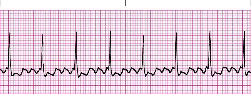
9 QT
10 QT

11 QT


12 Where are pacer spikes? Before the P wave or before the QRS complex? Did the heart capture the impulse (do you see an appropriate complex)? Is there any underlying rhythm we can see? If so, go through the rules of regularity, rate, P waves, PRI, QRS, QT Interpretation?
? Is there any underlying rhythm we can see?")

13 25. Where are pacer spikes? Before the P wave or before the QRS complex? Did the heart capture the impulse (do you see an appropriate complex)? Is there any underlying rhythm we can see? If so, go through the rules of regularity, rate, P waves, PRI, QRS, QT Interpretation? 26. Where are pacer spikes? Before the P wave or before the QRS complex? Did the heart capture the impulse (do you see an appropriate complex)? Is there any underlying rhythm we can see? If so, go through the rules of regularity, rate, P waves, PRI, QRS, QT Interpretation?
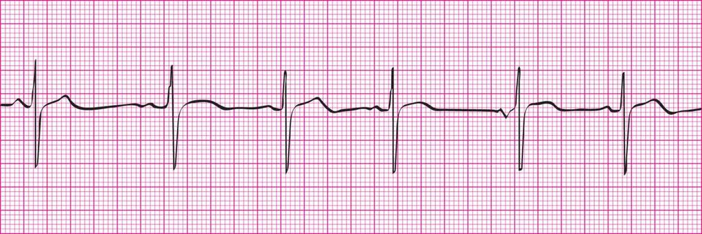
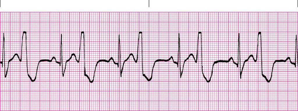
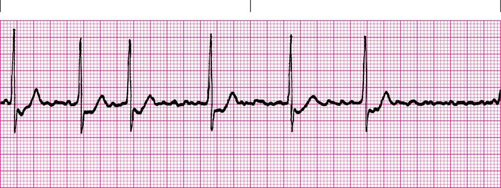
14 Basic Dysrhythmias Review Answers 1. Regular Rate: 120 P waves: upright and uniform, 1 P wave for each QRS PRI: 0.12 (0.10, 0.12, 0.14 acceptable) QRS: 0.12 (0.10, 0.12, 0.14 acceptable) Sinus Tachycardia (wide QRS if your measurement was 0.14) 2. Regular Rate: 240 P waves: none PRI: none QRS: do not worry about measuring a QRS do quick look to determine it is wide and bizarre Ventricular Tachycardia 3. Irregular Rate: 60 P waves: different morphologies PRI: varies between 0.12 to 0.20 QRS: 0.12 (0.10, 0.12, 0.14 acceptable) Wandering Pacemaker (wide QRS if your measurement was 0.14) 4. Regular Rate: 50 P waves: upright, uniform, 1 P wave for each QRS PRI: 0.16 (0.14, 0.16, 0.18 acceptable) QRS: 0.08 (0.06, 0.08, 0.10 acceptable) Sinus Bradycardia 5. Regular Rate: 80 P waves: inverted before QRS PRI: 0.08 (0.06, 0.08, 0.10 acceptable) QRS: 0.08 (0.06, 0.08, 0.10 acceptable) QT: 0.38 (0.36, 0.38, 0.40 acceptable) ( for rate of 80 should be 0.32 to 0.39 so OK) Accelerated Junctional Rhythm 6. Irregular Rate: 100 with ectopics (do not include last QRS because not a full complex) or 50 without ectopics P waves : upright, uniform and 1 P wave for each QRS in underlying rhythm. No P waves on ectopics PRI: 0.18 (0.16, 0.18, 0.20 acceptable) QRS: 0.14 (0.12, 0.14, 0.16 acceptable) in underlying rhythm and ectopics but notice different morphology of QRS on ectopics compared to underlying rhythm QRS Sinus Rhythm with wide QRS and Bigeminy, Unifocal Premature Ventricular Contractions (PVCs) if stated rate is 100 or Sinus Bradycardia with wide QRS and Bigeminy, Unifocal PVC s if stated rate is 50 (would not have wide QRS if 0.12)
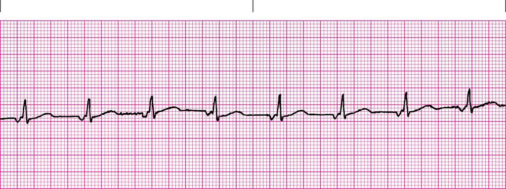
15 7. Regular Rate: 80 P waves: sawtooth/flutter PRI: none QRS: 0.10 (0.08, 0.10, 0.12 acceptable) Controlled Atrial Flutter, 4:1 conduction 8. Irregular Rate: 60 P waves: fibrillatory or no P waves PRI: none QRS: 0.08 (0.06, 0.08, 0.10 acceptable) Controlled Atrial Fibrillation 9. Irregular Rate: 110 with ectopics or 90 without ectopics P waves: upright, uniform and 1 P wave for each QRS in underlying rhythm. P waves are hidden in T waves on ectopics PRI: 0.16 (0.14, 0.16, 0.18 acceptable) QRS: 0.10 (0.08, 0.10, 0.12 acceptable) Sinus Tachycardia with 2 Premature Atrial Contractions (PAC s) if stated rate is 110 or Sinus Rhythm with 2 PAC s if stated rate is Regular Rate: 90 P waves: upright, uniform, 1 P wave for each QRS PRI: 0.28 (0.26, 0.28, 0.30 acceptable) QRS: 0.08 (0.06, 0.08, 0.10 acceptable) QT: 0.40 (0.38, 0.40, 0.42 acceptable) (for rate of 90 should be 0.30 to 0.36 so this is a little prolonged for this rate) Sinus Rhythm with First Degree Heart Block, Prolonged QT and ST segment depressiom 11. Irregular Rate: 100 with ectopics or 80 without ectopics P waves: upright, uniform and 1 P wave for each QRS in underlying rhythm. No P waves on ectopics PRI: 0.20 (0.18, 0.20, 0.22 acceptable) QRS: 0.12 (0.10, 0.12, 0.14 acceptable) in underlying and ectopics but notice different morphology of QRS on ectopics compared to underlying rhythm QRS Sinus Rhythm with 2 Unifocal Premature Ventricular Contractions (PVC s) (add First Degree Heart Block if your PRI was 0.22) (add wide QRS if your underlying QRS was 0.14) 12. Irregular Rate: 70 P waves: upright, uniform, 1 P wave for each QRS PRI: 0.12 (0.10, 0.12, 0.14 acceptable) QRS: 0.08 (0.06, 0.08, 0.10 acceptable) Sinus Arrhythmia
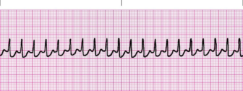
16 13. Regular Rate: 200 P waves: none PRI: none QRS: 0.06 (0.04, 0.06, 0.08 acceptable) Supraventricular Tachycardia with ST segment depression 14. Slightly Irregular (Not R-R is off by 2 boxes, but does not change final interpretation) Rate: 30 P waves: upright, more than 1 P wave for each QRS PRI: very different QRS: 0.12 (0.10, 0.12, 0.14 acceptable) Third Degree/Complete Heart Block 15. None None None None None Ventricular Fibrillation 16. Irregular Rate: 80 with ectopics or 60 without ectopics P waves: upright, uniform and 1 P wave for each QRS in underlying rhythm No P waves on ectopics PRI: 0.18 (0.16, 0.18, 0.20 acceptable) QRS: 0.04 (0.02, 0.04, 0.06 acceptable) in underlying rhythm and 0.16 (0.14, 0.16, 0.18 acceptable) for ectopics Sinus Rhythm with Multifocal Premature Ventricular Contractions (PVCs) 17. Irregular Rate: 80 with ectopic or 70 without ectopic P waves: upright, uniform and 1 P wave for each QRS in underlying rhythm. Have upright P wave on ectopic beat PRI: 0.14 (0.12, 0.14, 0.16 acceptable) in underlying and 0.16 (0.14, 0.16, 0.18 acceptable) on ectopic QRS: 0.10 (0.08, 0.10, 0.12 acceptable) in underlying rhythm and ectopic QT: 0.38 (0.36, 0.38, 0.40 acceptable) (for rate of 70 should be 0.33 to 0.41 and for rate of 80 should be 0.32 to 0.39, so overall QT not a big concern) Sinus Rhythm with Premature Atrial Contraction (PAC) 18. Regular Rate: 40 P waves: upright and two P waves for each QRS PRI: 0.28 (0.26, 0.28, 0.30 acceptable) QRS: 0.10 (0.08, 0.10, 0.12 acceptable) Second Degree Type II Classical Heart Block and ST segment depression
17 19. Regular Rate: 50 P waves: none PRI: none QRS: 0.10 (0.08, 0.10, 0.12 acceptable) Junctional Escape 20. Irregular Rate: 60 with ectopic or 50 without ectopic P waves: upright and 1 P wave for each QRS no P wave on ectopic beat PRI: 0.16 (0.14, 0.16, 0.18 acceptable) QRS: 0.06 (0.04, 0.06, 0.08 acceptable) QT: 0.40 (0.38, 0.40, 0.42 acceptable) (for rate of 50 should be 0.38 to 0.46 and for rate of 60 should be 0.35 to 0.43, so OK) Sinus Rhythm with Premature Junctional Contraction (PJC) if rate 60. Sinus Bradycardia with PJC if rate Slightly Irregular Rate: 60 P waves: P wave morphology changes PRI: First 3 P waves are 0.16 (0.14, 0.16, 0.18 acceptable) and last 3 P waves are 0.12 (0.10, 0.12, 0.14 acceptable) QRS: 0.12 (0.10, 0.12, 0.14 acceptable) QT: 0.52 ( , 0.54 acceptable) (for rate of 60 should be 0.35 to 0.43, so prolonged) Wandering Atrial Pacemaker with prolonged QT (add wide QRS if your QRS was 0.14) 22. Irregular Rate: 90 P waves: upright and more P waves than QRS complexes (some P waves hiding in T waves) PRI: varies, gets longer and drops a QRS and then pattern starts back over QRS: 0.08 (0.06, 0.08, 0.10 acceptable) Second Degree Type I Wenckebach Heart Block 23. Slightly Irregular Rate: 30 P waves: none PRI: none QRS: wide and bizarre Idioventricular 24. Pacer spikes are before the QRS, so trying to pace the ventricles For each pacer spike, you see a QRS, so it captured No underlying rhythm visible Interpretation: Ventricular Pacing with 100% Capture
18 25. Pacer spikes are before the P wave and before the QRS, so trying to pace the atria and the ventricles For each pacer spike, you see an appropriate complex (with pacers, you do not get a normal looking P wave and QRS complexes are usually wide and bizarre. You can also see normal QRS complexes as well with certain types of pacemakers) Interpretation: Dual Chamber Pacing with 100% Capture 26. Pacer spikes are before the QRS, so trying to pace the ventricles For each pacer spike, you see a QRS, so it captured You can see P waves that the heart generated on its own, so the SA node of the patient works fine. There is a problem. If you look at the PRIs on the strip, they are very different, which means the pacemaker is not sensing the P waves from the patient. So the P waves and QRS complexes are not related Interpretation: 100% Ventricular Pacing with Capture with an Underlying Third Degree Heart Block Regarding pacemakers, loss of capture is when the heart does not respond to an electrical stimulus from the pacemaker, so there will not be a complex after the pacer spike. Please note, you will not need to do any QT calculations (QTc) for the test. You also do not need to memorize any QT ranges for your test. If a strip has a prolonged QT interval, every multiple choice answer for that strip will have prolonged QT on it.
Rate: The atrial and ventricular rates are equal; heart rate is greater than 100 bpm (usually between bpm).
.") Sinus Bradycardia Regularity: The R-R intervals are constant; the rhythm is regular. Rate: The atrial and ventricular rates are equal; heart rate is less than 60 bpm. P wave: There is a uniform P wave
Sinus Bradycardia Regularity: The R-R intervals are constant; the rhythm is regular. Rate: The atrial and ventricular rates are equal; heart rate is less than 60 bpm. P wave: There is a uniform P wave
Basic Dysrhythmia Interpretation
 Basic Dysrhythmia Interpretation Objectives 2 To understand the Basic ECG To understand the meaning of Dysrhythmia To describe the normal heart conduction system. To describe the normal impulse pathways.
-RHYTHM PRACTICE- By Dr.moanes Msc.cardiology Assistant Lecturer of Cardiology Al Azhar University. OBHG Education Subcommittee
 -RHYTHM PRACTICE- By Dr.moanes Msc.cardiology Assistant Lecturer of Cardiology Al Azhar University The Normal Conduction System Sinus Node Normal Sinus Rhythm (NSR) Sinus Bradycardia Sinus Tachycardia
ABCs of ECGs. Shelby L. Durler
 ABCs of ECGs Shelby L. Durler Objectives Review the A&P of the cardiac conduction system Placement and obtaining 4-lead and 12-lead ECGs Overview of the basics of ECG rhythm interpretation Intrinsic
Electrocardiography for Healthcare Professionals
 Electrocardiography for Healthcare Professionals Chapter 7: Junctional Dysrhythmias 2012 The Companies, Inc. All rights reserved. Learning Outcomes 7.1 Describe the various junctional dysrhythmias 7.2
8/20/2012. Learning Outcomes (Cont d)
") 1 2 3 4 Electrocardiography for Healthcare Professionals Chapter 7: Junctional Dysrhythmias Learning Outcomes 7.1 Describe the various junctional dysrhythmias 7.2 Identify premature junctional complexes
1 2 3 4 Electrocardiography for Healthcare Professionals Chapter 7: Junctional Dysrhythmias Learning Outcomes 7.1 Describe the various junctional dysrhythmias 7.2 Identify premature junctional complexes
EKG Intermediate Tips, tricks, tools
 Birmingham Regional Emergency Medical Services System 2018 ALCTE Summer Conference EKG Intermediate Tips, tricks, tools Brian Gober, MAT, ATC, NRP, CSCS Education Services Manager ECC Training Center Coordinator
Course Objectives. Proper Lead Placements. Review the ECG print paper. Review the mechanics of the Myocardium. Review basics of ECG Rhythms
 ECG Interpretations Course Objectives Proper Lead Placements Review the ECG print paper Review the mechanics of the Myocardium Review basics of ECG Rhythms How Leads Work The ECG Leads we use are Bipolar
EKG Competency for Agency
 EKG Competency for Agency Name: Date: Agency: 1. The upper chambers of the heart are known as the: a. Atria b. Ventricles c. Mitral Valve d. Aortic Valve 2. The lower chambers of the heart are known as
Arrhythmia Study Guide 3 Junctional and Ventricular Rhythms
 Arrhythmia Study Guide 3 Junctional and Ventricular Rhythms JUNCTIONAL RHYTHMS The AV Junction (Bundle of His and surrounding cells) only acts as pacemaker of the heart when the SA Node is not firing normally
BEDSIDE ECG INTERPRETATION
 BEDSIDE ECG INTERPRETATION Presented by: Ryan Dean, RN, MSN, CCRN, CCNS, CFRN Flight Nurse 2017 Based on presentations originally by Gennifer DePaoli, RN Objectives Hospital policies Electrical conduction
UNDERSTANDING YOUR ECG: A REVIEW
 UNDERSTANDING YOUR ECG: A REVIEW Health professionals use the electrocardiograph (ECG) rhythm strip to systematically analyse the cardiac rhythm. Before the systematic process of ECG analysis is described
ECG interpretation basics
 ECG interpretation basics Michał Walczewski, MD Krzysztof Ozierański, MD 21.03.18 Electrical conduction system of the heart Limb leads Precordial leads 21.03.18 Precordial leads Precordial leads 21.03.18
CORONARY ARTERIES. LAD Anterior wall of the left vent Lateral wall of left vent Anterior 2/3 of interventricluar septum R & L bundle branches
 CORONARY ARTERIES RCA Right atrium Right ventricle SA node 55% AV node 90% Posterior wall of left ventricle in 90% Posterior third of interventricular septum 90% LAD Anterior wall of the left vent Lateral
Rhythm ECG Characteristics Example. Normal Sinus Rhythm (NSR)
") Normal Sinus Rhythm (NSR) Rate: 60-100 per minute Rhythm: R- R = P waves: Upright, similar P-R: 0.12-0.20 second & consistent P:qRs: 1P:1qRs Sinus Tachycardia Exercise Hypovolemia Medications Fever Substances
Normal Sinus Rhythm (NSR) Rate: 60-100 per minute Rhythm: R- R = P waves: Upright, similar P-R: 0.12-0.20 second & consistent P:qRs: 1P:1qRs Sinus Tachycardia Exercise Hypovolemia Medications Fever Substances
Basic EKG Interpretation. Nirja Parikh, PT, DPT
 Basic EKG Interpretation Nirja Parikh, PT, DPT Electrocardiogram (EKG) Using surface electrodes record the electrical activity of the heart electrical activity (usually) correlates to mechanical function
COURSE DESCRIPTION. Rev 2.0 7/2013. Page 1 of 26
 COURSE DESCRIPTION Ventricular dysrhythmias (arrhythmias) are unique and potentially dangerous cardiac rhythms. They are often associated with Code Blue calls, and life and death situations. The only two
EKG Rhythm Interpretation Exam
 as EKG Rhythm Interpretation Exam Name: Date: ID# Unit Assume each strip is a 6 second strip. Passing is 80%. 1. Identify the following rhythm: a. Asystole b. Ventricular fibrillation c. Atrial fibrillation
Electrocardiography for Healthcare Professionals
 Electrocardiography for Healthcare Professionals Chapter 9: Ventricular Dysrhythmias 2012 The Companies, Inc. All rights reserved. Learning Outcomes 9.1 Describe the various ventricular dysrhythmias 9.2
TEST BANK FOR ECGS MADE EASY 5TH EDITION BY AEHLERT
 Link download full: http://testbankair.com/download/test-bank-for-ecgs-made-easy-5thedition-by-aehlert/ TEST BANK FOR ECGS MADE EASY 5TH EDITION BY AEHLERT Chapter 5 TRUE/FALSE 1. The AV junction consists
PACIFIC MEDICAL TRAINING Arrhythmia Interpretation
 PACIFIC MEDICAL TRAINING Arrhythmia Interpretation Introduction Activity Summary Target Audience Educational Objectives Nursing Educational Objective Faculty Physician Continuing Medical Education Nursing
I have no conflicts of interest relative to this lecture.
 PATHWAYS AROUND THE PITFALLS OF EKG RHYTHM INTERPRETATION Donald D. Brown, MD April 4, 2019 I have no conflicts of interest relative to this lecture. OBJECTIVES FOR PATHWAYS AROUND PITFALLS OF EKG RHYTHM
CORONARY ARTERIES HEART
 CARDIAC/ECG MODULE THE HEART CORONARY ARTERIES FIBRILLATING HEART CORONARY ARTERIES HEART PRACTICE RHYTHMS PRACTICE RHYTHMS ELECTRICAL CONDUCTION SA Node (60 100) Primary pacemaker AV Node (40 60) ***Creates
PATIENT WITH ARRHYTHMIA IN DENTIST S OFFICE. Małgorzata Kurpesa, MD., PhD. Chair&Department of Cardiology
 PATIENT WITH ARRHYTHMIA IN DENTIST S OFFICE Małgorzata Kurpesa, MD., PhD. Chair&Department of Cardiology Medical University of Łódź The heart is made up of four chambers Left Atrium Right Atrium Left Ventricle
ECG Interpretation. Introduction to Cardiac Telemetry. Michael Peters, RN, CCRN, CFRN CALSTAR Air Medical Services
 ECG Interpretation Introduction to Cardiac Telemetry Michael Peters, RN, CCRN, CFRN CALSTAR Air Medical Services Disclosures Nothing to disclose Objectives Describe the electrical conduction pathway in
Electrocardiography for Healthcare Professionals
 Electrocardiography for Healthcare Professionals Kathryn A. Booth Thomas O Brien Chapter 5: Rhythm Strip Interpretation and Sinus Rhythms Learning Outcomes 5.1 Explain the process of evaluating ECG tracings
Cardiac Arrhythmias in Sleep
 Cardiac Arrhythmias in Sleep Only 53 Slides! Gauresh H Kashyap, MD, FACP, FCCP, FAASM 1 2 Cardiac Arrhythmias in Sleep Out of 400 Patients with OSA, 48% had some Arrhythmias 20% - 2 PVCs/min 7% - Bradycardia
Electrocardiography Biomedical Engineering Kaj-Åge Henneberg
 Electrocardiography 31650 Biomedical Engineering Kaj-Åge Henneberg Electrocardiography Plan Function of cardiovascular system Electrical activation of the heart Recording the ECG Arrhythmia Heart Rate
Review Packet EKG Competency This packet is a review of the information you will need to know for the proctored EKG competency test.
 Review Packet EKG Competency 2015 This packet is a review of the information you will need to know for the proctored EKG competency test. Normal Sinus Rhythm Rhythm: Regular Ventricular Rate: 60-100 bpm
ECG Interpretation and Clinical Significance
 Confirming Pages ECG Interpretation and Clinical Significance Chapter Outline 5.1 5.2 5.3 5.4 5.5 5.6 5.7 5.8 5.9 5.10 Learning Outcomes Key Terms Introduction (p. 114) Identifying the Components of the
Reading Assignment (p1-91 in Outline ) Objectives What s in an ECG?
 Objectives What s in an ECG?") Reading Assignment (p1-91 in Outline ) Objectives What s in an ECG? The 5-Step Method ECG #: Mearurements: Rhythm (s): Conduction: Waveform: Interpretation: A= V= PR= QRS= QT= Axis= 1. Compute the 5 basic
Reading Assignment (p1-91 in Outline ) Objectives What s in an ECG? The 5-Step Method ECG #: Mearurements: Rhythm (s): Conduction: Waveform: Interpretation: A= V= PR= QRS= QT= Axis= 1. Compute the 5 basic
CRC 431 ECG Basics. Bill Pruitt, MBA, RRT, CPFT, AE-C
 CRC 431 ECG Basics Bill Pruitt, MBA, RRT, CPFT, AE-C Resources White s 5 th ed. Ch 6 Electrocardiography Einthoven s Triangle Chest leads and limb leads Egan s 10 th ed. Ch 17 Interpreting the Electrocardiogram
Lab Activity 24 EKG. Portland Community College BI 232
 Lab Activity 24 EKG Reference: Dubin, Dale. Rapid Interpretation of EKG s. 6 th edition. Tampa: Cover Publishing Company, 2000. Portland Community College BI 232 Graph Paper 1 second equals 25 little boxes
EKG Abnormalities. Adapted from:
 EKG Abnormalities Adapted from: http://www.bem.fi/book/19/19.htm Some key terms: Arrhythmia-an abnormal rhythm or sequence of events in the EKG Flutter-rapid depolarizations (and therefore contractions)
a lecture series by SWESEMJR
 Electrolyte disturbances Hypokalaemia Decreased extracellular potassium increases excitability in the myocardial cells and consequently the effect of very severe hypokalaemia is ventricular arrhythmia.
INDEX. Kingston General Hospital May Introduction Page 2. Cardiac Conduction System Page 3. Introduction to Cardiac Monitoring Page 5
 Kingston General Hospital May 2008 INDEX Introduction Page 2 Cardiac Conduction System Page 3 Introduction to Cardiac Monitoring Page 5 Recording Electrical Events Page 9 Waves of the ECG Complex Page
Electrocardiography for Healthcare Professionals
 Electrocardiography for Healthcare Professionals Kathryn A. Booth Thomas O Brien Chapter 10: Pacemaker Rhythms and Bundle Branch Block Learning Outcomes 10.1 Describe the various pacemaker rhythms. 10.2
Answer: The electrical current has difficulty traveling down the normal conduction pathway.
 1 Electrocardiography for Healthcare Professionals Chapter 8: Heart Block Dysrhythmias 2 3 4 5 6 Learning Outcomes 8.1 Describe the various heartblock dysrhythmias 8.2 Identify first degree heartblock
Appendix D Output Code and Interpretation of Analysis
 Appendix D Output Code and Interpretation of Analysis 8 Arrhythmia Code No. Description 8002 Marked rhythm irregularity 8110 Sinus rhythm 8102 Sinus arrhythmia 8108 Marked sinus arrhythmia 8120 Sinus tachycardia
How To Think About Rhythms and Conduction
 How To Think About Rhythms and Conduction Frank Yanowitz, MD Professor of Medicine (Cardiology) University of Utah School of Medicine Medical Director, IHC ECG Services Intermountain Healthcare http://ecg.utah.edu
Supraventricular Arrhythmias. Reading Assignment. Chapter 5 (p17-30)
") Supraventricular Arrhythmias Reading Assignment Chapter 5 (p17-30) The Supraventricular Rhythms In Our Lives Site of Origin Single Events Slow Rates Intermediate Rates Fast Rates (>100 bpm) Sinus Sinus
Supraventricular Arrhythmias Reading Assignment Chapter 5 (p17-30) The Supraventricular Rhythms In Our Lives Site of Origin Single Events Slow Rates Intermediate Rates Fast Rates (>100 bpm) Sinus Sinus
2) Heart Arrhythmias 2 - Dr. Abdullah Sharif
 Heart Arrhythmias 2 - Dr. Abdullah Sharif") 2) Heart Arrhythmias 2 - Dr. Abdullah Sharif Rhythms from the Sinus Node Sinus Tachycardia: HR > 100 b/m Causes: o Withdrawal of vagal tone & Sympathetic stimulation (exercise, fight or flight) o Fever
2) Heart Arrhythmias 2 - Dr. Abdullah Sharif Rhythms from the Sinus Node Sinus Tachycardia: HR > 100 b/m Causes: o Withdrawal of vagal tone & Sympathetic stimulation (exercise, fight or flight) o Fever
Please check your answers with correct statements in answer pages after the ECG cases.
 ECG Cases ECG Case 1 Springer International Publishing AG, part of Springer Nature 2018 S. Okutucu, A. Oto, Interpreting ECGs in Clinical Practice, In Clinical Practice, https://doi.org/10.1007/978-3-319-90557-0
Step by step approach to EKG rhythm interpretation:
 Sinus Rhythms Normal sinus arrhythmia Small, slow variation of the R-R interval i.e. variation of the normal sinus heart rate with respiration, etc. Sinus Tachycardia Defined as sinus rhythm with a rate
HR: 50 bpm (Sinus) PR: 280 ms QRS: 120 ms QT: 490 ms Axis: -70. Sinus bradycardia with one ventricular escape (*)
 PR: 280 ms QRS: 120 ms QT: 490 ms Axis: -70. Sinus bradycardia with one ventricular escape (*)") 1? HR: 50 bpm (Sinus) PR: 280 ms QRS: 120 ms QT: 490 ms Axis: -70 1 Sinus P waves? 2 sinus cycles The pause (2 sinus cycles) suggests that the sinus fired (?) but did not conduct to the atria (i.e., missing
1? HR: 50 bpm (Sinus) PR: 280 ms QRS: 120 ms QT: 490 ms Axis: -70 1 Sinus P waves? 2 sinus cycles The pause (2 sinus cycles) suggests that the sinus fired (?) but did not conduct to the atria (i.e., missing
Cardiology Flash Cards
 Cardiology Flash Cards EKG in a nut shell www.brain101.info Conduction System www.brain101.info 2 Analyzing EKG Step by step Steps in Analyzing ECG'S 1. Rhythm: - Regular _ Sinus, Junctional or Ventricular.
Basic ECG Interpretation Module Notebook
 Basic ECG Interpretation Module Notebook ECG_Notebook_04.27.05 Page 1 of 142 Basic ECG Interpretation Table of Contents Module Objectives... 3 Module Outline... 6 Lesson I... 6 Lesson II... 8 Lesson III...
Diploma in Electrocardiography
 The Society for Cardiological Science and Technology Diploma in Electrocardiography The Society makes this award to candidates who can demonstrate the ability to accurately record a resting 12-lead electrocardiogram
Anesthesia Assistants Review Course
 American Association of Oral and Maxillofacial Surgeons Anesthesia Assistants Review Course Four Seasons Las Vegas February 24-25, 2018 Las Vegas, Nevada Anesthesia Assistants Review Course EKG Lecture
The ECG Course. Boone County Fire Protection District EMS Education
 The ECG Course Level I G rated material AV Blocks What Causes AV Block? Long list of bad things that includes ischemia and.. Old age / disease Medications or drugs Electrolyte imbalances Physiologic Blocks
Miscellaneous Stuff Keep reading the Outline
 Miscellaneous Stuff Keep reading the Outline Welcome to the 5-Step Method ECG #: Mearurements: Rhythm (s): Conduction: Waveform: Interpretation: A= V= PR= QRS= QT= Axis= 1. Compute the 5 basic measurements:
12/2/2016. Basic Cardiac Anatomy. Blood Flow Through the Heart. Fun Fact.. Layers of the Heart. Layers of the Heart
 Basic Cardiac Anatomy Blood Flow Through the Heart Septum 1. Blood enters right atrium via inferior & superior vena cava 2. Right atrium contracts, sending blood through the tricuspid valve and into the
Dr.Binoy Skaria 13/07/15
 Dr.Binoy Skaria binoyskaria@hotmail.com binoy.skaria@heartofengland.nhs.uk 13/07/15 Acknowledgement Medtronic, Google images & Elsevier for slides Natalie Ryan, Events Manager, HEFT- for organising the
MAT vs AFIB. Henry Clemo. Fast & Easy ECGs, 2E 2013 The McGraw-Hill Companies, Inc. All rights reserved.
 MAT vs AFIB Henry Clemo 1 Multifocal Atrial Tachycardia (MAT) > 3 P wave morphologies HR > 100 HR < 100 wandering pacemaker I 2 Multifocal Atrial Tachycardia 3 Multifocal Atrial Tachycardia 4 Multifocal
FLB s What Are Those Funny-Looking Beats?
 FLB s What Are Those Funny-Looking Beats? Reading Assignment (pages 27-45 in Outline ) The 5-Step Method ECG #: Mearurements: Rhythm (s): Conduction: Waveform: Interpretation: A= V= PR= QRS= QT= Axis=
ECG Interpretation Made Easy
 ECG Interpretation Made Easy Dr. A Tageldien Abdellah, MSc MD EBSC Lecturer of Cardiology- Hull University Hull York Medical School 2007-2008 ECG Interpretation Made Easy Synopsis Benefits Objectives Process
ACLS Study Guide for Precourse Self-Assessment
 20 rhythm strips on Precourse Self-Assessment with the following matching choices: Agonal Rhythm/Asystole Atrial Fibrillation Flutter Ventricular Fibrillation Monomorphic Ventricular Normal Sinus Rhythm
12-Lead ECG Interpretation. Kathy Kuznar, RN, ANP
 12-Lead ECG Interpretation Kathy Kuznar, RN, ANP The 12-Lead ECG Objectives Identify the normal morphology and features of the 12- lead ECG. Perform systematic analysis of the 12-lead ECG. Recognize abnormalities
4/14/15 HTEC 91. Topics for Today. Guess That Rhythm. Premature Ventricular Contractions (PVCs) Ventricular Rhythms
 Ventricular Rhythms") 4/14/15 Topics for Today HTEC 91 Medical Office Diagnostic Tests Week 5 Ventricular Rhythms PVCs: Premature Ventricular Contractions VT: Ventricular Tachycardia VF: Ventricular Fibrillation Asystole Study
4/14/15 Topics for Today HTEC 91 Medical Office Diagnostic Tests Week 5 Ventricular Rhythms PVCs: Premature Ventricular Contractions VT: Ventricular Tachycardia VF: Ventricular Fibrillation Asystole Study
Interpreting Electrocardiograms (ECG) Physiology Name: Per:
 Physiology Name: Per:") Interpreting Electrocardiograms (ECG) Physiology Name: Per: Introduction The heart has its own system in place to create nerve impulses and does not actually require the brain to make it beat. This electrical
Interpreting Electrocardiograms (ECG) Physiology Name: Per: Introduction The heart has its own system in place to create nerve impulses and does not actually require the brain to make it beat. This electrical
Cardiac Arrhythmia How to approach นพ.พ น จ แกวส วรรณะ หน วยโรคห วใจและหลอดเล อด
 Cardiac Arrhythmia How to approach นพ.พ น จ แกวส วรรณะ หน วยโรคห วใจและหลอดเล อด EKG paper is a grid where time is measured along the horizontal axis. Each small square is 1 mm in length and represents
Dr. Schroeder has no financial relationships to disclose
 Valerie A Schroeder MD MS Assistant Professor University of Kansas Medical Center READING THE WAVES- THE HEART S ELECTRICAL MESSAGE FINANCIAL DISCLOSURE Dr. Schroeder has no financial relationships to
ECG ABNORMALITIES D R. T AM A R A AL Q U D AH
 ECG ABNORMALITIES D R. T AM A R A AL Q U D AH When we interpret an ECG we compare it instantaneously with the normal ECG and normal variants stored in our memory; these memories are stored visually in
Lake EMS Basic EKG Review: Atrial Rhythms. The Lake EMS Quality Development Team
 Lake EMS Basic EKG Review: Atrial Rhythms The Lake EMS Quality Development Team This program is the Intellectual Property of Lake Emergency Medical Services Use of this program is limited to training and
Module 1: Introduction to ECG & Normal ECG
 Module 1: Introduction to ECG & Normal ECG Importance of Correct anatomical positions Measurements & Morphologies ONLY accurate if Precise anatomical positions adhered to Standardised techniques are used
Hatim Al Lawati. MD, FRCPC, DABIM(CV), FACC
, FACC") Hatim Al Lawati. MD, FRCPC, DABIM(CV), FACC Consultant Interventional Cardiology & Structural Heart Disease Department of Medicine Sultan Qaboos University Hospital hatim.al.lawati@gmail.com April 2017
Hatim Al Lawati. MD, FRCPC, DABIM(CV), FACC Consultant Interventional Cardiology & Structural Heart Disease Department of Medicine Sultan Qaboos University Hospital hatim.al.lawati@gmail.com April 2017
TELEMETRY BASICS FOR NURSING STUDENTS
 TELEMETRY BASICS FOR NURSING STUDENTS Accuracy of cardiac monitoring is an important component of patient safety in hospitalized patients who meet the criteria for dysrhythmia monitoring. (AACN, 2016,
Chapter 28, Part 1 Cardiology. Cardiac Physiology. Cardiovascular Anatomy
 Chapter 28, Part 1 Cardiology Part 1: Cardiovascular Anatomy & Physiology, ECG Monitoring, and Dysrhythmia Analysis 1 2 Cardiovascular Anatomy Coronary Circulation Cardiac Physiology The cardiac cycle
Northwest Community Healthcare Paramedic Education Program AV Conduction Defects/AV Blocks Connie J. Mattera, M.S., R.N., EMT-P
 Northwest Community Healthcare Paramedic Education Program AV Conduction Defects/ Connie J. Mattera, M.S., R.N., EMT-P Reading assignments: Bledsoe Vol. 3: pp. 88-93; 120-121 (atropine, norepinephrine,
The ABCs of EKGs/ECGs for HCPs. Al Heuer, PhD, MBA, RRT, RPFT Professor, Rutgers School of Health Related Professions
 The ABCs of EKGs/ECGs for HCPs Al Heuer, PhD, MBA, RRT, RPFT Professor, Rutgers School of Health Related Professions Learning Objectives Review the basic anatomy of the heart Describe the cardiac conducting
BASIC ECG REVIEW. Overview of Conduction, Review of Strip Reading, Commonly seen arrhythmias
 BASIC ECG REVIEW Overview of Conduction, Review of Strip Reading, Commonly seen arrhythmias OBJECTIVES Review: Anatomy of heart Conduction systems Identification of rhythms Arrhythmias ANATOMY OVERIEW
1 st Degree Block Prolonged P-R interval caused by first degree heart block (lead II)
") AV Heart Blocks 1 st degree A condition of a rhythm, not a true rhythm Need to always state underlying rhythm 2 nd degree Type I - Wenckebach Type II Classic dangerous to the patient Can be variable (periodic)
AV Heart Blocks 1 st degree A condition of a rhythm, not a true rhythm Need to always state underlying rhythm 2 nd degree Type I - Wenckebach Type II Classic dangerous to the patient Can be variable (periodic)
Chad Morsch B.S., ACSM CEP
 What Is Cardiac Stress Testing? Chad Morsch B.S., ACSM CEP A Cardiac Stress Test is a test used to measure the heart's ability to respond to external stress in a controlled clinical environment. Cardiac
ECG Practice Strips Discussion part 1:
 ECG Practice Strips Discussion part 1: The first 20 strips are for teaching various abnormalities of the morphology of the waves of the ECG. Strips 21 and following are for teaching some abnormalities
ECGs and Arrhythmias: Family Medicine Board Review 2009
 Rate Rhythm Intervals Hypertrophy ECGs and Arrhythmias: Family Medicine Board Review 2009 Axis Jess (Fogler) Waldura, MD University of California, San Francisco walduraj@nccc.ucsf.edu Ischemia Overview
Blocks & Dissociations. Reading Assignment (p47-52 in Outline )
") Blocks & Dissociations Reading Assignment (p47-52 in Outline ) Objectives Who are Wenckebach and Mobitz? Review SA and AV Blocks AV Dissociations: learning who s the boss and why 2 nd degree SA Block:
Blocks & Dissociations Reading Assignment (p47-52 in Outline ) Objectives Who are Wenckebach and Mobitz? Review SA and AV Blocks AV Dissociations: learning who s the boss and why 2 nd degree SA Block:
KERN COMMUNITY COLLEGE DISTRICT CERRO COSO COLLEGE HCRS C255 COURSE OUTLINE OF RECORD
 KERN COMMUNITY COLLEGE DISTRICT CERRO COSO COLLEGE HCRS C255 COURSE OUTLINE OF RECORD 1. DISCIPLINE AND COURSE NUMBER: HCRS C255 2. COURSE TITLE: Basic Cardiac Rhythm Interpretation 3. SHORT BANWEB TITLE:
Cardiac Arrhythmias. Cathy Percival, RN, FALU, FLMI VP, Medical Director AIG Life and Retirement Company
 Cardiac Arrhythmias Cathy Percival, RN, FALU, FLMI VP, Medical Director AIG Life and Retirement Company The Cardiovascular System Three primary functions Transport of oxygen, nutrients, and hormones to
1. Normal sinus rhythm 2. SINUS BRADYCARDIA
 1. Normal sinus rhythm 2. SINUS BRADYCARDIA No signs and symptoms observe There are severe signs or symptoms o What are the signs and symptom Hypotension
If the P wave > 0.12 sec( 3 mm) usually in any lead. Notched P wave usually in lead I,aVl may be lead II Negative terminal portion of P wave in V1, 1
 usually in any lead. Notched P wave usually in lead I,aVl may be lead II Negative terminal portion of P wave in V1, 1") If the P wave > 0.12 sec( 3 mm) usually in any lead. Notched P wave usually in lead I,aVl may be lead II Negative terminal portion of P wave in V1, 1 mm depth and 3 mm width( most specific) Since Mitral
If the P wave > 0.12 sec( 3 mm) usually in any lead. Notched P wave usually in lead I,aVl may be lead II Negative terminal portion of P wave in V1, 1 mm depth and 3 mm width( most specific) Since Mitral
MA-1600: EKG - ELECTROCARDIOGRAM FUNDAMENTALS
 MA-1600: EKG - Electrocardiogram Fundamentals 1 MA-1600: EKG - ELECTROCARDIOGRAM FUNDAMENTALS Cuyahoga Community College Viewing:MA-1600 : EKG - Electrocardiogram Fundamentals Board of Trustees: May 2018
Minding your P s and Q s Understanding ECG s. Samantha Knell, RVT
 Minding your P s and Q s Understanding ECG s Samantha Knell, RVT In Veterinary field for 20 years RVT since 7/2015 Team Leader at Rockville CVCA since 8/2016 Vice President of Maryland Veterinary Technician
Full file at
 MULTIPLE CHOICE. Choose the one alternative that best completes the statement or answers the question. 1) What electrical event must occur for atrial kick to occur? 1) A) Atrial repolarization B) Ventricular
EHRA Accreditation Exam - Sample MCQs Cardiac Pacing and ICDs
 EHRA Accreditation Exam - Sample MCQs Cardiac Pacing and ICDs Dear EHRA Member, Dear Colleague, As you know, the EHRA Accreditation Process is becoming increasingly recognised as an important step for
Dysrhythmias that every Learn how to recognize an abnormal cardiac rhythm and intervene appropriately. By AnneMarie Palatnik, RN, APN-BC, MSN
 Too fast, too slow, too ugly: Dysrhythmias that every Learn how to recognize an abnormal cardiac rhythm and intervene appropriately. By AnneMarie Palatnik, RN, APN-BC, MSN 2.5 ANCC CONTACT HOURS CONTINUOUS
ARRHYTHMIAS IN THE ICU: DIAGNOSIS AND PRINCIPLES OF MANAGEMENT
 ARRHYTHMIAS IN THE ICU: DIAGNOSIS AND PRINCIPLES OF MANAGEMENT Nora Goldschlager, M.D. MACP, FACC, FAHA, FHRS SFGH Division of Cardiogy UCSF CLINICAL VARIABLES IN ARRHYTHMOGENESIS Ischemia/infarction (scar)
Contents ECG Study Guide American Heart Association (AHA) Guidelines Highlights Update for Cardiopulmonary Resuscitation (CPR) and
 Guidelines Highlights Update for Cardiopulmonary Resuscitation (CPR) and") ECG Study Guide Contents ECG Study Guide... 4 2015 American Heart Association (AHA) Guidelines Highlights Update for Cardiopulmonary Resuscitation (CPR) and Emergency Cardiovascular Care (ECC).... 4 ECG
ECG Study Guide Contents ECG Study Guide... 4 2015 American Heart Association (AHA) Guidelines Highlights Update for Cardiopulmonary Resuscitation (CPR) and Emergency Cardiovascular Care (ECC).... 4 ECG
Arrhythmic Complications of MI. Teferi Mitiku, MD Assistant Clinical Professor of Medicine University of California Irvine
 Arrhythmic Complications of MI Teferi Mitiku, MD Assistant Clinical Professor of Medicine University of California Irvine Objectives Brief overview -Pathophysiology of Arrhythmia ECG review of typical
physiology 6 Mohammed Jaafer Turquoise team
 15 physiology 6 Mohammed Jaafer 22-3-2016 Turquoise team Cardiac Arrhythmias and Their Electrocardiographic Interpretation Today, we are going to talk about the abnormal excitation. As we said before,
Paroxysmal Supraventricular Tachycardia PSVT.
 Atrial Tachycardia; is the name for an arrhythmia caused by a disorder of the impulse generation in the atrium or the AV node. An area in the atrium sends out rapid signals, which are faster than those
Cardiac Telemetry Self Study: Part One Cardiovascular Review 2017 THINGS TO REMEMBER
 Please review the above anatomy of the heart. THINGS TO REMEMBER There are 3 electrolytes that affect cardiac function o Sodium, Potassium, and Calcium When any of these electrolytes are out of the normal
Sinus rhythm with premature atrial beats 2 and 6 (see Lead II).
.") Cardiac Pacemaker Premature Beats When one of ectopic foci becomes irritable, it may spontaneously fire, leading to one or more premature beats. Atrial and junctional foci may become irritable from excess
Cardiac Pacemaker Premature Beats When one of ectopic foci becomes irritable, it may spontaneously fire, leading to one or more premature beats. Atrial and junctional foci may become irritable from excess
Ventricular Parasystole
 Ventricular Parasystole 1 WHAT IS IT? In addition to the sinus node, there are many accessory pacemakers throughout the conducting system of the atria, junction and ventricles that are ready to assume
Pennsylvania Academy of Family Physicians Foundation & UPMC 43rd Refresher Course in Family Medicine CME Conference March 10-13, 2016
 Pennsylvania Academy of Family Physicians Foundation & UPMC 43rd Refresher Course in Family Medicine CME Conference March 10-13, 2016 Disclosures: EKG Workshop Louis Mancano, MD Speaker has no disclosures
Arrhythmia Management Joshua M. Cooper, MD, FHRS, FACC
 Arrhythmia Management Joshua M. Cooper, MD, FHRS, FACC Professor of Medicine Director of Cardiac Electrophysiology Temple University Health System Plumbing Electrical System Bradyarrhythmias Sinus Node
The Electrocardiogram
 The Electrocardiogram Chapters 11 and 13 AUTUMN WEDAN AND NATASHA MCDOUGAL The Normal Electrocardiogram P-wave Generated when the atria depolarizes QRS-Complex Ventricles depolarizing before a contraction
Bradydysrhythmias and Atrioventricular Conduction Blocks
 Emerg Med Clin N Am 24 (2006) 1 9 Bradydysrhythmias and Atrioventricular Conduction Blocks Jacob W. Ufberg, MD*, Jennifer S. Clark, MD Department of Emergency Medicine, Temple University School of Medicine,
Puzzling Pacemakers Cheryl Herrmann, APN, CCRN, CCNS-CSC-CMC
 Puzzling Pacemakers Cheryl Herrmann, APN, CCRN, CCNS-CSC-CMC Pacemaker: An electric device implanted in the body to regulate the heart beat. Delivers electrical stimuli over leads with electrodes in contact
PA-1350: ELECTROCARDIOGRAPHY
 PA-1350: Electrocardiography 1 PA-1350: ELECTROCARDIOGRAPHY Cuyahoga Community College Viewing:PA-1350 : Electrocardiography Board of Trustees: 2015-05-28 Academic Term: 2015-08-24 Subject Code PA - Physician
Understanding the 12-lead ECG, part II
 Bundle-branch blocks Understanding the 12-lead ECG, part II Most common electrocardiogram (ECG) abnormality Appears as a wider than normal S complex Occurs when one of the two bundle branches can t conduct