The Future of intravascular Is there light or sound at the end of the tunnel? Hybrid Imaging
|
|
|
- Camilla Page
- 6 years ago
- Views:
Transcription
1 The Future of intravascular imaging : Is there light or sound at the end of the tunnel? Hybrid Imaging Jung Ho Heo MD, PhD Kosin University Hospital Busan, Korea


2 From: A Heart With 67 Stents J Am Coll Cardiol. 2010;56(19): doi: /j.jacc
3 To improve cardiovascular outcomes Progress of coronary stent technology Antithrombotic therapy The use of novel diagnostic approaches


4 Invasive imaging!! FFR!! Noninvasive imaging!!
 98.5 98.0 92.6 97.7 94.3 95.7 94.3 96.7 96.7 97.5 97.0 96.7 MACE = 4.8% MACE = 2.8% IVUS 37.5 47.8 47.7 41.7 53.7 58.2 57.6 57.8 FFR 13.")
5 Between January 2008 and December 2011, 5097 pts underwent PCI at Asan Medical Center, Seoul, Korea and were followed for 1 year. Before Routine Use of FFR (N=2699) After Routine Use of FFR (N=2398) MACE = 4.8% MACE = 2.8% IVUS FFR 13.3 The strategy of (1) FFR to assess lesion severity and (2) IVUS to optimize stent implantation was associated with a 1.3% rate of death/mi and a % rate of any repeat revascularization at 1 yr. Park et al. Eur Heart J, in press


6 Tools in the Cath Lab: Physiology, Anatomy, and Biology FFR IVUS CFR Endothelial Fx Vasospasm NIR OCT VH -IVUS
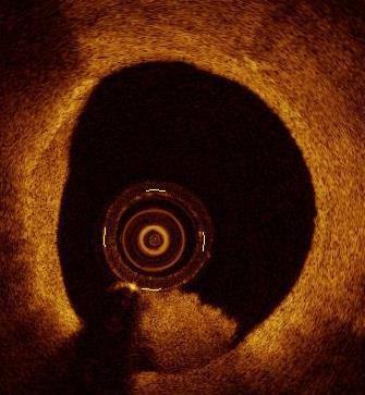
7 Optical Coherence Tomography The new watchful eye of interventional cardiologists.. 7


8 Intravascular Imaging QCA D1 IVUS Axial Resolution: μm Lat. Resolution: μm OCT Axial Resolution: 12-18μm Lat. Resolution: μm


9 OCT vs Other Imaging Modalities OCT IVUS CA MSCT MRI Angioscopy Resolution (µm) Time aspect I Time aspect II Type of scan source Real-time Real-time Real-time Real-time 2-50 sec sec 30 sec IR-light Ultrasound X-Ray X-Ray Magnetic res Visible light Layer Layer Bloodflow Density Density Surface Imaging target

10 Some people think that


11 N Engl J Med 2011; 364:
12 Lesson learnt A quarter of century later, IVUS is a IIB C guideline recommendation for LM. That s all ESC guidelines for myocardial revascularization: Recommendations for specific percutaneous coronary intervention devices and pharmacotherapy IVUS-guided stent implantation may be considered for unprotected left main PCI. IIB C
13 Meta-analysis of Randomized Trials of IVUS vs Angiographic Guided BMS implantation (n=2193 pts) IVUS guidance was associated with significantly lower rate of Angiographic restenosis (22.2% vs. 28.9%; OR 0.64, p=0.02) Repeat revascularization (12.6% vs. 18.4%; OR 0.66, p=0.004) Overall MACE (19.1% vs. 23.1%; OR 0.69, p=0.03) But no significant effect on MI (p=0.51) or mortality (p=0.18). ST was not reported TULIP DIPOL Gaster RESIST SIPS AVID OPTICUS Combined (RE) Combined (FE) Favors IVUS MACE Odds Ratio Favors Non-IVUS Parise et al. Am J Cardiol. 2011;107:374-82
14 Study Year Death HR (95% CI) Weight % P Roy SJ Park SH Kim J Jakabcin JS Kim BE Claessen SH Hur K Ahmed KW Park SL Chen Overall Favors IVUS Favors Non-IVUS Stent Thrombosis 0.81 (0.55, 1.20) 0.39 (0.15, 1.02) 0.21 (0.06, 0.73) 1.50 (0.15, 15.42) 0.58 (0.21, 1.61) 0.74 (0.37, 1.47) 0.49 (0.35, 0.69) 0.49 (0.28, 0.86) 1.56 (0.48, 1.59) 0.55 (0.19, 1.57) 0.59 (0.48, 0.72) P Roy (0.39, 0.89) SJ Park (0.12, 76.85) 0.82 J Jakabcin (0.15, 3.00) 3.82 SH Kim (0.06, 1.28) 3.73 BE Claessen (0.10, 3.51) 2.75 JS Kim (0.04, 2.96) 1.79 SH Hur (0.44, 1.17) KW Park (0.10, 2.68) 2.93 SL Chen (0.05, 0.61) 5.19 Overall 0.58 (0.44, 0.77) Favors IVUS 1 10 Favors Non- IVUS 100 Meta-Analysis of 11 Studies (n=19,619 patients) Compared with angiographyguidance, IVUS-guided DES implantation was associated with a reduced incidence of Death (HR: 0.59, 95% CI: , p<0.001) Stent thrombosis (HR: 0.58, 95% CI: , p<0.0001) Major adverse cardiac events (HR: 0.87, 95% CI: , p=0.008) Zhang et al. Eurointervention 2012;8:855-65

15 In the meta-analysis of 19,619 patients, mortality w a s significantly reduced by 27%... Mortality IVUS Guidance Non IVUS Guidance Zhang et al. EuroIntervention
16 Impact of Intravascular Ultrasound-Guided Percutaneous Coronary Intervention on Long-Term Clinical Outcomes in a Real World Population Hur et al. CCI 2013
17 Impact of intravascular ultrasound guidance in routine percutaneous coronaryintervention for conventional lesions: data from the EXCELLENT trial Park et al. IJC 2012


18 ADAPT-DES Assessment of Dual AntiPlatelet Therapy with Drug-Eluting Stents 8,575 pts prospectively enrolled No clinical or anatomic exclusion criteria Succesful and uncomplicated PCI with 1 noninvestigational DES Pre-specified IVUS vs no IVUS substudy IVUS: 3349 pts No IVUS: 5234 pts Clinical FU at 30 days, 1 year, 2 years Witzenbichler, et al. Circulation, Nov 2013

 (%) How IVUS changed the procedure?")

19 Larger Size of Stent/Balloon Higher Pressure Longer Stent Reason for IVUS Use Document Procedure (26%) Guide and Optimize Procedure (74%) (%) How IVUS changed the procedure? Post Dilation Underexpansion Malapposition Additional Stent Witzenbichler, et al. Circulation Nov 2013
20 IVUS Use and Definite/Probable ST Within 2 Years Definite/Probable ST (%) 2 HR: 0.47 [95% CI: 0.28, 0.80] P = IVUS Not Used 1.16% IVUS Used 0.55% Number at risk: Time in Months IVUS Used IVUS Not Used
![Myocardial Infarction (%) IVUS Use and MI Within 2 Years 10 HR: 0.62 [95% CI: 0.49, 0.77] P < 0.001 5 IVUS Not Used IVUS Used 5.](/docs-images/80/81438247/images/21-1.jpg "59% 3.")
21 Myocardial Infarction (%) IVUS Use and MI Within 2 Years 10 HR: 0.62 [95% CI: 0.49, 0.77] P < IVUS Not Used IVUS Used 5.59% 3.47% Number at risk: Time in Months IVUS Used IVUS Not Used


22 MACE (%) IVUS Use and MACE (Definite/Probable ST, Cardiac Death, MI) Within 2 Years 10 HR: 0.65 [95% CI: 0.54, 0.78] P < IVUS Not Used 7.4% 5 4.9% IVUS Used 0 Number at risk: T ime in Months IVUS Used IVUS Not Used
23 Association of IVUS Use with MACE (Definite/Probable ST, Cardiac Death, MI) in Relation to Index Presentation Event Rate (n) IVUS vs Angio HR [95%CI] P-Value All 4.9% (158) vs 7.5% (373) 0.65 [0.54, 0.78] < STEMI 3.7% (15) vs 6.4% (24) 0.56 [0.29, 1.07] 0.07 NSTEMI/UA 6.1% (82) vs 8.8% (184) 0.68 [0.52, 0.88] Stable CAD 4.2% (61) vs 6.5% (165) 0.63 [0.47, 0.85] Favors IVUS Use Favors Angio Use


24

25 Lesson learnt Intravascular OCT allow s for accurate assessment of vessel structures close to the luminal side. Clinical application is feasible. To date, how ever, the clinical relevance of OCT findings in coronary arteries is unclear and further validation of OCT imaging is mandatory. A decade later, OCT is still a valuable research tool. That s all ESC guidelines for myocardial revascularization: Recom m endations for specific percutaneous coronary intervention devices and pharmacotherapy Optical coherence tomography (OCT) is a light-based modality of intravascular imaging w ith higher spatial resolution than IVUS (15 vs. 100 μm). Its penetration is lower than IVUS but it provides detailed imaging of th e endoluminal borders. At present, OCT is a valuable research tool.


26 Greek mythology of Icarus


27 Lesson learnt Although s o me evidences are em erging In the analysis of matched pair (6 70 patients), mortality was significantly reduced by 5 2 %...


28 Safety and Feasibility of FD-OCT Imaging Imola F et al. EuroIntervention 2010;6: Success 99% Duration 2.1±0.5 min Contrast 49±19 ml VT/VF 0% MACE 0%
29 OCT for Intracoronary Imaging Atherosclerosis Normal Vessel Wall Different Plaque Types Plaque Progression Plaque Rupture Stent Evaluation Neointimal Hyperplasia Pattern of Restenosis Strut Coverage and Apposition Stent thrombosis


30 Tunica Intima A1 B1 C1 A2 B2 C2 i a m i m a a m i
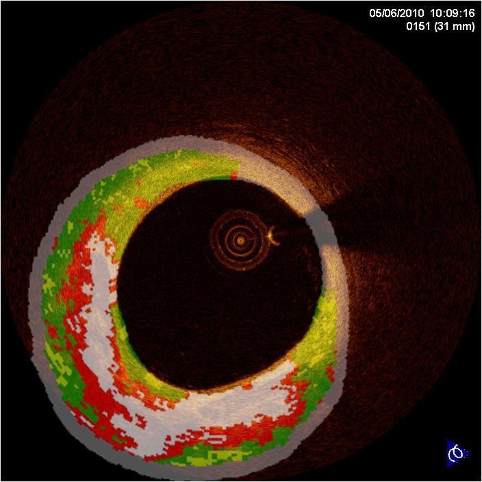
31 Value of Combined Assessment Grayscale IVUS, IVUS-VH and OCT Vulnerable Plaque characteristics OCT IVUS-VH Fibrous cap thickness Detailed surface morphology Visualization of entire plaques Plaque composition Differentiation lipid vs calcium Thin cap fibroma-atheroma TCFA Detection Algorithm?
32 OCT/IVUS VH TCFA Detection Algorithm Plaque thickness >600 µ m, >3 frames Comfortable IBIS 4 AIT AIT No Yes Courtesy: Hector Garcia Confluent C Confluent NC NC >10% NC >10% >10% (V (V H) No Yes >15% >15% FF FF (VH) FF (VH) Fibrous In direct cap <65 contact <65 microns w ith by lumen O by OC CT T PIT PIT Yes No No Yes >10% DC (VH) >10% DC (VH) >10% DC (VH) PI T Yes No Yes No Yes No FC FT Ca FA FA Ca TCFA TCFA FC FT Ca FA FA AIT NC FF DC FC = adaptive intimal thickening = necrotic core = fibrofatty = dense calcium = fibrocalcic Comfortable FT = fibrotic Ca FA = calcium fibroatheroma FA = fibroatheroma Ca TCFA = ca-thin-cap-fibroatheroma TCFA = thin-cap-fibroatheroma


33
34 OCT/IVUS VH TCFA Detection Algorithm Plaque thickness >600 µ m, >3 frames Comfortable IBIS 4 AIT AIT No Yes Courtesy: Hector Garcia Confluent C Confluent NC NC >10% NC >10% >10% (V (V H) No Yes >15% >15% FF FF (VH) FF (VH) Fibrous In direct cap <65 contact <65 microns w ith by lumen O by OC CT T PIT PIT Yes No No Yes >10% DC (VH) >10% DC (VH) >10% DC (VH) PI T Yes No Yes No Yes No FC FT Ca FA FA Ca TCFA TCFA FC FT Ca FA FA AIT NC FF DC FC = adaptive intimal thickening = necrotic core = fibrofatty = dense calcium = fibrocalcic Comfortable FT = fibrotic Ca FA = calcium fibroatheroma FA = fibroatheroma Ca TCFA = ca-thin-cap-fibroatheroma TCFA = thin-cap-fibroatheroma


35 A1 Fibrous plaque A2 A3 Fibrocalcific plaque B1 B2 B3
 No Yes >15% >15% FF FF (VH)")
 >10% DC (VH) >10% DC (VH) PI T Yes No Yes No Yes No FC FT Ca FA FA Ca TCFA TCFA FC FT Ca FA FA AIT NC FF DC")

36 OCT/IVUS VH TCFA Detection Algorithm Hector Garcia Plaque thickness >600 µ m, >3 frames Comfortable IBIS 4 AIT AIT No Yes Courtesy: Hector Garcia Confluent C Confluent NC NC >10% NC >10% >10% (V (V H) No Yes >15% >15% FF FF (VH) FF (VH) Fibrous In direct cap <65 contact <65 microns w ith by lumen O by OC CT T PIT PIT Yes No No Yes >10% DC (VH) >10% DC (VH) >10% DC (VH) PI T Yes No Yes No Yes No FC FT Ca FA FA Ca TCFA TCFA FC FT Ca FA FA AIT NC FF DC FC = adaptive intimal thickening = necrotic core = fibrofatty = dense calcium = fibrocalcic Comfortable FT = fibrotic Ca FA = calcium fibroatheroma FA = fibroatheroma Ca TCFA = ca-thin-cap-fibroatheroma TCFA = thin-cap-fibroatheroma


37 Thick Cap Fibroatheroma Thin Cap Fibroatheroma (TCFA)

38 Broad Inclusion Criteria: Age > 18 years ECG and Chest pain Primary PCI within 24 hours Screening of all incoming STEMI patients Recanalisation, aspiration, intra- and extracoronary blood sampling Imaging Candidate if -Hemodynamic stable -Creatinine Clearance >50ml/min -Age <90 -OCT/IVUS feasible -No stent at the site of infarct lesion Comfortable AMI IBIS 4 n=1100 1:1 R IBIS 4 n=100 R 1:1 Principal Investigators Lorenz Räber Hector Garcia Garcia Stephan Windecker DES-Biomatrix N=500 BMS-Gazelle N=500 DES-Biomatrix N=50 BMS-Gazelle N=50 3-vessel IVUS and OCT at baseline Participating sites Bern (60) Copenhagen (21) Geneva (13) Lugano (6) Zurich (3) 30 day telephone (n=1100) 12 month visit or telephone 2 year telephone Final 5 year telephone Rosuvastatin 40mg 3-vessel IVUS and OCT at 13 months
39 Comfortable IBIS 4 study 3 vessel imaging using IVUS Virtual Histology: - Culprit vessel: Post-treatment imaging of the stented segment and distal segment - Non-culprit vessel: >40 mm of proximal coronary artery
40 Comfortable IBIS 4 study 3 vessel imaging using IVUS Virtual Histology: - Culprit vessel: Post-treatment imaging of the stented segment and distal segment - Non-culprit vessel: >40 mm of proximal coronary artery Primary Objective To determine the effects of rosuvastatin on compositional measures of coronary plaque in a non-intervened coronary segment. Specifically, the change from baseline in Virtual Histology TM necrotic core volume at the end of week 52.
41 Comfortable IBIS 4 study 3 vessel imaging using OCT: - Culprit vessel: Post-treatment imaging of the stented segment and distal segment - Non-culprit vessel: >40 mm of proximal coronary artery
42 Comfortable IBIS 4 study 3 vessel imaging using OCT: - Culprit vessel: Post-treatment imaging of the stented segment and distal segment - Non-culprit vessel: >40 mm of proximal coronary artery Secondary Objective To determine the effects of rosuvastatin on fibrous cap thickness of coronary plaque in a nonintervened coronary segment. Specifically, the change from baseline in OCT cap thickness at the end of week 52.

43


44


45

46 IVUS Stent underexpansion Geographical miss OCT (Minor) findings not seen on IVUS Malapposition Tissue protrusion Edge dissections
47 What are the intravascular imaging modalities in interventional cardiology: current and future technologies Current Future Intravascular ultrasound (IVUS) Intravascular ultrasound (IVUS) High-frequency Focused acoustic tomography (FACT) Grey Scale (20, 4 0, 45 Mhz) Radiofrequency backscattering (Virtual histology, Palpography, Integrated backscatter IVUS and imap) Optical coherence tomography 2 D (10 0, 160, 180 frames / s e c ) 3 D (offli n e / on line) Hemodynamic parameter derived from 3 D imaging NIR infrared spectroscopy Combined IVUS and NIRS (FDA approved) Intravascular imaging combined w ith interventional devices Optical coherence tomography 3 D OCT w ith tissue characterization Micro OCT Super fast OCT (10 0 m m / s ) Hybrid intravascular imaging OCT + NIR OCT + IVUS IVUS + TRFS IVUS + IVPA


48 #current technology: Hemodynamic parameter derived from 3 D imaging Fusion of 3D-QCA and OCT Tu et al. Circ Interv. 2013


49 #Future technology: Intravascular imaging combined w ith interventional devices Atherectomy with OCT Avinger Ocelot IVUS guided subintimal reentry device Forward looking IVUS ~5 mm radius ~3.5 mm forward

50 MGH Courtesy of Prof. Tearney Artery wall Lipid Calcium Macrophages Stent Guidewire


51 1 A. Proximal recrossing 2 A. Distal recrossing * * 1 B. Results after KB 2 B. Results after KB * *


52


53 Micro OCT Leukocytes tethered to the endothelium Cholesterol crystals and Necrotic Core Leukocytes Cluster at the Site of Polymer Fracture
-IVUS, VH-IVUS, NIRS, OCT, IVPA, IV- MRI")

54 Intravascular imaging allowed us for the first time to study in vivo plaque morphology and atherosclerotic evolution Today numerous invasive modalities are available: integrated backscatter analysis (IB)-IVUS, VH-IVUS, NIRS, OCT, IVPA, IV- MRI spectroscopy, IV-MRI, Raman spectroscopy and time resolved fluorescence spectroscopic (TRFS) that permit imaging of the plaque IB-IVUS, VH-IVUS, NIRS, OCT and CTCA have been used in clinical studies and allowed detection of plaque features related with future adverse events
55
56 Intravascular ultrasound (IVUS). Definitely Beneficial. IVUS is an accurate method for determining optimal stent deployment (complete stent expansion and apposition and lack of edge dissection or other complications after implantation), and the size of the vessel undergoing stent implantation. Probably Beneficial. IVUS can be used to appraise the significance of LMCA stenosis and, employing a cutoff MLA¼ 6 mm2, assess whether revascularization is warranted. Possibly Beneficial. IVUS can be useful for the assessment of plaque morphology. No Proven Value/Should be Discouraged. IVUS measurements for determination of non-lmca lesion severity should not be relied upon, in the absence of additional functional evidence, for recommending revascularization.
57 Optical Coherence Tomography (OCT). Probably Beneficial. Determination of optimal stent deployment (sizing, apposition, and lack of edge dissection), with improved resolution compared with IVUS. Possibly Beneficial. OCT can be useful for the assessment of plaque morphology. No Proven Value/Should be Discouraged. OCT should not be performed to determine stenosis functional significance.


58
Optical Coherence Tomography for Intracoronary Imaging
 Optical Coherence Tomography for Intracoronary Imaging Lorenz Räber Stephan Windecker Department of Cardiology Swiss Cardiovascular Center and Clinical Trials Unit Bern Bern University Hospital, Switzerland
Imaging Atheroma The quest for the Vulnerable Plaque
 Imaging Atheroma The quest for the Vulnerable Plaque P.J. de Feijter 1. Department of Cardiology 2. Department of Radiology Coronary Heart Disease Remains the Leading Cause of Death in the U.S, Causing
The PROSPECT Trial. A Natural History Study of Atherosclerosis Using Multimodality Intracoronary Imaging to Prospectively Identify Vulnerable Plaque
 The PROSPECT Trial Providing Regional Observations to Study Predictors of Events in the Coronary Tree A Natural History Study of Atherosclerosis Using Multimodality Intracoronary Imaging to Prospectively
INSIDE INFORMATION YOU CAN T IGNORE
 INSIDE INFORMATION YOU CAN T IGNORE Volcano, the Volcano logo and SyncVision are registered trademarks of Volcano Corporation. All other trademarks set-forth are properties of their respective owners.
Invasive Coronary Imaging Modalities for Vulnerable Plaque Detection
 Invasive Coronary Imaging Modalities for Vulnerable Plaque Detection Gary S. Mintz, MD Cardiovascular Research Foundation New York, NY Greyscale IVUS studies have shown Plaque ruptures do not occur randomly
The PROSPECT Trial. A Natural History Study of Atherosclerosis Using Multimodality Intracoronary Imaging to Prospectively Identify Vulnerable Plaque
 The PROSPECT Trial Providing Regional Observations to Study Predictors of Events in the Coronary Tree A Natural History Study of Atherosclerosis Using Multimodality Intracoronary Imaging to Prospectively
Imaging Overview for Vulnerable Plaque: Data from IVUS Trial and An Introduction to VH-IVUS Imgaging
 Imaging Overview for Vulnerable Plaque: Data from IVUS Trial and An Introduction to VH-IVUS Imgaging Gary S. Mintz,, MD Cardiovascular Research Foundation New York, NY Today, in reality, almost everything
CLINICAL APPLICATIONS OF OPTICAL COHERENCE TOMOGRAPHY. Konstantina P. Bouki, FESC 2 nd Department of Cardiology General Hospital Of Nikea, Pireaus
 CLINICAL APPLICATIONS OF OPTICAL COHERENCE TOMOGRAPHY Konstantina P. Bouki, FESC 2 nd Department of Cardiology General Hospital Of Nikea, Pireaus OPTICAL COHERENCE TOMOGRAPHY (OCT) IVUS and OCT IVUS OCT
Can IVUS Define Plaque Features that Impact Patient Care?
 Can IVUS Define Plaque Features that Impact Patient Care? A Pichard L Satler, K Kent, R Waksman, W Suddath, N Bernardo, N Weissman, M Angelo, D Harrington, J Lindsay, J Panza. Washington Hospital Center
Cardiovascular Research Foundation and Columbia University Medical Center, New York.
 Virtual Histology Intravascular Ultrasound Analysis of Non-culprit Attenuated Plaques Detected by Grayscale Intravascular Ultrasound in Patients with Acute Coronary Syndromes Xiaofan Wu, Akiko Maehara,
OCT; Comparative Imaging Results with IVUS, VH and Angioscopy
 OCT; Comparative Imaging Results with IVUS, VH and Angioscopy Takashi Akasaka, M.D. Department of Cardiovascular Medicine Wakayama, Japan Comparison among coronary imaging techniques OCT IVUS MRI CAG Angioscopy
Clinical Value of OCT. Guidance for Coronary Stenting. Giulio Guagliumi, MD
 Clinical Value of OCT Guidance for Coronary Stenting Giulio Guagliumi, MD 100 % Endovascular Imaging Indications of use 87.5 % 75 % 57.5 % 50 % 45 % 25 % 15 % 0 Lesion morphology Stent optimization Lesion
OCT Findings: Lesson from Stable vs Unstable Plaques
 ANGIOPLASTY SUMMIT TCTAP 2010 Imaging Workshop OCT Findings: Lesson from Stable vs Unstable Plaques Giulio Guagliumi MD Ospedali Riuniti di Bergamo, Italy DISCLOSURE OF FINANCIAL INTERESTS Consultant Boston
Intracoronary Imaging For Complex PCI A Pichard, L Satler, Ron Waksman, I Ben-Dor, W Suddath, N Bernardo, D Harrington.
 Intracoronary Imaging For Complex PCI A Pichard, L Satler, Ron Waksman, I Ben-Dor, W Suddath, N Bernardo, D Harrington. Medstar Washington Hospital Center Washington, DC Conflict of Interest None for this
IVUS vs FFR Debate: IVUS-Guided PCI
 IVUS vs FFR Debate: IVUS-Guided PCI Gary S. Mintz, MD Cardiovascular Research Foundation New York, NY Disclosure Statement of Financial Interest Within the past 12 months, I have had a financial interest/arrangement
Assessment of Vulnerable Plaque by IVUS and VH-IVUS
 Assessment of Vulnerable Plaque by IVUS and VH-IVUS Akiko Maehara, MD Director of Intravascular Imaging & Physiology Core Laboratories Associate Director of MRI/MDCT Core Laboratory Cardiovascular Research
Plaque Characteristics in Coronary Artery Disease. Chourmouzios Arampatzis MD, PhD, FESC
 Plaque Characteristics in Coronary Artery Disease Chourmouzios Arampatzis MD, PhD, FESC Disclosure Statement of Financial Interest Regarding this Presentation NONE Atherosclerosis Model proposed by Stary
Integrated Use of IVUS and FFR for LM Stenting
 Integrated Use of IVUS and FFR for LM Stenting Gary S. Mintz, MD Cardiovascular Research Foundation Four studies have highlighted the inaccuracy of angiography in the assessment of LMCA disease Fisher
2yrs 2-6yrs >6yrs BMS 0% 22% 42% DES 29% 41% Nakazawa et al. J Am Coll Cardiol 2011;57:
 Pathology of In-stent Neoatherosclerosis in BMS and DES 197 BMS, 103 SES, and 106 PES with implant duration >30 days The incidence of neoatherosclerosis was significantly greater in DES (31%) than BMS
Κλινική Χρήση IVUS και OCT PERIKLIS A. DAVLOUROS ASSOCIATE PROFESSOR OF CARDIOLOGY INVASIVE CARDIOLOGY & CONGENITAL HEART DISEASE
 Κλινική Χρήση IVUS και OCT PERIKLIS A. DAVLOUROS ASSOCIATE PROFESSOR OF CARDIOLOGY INVASIVE CARDIOLOGY & CONGENITAL HEART DISEASE Conflict of interest None to declare While IVUS is the most used intravascular
Intervention: How and to which extent is technology helping us?
 Cardiological Society of India Congress 12th February 2016 Chennai, India Intervention: How and to which extent is technology helping us? SIMONE BISCAGLIA MD CARDIOVASCULAR INSTITUTE, FERRARA, ITALY Introduction
Optical Coherence Tomography
 Optical Coherence Tomography Disclosure Information Demetrius Lopes MD The following relationships exist related to this presentation: University Grant/Research Support: Rush University Industry Grant
as a Mechanism of Stent Failure
 In-Stent t Neoatherosclerosis e osc e os s as a Mechanism of Stent Failure Soo-Jin Kang MD., PhD. University of Ulsan College of Medicine, Heart Institute Asan Medical Center, Seoul, Korea Disclosure I
Added Value of Invasive Coronary Imaging for Plaque Rupture and Erosion
 Assessment of Coronary Plaque Rupture and Erosion Added Value of Invasive Coronary Imaging for Plaque Rupture and Erosion Yukio Ozaki, MD, PhD, FACC, FESC Cardiology Dept., Fujita Health Univ. Toyoake,
Integrating IVUS, FFR, and Noninvasive Imaging to Optimize Outcomes. Gary S. Mintz, MD Cardiovascular Research Foundation
 Integrating IVUS, FFR, and Noninvasive Imaging to Optimize Outcomes Gary S. Mintz, MD Cardiovascular Research Foundation COURAGE Nuclear Substudy (n=314) Death/MI according the residual ischemia (SPECT)
Invasive Imaging (IVUS, VH-IVUS, and OCT): How I Implement into My
: How I Implement into My") Invasive Imaging (IVUS, VH-IVUS, and OCT): How I Implement into My Practice Gary S. Mintz, MD Cardiovascular Research Foundation Modalities FFR IVUS (with or without VH, imap, or IB-IVUS) OCT NIRS (with
Invasive Imaging (IVUS, VH-IVUS, and OCT): How I Implement into My Practice Gary S. Mintz, MD Cardiovascular Research Foundation Modalities FFR IVUS (with or without VH, imap, or IB-IVUS) OCT NIRS (with
State of the Art. Advances in Cardiovascular Imaging. ESC Congres Stockholm September 1, 2010 Frank E. Rademakers, MD, PhD, FESC
 State of the Art Advances in Cardiovascular Imaging ESC Congres Stockholm September 1, 2010 Frank E. Rademakers, MD, PhD, FESC Coronary Artery Disease Content Patho Physiology Imaging requirements Economical
Post PCI functional testing and imaging: case based lessons from FFR React
 Post PCI functional testing and imaging: case based lessons from FFR React Joost Daemen, MD, PhD, FESC Optics in Cardiology 2018 April 21st, 2018 10.15 10.30h Disclosure Statement of Financial Interest
Evaluation of Intermediate Coronary lesions: Can You Handle the Pressure? Jeffrey A Southard, MD May 4, 2013
 Evaluation of Intermediate Coronary lesions: Can You Handle the Pressure? Jeffrey A Southard, MD May 4, 2013 Disclosures Consultant- St Jude Medical Boston Scientific Speaker- Volcano Corporation Heart
A Novel Low Pressure Self Expanding Nitinol Coronary Stent (vprotect): Device Design and FIH Experience
: Device Design and FIH Experience") A Novel Low Pressure Self Expanding Nitinol Coronary Stent (vprotect): Device Design and FIH Experience Juan F. Granada, MD Medical Director, Skirball Center for Cardiovascular Research The Cardiovascular
A Novel Low Pressure Self Expanding Nitinol Coronary Stent (vprotect): Device Design and FIH Experience Juan F. Granada, MD Medical Director, Skirball Center for Cardiovascular Research The Cardiovascular
Technical considerations in the Treatment of Left Main Lesions Ioannis Iakovou, MD, PhD
 Technical considerations in the Treatment of Left Main Lesions Ioannis Iakovou, MD, PhD Onassis Cardiac Surgery Center, Athens, Greece Critical issues in LM PCI Anatomic variability Techniques Variability
Effect of Intravascular Ultrasound- Guided vs. Angiography-Guided Everolimus-Eluting Stent Implantation: the IVUS-XPL Randomized Clinical Trial
 Effect of Intravascular Ultrasound- Guided vs. Angiography-Guided Everolimus-Eluting Stent Implantation: the IVUS-XPL Randomized Clinical Trial Myeong-Ki Hong, MD. PhD on behalf of the IVUS-XPL trial investigators
Béla MERKELY MD, PhD, DSc, FESC. Stent thrombosis: patophysiology, predisposing factors, definition, classification, prevention and treatment
 Semmelweis University Heart Center Budapest, Hungary Béla MERKELY MD, PhD, DSc, FESC Stent thrombosis: patophysiology, predisposing factors, definition, classification, prevention and treatment 10th Interventional
대한심장학회춘계학술대회 Satellite Symposium
 대한심장학회춘계학술대회 Satellite Symposium Coronary Plaque Regression and Compositional Changes by Lipid-Lowering Therapy: IVUS Substudy in Livalo (Pitavastatin) in Acute Myocardial Infarction Study (LAMIS) Livalo
Intravascular Ultrasound
 May 2008 Beth Israel Deaconess Medical Center Harvard Medical School Intravascular Ultrasound Matthew Altman, HMS III Gillian Lieberman, MD BIDMC Department of Radiology Presentation Overview 1. Patient
Cover Page. Author: Wang, Ancong Title: Automatic quantification of intravascular optical coherence tomography Issue Date:
 Cover Page The handle http://hdl.handle.net/1887/29690 holds various files of this Leiden University dissertation Author: Wang, Ancong Title: Automatic quantification of intravascular optical coherence
malapposition assessed by OCT
 Stent t coverage and malapposition assessed by OCT Myeong-Ki Hong, M.D. Ph D Professor of Medicine Division of Cardiology, Severance Cardiovascular Hospital Yonsei University College of Medicine, Seoul,
Left main coronary artery (LMCA): The proximal segment
: The proximal segment") Anatomy and Pathology of Left main coronary artery G Nakazawa Tokai Univ. Kanagawa, Japan 1 Anatomy Difinition Left main coronary artery (LMCA): The proximal segment RCA AV LAD LM LCX of the left coronary
Anatomy and Pathology of Left main coronary artery G Nakazawa Tokai Univ. Kanagawa, Japan 1 Anatomy Difinition Left main coronary artery (LMCA): The proximal segment RCA AV LAD LM LCX of the left coronary
PCI for Left Main Coronary Artery Stenosis. Jean Fajadet Clinique Pasteur, Toulouse, France
 PCI for Left Main Coronary Artery Stenosis Jean Fajadet Clinique Pasteur, Toulouse, France Athens, October 19, 2018 Left Main Coronary Artery Disease Significant unprotected left main coronary artery disease
Assessment of plaque morphology by OCT in patients with ACS
 Assessment of plaque morphology by OCT in patients with ACS Takashi Akasaka, M.D. Department of Cardiovascular Medicine Wakayama, Japan Unstable plaque Intima Lipid core Plaque rupture and coronary events
Can We Identify Vulnerable Patients & Vulnerable Plaque?
 Can We Identify Vulnerable Patients & Vulnerable Plaque? We Know Enough to Treat High-Risk Lesions? Takashi Akasaka, MD, PhD Department of Cardiovascular Medicine, Japan Disclosure Statement of Financial
Insights in Thrombosis and In-Stent Restenosis
 Clinical Value of OCT Insights in Thrombosis and In-Stent Restenosis Fernando Alfonso MD, PhD, FESC Interventional Cardiology. Cardiovascular Institute. Clinico San Carlos University Hospital. Madrid.
Que nos puede aportar el OCT intracoronario
 XXXI Jornadas SOLACI. 10ª Región CONOSUR LIIIº Congreso Chileno de Cardiología y Cirugía Cardiovascular Hotel Patagónico. Puerto Varas. Chile (30 Nov 1 Dic 2016) Que nos puede aportar el OCT intracoronario
1st Department of Cardiology, University of Athens, Hippokration Hospital, Athens, Greece
 Konstantinos Toutouzas, Maria Riga, Antonios Karanasos, Eleftherios Tsiamis, Andreas Synetos, Maria Drakopoulou, Chrysoula Patsa, Georgia Triantafyllou, Aris Androulakis, Christodoulos Stefanadis 1st Department
Evolution In Interventional Cardiology. Jawed Polad Jeroen Bosch Hospital s-hertogenbosch The Netherlands
 Evolution In Interventional Cardiology Jawed Polad Jeroen Bosch Hospital s-hertogenbosch The Netherlands 25 November 2010 Coronary Atherosclerosis Timeline in interventional cardiology Indications for
Carotid Intravascular Imaging Technique and Indication
 Nurse and Technician Forum Carotid Intravascular Imaging Technique and Indication Gianmarco de Donato Assistant Professor Vascular and Endovascular Surgery University of Siena - Italy Disclosure Speaker
Quick guide. Core. precision guided therapy system
 Quick guide Core precision guided therapy system The Philips Volcano imaging system should only be operated by trained personnel. The following information is presented for your convenience and is not
Usefulness of OCT during coronary intervention
 Usefulness of OCT during coronary intervention Takashi Akasaka, M.D. Department of Cardiovascular Medicine Wakayama, Japan Predictors at 12 Months of Stent Thrombosis and Target Lesion Revascularization
PCI for Left Anterior Descending Artery Ostial Stenosis
 PCI for Left Anterior Descending Artery Ostial Stenosis Why do you hesitate PCI for LAD ostial stenosis? LAD Ostial Lesion Limitations of PCI High elastic recoil Involvement of the distal left main coronary
New Insight about FFR and IVUS MLA
 New Insight about FFR and IVUS MLA Can IVUS MLA Predict FFR
Left Main Intervention: Will it become standard of care?
 Left Main Intervention: Will it become standard of care? David Cox, MD FSCAI, FACC Director, Interventional Cardiology Research Associate Director, Cardiac Cath Lab Lehigh Valley Health Network Allentown,
IVUS Analysis. Myeong-Ki. Hong, MD, PhD. Cardiac Center, Asan Medical Center University of Ulsan College of Medicine, Seoul, Korea
 IVUS Analysis Myeong-Ki Hong, MD, PhD Cardiac Center, Asan Medical Center University of Ulsan College of Medicine, Seoul, Korea Intimal disease (plaque) is dense and will appear white Media is made of
PCI for LMCA lesions A Review of latest guidelines and relevant evidence
 HCS Working Group Seminars Met Hotel, Thursday 14 th February 2013 PCI for LMCA lesions A Review of latest guidelines and relevant evidence Vassilis Spanos Interventional Cardiologist, As. Director 3 rd
Culprit Lesion Remodeling and Long-term (> 5years) Prognosis in Patients with Acute Coronary Syndrome
 Prognosis in Patients with Acute Coronary Syndrome") Culprit Lesion Remodeling and Long-term (> 5years) Prognosis in Patients with Acute Coronary Syndrome Hiroyuki Okura*, MD; Nobuya Matsushita**,MD Kenji Shimeno**, MD; Hiroyuki Yamaghishi**, MD Iku Toda**,
Culprit Lesion Remodeling and Long-term (> 5years) Prognosis in Patients with Acute Coronary Syndrome Hiroyuki Okura*, MD; Nobuya Matsushita**,MD Kenji Shimeno**, MD; Hiroyuki Yamaghishi**, MD Iku Toda**,
OCT GUIDED TREATMENT OF CALCIFIED LESIONS RICHARD SHLOFMITZ, MD CHAIRMAN OF DEPT. OF CARDIOLOGY ST. FRANCIS HOSPITAL ROSLYN, NEW YORK
 OCT GUIDED TREATMENT OF CALCIFIED LESIONS RICHARD SHLOFMITZ, MD CHAIRMAN OF DEPT. OF CARDIOLOGY ST. FRANCIS HOSPITAL ROSLYN, NEW YORK Disclosure Statement of Financial Interest Within the past 12 months,
Analysis of neointimal coverage after silolimus-eluting stent implantation using optical coherence tomography.
 Analysis of neointimal coverage after silolimus-eluting stent implantation using optical coherence tomography. Division of Cardiology, Department of Internal Medicine, Fasculty of Medicine, Kinki University,
DESolve NX Trial Clinical and Imaging Results
 DESolve NX Trial Clinical and Imaging Results Alexandre Abizaid, MD, PhD, Instituto Dante Pazzanese, Sao Paulo, Brazil On behalf of the DESolve Nx Trial Investigators Please refer to the TCT2014 App or
HCS Working Group Seminars Macedonia Pallas Hotel, Friday 21 st February Drug-eluting stents Are they all equal?
 HCS Working Group Seminars Macedonia Pallas Hotel, Friday 21 st February 2014 Drug-eluting stents Are they all equal? Vassilis Spanos Interventional Cardiologist, As. Director 3 rd Cardiology Clinic Euroclinic
Perspective of LM stenting with Current registry and Randomized Clinical Data
 Asian Pacific TCT Perspective of LM stenting with Current registry and Randomized Clinical Data Patrick W. Serruys MD PhD Yoshinobu Onuma MD Seung-Jung Park MD, PhD 14:48-15:00, 2009 Symposium Arena, Level
CPIS So-Yeon Choi, MD., PhD. Department of Cardiology Ajou University School of MedicineSuwon, Korea
 So-Yeon Choi, MD., PhD. Department of Cardiology Ajou University School of MedicineSuwon, Korea Coronary Artery Imaging The ideal coronary imaging technology would be capable of identifying not only vessel
Left Main Intervention: Where are we in 2015?
 Left Main Intervention: Where are we in 2015? David A. Cox, MD FSCAI Director, Cardiology Research Associate Director, Cardiac Cath Lab Lehigh Valley Health Network Allentown, PA Fall Fellows Course Laa
OCT guidance for distal LM lesions
 OCT guidance for distal LM lesions FRANCESCO BURZOTTA INSTITUTE OF CARDIOLOGY CATHOLIC UNIVERSITY OF THE SACRED HEART ROME, ITALY LM suitability for OCT At FU in stented LM Parodi G et al. Eurointervention
Gary S. Mintz,, MD. IVUS Observations in Acute (vs Chronic) Coronary Artery Disease: Structure vs Function
 Coronary Artery Disease: Structure vs Function") Gary S. Mintz,, MD IVUS Observations in Acute (vs Chronic) Coronary Artery Disease: Structure vs Function Important IVUS Observations: Remodeling Originally used (first by Glagov) ) to explain atherosclerosis
Gary S. Mintz,, MD IVUS Observations in Acute (vs Chronic) Coronary Artery Disease: Structure vs Function Important IVUS Observations: Remodeling Originally used (first by Glagov) ) to explain atherosclerosis
Catch-up Phenomenon: Insights from Pathology
 Catch-up Phenomenon: Insights from Pathology Michael Joner, MD CVPath Institute Inc. Gaithersburg, MD USA Path Lessons learned from the BMS and DES (1 st Gen) era Neointimal Thickness [mm] In Stent Re
New Generation Drug- Eluting Stent in Korea
 New Generation Drug- Eluting Stent in Korea Young-Hak Kim, MD, PhD Department of Cardiology, Asan Medical Center, University of Ulsan College of Medicine, Seoul, Korea Purpose To briefly introduce the
Coronary Artery Thermography
 Coronary Artery Thermography The 10th Anniversary, Interventional Vascular Therapeutics Angioplasty Summit 2005 TCT Asia Pacific Christodoulos Stefanadis Professor of Cardiology Athens Medical School In
LM stenting - Cypher
 LM stenting - Cypher Left main stenting with BMS Since 1995 Issues in BMS era AMC Restenosis and TLR (%) 3 27 TLR P=.282 Restenosis P=.71 28 2 1 15 12 Ostium 5 4 Shaft Bifurcation Left main stenting with
Stent Fracture and Longitudinal Compression on CT Angiography between the
 2014 ASCI Stent Fracture and Longitudinal Compression on CT Angiography between the First- and New-Generation Drug-Eluting Stent Mi Sun Chung, Dong Hyun Yang,Young-Hak Kim, Jae-Hyung Roh, Joon-Won Kang,
Review Article Optical Coherence Tomography Imaging in Acute Coronary Syndromes
 SAGE-Hindawi Access to Research Cardiology Research and Practice Volume 2011, Article ID 312978, 7 pages doi:10.4061/2011/312978 Review Article Optical Coherence Tomography Imaging in Acute Coronary Syndromes
Side Branch Occlusion
 Side Branch Occlusion Mechanism, Outcome, and How to avoid it From COBIS II Registry Hyeon-Cheol Gwon Cardiac&Vascular Center, Samsung Medical Center Sungkyunkwan University School of Medicine SB occlusion
Percutaneous Intervention of Unprotected Left Main Disease
 Percutaneous Intervention of Unprotected Left Main Disease Technical feasibility and Clinical outcomes Seung-Jung Park, MD, PhD, FACC Professor of Internal Medicine Asan Medical Center, Seoul, Korea Unprotected
Drug eluting stents From revolution to evolution. Current limitations
 Drug eluting stents From revolution to evolution Current limitations Eric Eeckhout Centre Hospitalier Universitaire Vaudois Lausanne - Switzerland eric.eeckhout@chuv.ch Overview Historical perspective
Basics of Angiographic Interpretation Analysis of Angiography
 Basics of Angiographic Interpretation Analysis of Angiography Young-Hak Kim, MD, PhD Cardiac Center, University of Ulsan College of Medicine, Seoul, Korea What made us nervous Supervisors Stent Contrast
Prognostic Value of Gated Myocardial Perfusion SPECT
 Current Use of IVUS & FFR George D. Dangas, MD, PhD, FACC, FSCAI Professor of Medicine Mount Sinai School of Medicine Prognostic Value of Gated Myocardial Perfusion SPECT 0.6% / year, Cardiac Death and
Machine Learning in Precision Medicine Coronary Health Prediction - Cardiac Events (Atherosclerosis) - Heart Transplant (Vasculopathy)
 - Heart Transplant (Vasculopathy)") Machine Learning in Precision Medicine Coronary Health Prediction - Cardiac Events (Atherosclerosis) - Heart Transplant (Vasculopathy) M. Sonka + IIBI, Charles University, IKEM, CKTCH The University of
Machine Learning in Precision Medicine Coronary Health Prediction - Cardiac Events (Atherosclerosis) - Heart Transplant (Vasculopathy) M. Sonka + IIBI, Charles University, IKEM, CKTCH The University of
Upgrade of Recommendation
 Challenges in LM PCI Decision-making process for stenting Young-Hak Kim, MD, PhD, Heart Institute, University of Ulsan College of Medicine Asan Medical Center, Seoul, Korea Upgrade of Recommendation for
RESTENOSIS Facing up to the problem
 RESTENOSIS Facing up to the problem Petr Kala University Hospital Brno Czech Republic ESC 2011, Paris Disclosure Scientific Advisory Boards or Education presentations fee Abbott, Boston Scientific, Cordis
DES In-stent Restenosis
 DES In-stent Restenosis Roxana Mehran, MD Columbia University Medical Center The Cardiovascular Research Foundation DES Restenosis Mechanisms Predictors Morphological patterns Therapy approach Mechanisms
Αγγειοπλαστική σε Eπαναστενωτικές Bλάβες
 Αγγειοπλαστική σε Eπαναστενωτικές Bλάβες Βάιος Π. Τζίφος Δ/ντής Γ Καρδιολογικής Κλινικής - Επεμβατικής Καρδιολογίας. Ερρίκος Ντυνάν HC The Mehran s Classification for BMS-ISR Prognostic Value Pattern (1)
FFR Incorporating & Expanding it s use in Clinical Practice
 FFR Incorporating & Expanding it s use in Clinical Practice Suleiman Kharabsheh, MD Consultant Invasive Cardiology Assistant professor, Alfaisal Univ. KFHI - KFSHRC Concept of FFR Maximum flow down a vessel
Between Coronary Angiography and Fractional Flow Reserve
 Visual-Functional Mismatch Between Coronary Angiography and Fractional Flow Reserve Seung-Jung Park, MD., PhD. University of Ulsan, College of Medicine Asan Medical Center, Seoul, Korea Visual - Functional
Dynamic Nature of Nonculprit Coronary Artery Lesion Morphology in STEMI
 JACC: CARDIOVASCULAR IMAGING VOL. 6, NO. 1, 2013 2013 BY THE AMERICAN COLLEGE OF CARDIOLOGY FOUNDATION ISSN 1936-878X/$36.00 PUBLISHED BY ELSEVIER INC. http://dx.doi.org/10.1016/j.jcmg.2012.08.010 Dynamic
Medical sciences 1 (2017) 1 9
 1 9") Medical sciences 1 (2017) 1 9 TISSUE CHARACTERISTICS OF CULPRIT CORONARY LESIONS IN ACUTE CORONARY SYNDROME AND TARGET CORONARY LESIONS IN STABLE ANGINA PECTORIS: VIRTUAL HISTOLOGY AND INTRAVASCULAR ULTRASOUND
Medical sciences 1 (2017) 1 9 TISSUE CHARACTERISTICS OF CULPRIT CORONARY LESIONS IN ACUTE CORONARY SYNDROME AND TARGET CORONARY LESIONS IN STABLE ANGINA PECTORIS: VIRTUAL HISTOLOGY AND INTRAVASCULAR ULTRASOUND
DID OCT change our experience on coronary arteries?
 DID OCT change our experience on coronary arteries? Istanbul June 2012 F Prati San Giovanni Hospital, Rome Rome Heart Research Use imaging modalities to.. Avoid useless procedures! MLA of 2.0 mm 2 best
IVUS Assessment of the Mechanism of In-stent Restenosis? Gary S. Mintz, MD Cardiovascular Research Foundation
 IVUS Assessment of the Mechanism of In-stent Restenosis? Gary S. Mintz, MD Cardiovascular Research Foundation SURE Trial: Restenosis in non-stented lesions Average of the two image slices with the smallest
FFR and intravascular imaging, which of which?
 FFR and intravascular imaging, which of which? Ayman Khairy MD, PhD, FESC Associate professor of Cardiovascular Medicine Vice Director of Assiut University Hospitals Assiut, Egypt Diagnostic assessment
OCT in the Evaluation of Vascular Healing Following DES Implantation: Will It Be a Helpful Tool to Reduce Stent Thrombosis?
 OCT in the Evaluation of Vascular Healing Following DES Implantation: Will It Be a Helpful Tool to Reduce Stent Thrombosis? Juan F. Granada, MD Medical Director, Skirball Center for Cardiovascular Research
IVUS-Guided d Provisional i Stenting: Plaque or Carina Shift. Soo-Jin Kang, MD., PhD.
 Left Main and Bifurcation Summit IVUS-Guided d Provisional i Stenting: ti Plaque or Carina Shift Soo-Jin Kang, MD., PhD. Department of Cardiology, University of Ulsan College of Medicine Asan Medical Center,
How to evaluate heart disease - Do we need new tools Focus on myocardial circulation
 How to evaluate heart disease - Do we need new tools Focus on myocardial circulation Alf Inge Larsen Professor department of clinical science University of Bergen Head of Cardiovascular Research Group
Neointimal coverage of bare-metal and sirolimuseluting stents evaluated with optical coherence tomography
 Neointimal coverage of bare-metal and sirolimuseluting stents evaluated with optical coherence tomography B X Chen, F Y Ma, W Luo, J H Ruan, W L Xie, X Z Zhao, S H Sun, X M Guo, F Wang, T Tian, X W Chu
EXCEL vs. NOBLE: How to Treat Left Main Disease in 2017 AATS International Cardiovascular Symposium December 8-9, 2017
 EXCEL vs. NOBLE: How to Treat Left Main Disease in 2017 AATS International Cardiovascular Symposium December 8-9, 2017 Igor F. Palacios, MD Director of Interventional Cardiology Professor of Medicine Massachusetts
OCT Technology: Differences between Biodegradable and Durable Polymers: Insights from the LEADERS Trial LEADERS OCT
 OCT Technology: Differences between Biodegradable and Durable Polymers: Insights from the LEADERS Trial LEADERS OCT Substudy Carlo Di Mario, MD Peter Barlis, MD Evelyn Regar, MD Peter Juni, MD Patrick
Unprotected LM intervention
 Unprotected LM intervention Guideline for COMBAT Seung-Jung Park, MD, PhD Professor of Internal Medicine, Seoul, Korea Current Recommendation for unprotected LMCA Stenosis Class IIb C in ESC guideline
Shockwave Intravascular Lithotripsy System treatment of calcified lesions: Intravascular OCT analysis
 Shockwave Intravascular Lithotripsy System treatment of calcified lesions: Intravascular OCT analysis Andrew Holden, MBChB, FRANZCR, EBIR Director of Interventional Radiology Auckland, New Zealand LINC
What do the guidelines say?
 Percutaneous coronary intervention in 3-vessel disease and main stem What do the guidelines say? Nothing to disclose Dariusz Dudek Institute of Cardiology, Jagiellonian University Krakow, Poland The European
PCI for Long Coronary Lesion
 PCI for Long Coronary Lesion Shift of a General Idea with the Introduction of DES In the Bare Metal Stent Era Higher Restenosis Rate With Increasing Stent Length and Decreasing Stent Area Restenosis.6.4.2
Bioabsorbable stents: early clinical results. Dr Angela Hoye MB ChB, PhD Senior Lecturer in Cardiology Hull & East Yorkshire Hospitals
 Bioabsorbable stents: early clinical results Dr Angela Hoye MB ChB, PhD Senior Lecturer in Cardiology Hull & East Yorkshire Hospitals MY CONFLICTS OF INTEREST ARE: Clinical Events Committee member for
High-risk vulnerable plaques. Kostis Raisakis G.Gennimatas General Hospital of Athens
 High-risk vulnerable plaques. Kostis Raisakis G.Gennimatas General Hospital of Athens Overview: 1 Definition-Pathology 2 3 Diagnostic Strategies Invasive Non Invasive Prognostic Value of Detection 4 Treatment
Noninvasive Coronary Imaging: Plaque Imaging by MDCT
 Coronary Physiology & Imaging Summit 2007 Noninvasive Coronary Imaging: Plaque Imaging by MDCT Byoung Wook Choi Department of Radiology Yonsei University, Seoul, Korea Stary, H. C. et al. Circulation
Stephen G. Ellis, M.D. Professor of Medicine Director Invasive Services Co-Director Cardiac Gene Bank
 From ABSORB Cohort A to ABSORB III and IV Randomized Trials Stephen G. Ellis, M.D. Professor of Medicine Director Invasive Services Co-Director Cardiac Gene Bank Disclosures Consultant, Abbott Vascular