Cardiac Physiology an Overview
|
|
|
- Pierce Bruce
- 5 years ago
- Views:
Transcription
1 Cardiac Physiology an Overview Dr L J Solomon Department of Paediatrics and Child Health School of Medicine Faculty of Health Sciences University of the Free State and PICU Universitas Academic Hospital Bloemfontein
2 Overview Haemodynamic Principles Cardiac Cycle events and (LV) Pressure and volume changes Whole Heart flow Length tension relationships (Starling Mechanisms) Subcellular E-C coupling mechanisms Determinants of myocardial performance Adrenergic effects Starling effects Force-frequency relation Vascular function Pleural Pressure and Cardiac function Software simulator to illustrate priciples

3 The heart is just a pump! Dr. Christiaan Barnard of South Africa, left, with Dr. Michael E. DeBakey and Dr. Adrian Kantrowitz of Brooklyn in 1967 July 13, 2008
4 Cardiac Physiology Theoretical Model: Pumps Left side Right side Pipes Pulmonary Systemic Central Peripheral Resistance (variable) Tissue arteriolar and capillary networks Preload Contractility HR Afterload Peripheral Resistance CO = HR X SV
5 Haemodynamic principles Flow (Q), Velocity (v), Cross sectional area (A) v = Q/A v Aorta Cap bed Large veins
6 Haemodynamic principles
7 Haemodynamic principles Resistances in series = R1 +R2 + R3 etc. Resistances in parallel = 1/R1 + 1/R2 + 1/R3 etc. P Aorta Arterioles Cap bed Large veins
8 Model limitations Non-Newtonian Fluids Blood: viscosity decreases with increased shear rate Non-linear Pressure Flow relationships Curved, branched, tapered, distensible arteries Non-laminar, pulsatile flow Cardiopulmonary interactions Complexities of flow through heart Twist Asymmetric redirection of flow

9 Cardiac Physiology Cardiac Cycle Systole Diastole Artificial Organs 35(5):
10 Cardiac Physiology Asymmetric redirection of flow through the heart Philip J. Kilner, Guang-Zhong Yang, A. John Wilkes, Raad H. Mohiaddin, David N. Firmin & Magdi H. Yacoub Nature 404, (13 April 2000) doi: / a, In the linear arrangement in systole, contraction of the ventricle (thick boundary) pulls on the atrium (white arrowheads), contributing to atrial expansion. Inflow gives rise to recirculation bilaterally (thin arrows), a relatively unstable pattern of flow that redirects blood inappropriately for subsequent ventricular filling. Any recoil of the ventricle (black arrowheads) away from blood accelerated to the outflow (thick arrows) would push back against the atrium, counteracting atrial expansion. b, In diastole, recirculation in the expanding ventricle redirects fluid away from the outflow tract.
11 Cardiac Physiology Asymmetric redirection of flow through the heart Philip J. Kilner, Guang-Zhong Yang, A. John Wilkes, Raad H. Mohiaddin, David N. Firmin & Magdi H. Yacoub Nature 404, (13 April 2000) doi: / c, In a sinuously looped arrangement in systole, ventricular contraction also expands the adjacent atrium (white arrowheads). Atrial filling is now asymmetric and more stable, streamlines being accommodated by wall curvatures in a way that redirects momentum towards the atrio-ventricular valve. Recoil of the ventricle away from ejected blood (black arrowheads) now adds to the pull on the atrio-ventricular junction, enhancing rather than suppressing atrial expansion. d, In diastole, redirected intra-atrial momentum can contribute to ventricular filling, which occurs with asymmetric recirculation, redirecting flow preferentially towards the outflow tract. Looped curvature thus allows the sinuous redirection of momentum and dynamically enhanced reciprocation between atrial and ventricular function.

 through the open tricuspid valve.")
12 Cardiac Physiology a, In the right atrium in ventricular systole, viewed in a sagittal plane from the subject's right side, blood entering from superior and inferior caval veins contributes to the forward rotation of blood in the expanding chamber. Coloured streamlines computed from magnetic resonance velocity acquisitions show local speed, as indicated by the colour scale. b, In early ventricular diastole, further inflow of blood is again redirected forwards and down the front of the right atrium, and away from the viewer (out of plane) through the open tricuspid valve. Asymmetric redirection of flow through the heart Philip J. Kilner, Guang-Zhong Yang, A. John Wilkes, Raad H. Mohiaddin, David N. Firmin & Magdi H. Yacoub Nature 404, (13 April 2000) doi: /


13 Cardiac Physiology c, In the left atrium in ventricular systole, blood enters from the upper and lower pulmonary veins on each side, indicated by arrows in this coronal plane viewed from the front (lower veins lie out of plane posteriorly). Streamlines are redirected asymmetrically round towards the mitral valve, which is closed, but pulled by contraction of the left ventricle. d, In early ventricular diastole, there is further inflow from veins while blood passes through the open mitral valve to the left ventricle. Vertical and oblique dotted lines indicate planes orthogonal to this panel Asymmetric redirection of flow through the heart Philip J. Kilner, Guang-Zhong Yang, A. John Wilkes, Raad H. Mohiaddin, David N. Firmin & Magdi H. Yacoub Nature 404, (13 April 2000) doi: /

 round the anterior leaflet")
14 Cardiac Physiology e, In the left ventricle in systole, streamlines pass from the left ventricle through the aortic valve, viewed here in an oblique long-axis plane from above left, the front of the subject being located to the left of the image. f, In early diastole, streamlines pass from the left atrium through the open mitral valve to the left ventricle, with asymmetric recirculation (curved arrow) round the anterior leaflet of the mitral valve. Asymmetric redirection of flow through the heart Philip J. Kilner, Guang-Zhong Yang, A. John Wilkes, Raad H. Mohiaddin, David N. Firmin & Magdi H. Yacoub Nature 404, (13 April 2000) doi: /
 Heart http://en.wikipedia.")
.")
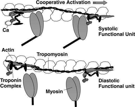
15 Frank-Starling Law / mechanism of the Heart Otto Frank Ernest Starling 1866 to 1927 Stroke Volume increases with increasing End Diastolic Volume which stretches the ventricular wall, causing cardiac muscle to contract more forcefully (the so-called Frank-Starling mechanism). Stroke volume may also increase due to greater contractility of the cardiac muscle during exercise, independent of end-diastolic volume. Stroke volume is influenced more by the Frank-Starling mechanism at lower work rates while contractility has a greater influence at higher work rates.

16 Length Tension Sarcomere (Starling mechanism) R. John Solaro Advan in Physiol Edu 277:S155-S163, 1999

17 Subcellular mechanism Ca ++ Flux Influx during systole Efflux during diastole influx efflux Donna H. Korzick Adv Physiol Educ 35: 22 27, 2011
18 Contractile mechanism:
19 Sarcomere Length Tension and Ca++
20 Pump Performance Determinants Neuro-humeral tone Adrenergic receptor regulation Frank Starling Effects Force Frequency Relation Vascular Function

21 Adrenergic receptor regulation
22 Frank Starling effects Cardiac output CO = HR X SV SV ~ Contractility, Preload, Afterload Decreased Afterload or Increased contractility CO or SV Normal Increased Afterload or Decreased contractility (Preload)
23 Force frequency relation force-frequency relationship represents an intrinsic regulatory factor essential for immediate adjustment of cardiac contractile function to rapidly changing requirements of blood supply. This frequency-dependent gain in contractility is intrinsic to mammalian cardiac muscle and allows for greater contractile force [Eur J Pharmacol 2004, 500:73-861]. The heart generally beats stronger when it is stimulated to contract faster, AND it displays frequency-dependent acceleration of relaxation (FDAR) [Eur J Pharmacol 2004, 500:73-861, J Mol Cell Cardiol 2007, 43: ]. From a physiological perspective, FDAR participates in the maintenance of efficient ventricular filling and coronary blood flow at higher heart rates, despite a decreased diastolic time interval. Critical Care 2009, 13:R14
24 Force recordings from isolated muscle strips from RA and LV from explanted human hearts. Münch G et al. Am J Physiol Heart Circ Physiol 2000;278:H1924-H by American Physiological Society
25 Vascular Function Curve CO effect on CVP CO CVP Peripheral Resistance CVP CO
26 Vascular Function Curve CO effect on CVP CO CVP Peripheral Resistance CVP CO
27 Vascular Function Curve CO effect on CVP CO CVP Peripheral Resistance CVP CO

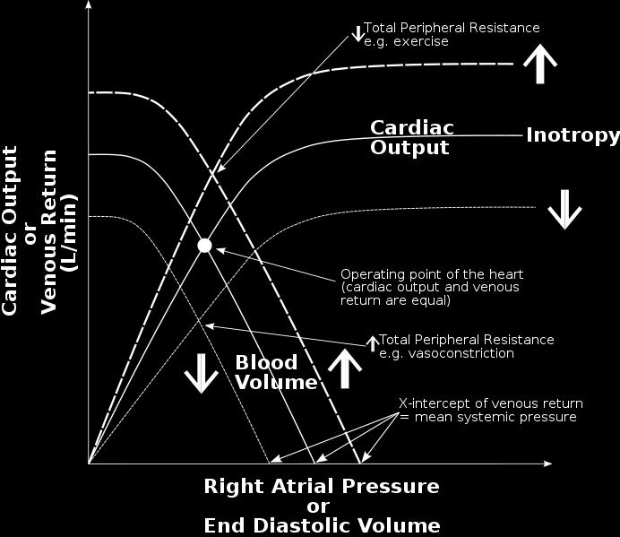
28 Cardiac Physiology Interactions between Vascular function curve and Starling curve
29 Cardiac Physiology Functional Anatomy Circulation Compartments: Intra-thoracic Abdominal Peripheral Pressure gradients Flow From Shekerdemian and Bohn: Arch. Dis. Child 1999;80;
30 Cardiac Physiology Right Ventricular Preload, Pleural Pressure, Cardiac output Pleural Pressure +2 CO Right Atrial Pressure
31 Cardiac Physiology Right Ventricular Afterload and Lung Volume Total PVR Alveolar Vessels Parenchymal Vessels RV FRC TLC Lung Volume
32 Cardiac Physiology Left Ventricular Afterload Transmural Pressure = Pressure inside pressure outside Therefore P LVTM = P Ao P PL Afterload increases with: High P Ao Low P PL

33 Cardiac Physiology Artificial Organs 35(5): ,
SIKLUS JANTUNG. Rahmatina B. Herman
 SIKLUS JANTUNG Rahmatina B. Herman The Cardiac Cycle Definition: The cardiac events that occur from the beginning of one heartbeat to the beginning of the next The cardiac cycle consists of: - Diastole
Cardiovascular Physiology. Heart Physiology. Introduction. The heart. Electrophysiology of the heart
 Cardiovascular Physiology Heart Physiology Introduction The cardiovascular system consists of the heart and two vascular systems, the systemic and pulmonary circulations. The heart pumps blood through
Principles of Biomedical Systems & Devices. Lecture 8: Cardiovascular Dynamics Dr. Maria Tahamont
 Principles of Biomedical Systems & Devices Lecture 8: Cardiovascular Dynamics Dr. Maria Tahamont Review of Cardiac Anatomy Four chambers Two atria-receive blood from the vena cave and pulmonary veins Two
SymBioSys Exercise 2 Cardiac Function Revised and reformatted by C. S. Tritt, Ph.D. Last updated March 20, 2006
 SymBioSys Exercise 2 Cardiac Function Revised and reformatted by C. S. Tritt, Ph.D. Last updated March 20, 2006 The goal of this exercise to explore the behavior of the heart as a mechanical pump. For
Cardiovascular System. Heart Anatomy
 Cardiovascular System Heart Anatomy 1 The Heart Location & general description: Atria vs. ventricles Pulmonary vs. systemic circulation Coverings Walls The heart is found in the mediastinum, the medial
The circulatory system
 Introduction to Physiology (Course # 72336) 1 הלב עקרונות בסיסיים (הכנה למעבדת לב) Adi Mizrahi mizrahia@cc.huji.ac.il Textbook Chapter 12 2 The circulatory system To the heart Away from the heart 3 L 2.5
GIGA - In Silico Medicine, University of Liege, Belgium, 2
 S. Kosta 1,*, A. Pironet 1, J.A. Negroni 2, E.C. Lascano 2, P.C. Dauby 1 1 GIGA - In Silico Medicine, University of Liege, Belgium, 2 Department of Comparative Cellular and Molecular Biology, Favaloro
The Heart. Happy Friday! #takeoutyournotes #testnotgradedyet
 The Heart Happy Friday! #takeoutyournotes #testnotgradedyet Introduction Cardiovascular system distributes blood Pump (heart) Distribution areas (capillaries) Heart has 4 compartments 2 receive blood (atria)
Introduction to Physiology (Course # 72336) 1. Adi Mizrahi Textbook Chapter 12
 1. Adi Mizrahi Textbook Chapter 12") Introduction to Physiology (Course # 72336) 1 עקרונות בסיסיים (הכנה למעבדת לב) הלב Adi Mizrahi mizrahia@cc.huji.ac.il Textbook Chapter 12 2 The circulatory system To the heart Away from the heart 3 L 2.5
Introduction to Physiology (Course # 72336) 1 עקרונות בסיסיים (הכנה למעבדת לב) הלב Adi Mizrahi mizrahia@cc.huji.ac.il Textbook Chapter 12 2 The circulatory system To the heart Away from the heart 3 L 2.5
Chapter 13 The Cardiovascular System: Cardiac Function
 Chapter 13 The Cardiovascular System: Cardiac Function Overview of the Cardiovascular System The Path of Blood Flow through the Heart and Vasculature Anatomy of the Heart Electrical Activity of the Heart
Major Function of the Cardiovascular System. Transportation. Structures of the Cardiovascular System. Heart - muscular pump
 Structures of the Cardiovascular System Heart - muscular pump Blood vessels - network of tubes Blood - liquid transport vehicle brachiocephalic trunk superior vena cava right pulmonary arteries right pulmonary
Cardiac Cycle MCQ. Professor of Cardiovascular Physiology. Cairo University 2007
 Cardiac Cycle MCQ Abdel Moniem Ibrahim Ahmed, MD Professor of Cardiovascular Physiology Cairo University 2007 1- Regarding the length of systole and diastole: a- At heart rate 75 b/min, the duration of
Cardiovascular Physiology
 Cardiovascular Physiology Lecture 1 objectives Explain the basic anatomy of the heart and its arrangement into 4 chambers. Appreciate that blood flows in series through the systemic and pulmonary circulations.
From PV loop to Starling curve. S Magder Division of Critical Care, McGill University Health Centre
 From PV loop to Starling curve S Magder Division of Critical Care, McGill University Health Centre Otto Frank 1890 s Frank-Starling Relationship ( The Law of the Heart ) The greater the initial stretch
The Cardiovascular System
 Essentials of Human Anatomy & Physiology Elaine N. Marieb Seventh Edition Chapter 11 The Cardiovascular System Slides 11.1 11.19 Lecture Slides in PowerPoint by Jerry L. Cook The Cardiovascular System
Cardiovascular System
 Cardiovascular System The Heart Cardiovascular System The Heart Overview What does the heart do? By timed muscular contractions creates pressure gradients blood moves then from high pressure to low pressure
Blood flows away from the heart in arteries, to the capillaries and back to the heart in the veins
 Cardiovascular System Summary Notes The cardiovascular system includes: The heart, a muscular pump The blood, a fluid connective tissue The blood vessels, arteries, veins and capillaries Blood flows away
BME 5742 Bio-Systems Modeling and Control. Lecture 41 Heart & Blood Circulation Heart Function Basics
 BME 5742 Bio-Systems Modeling and Control Lecture 41 Heart & Blood Circulation Heart Function Basics Dr. Zvi Roth (FAU) 1 Pumps A pump is a device that accepts fluid at a low pressure P 1 and outputs the
DESCRIBE THE FACTORS AFFECTING CARDIAC OUTPUT.
 DESCRIBE THE FACTORS AFFECTING CARDIAC OUTPUT. BY: DISHA PRAKASH I MBBS, ROLL NO: 16M069 OBJECTIVES OF LEARNING Terminology and conceptual understanding of Cardiac Output. Factors regulating Cardiac Output.
During exercise the heart rate is 190 bpm and the stroke volume is 115 ml/beat. What is the cardiac output?
 The Cardiovascular System Part III: Heart Outline of class lecture After studying part I of this chapter you should be able to: 1. Be able to calculate cardiac output (CO) be able to define heart rate
Cardiac Output (C.O.) Regulation of Cardiac Output
 Regulation of Cardiac Output") Cardiac Output (C.O.) Is the volume of the blood pumped by each ventricle per minute (5 Litre) Stroke volume: Is the volume of the blood pumped by each ventricle per beat. Stroke volume = End diastolic
Cardiac Output (C.O.) Is the volume of the blood pumped by each ventricle per minute (5 Litre) Stroke volume: Is the volume of the blood pumped by each ventricle per beat. Stroke volume = End diastolic
Cardiovascular Physiology
 Cardiovascular Physiology Introduction The cardiovascular system consists of the heart and two vascular systems, the systemic and pulmonary circulations. The heart pumps blood through two vascular systems
The Cardiac Cycle Clive M. Baumgarten, Ph.D.
 The Cardiac Cycle Clive M. Baumgarten, Ph.D. OBJECTIVES: 1. Describe periods comprising cardiac cycle and events within each period 2. Describe the temporal relationships between pressure, blood flow,
Contents 1 Computational Haemodynamics An Introduction 2 The Human Cardiovascular System
 Contents 1 Computational Haemodynamics An Introduction... 1 1.1 What is Computational Haemodynamics (CHD)... 1 1.2 Advantages of CHD... 3 1.3 Applications in the Cardiovascular System... 4 1.3.1 CHD as
Topics to be Covered. Cardiac Measurements. Distribution of Blood Volume. Distribution of Pulmonary Ventilation & Blood Flow
 Topics to be Covered MODULE F HEMODYNAMIC MONITORING Cardiac Output Determinants of Stroke Volume Hemodynamic Measurements Pulmonary Artery Catheterization Control of Blood Pressure Heart Failure Cardiac
FUNDAMENTALS OF HEMODYNAMICS, VASOACTIVE DRUGS AND IABP IN THE FAILING HEART
 FUNDAMENTALS OF HEMODYNAMICS, VASOACTIVE DRUGS AND IABP IN THE FAILING HEART CINDY BITHER, MSN, ANP, ANP, AACC, CHFN CHIEF NP, ADV HF PROGRAM MEDSTAR WASHINGTON HOSPITAL CENTER CONFLICTS OF INTEREST NONE
2.6 Cardiovascular Computer Simulation
 2.6 Cardiovascular Computer Simulation ROOM 23G22 Contents 1. INTRODUCTION... 4 1.1. GENERAL REMARKS... 4 1.2. LEARNING GOALS... 4 1.3. PHYSIOLOGICAL PARAMETERS... 5 1.4. GLOSSARY... 5 2. USING THE COMPUTER
11/10/2014. Muscular pump Two atria Two ventricles. In mediastinum of thoracic cavity 2/3 of heart's mass lies left of midline of sternum
 It beats over 100,000 times a day to pump over 1,800 gallons of blood per day through over 60,000 miles of blood vessels. During the average lifetime, the heart pumps nearly 3 billion times, delivering
Lecture 13: The Cardiovascular System ref: Cardiovascular Physiology, D. Mohrman and L. Heller, 4th ed. McGraw-Hill (1997)
") Lecture 13: The Cardiovascular System ref: Cardiovascular Physiology, D. Mohrman and L. Heller, 4th ed. McGraw-Hill (1997) Blood Heart Blood Vessels Arteries - capillaries - Veins Ventilation-Perfusion
Lecture 13: The Cardiovascular System ref: Cardiovascular Physiology, D. Mohrman and L. Heller, 4th ed. McGraw-Hill (1997) Blood Heart Blood Vessels Arteries - capillaries - Veins Ventilation-Perfusion
TEACH Lesson Plan Manual for Herlihy s The Human Body in Health and Illness 5 th edition
 TEACH Lesson Plan Manual for Herlihy s The Human Body in Health and Illness 5 th edition Chapter 17 Function of the Heart Lesson 17.1 Function of the Heart 1. Define cardiac cycle with respect to systole
cardiac imaging planes planning basic cardiac & aortic views for MR
 cardiac imaging planes planning basic cardiac & aortic views for MR Dianna M. E. Bardo, M. D. Assistant Professor of Radiology & Cardiovascular Medicine Director of Cardiac Imaging cardiac imaging planes
Circulatory Systems. All cells need to take in nutrients and expel metabolic wastes.
 Circulatory Systems All cells need to take in nutrients and expel metabolic wastes. Single celled organisms: nutrients from the environment can diffuse (or be actively transported) directly in to the cell
Cardiovascular system
 BIO 301 Human Physiology Cardiovascular system The Cardiovascular System: consists of the heart plus all the blood vessels transports blood to all parts of the body in two 'circulations': pulmonary (lungs)
Heart Dissection. 5. Locate the tip of the heart or the apex. Only the left ventricle extends all the way to the apex.
 Heart Dissection Page 1 of 6 Background: The heart is a four-chambered, hollow organ composed primarily of cardiac muscle tissue. It is located in the center of the chest in between the lungs. It is the
Blood must move! 4/15/2014. Heart Basics
 What is the CARDIOVASCULAR system? The cardiovascular system carries blood and dissolved substances to and from different places in the body. The Heart has the job of pumping these things around the body.
Circulation: Chapter 25. Cardiac Output. The Mammalian Heart Fig Right side of the heart
 Circulation: Chapter 25 1. Limits of Diffusion A. Small organisms use diffusion B. rapid over small distances 2. Most animals have circulatory systems A. Blood B. Pump (Heart) or propulsive structures
Anatomy Review: The Heart Graphics are used with permission of A.D.A.M. Software, Inc. and Benjamin/Cummings Publishing Co.
 Anatomy Review: The Heart Graphics are used with permission of A.D.A.M. Software, Inc. and Benjamin/Cummings Publishing Co. Anatomy Views Label the diagrams of the heart below: Interactive Physiology Study
Cardiovascular System Notes: Physiology of the Heart
 Cardiovascular System Notes: Physiology of the Heart Interesting Heart Fact Capillaries are so small it takes ten of them to equal the thickness of a human hair. Review What are the 3 parts of the cardiovascular
The cardiovascular system is composed of a pump the heart and blood
 5 E X E R C I S E Cardiovascular Dynamics O B J E C T I V E S 1. To understand the relationships among blood flow, pressure gradient, and resistance 2. To define resistance and describe the main factors
End of chapter exercises
 End of chapter exercises Problem 1: The following diagrams show the heart during the cardiac cycle. The arrows represent the flow of blood. Study the diagrams and answer the questions that follow: Figure
10/23/2017. Muscular pump Two atria Two ventricles. In mediastinum of thoracic cavity 2/3 of heart's mass lies left of midline of sternum
 It beats over 100,000 times a day to pump over 1,800 gallons of blood per day through over 60,000 miles of blood vessels. During the average lifetime, the heart pumps nearly 3 billion times, delivering
Atrial Septal Defects
 Supplementary ACHD Echo Acquisition Protocol for Atrial Septal Defects The following protocol for echo in adult patients with atrial septal defects (ASDs) is a guide for performing a comprehensive assessment
CARDIOVASCULAR SYSTEM
 CARDIOVASCULAR SYSTEM Overview Heart and Vessels 2 Major Divisions Pulmonary Circuit Systemic Circuit Closed and Continuous Loop Location Aorta Superior vena cava Right lung Pulmonary trunk Base of heart
How does the heart pump? From sarcomere to ejection volume
 How does the heart pump? From sarcomere to ejection volume Piet Claus Cardiovascular Imaging and Dynamics Department of Cardiovascular Diseases University Leuven, Leuven, Belgium Course on deformation
Chapter 9, Part 2. Cardiocirculatory Adjustments to Exercise
 Chapter 9, Part 2 Cardiocirculatory Adjustments to Exercise Electrical Activity of the Heart Contraction of the heart depends on electrical stimulation of the myocardium Impulse is initiated in the right
Appendix II: ECHOCARDIOGRAPHY ANALYSIS
 Appendix II: ECHOCARDIOGRAPHY ANALYSIS Two-Dimensional (2D) imaging was performed using the Vivid 7 Advantage cardiovascular ultrasound system (GE Medical Systems, Milwaukee) with a frame rate of 400 frames
PHYSIOLOGY MeQ'S (Morgan) All the following statements related to blood volume are correct except for: 5 A. Blood volume is about 5 litres. B.
 All the following statements related to blood volume are correct except for: 5 A. Blood volume is about 5 litres. B.") PHYSIOLOGY MeQ'S (Morgan) Chapter 5 All the following statements related to capillary Starling's forces are correct except for: 1 A. Hydrostatic pressure at arterial end is greater than at venous end.
PHYSIOLOGY MeQ'S (Morgan) Chapter 5 All the following statements related to capillary Starling's forces are correct except for: 1 A. Hydrostatic pressure at arterial end is greater than at venous end.
Chapter 18 - Heart. I. Heart Anatomy: size of your fist; located in mediastinum (medial cavity)
") Chapter 18 - Heart I. Heart Anatomy: size of your fist; located in mediastinum (medial cavity) A. Coverings: heart enclosed in double walled sac called the pericardium 1. Fibrous pericardium: dense connective
Chapter 18 - Heart I. Heart Anatomy: size of your fist; located in mediastinum (medial cavity) A. Coverings: heart enclosed in double walled sac called the pericardium 1. Fibrous pericardium: dense connective
Electrical Conduction
 Sinoatrial (SA) node Electrical Conduction Sets the pace of the heartbeat at 70 bpm AV node (50 bpm) and Purkinje fibers (25 40 bpm) can act as pacemakers under some conditions Internodal pathway from
The Cardiovascular System
 11 PART A The Cardiovascular System PowerPoint Lecture Slide Presentation by Jerry L. Cook, Sam Houston University ESSENTIALS OF HUMAN ANATOMY & PHYSIOLOGY EIGHTH EDITION ELAINE N. MARIEB The Cardiovascular
The Anatomy and Physiology of the Circulatory System
 CHAPTER 5 The Anatomy and Physiology of the Circulatory System The Circulatory System Blood Heart Vascular System THE BLOOD Formed Elements of Blood Table 5-1 Cell Type Erythrocytes (Red Blood Cells, RBCs)
ADVANCED ASSESSMENT Cardiovascular System
 ONTARIO BASE HOSPITAL GROUP QUIT ADVANCED ASSESSMENT Cardiovascular System 2007 Ontario Base Hospital Group ADVANCED ASSESSMENT Cardiovascular System AUTHORS Mike Muir AEMCA, ACP, BHSc Paramedic Program
BIPN100 F15 Human Physiol I (Kristan) Lecture 14 Cardiovascular control mechanisms p. 1
 Lecture 14 Cardiovascular control mechanisms p. 1") BIPN100 F15 Human Physiol I (Kristan) Lecture 14 Cardiovascular control mechanisms p. 1 Terms you should understand: hemorrhage, intrinsic and extrinsic mechanisms, anoxia, myocardial contractility, residual
BIPN100 F15 Human Physiol I (Kristan) Lecture 14 Cardiovascular control mechanisms p. 1 Terms you should understand: hemorrhage, intrinsic and extrinsic mechanisms, anoxia, myocardial contractility, residual
Approximately the size of your fist Location. Pericardial physiology
 Heart Anatomy Approximately the size of your fist Location Superior surface of diaphragm Left of the midline Anterior to the vertebral column, posterior to the sternum Wednesday, March 28, 2012 Muscle
BIOL 219 Spring Chapters 14&15 Cardiovascular System
 1 BIOL 219 Spring 2013 Chapters 14&15 Cardiovascular System Outline: Components of the CV system Heart anatomy Layers of the heart wall Pericardium Heart chambers, valves, blood vessels, septum Atrioventricular
The Cardiovascular System (Heart)
") The Cardiovascular System The Cardiovascular System (Heart) A closed system of the heart and blood vessels The heart pumps blood Blood vessels allow blood to circulate to all parts of the body The function
The Cardiovascular System The Cardiovascular System (Heart) A closed system of the heart and blood vessels The heart pumps blood Blood vessels allow blood to circulate to all parts of the body The function
The Cardiovascular System
 The Cardiovascular System The Cardiovascular System A closed system of the heart and blood vessels The heart pumps blood Blood vessels allow blood to circulate to all parts of the body The function of
P215 SPRING 2019: CIRCULATORY SYSTEM Chaps 13, 14 & 15: pp , , , I. Major Functions of the Circulatory System
 P215 SPRING 2019: CIRCULATORY SYSTEM Chaps 13, 14 & 15: pp 360-390, 395-404, 410-428 433-438, 441-445 I. Major Functions of the Circulatory System 1. 2. 3. 4. II. Structure of the Heart 1. atria 2. ventricles
The Mammalian Circulatory System
 The Mammalian Heart The Mammalian Circulatory System Recall: What are the 3 cycles of the mammalian circulatory system? What are their functions? What are the three main vessel types in the mammalian circulatory
Chapter 20 (1) The Heart
 The Heart") Chapter 20 (1) The Heart Learning Objectives Describe the location and structure of the heart Describe the path of a drop of blood from the superior vena cava or inferior vena cava through the heart out
Chapter 20 (1) The Heart Learning Objectives Describe the location and structure of the heart Describe the path of a drop of blood from the superior vena cava or inferior vena cava through the heart out
Mechanisms of heart failure with normal EF Arterial stiffness and ventricular-arterial coupling. What is the pathophysiology at presentation?
 Mechanisms of heart failure with normal EF Arterial stiffness and ventricular-arterial coupling What is the pathophysiology at presentation? Ventricular-arterial coupling elastance Central arterial pressure
(D) (E) (F) 6. The extrasystolic beat would produce (A) increased pulse pressure because contractility. is increased. increased
 (E) (F) 6. The extrasystolic beat would produce (A) increased pulse pressure because contractility. is increased. increased") Review Test 1. A 53-year-old woman is found, by arteriography, to have 5% narrowing of her left renal artery. What is the expected change in blood flow through the stenotic artery? Decrease to 1 2 Decrease
Review Test 1. A 53-year-old woman is found, by arteriography, to have 5% narrowing of her left renal artery. What is the expected change in blood flow through the stenotic artery? Decrease to 1 2 Decrease
Left atrial function. Aliakbar Arvandi MD
 In the clinic Left atrial function Abstract The left atrium (LA) is a left posterior cardiac chamber which is located adjacent to the esophagus. It is separated from the right atrium by the inter-atrial
AS Level OCR Cardiovascular System
 AS Level OCR Cardiovascular System Learning Objectives The link between the Cardiac Cycle and the Conduction system of the heart. The relationship between Stroke volume, Heart rate and Cardiac Output.
Circulation. Blood Pressure and Antihypertensive Medications. Venous Return. Arterial flow. Regulation of Cardiac Output.
 Circulation Blood Pressure and Antihypertensive Medications Two systems Pulmonary (low pressure) Systemic (high pressure) Aorta 120 mmhg Large arteries 110 mmhg Arterioles 40 mmhg Arteriolar capillaries
10. Thick deposits of lipids on the walls of blood vessels, called, can lead to serious circulatory issues. A. aneurysm B. atherosclerosis C.
 Heart Student: 1. carry blood away from the heart. A. Arteries B. Veins C. Capillaries 2. What is the leading cause of heart attack and stroke in North America? A. alcohol B. smoking C. arteriosclerosis
Heart. Structure Physiology of blood pressure and heartbeat
 Heart Structure Physiology of blood pressure and heartbeat Location and Anatomy Location and Anatomy Pericardial cavity: surrounds, isolates, and anchors heart Parietal pericardium lined with serous membrane
Heart Pump and Cardiac Cycle. Faisal I. Mohammed, MD, PhD
 Heart Pump and Cardiac Cycle Faisal I. Mohammed, MD, PhD 1 Objectives To understand the volume, mechanical, pressure and electrical changes during the cardiac cycle To understand the inter-relationship
- what other structures, besides the heart, does the mediastinum contain?
 Basic A & P II Dr. L. Bacha Chapter Outline (Martini & Nath 2010) An Introduction to the Cardiovascular System - read the paragraphs under this heading on page 580 The Heart is a Four Chambered Organ describe
Lab Activity 23. Cardiac Anatomy. Portland Community College BI 232
 Lab Activity 23 Cardiac Anatomy Portland Community College BI 232 Cardiac Muscle Histology Branching cells Intercalated disc: contains many gap junctions connecting the adjacent cell cytoplasm, creates
The Cardiovascular System
 The Cardiovascular System The Manila Times College of Subic Prepared by: Stevens B. Badar, RN, MANc THE HEART Anatomy of the Heart Location and Size approx. the size of a person s fist, hollow and cone-shaped,
Chapter 27 The Heart and Blood Vessels
 Chapter 27 The Heart and Blood Vessels Most animals have a closed blood system. The blood flows continuously in vessels back to the heart. In an open system the blood is pumped into open ended tubes and
LECTURE 5. Anatomy of the heart
 LECTURE 5. Anatomy of the heart Main components of the CVS: Heart Blood circulatory system arterial compartment haemomicrocirculatory (=microvascular) compartment venous compartment Lymphatic circulatory
Lab 16. The Cardiovascular System Heart and Blood Vessels. Laboratory Objectives
 Lab 16 The Cardiovascular System Heart and Blood Vessels Laboratory Objectives Describe the anatomical structures of the heart to include the pericardium, chambers, valves, and major vessels. Describe
The Heart. Size, Form, and Location of the Heart. 1. Blunt, rounded point; most inferior part of the heart.
 12 The Heart FOCUS: The heart is composed of cardiac muscle cells, which are elongated, branching cells that appear striated. Cardiac muscle cells behave as a single electrical unit, and the highly coordinated
Mr. Epithelium s Anatomy and Physiology Test SSSS
 Mr. Epithelium s Anatomy and Physiology Test SSSS You have 50 minutes to complete this test packet. One 8.5 x 11 cheat sheet is allowed, along with 1 non-programmable calculator dedicated to computation.
COMPREHENSIVE EVALUATION OF FETAL HEART R. GOWDAMARAJAN MD
 COMPREHENSIVE EVALUATION OF FETAL HEART R. GOWDAMARAJAN MD Disclosure No Relevant Financial Relationships with Commercial Interests Fetal Echo: How to do it? Timing of Study -optimally between 22-24 weeks
Anaesthesia. Update in. An Introduction to Cardiovascular Physiology. James Rogers Correspondence
 Update in Anaesthesia Originally published in Update in Anaesthesia, edition 10 (1999) An Introduction to Cardiovascular Physiology Correspondence Email: James.Rogers@nbt.nhs.uk INTRODUCTION The cardiovascular
Cardiac output and Venous Return. Faisal I. Mohammed, MD, PhD
 Cardiac output and Venous Return Faisal I. Mohammed, MD, PhD 1 Objectives Define cardiac output and venous return Describe the methods of measurement of CO Outline the factors that regulate cardiac output
Circulation. Sinoatrial (SA) Node. Atrioventricular (AV) Node. Cardiac Conduction System. Cardiac Conduction System. Linked to the nervous system
 Node. Atrioventricular (AV) Node. Cardiac Conduction System. Cardiac Conduction System. Linked to the nervous system") Circulation Cardiac Conduction System AHS A H S Your body resembles a large roadmap. There are routes or arteries that take you downtown to the heart of the city and veins that take you to the outskirts
Circulation Cardiac Conduction System AHS A H S Your body resembles a large roadmap. There are routes or arteries that take you downtown to the heart of the city and veins that take you to the outskirts
Chapter 27 -The Heart & Blood Vessels
 Chapter 27 -The Heart & Blood Vessels 3.2 Learning Objectives 3.2.2 Organisational Complexity of the human 1. Describe the structures and organisation of tissues in the closed circulatory system. 2. Discuss
Case 47 Clinical Presentation
 93 Case 47 C Clinical Presentation 45-year-old man presents with chest pain and new onset of a murmur. Echocardiography shows severe aortic insufficiency. 94 RadCases Cardiac Imaging Imaging Findings C
IB TOPIC 6.2 THE BLOOD SYSTEM
 IB TOPIC 6.2 THE BLOOD SYSTEM THE BLOOD SYSTEM TERMS TO KNOW circulation ventricle artery vein 6.2.U1 - Arteries convey blood at high pressure from the ventricles to the tissues of the body Circulation
CVS Hemodynamics. Faisal I. Mohammed, MD,PhD.
 CVS Hemodynamics Faisal I. Mohammed, MD,PhD. Objectives point out the physical characteristics of the circulation: distribution of blood volume total cross sectional area velocity blood pressure List the
THE CARDIOVASCULAR SYSTEM
 THE CARDIOVASCULAR SYSTEM AND RESPONSES TO EXERCISE Mr. S. Kelly PSK 4U North Grenville DHS THE HEART: A REVIEW Cardiac muscle = myocardium Heart divided into two sides, 4 chambers (L & R) RS: pulmonary
IB TOPIC 6.2 THE BLOOD SYSTEM
 IB TOPIC 6.2 THE BLOOD SYSTEM TERMS TO KNOW circulation ventricle artery vein THE BLOOD SYSTEM 6.2.U1 - Arteries convey blood at high pressure from the ventricles to the tissues of the body Circulation
1. Which of the following blood vessels has a thin elastic layer? A. Aorta. B. Pulmonary artery. C. Posterior vena cava. D. Mesenteric capillary.
 CIRCULATORY SYSTEM 1. Which of the following blood vessels has a thin elastic layer? A. Aorta. B. Pulmonary artery. C. Posterior vena cava. D. Mesenteric capillary. 2. Capillary beds are equipped with
d) Cardiovascular System Higher Human Biology
 Cardiovascular System Higher Human Biology") d) Cardiovascular System Higher Human Biology What can your remember about the heart and blood vessels? What is the Cardiovascular System? The cardiovascular system, also known as the circulatory system,
d) Cardiovascular System Higher Human Biology What can your remember about the heart and blood vessels? What is the Cardiovascular System? The cardiovascular system, also known as the circulatory system,
Circulatory System 10.1
 1 Circulatory System 10.1 2 ARTERIES Arteries-blood vessels that carry blood away from the heart Thick walls Inner & Outer layers: connective tissue Middle layers are muscle and elastic connective tissue
Danil Hammoudi.MD 1/12/2009
 Danil Hammoudi.MD Aorta the biggest and longest artery (a blood vessel carrying blood away from the heart) in the body. It carries oxygen rich blood from the left ventricle of the heart to the body.inferior
McHenry Western Lake County EMS System Paramedic, EMT-B and PHRN Optional Continuing Education 2018 #12 Understanding Preload and Afterload
 McHenry Western Lake County EMS System Paramedic, EMT-B and PHRN Optional Continuing Education 2018 #12 Understanding Preload and Afterload Cardiac output (CO) represents the volume of blood that is delivered
Heart and Lung Dissection
 Heart and Lung Dissection Name(s) Before you begin any work or dissection of your specimen, please try to identify the following. You will need to illustrate what you see, so make sure to note size, texture
Cardiac Output 1 Fox Chapter 14 part 1
 Vert Phys PCB3743 Cardiac Output 1 Fox Chapter 14 part 1 T. Houpt, Ph.D. Regulation of Heart & Blood Pressure Keep Blood Pressure constant if too low, not enough blood (oxygen, glucose) reaches tissues
Chapter 20: Cardiovascular System: The Heart
 Chapter 20: Cardiovascular System: The Heart I. Functions of the Heart A. List and describe the four functions of the heart: 1. 2. 3. 4. II. Size, Shape, and Location of the Heart A. Size and Shape 1.
Principles of Anatomy and Physiology
 Principles of Anatomy and Physiology 14 th Edition CHAPTER 20 The Cardiovascular System: The Heart Introduction The purpose of the chapter is to: 1. Learn about the components of the cardiovascular system
THE HEART OBJECTIVES: LOCATION OF THE HEART IN THE THORACIC CAVITY CARDIOVASCULAR SYSTEM
 BIOLOGY II CARDIOVASCULAR SYSTEM ACTIVITY #3 NAME DATE HOUR THE HEART OBJECTIVES: Describe the anatomy of the heart and identify and give the functions of all parts. (pp. 356 363) Trace the flow of blood