Chapter 20 (1) The Heart
|
|
|
- Liliana Lambert
- 5 years ago
- Views:
Transcription
 The")
1 Chapter 20 (1) The Heart
2 Learning Objectives Describe the location and structure of the heart Describe the path of a drop of blood from the superior vena cava or inferior vena cava through the heart out of the aortic arch, listing all chambers, valves, and vessels involved. State the function of the atria, valves, chordae tendinae, papillary muscles, and ventricles of the heart
3
4
5 Circulatory Circuits of the Heart Pulmonary circuit CO 2 O 2 right side of heart to lungs carries blood to lungs for gas exchange and back to heart // low pressure O 2 -poor, CO 2 -rich blood Pulmonary circuit O 2 -rich, CO 2 -poor blood Systemic circuit Systemic circuit left side of heart to all tissue of body except lungs supplies oxygenated blood to all tissues of the body and returns it to the heart // high pressure CO 2 O 2
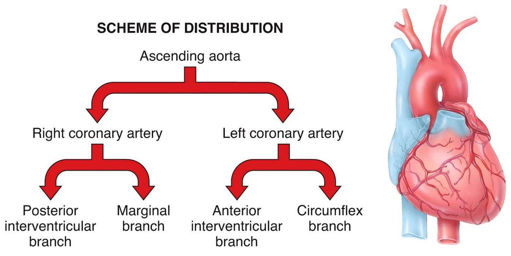
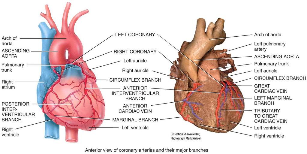
 with blood circulation RCA divides into the marginal artery branch and the posterior interventricular branch LCA divides into circumflex brach and the anterior interventrcicular")
6 Circulatory Circuits of the Heart Coronary Circuit part of the systemic circuit aortic recoil moves blood into the coronary arteries supply the myocardiocytes (the cells making the walls of the heart) with blood circulation RCA divides into the marginal artery branch and the posterior interventricular branch LCA divides into circumflex brach and the anterior interventrcicular brach
7

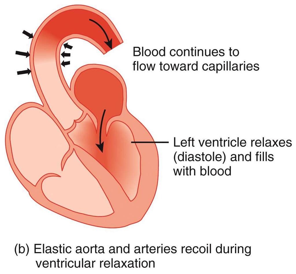
8 At the start of ventricular diastole accompanied with aortic recoil, blood moves into the systemic circuit and blood flows back towards the left ventricle. This movement of blood towards the left ventricle closes the aortic semilunar valve. As the semilunar valves close they uncovers the openings into the right and left coronary arteries. The force of the recoiling aorta moves blood into the coronary circuit as the myocardium is in a relaxed state.
9

10 Cardiovascular System Circuit Left side of heart CO 2 O 2 fully oxygenated blood arrives from lungs via pulmonary veins (100% Hb-O2) Pulmonary circuit blood sent to all organs of the body via aorta O 2 -poor, CO 2 -rich blood O 2 -rich, CO 2 -poor blood high pressure side Right side of heart Systemic circuit CO 2 O 2 lesser oxygenated blood arrives from inferior and superior vena cava (75% Hb- O2) blood sent to lungs via pulmonary trunk low pressure side
11
 Pericardial sac (cut) Aorta")
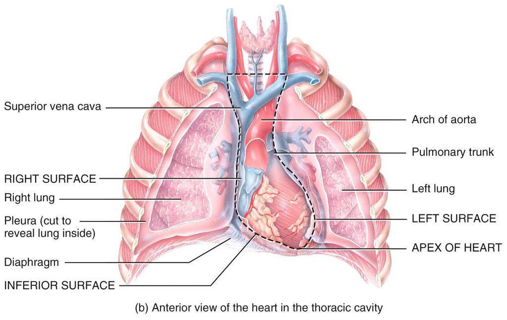
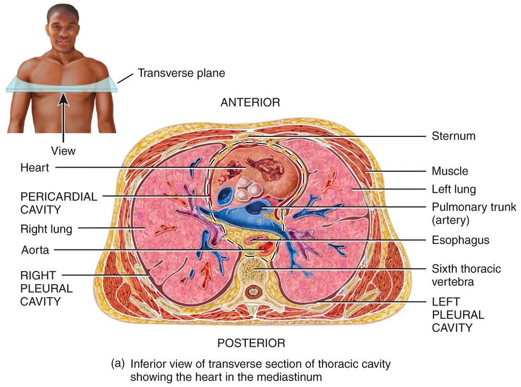
12 Position, Size, and Shape heart located in mediastinum, between lungs Copyright The McGraw-Hill Companies, Inc. Permission required for reproduction or display. base wide, superior portion of heart, blood vessels attach here apex - inferior end, tilts to the left, tapers to point Superior vena cava Right lung Parietal pleura (cut) Pericardial sac (cut) Aorta Pulmonary trunk Base of heart Apex of heart 3.5 in. wide at base (c) Diaphragm 5 in. from base to apex 2.5 in. anterior to posterior weighs 10 oz.
13
14
15
 Sternum")
16 Heart Position Copyright The McGraw-Hill Companies, Inc. Permission required for reproduction or display. Sternum 3rd rib Lungs Posterior Diaphragm Thoracic vertebra Pericardial cavity Right ventricle Left ventricle (a) Sternum Interventricular septum (b) Anterior
17
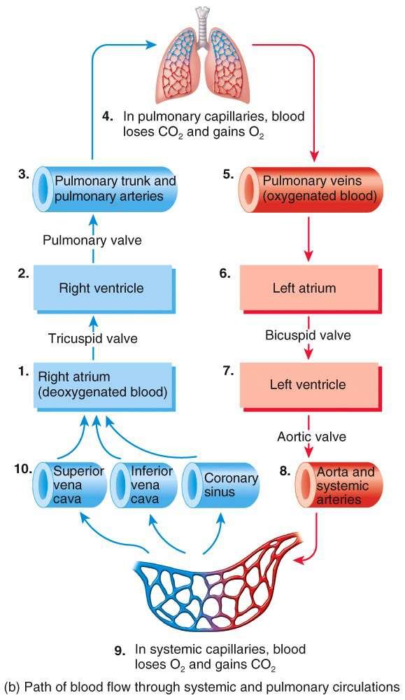
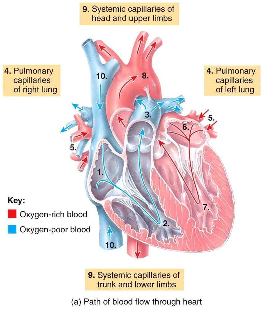
18 Blood Flow Through Heart Right atria Right ventricle Pulmonary truck into pulmonary circulation (lungs) Left atria Left ventricle Aorta to systemic circulation (all tissues of body except lungs) Note: blood enters the right atria via superior vena cava, inferior vena cava, coronary sinus, and thebesian veins (highest concentration in right atria and lowest concentration in left ventricle) Note: blood flow through the heart is different than the pumping action of the heart // pumping action occurs as two separate pumps working in phase /// first action is to move blood downward in both pumps followed by both pumps moving blood upward (see next slide)

19 Blood Flow Through Heart Copyright The McGraw-Hill Companies, Inc. Permission required for reproduction or display Blood enters right atrium from superior and inferior venae cavae. Aorta Left pulmonary artery 2 3 Blood in right atrium flows through right AV valve into right ventricle. Contraction of right ventricle forces pulmonary valve open. Superior vena cava Right pulmonary veins Right atrium Right AV (tricuspid) valve Right ventricle Inferior vena cava Pulmonary trunk Left pulmonary veins Left atrium Aortic valve Left AV (bicuspid) valve Left ventricle Blood flows through pulmonary valve into pulmonary trunk. Blood is distributed by right and left pulmonary arteries to the lungs, where it unloads CO 2 and loads O 2. Blood returns from lungs via pulmonary veins to left atrium. Blood in left atrium flows through left AV valve into left ventricle. Contraction of left ventricle (simultaneous with step 3 ) forces aortic valve open. Blood flows through aortic valve into ascending aorta. Blood in aorta is distributed to every organ in the body, where it unloads O 2 and loads CO Blood returns to heart via venae cavae. blood starts transit through heart in right atria / blood pressure and heart valves direct flow of blood / no valves associated with inferior or superior vena cava
20

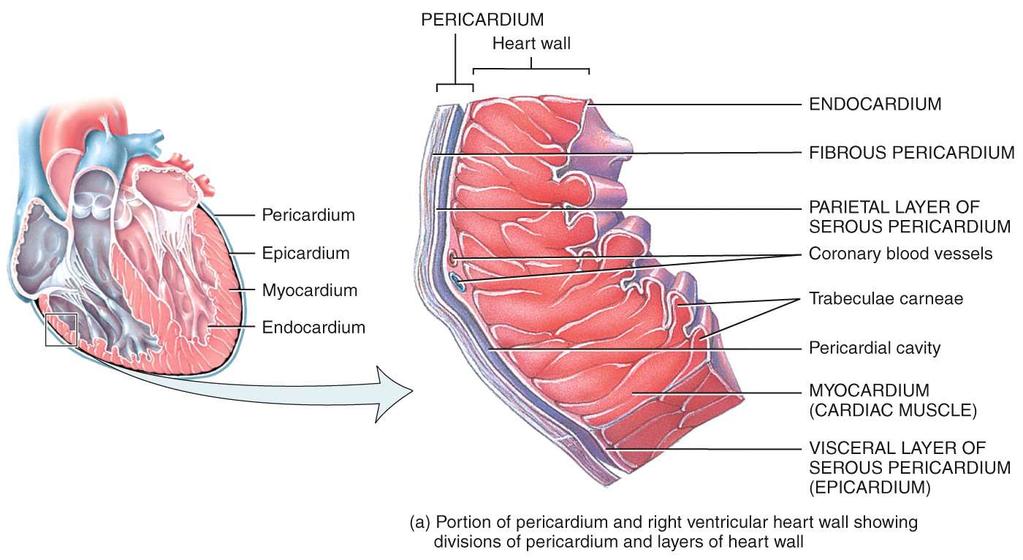
21 Pericardium and Heart Wall Pericardial cavity Pericardial sac: Fibrous layer Serous layer Epicardium Myocardium Endocardium Epicardium Pericardial sac
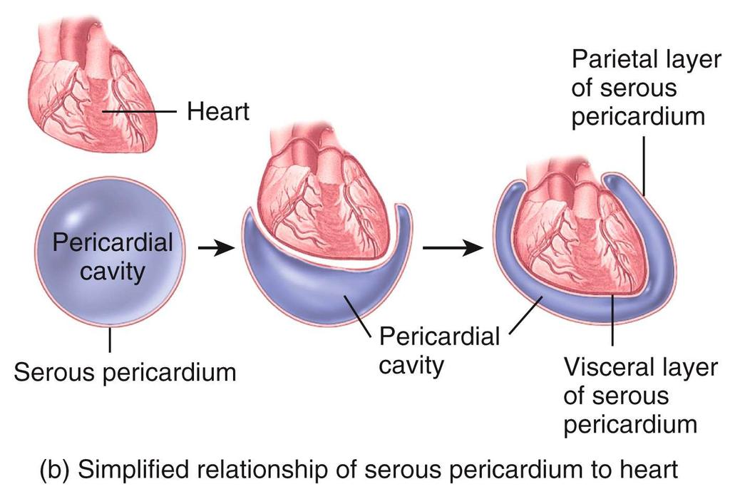
22
23 Pericardium Pericardium double-walled sac (pericardial sac) that encloses the heart // fibrous pericardium with inner and outer surface allows heart to beat without friction, provides room to expand // resists excessive expansion anchored to diaphragm inferiorly and sternum on anterior surface Parietal pericardium outer wall of sac superficial fibrous layer of connective tissue a deep, thin serous layer
24 Pericardium Visceral pericardium (epicardium) simple squamous epithelium / heart covering serous lining of pericardial sac // turns inward at base of heart to cover the heart surface Pericardial cavity // space inside the pericardial sac filled with 5-30 ml of pericardial fluid Pericarditis // inflammation of the membranes // painful friction rub with each heartbeat // excess fluid accumulates in pericardial cavity
25 Heart Wall Epicardium (visceral pericardium) serous membrane covering heart adipose in thick layer in some places coronary blood vessels travel through this layer Myocardium layer of cardiac muscle proportional to work load muscle spirals around heart which produces wringing motion
26 Heart Wall Endocardium smooth inner lining of heart and blood vessels covers the valve surfaces and continuous with endothelium of blood vessels Fibrous skeleton of the heart framework of collagenous and elastic fibers provides structural support and attachment for cardiac muscle and anchor for valve tissue electrical insulation between atria and ventricles important in timing and coordination of contractile activity
27
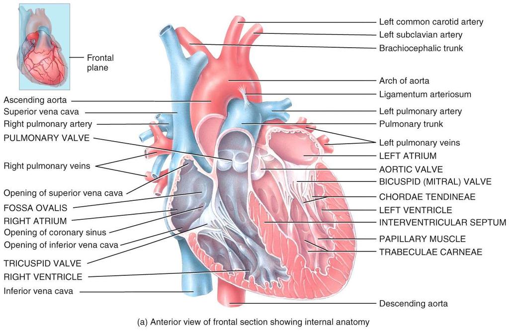
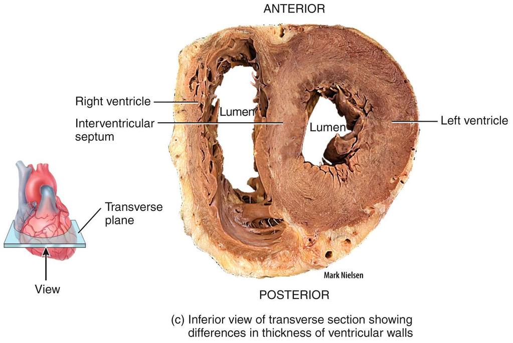
28 Four Chambers of he Heart Superior chambers right and left atria Copyright The McGraw-Hill Companies, Inc. Permission required for reproduction or display. two superior chambers receive blood returning to heart from pulmonary veins and vena cava auricles (seen on surface) allow chambers to expand volume Aorta Right pulmonary artery Superior vena cava Right pulmonary veins Interatrial septum Right atrium Fossa ovalis Pectinate muscles Right AV (tricuspid) valve Tendinous cords Trabeculae carneae Right ventricle Inferior vena cava Left pulmonary artery Pulmonary trunk Left pulmonary veins Pulmonary valve Left atrium Aortic valve Left AV (bicuspid) valve Left ventricle Papillary muscle Interventricular septum Endocardium Myocardium Epicardium both upper chambers contract together to move blood downward into the right and left ventricles
29 Four Chambers of he Heart Inferior chambers Aorta right and left ventricles both ventricles contract together right ventricle pump blood into pulmonary truck left ventricle pump blood into aorta Right pulmonary artery Superior vena cava Right pulmonary veins Interatrial septum Right atrium Fossa ovalis Pectinate muscles Right AV (tricuspid) valve Tendinous cords Trabeculae carneae Right ventricle Inferior vena cava Left pulmonary artery Pulmonary trunk Left pulmonary veins Pulmonary valve Left atrium Aortic valve Left AV (bicuspid) valve Left ventricle Papillary muscle Interventricular septum Endocardium Myocardium Epicardium Note: heart is a four chamber pump // low pressure and high pressure sides // blood circulates through heart however blood flow moves as an atrial downward phase followed by a ventricular upward phase

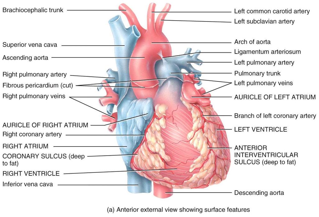
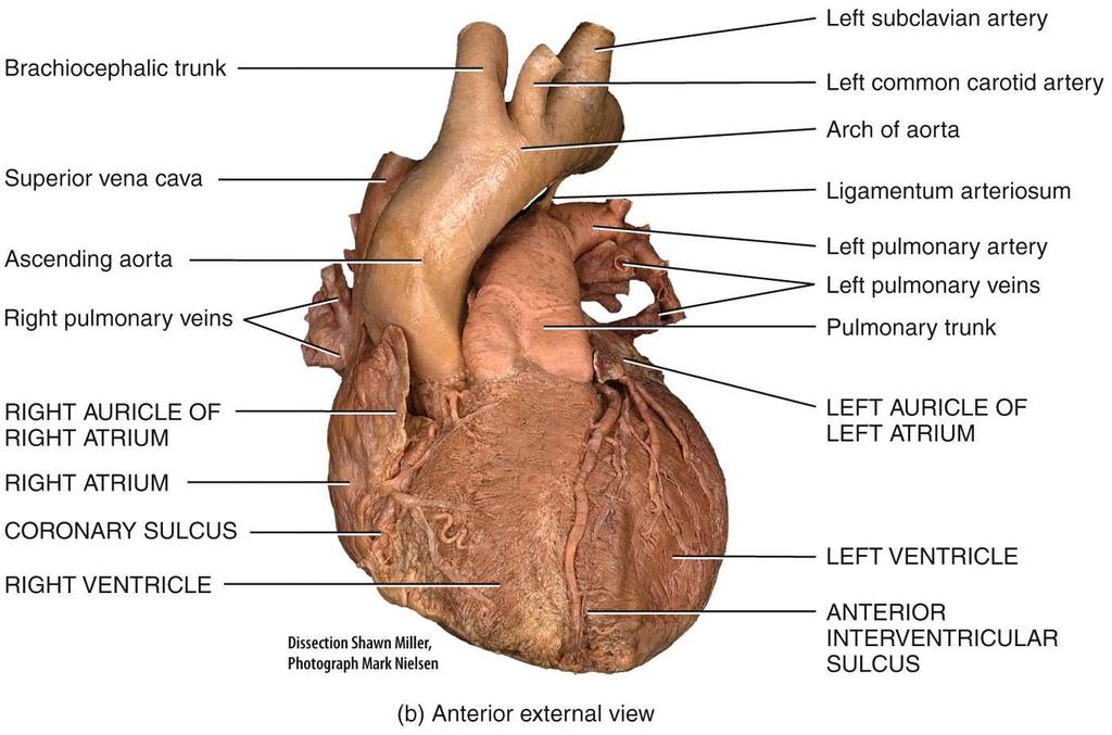
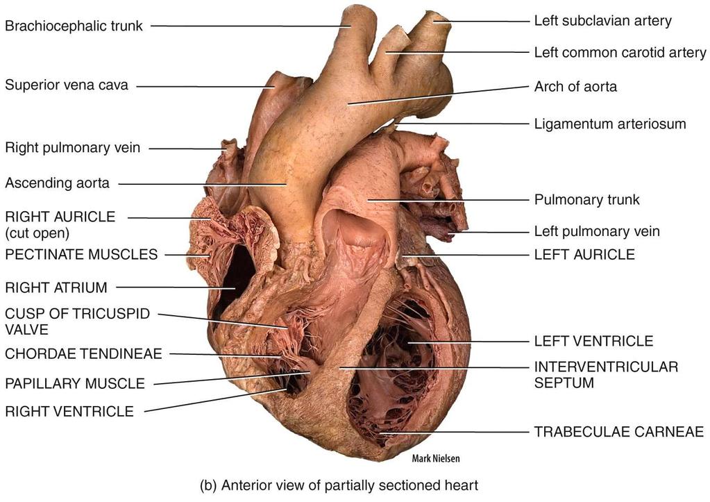
30 External Anatomy - Anterior Aortic arch Superior vena cava Copyright The McGraw-Hill Companies, Inc. Permission required for reproduction or display. Ligamentum arteriosum Ascending aorta atrioventricular sulcus separates atria and ventricles Branches of the right pulmonary artery Right pulmonary veins Right auricle Right atrium Left pulmonary artery Pulmonary trunk Left pulmonary veins Left auricle interventricular sulcus lies over the interventricular septum that divides the right ventricle from the left anterior and posterior Coronary sulcus Right ventricle Inferior vena cava (a) Anterior view Anterior interventricular sulcus Left ventricle Apex of heart Note: sulci are grooves on surface of heart that contain coronary arteries, as well as other heart arteries and veins
31
 Posterior")
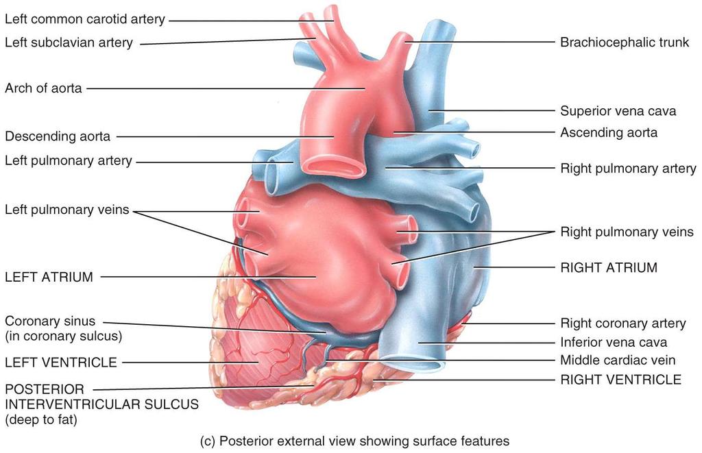
32 External Anatomy - Posterior Aorta Left pulmonary artery Left pulmonary veins Left atrium Coronary sulcus Coronary sinus Superior vena cava Right pulmonary artery Right pulmonary veins Right atrium Inferior vena cava Fat Left ventricle Apex of heart Posterior interventricular sulcus Right ventricle (b) Posterior view
33
34
35
36
37 Internal Anatomy - Anterior Aorta Ligamentum arteriosum Ligament Atriosum (2) Interatrial septum Right atrium Fossa ovalis (1) Right pulmonary artery Superior vena cava Right pulmonary veins Pectinate muscles Right AV (tricuspid) valve Tendinous cords Trabeculae carneae Right ventricle Inferior vena cava Left pulmonary artery Pulmonary trunk Left pulmonary veins Pulmonary valve Left atrium Aortic valve Left AV (bicuspid) valve Left ventricle Papillary muscle Interventricular septum Endocardium Myocardium Epicardium Notes: (1) foramine ovalis / (2) Dutus Atriosum / fetal structures to redirect blood into systemic circulation
38 Heart Chambers Internal Structures interatrial septum wall that separates atria pectinate muscles internal ridges of myocardium in right atrium and both auricles interventricular septum muscular wall that separates ventricles trabeculae carneae internal ridges in both ventricles
39 Internal Anatomy - Anterior Aorta Ligamentum arteriosum Ligament Atriosum (2) Interatrial septum Right atrium Fossa ovalis (1) Right pulmonary artery Superior vena cava Right pulmonary veins Pectinate muscles Right AV (tricuspid) valve Tendinous cords Trabeculae carneae Right ventricle Inferior vena cava Left pulmonary artery Pulmonary trunk Left pulmonary veins Pulmonary valve Left atrium Aortic valve Left AV (bicuspid) valve Left ventricle Papillary muscle Interventricular septum Endocardium Myocardium Epicardium Notes: (1) foramine ovalis / (2) Dutus Atriosum / fetal structures to redirect blood into systemic circulation
40
41
42

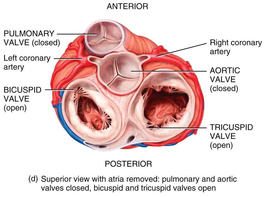
43 Heart Valves Posterior Left AV (bicuspid) valve Right AV (tricuspid) valve Fibrous skeleton Openings to coronary arteries Aortic valve Pulmonary valve Note: atria have been removed to show AV valves Anterior

44 Note: all valves are built into the atrioventricular septum / it is a strong connective tissue transverse plane / this also prevents action potentials from moving into ventricles via gap junctions
45 Left side

46 Note: All four valves are on same plane // the atrialventricular septum connective tissue no gap junctions unite atrial and ventricular myocariocytes!
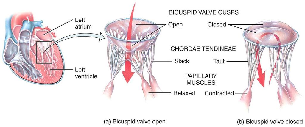
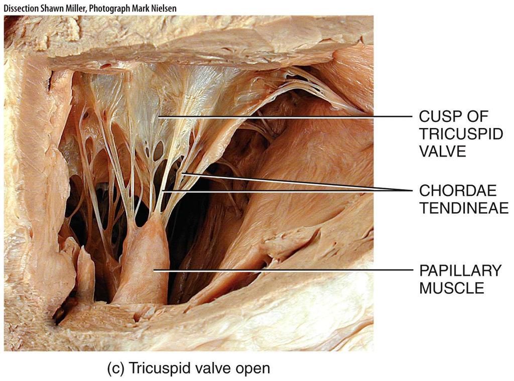
47 Heart Valve Functions / Atrioventricular controls blood flow between atria and ventricles Valves open and close due to blood pressure between chambers guarded by valves right AV valve has 3 cusps (tricuspid valve) left AV valve has 2 cusps (mitral or bicuspid valve) chordae tendineae connective tissue fibers that connect AV valves to papillary muscles on floor of ventricles papillary muscles prevent AV valves from flipping inside out or bulging into the atria when the ventricles contract
48
49 AV Valve Mechanics When ventricles relax pressure drops inside the ventricles semilunar valves close as blood attempts to back up into the ventricles from the vessels AV valves open blood flows from atria to ventricles When ventricles contract AV valves close as blood attempts to back up into the atria pressure rises inside of the ventricles semilunar valves open and blood flows into great vessels
50
51

52 Heart Valves / AV Valves Tendinous cords Papillary muscle
53 Heart Valve Function / Semilunar control flow into pulmonary truck and aorta // the great arteries of the heart these valves open and close because of blood flow and pressure pulmonary semilunar valve // between right ventricle and pulmonary trunk aortic semilunar valve // between left ventricle and aorta semilunar valves do not have chordae tenineae cusps of valves close as ventricles relax and blood starts to flow back towards ventricles // backflow of blood fill cusps which cause them to close

54 Endoscopic View of Semi Lunar Heart Valve

55 No papillary muscles or cordae tendinae

56 Function of Heart Valves Valves ensure a one-way flow of blood through the heart Blood flows from an area of high pressure to an area of low pressure The pressure difference across the valve open and close these valves
THE CARDIOVASCULAR SYSTEM. Part 1
 THE CARDIOVASCULAR SYSTEM Part 1 CARDIOVASCULAR SYSTEM Blood Heart Blood vessels What is the function of this system? What other systems does it affect? CARDIOVASCULAR SYSTEM Functions Transport gases,
Heart Anatomy. 7/5/02 Stephen G Davenport 1
 Heart Anatomy Copyright 1999, Stephen G. Davenport, No part of this publication may be reproduced, stored in a retrieval system, or transmitted, in any form without prior written permission. 7/5/02 Stephen
2. right heart = pulmonary pump takes blood to lungs to pick up oxygen and get rid of carbon dioxide
 A. location in thorax, in inferior mediastinum posterior to sternum medial to lungs superior to diaphragm anterior to vertebrae orientation - oblique apex points down and to the left 2/3 of mass on left
the Cardiovascular System I
 the Cardiovascular System I By: Dr. Nabil A Khouri MD, MsC, Ph.D MEDIASTINUM 1. Superior Mediastinum 2. inferior Mediastinum Anterior mediastinum. Middle mediastinum. Posterior mediastinum Anatomy of
CV Anatomy Quiz. Dr Ella Kim Dr Pip Green
 CV Anatomy Quiz Dr Ella Kim Dr Pip Green Q1 The location of the heart is correctly described as A) lateral to the lungs. B) medial to the sternum. C) superior to the diaphragm. D) posterior to the spinal
THE HEART. A. The Pericardium - a double sac of serous membrane surrounding the heart
 THE HEART I. Size and Location: A. Fist-size weighing less than a pound (250 to 350 grams). B. Located in the mediastinum between the 2 nd rib and the 5 th intercostal space. 1. Tipped to the left, resting
The Heart. The Heart A muscular double pump. The Pulmonary and Systemic Circuits
 C H A P T E R 19 The Heart The Heart A muscular double pump circuit takes blood to and from the lungs Systemic circuit vessels transport blood to and from body tissues Atria receive blood from the pulmonary
THE HEART OBJECTIVES: LOCATION OF THE HEART IN THE THORACIC CAVITY CARDIOVASCULAR SYSTEM
 BIOLOGY II CARDIOVASCULAR SYSTEM ACTIVITY #3 NAME DATE HOUR THE HEART OBJECTIVES: Describe the anatomy of the heart and identify and give the functions of all parts. (pp. 356 363) Trace the flow of blood
Anatomy of the Heart. Figure 20 2c
 Anatomy of the Heart Figure 20 2c Pericardium & Myocardium Remember, the heart sits in it s own cavity, known as the mediastinum. The heart is surrounded by the Pericardium, a double lining of the pericardial
Ch 19: Cardiovascular System - The Heart -
 Ch 19: Cardiovascular System - The Heart - Give a detailed description of the superficial and internal anatomy of the heart, including the pericardium, the myocardium, and the cardiac muscle. Trace the
Human Anatomy, First Edition
 Human Anatomy, First Edition McKinley & O'Loughlin Chapter 22 : Heart 1 Functions of the Heart Center of the cardiovascular system, the heart. Connects to blood vessels that transport blood between the
Chapter 14. The Cardiovascular System
 Chapter 14 The Cardiovascular System Introduction Cardiovascular system - heart, blood and blood vessels Cardiac muscle makes up bulk of heart provides force to pump blood Function - transports blood 2
Lab Activity 23. Cardiac Anatomy. Portland Community College BI 232
 Lab Activity 23 Cardiac Anatomy Portland Community College BI 232 Cardiac Muscle Histology Branching cells Intercalated disc: contains many gap junctions connecting the adjacent cell cytoplasm, creates
Human Anatomy and Physiology Chapter 19 Worksheet 1- The Heart
 Human Anatomy and Physiology Chapter 19 Worksheet 1- The Heart Name Date Period 1. The "double pump" function of the heart includes the right side, which serves as the circuit pump, while the left side
This lab activity is aligned with Visible Body s A&P app. Learn more at visiblebody.com/professors
 1 This lab activity is aligned with Visible Body s A&P app. Learn more at visiblebody.com/professors 2 PRE-LAB EXERCISES: A. Watch the video 29.1 Heart Overview and make the following observations: 1.
LAB 12-1 HEART DISSECTION GROSS ANATOMY OF THE HEART
 LAB 12-1 HEART DISSECTION GROSS ANATOMY OF THE HEART Because mammals are warm-blooded and generally very active animals, they require high metabolic rates. One major requirement of a high metabolism is
Anatomy of the Heart
 Biology 212: Anatomy and Physiology II Anatomy of the Heart References: Saladin, KS: Anatomy and Physiology, The Unity of Form and Function 8 th (2018). Required reading before beginning this lab: Chapter
LECTURE 5. Anatomy of the heart
 LECTURE 5. Anatomy of the heart Main components of the CVS: Heart Blood circulatory system arterial compartment haemomicrocirculatory (=microvascular) compartment venous compartment Lymphatic circulatory
The Cardiovascular System
 The Cardiovascular System The Manila Times College of Subic Prepared by: Stevens B. Badar, RN, MANc THE HEART Anatomy of the Heart Location and Size approx. the size of a person s fist, hollow and cone-shaped,
Read Me. covering the Heart Anatomy. Labs. textbook. use. car: you
 Heart Anatomy Lab Pre-Lab Exercises Read Me These exercises should be done before coming to lab, after watching the videos covering the Heart Anatomy Labs. Answer the questions in this guide using the
Read Chapters 21 & 22, McKinley et al
 ACTIVITY 9: BLOOD AND HEART OBJECTIVES: 1) How to get ready: Read Chapters 21 & 22, McKinley et al., Human Anatomy, 5e. All text references are for this textbook. Read dissection instructions BEFORE YOU
Chapter 18 - Heart. I. Heart Anatomy: size of your fist; located in mediastinum (medial cavity)
") Chapter 18 - Heart I. Heart Anatomy: size of your fist; located in mediastinum (medial cavity) A. Coverings: heart enclosed in double walled sac called the pericardium 1. Fibrous pericardium: dense connective
Chapter 18 - Heart I. Heart Anatomy: size of your fist; located in mediastinum (medial cavity) A. Coverings: heart enclosed in double walled sac called the pericardium 1. Fibrous pericardium: dense connective
The Heart. Happy Friday! #takeoutyournotes #testnotgradedyet
 The Heart Happy Friday! #takeoutyournotes #testnotgradedyet Introduction Cardiovascular system distributes blood Pump (heart) Distribution areas (capillaries) Heart has 4 compartments 2 receive blood (atria)
ACTIVITY 9: BLOOD AND HEART BLOOD
 ACTIVITY 9: BLOOD AND HEART OBJECTIVES: 1) How to get ready: Read Chapters 21 & 22, McKinley et al., Human Anatomy, 4e. All text references are for this textbook. Read dissection instructions BEFORE YOU
Cardiovascular System. Heart Anatomy
 Cardiovascular System Heart Anatomy 1 The Heart Location & general description: Atria vs. ventricles Pulmonary vs. systemic circulation Coverings Walls The heart is found in the mediastinum, the medial
Heart Dissection. 5. Locate the tip of the heart or the apex. Only the left ventricle extends all the way to the apex.
 Heart Dissection Page 1 of 6 Background: The heart is a four-chambered, hollow organ composed primarily of cardiac muscle tissue. It is located in the center of the chest in between the lungs. It is the
Cardiovascular System Module 3: Heart Anatomy *
 OpenStax-CNX module: m49683 1 Cardiovascular System Module 3: Heart Anatomy * Donna Browne Based on Heart Anatomy by OpenStax This work is produced by OpenStax-CNX and licensed under the Creative Commons
Middle mediastinum---- heart & pericardium. Dep. of Human Anatomy Zhou Hongying
 Middle mediastinum---- heart & pericardium Dep. of Human Anatomy Zhou Hongying eaglezhyxzy@163.com Subdivisions of the mediastinum Contents of Middle mediastinum Heart Pericardium: a serous sac enclosing
human anatomy 2016 lecture thirteen Dr meethak ali ahmed neurosurgeon
 Heart The heart is a hollow muscular organ that is somewhat pyramid shaped and lies within the pericardium in the mediastinum. It is connected at its base to the great blood vessels but otherwise lies
The Heart. Size, Form, and Location of the Heart. 1. Blunt, rounded point; most inferior part of the heart.
 12 The Heart FOCUS: The heart is composed of cardiac muscle cells, which are elongated, branching cells that appear striated. Cardiac muscle cells behave as a single electrical unit, and the highly coordinated
CJ Shuster A&P2 Lab Addenum Beef Heart Dissection 1. Heart Dissection. (taken from Johnson, Weipz and Savage Lab Book)
") CJ Shuster A&P2 Lab Addenum Beef Heart Dissection 1 Heart Dissection. (taken from Johnson, Weipz and Savage Lab Book) Introduction When you have finished examining the model, you are ready to begin your
CJ Shuster A&P2 Lab Addenum Beef Heart Dissection 1 Heart Dissection. (taken from Johnson, Weipz and Savage Lab Book) Introduction When you have finished examining the model, you are ready to begin your
CARDIOVASCULAR SYSTEM
 CARDIOVASCULAR SYSTEM Overview Heart and Vessels 2 Major Divisions Pulmonary Circuit Systemic Circuit Closed and Continuous Loop Location Aorta Superior vena cava Right lung Pulmonary trunk Base of heart
The Cardiovascular System: The Heart: Part A
 PowerPoint Lecture Slides prepared by Janice Meeking, Mount Royal College CHAPTER 18 The Cardiovascular System: The Heart: Part A Heart Anatomy Approximately the size of a fist Location In the mediastinum
Cardiovascular Anatomy Dr. Gary Mumaugh
 Cardiovascular Anatomy Dr. Gary Mumaugh Location of Heart Approximately the size of your fist Location o Superior surface of diaphragm o Left of the midline in mediastinum o Anterior to the vertebral column,
The Cardiovascular System. Chapter 15. Cardiovascular System FYI. Cardiology Closed systemof the heart & blood vessels. Functions
 Chapter 15 Cardiovascular System FYI The heart pumps 7,000 liters (4000 gallons) of blood through the body each day The heart contracts 2.5 billion times in an avg. lifetime The heart & all blood vessels
The cardiovascular system is composed of the heart and blood vessels that carry blood to and from the body s organs. There are 2 major circuits:
 1 The cardiovascular system is composed of the heart and blood vessels that carry blood to and from the body s organs. There are 2 major circuits: pulmonary and systemic. The pulmonary goes out to the
The Cardiovascular System (Heart)
") The Cardiovascular System The Cardiovascular System (Heart) A closed system of the heart and blood vessels The heart pumps blood Blood vessels allow blood to circulate to all parts of the body The function
The Cardiovascular System The Cardiovascular System (Heart) A closed system of the heart and blood vessels The heart pumps blood Blood vessels allow blood to circulate to all parts of the body The function
11/10/2014. Muscular pump Two atria Two ventricles. In mediastinum of thoracic cavity 2/3 of heart's mass lies left of midline of sternum
 It beats over 100,000 times a day to pump over 1,800 gallons of blood per day through over 60,000 miles of blood vessels. During the average lifetime, the heart pumps nearly 3 billion times, delivering
2. Obtain the following: eye guards gloves dissection tools: several blunt probes, scissors, a scalpel and forceps dissection pan sheep heart
 Week 04 Lab Heart Anatomy LEARNING OUTCOMES: Describe the gross external and internal anatomy of the heart. Identify and discuss the function of the valves of the heart. Identify the major blood vessels
The Cardiovascular System
 Essentials of Human Anatomy & Physiology Elaine N. Marieb Slides 11.1 11.19 Seventh Edition Chapter 11 The Cardiovascular System Functions of the Cardiovascular system Function of the heart: to pump blood
HUMAN HEART. Learn the following structures on the heart models.
 HUMAN HEART Learn the following structures on the heart models. The human heart has four chambers that consist of the right atrium, left atrium, right ventricle, and left ventricle. The atria are smaller
Chapter 20: Cardiovascular System: The Heart
 Chapter 20: Cardiovascular System: The Heart I. Functions of the Heart A. List and describe the four functions of the heart: 1. 2. 3. 4. II. Size, Shape, and Location of the Heart A. Size and Shape 1.
MODULE 2: CARDIOVASCULAR SYSTEM ANTOMY An Introduction to the Anatomy of the Heart and Blood vessels
 MODULE 2: CARDIOVASCULAR SYSTEM ANTOMY An Introduction to the Anatomy of the Heart and Blood vessels The cardiovascular system includes a pump (the heart) and the vessels that carry blood from the heart
The Cardiovascular System: The Heart
 PowerPoint Lecture Slides prepared by Meg Flemming Austin Community College C H A P T E R 12 The Cardiovascular System: The Heart Chapter 12 Learning Outcomes 12-1 12-2 Describe the anatomy of the heart,
The Cardiovascular System
 11 PART A The Cardiovascular System PowerPoint Lecture Slide Presentation by Jerry L. Cook, Sam Houston University ESSENTIALS OF HUMAN ANATOMY & PHYSIOLOGY EIGHTH EDITION ELAINE N. MARIEB The Cardiovascular
- what other structures, besides the heart, does the mediastinum contain?
 Basic A & P II Dr. L. Bacha Chapter Outline (Martini & Nath 2010) An Introduction to the Cardiovascular System - read the paragraphs under this heading on page 580 The Heart is a Four Chambered Organ describe
Circulation. Circulation = is a process used for the transport of oxygen, carbon! dioxide, nutrients and wastes through-out the body
 Circulation Circulation = is a process used for the transport of oxygen, carbon! dioxide, nutrients and wastes through-out the body Heart = muscular organ about the size of your fist which pumps blood.
The Heart. C h a p t e r. PowerPoint Lecture Slides prepared by Jason LaPres Lone Star College - North Harris
 C h a p t e r 20 The Heart PowerPoint Lecture Slides prepared by Jason LaPres Lone Star College - North Harris Copyright 2009 Pearson Education, Inc., publishing as Pearson Benjamin Cummings Introduction
BIOLOGY 2060 LECTURE NOTES ANATOMY & PHYSIOLOGY II (A. IMHOLTZ) HEART P1 OF 5
 HEART P1 OF 5") BIOLOGY 2060 LECTURE NOTES ANATOMY & PHYSIOLOGY II (A. IMHOLTZ) HEART P1 OF 5 1. Heart Functions a. Generates pressure that propels blood thru blood vessels. (Tissue perfusion.) b. Separates oxygenated
BIOLOGY 2060 LECTURE NOTES ANATOMY & PHYSIOLOGY II (A. IMHOLTZ) HEART P1 OF 5 1. Heart Functions a. Generates pressure that propels blood thru blood vessels. (Tissue perfusion.) b. Separates oxygenated
Cardiovascular System
 Cardiovascular System I. Structure of the Heart A. Average adult heart is 14 cm long and 9 cm wide. B. Lies in the mediastinum. C. Enclosed in the pericardium. 1. Fibrous pericardium- Outer, tough connective
The Cardiovascular System Part I: Heart Outline of class lecture After studying part I of this chapter you should be able to:
 The Cardiovascular System Part I: Heart Outline of class lecture After studying part I of this chapter you should be able to: 1. Describe the functions of the heart 2. Describe the location of the heart,
AP2 Lab 1 - Blood & Heart
 AP2 Lab 1 - Blood & Heart Project 1 - Formed Elements Identification & Recognition See fig. 17.10 and Table 17.2. Instructor may also provide other images. Note: See Fig. 17.11 All formed elements are
Approximately the size of your fist Location Superior surface of diaphragm Left of the midline in mediastinum Anterior to the vertebral column,
 Dr. Gary Mumaugh Approximately the size of your fist Location Superior surface of diaphragm Left of the midline in mediastinum Anterior to the vertebral column, posterior to the sternum Posteriorly the
INTRODUCTORY REMARKS:
 INTRODUCTORY REMARKS: The circulatory system provides a way for the blood to be transported throughout the body. This provides nutrients to the cells and allows wastes to be removed. Open vs. Closed Circulatory
Ch.15 Cardiovascular System Pgs {15-12} {15-13}
 Ch.15 Cardiovascular System Pgs {15-12} {15-13} E. Skeleton of the Heart 1. The skeleton of the heart is composed of rings of dense connective tissue and other masses of connective tissue in the interventricular
Part 1. Copyright 2011 Pearson Education, Inc. Figure Copyright 2011 Pearson Education, Inc.
 PowerPoint Lecture Slides prepared by Leslie Hendon University of Alabama, Birmingham C H A P T E R The Heart 19 Part 1 The Heart A muscular double pump circuit vessels transport blood to and from the
Chapter 14. Circulatory System Images. VT-122 Anatomy & Physiology II
 Chapter 14 Circulatory System Images VT-122 Anatomy & Physiology II The mediastinum Dog heart Dog heart Cat heart Dog heart ultrasound Can see pericardium as distinct bright line Pericardial effusion Fluid
Cardiovascular System. I. Structures of the heart A. : Pericardium sack that surrounds the heart
 Cardiovascular System I. Structures of the heart A. : Pericardium sack that surrounds the heart 1. : Pericardial Cavity serous fluid filled space between the heart and the pericardium B. Heart Wall 1.
ANATDMY. lecture # : Date : Lecturer : Maher Hadidi
 ANATDMY 27 lecture # : Date : Lecturer : Maher Hadidi Pericardium A double-walled fibroserous conical-shaped sac, within middle mediastinum. Enclose the heart and roots of its large vessels. Vagus nerves
10/23/2017. Muscular pump Two atria Two ventricles. In mediastinum of thoracic cavity 2/3 of heart's mass lies left of midline of sternum
 It beats over 100,000 times a day to pump over 1,800 gallons of blood per day through over 60,000 miles of blood vessels. During the average lifetime, the heart pumps nearly 3 billion times, delivering
BIOLOGY 2060 LECTURE NOTES ANATOMY & PHYSIOLOGY II (A. IMHOLTZ) HEART P1 OF 7
 HEART P1 OF 7") BIOLOGY 2060 LECTURE NOTES ANATOMY & PHYSIOLOGY II (A. IMHOLTZ) HEART P1 OF 7 1. Heart a. Generates the pressure that propels blood thru blood vessels. b. Separates oxygenated and deoxygenated blood separate.
BIOLOGY 2060 LECTURE NOTES ANATOMY & PHYSIOLOGY II (A. IMHOLTZ) HEART P1 OF 7 1. Heart a. Generates the pressure that propels blood thru blood vessels. b. Separates oxygenated and deoxygenated blood separate.
Major Function of the Cardiovascular System. Transportation. Structures of the Cardiovascular System. Heart - muscular pump
 Structures of the Cardiovascular System Heart - muscular pump Blood vessels - network of tubes Blood - liquid transport vehicle brachiocephalic trunk superior vena cava right pulmonary arteries right pulmonary
Lab 1 Blood Composition and formed elements
 Lab 1 Blood Composition and formed elements Plasma 55% of whole blood 90% water 8% proteins from liver 2% misc. Nutrients: AA, glucose, lipids vitamins, minerals Wastes: urea, uric acid, creatine, ammonium
Figure ) The specific chamber of the heart that is indicated by letter A is called the. Diff: 1 Page Ref: 364
 The specific chamber of the heart that is indicated by letter A is called the. Diff: 1 Page Ref: 364") Essentials of Anatomy and Physiology, 9e (Marieb) Chapter 11 The Cardiovascular System Short Answer Figure 11.1 Using Figure 11.1, identify the following: 1) The Purkinje fibers are indicated by label.
Essentials of Anatomy and Physiology, 9e (Marieb) Chapter 11 The Cardiovascular System Short Answer Figure 11.1 Using Figure 11.1, identify the following: 1) The Purkinje fibers are indicated by label.
STRUCTURES OF THE CARDIOVASCULAR SYSTEM
 STRUCTURES OF THE CARDIOVASCULAR SYSTEM CARDIOVASCULAR SYSTEM Also called the circulatory system Consists of the heart, arteries, veins, and capillaries Main function is to pump/circulate oxygenated blood
CIRCULATORY SYSTEM BLOOD VESSELS
 Name: Block: CIRCULATORY SYSTEM Multicellular organisms (above the level of roundworms) rely on a circulatory system to bring nutrients to, and take wastes away from, cells. In higher organisms such as
Collin County Community College. ! BIOL Anatomy & Physiology! WEEK 5. The Heart
 Collin County Community College! BIOL. 2402 Anatomy & Physiology! WEEK 5 The Heart 1 (1578-1657) A groundbreaking work in the history of medicine, English physician William Harvey s Anatomical Essay on
Heart. Heart 2-Tunica media: middle layer (media ='middle') muscle fibers (smooth or cardiac).
 muscle fibers (smooth or cardiac).") t. innermost lumenal General Circulatory system heart and blood vessels walls have 3 layers (inside to outside) 1-Tunica interna: aka tunica intima layer--lumenal layer epithelium--endothelium simple squamous
t. innermost lumenal General Circulatory system heart and blood vessels walls have 3 layers (inside to outside) 1-Tunica interna: aka tunica intima layer--lumenal layer epithelium--endothelium simple squamous
The Heart. PowerPoint Lecture Presentations prepared by Jason LaPres. Lone Star College North Harris Pearson Education, Inc.
 20 The Heart PowerPoint Lecture Presentations prepared by Jason LaPres Lone Star College North Harris An Introduction to the Cardiovascular System Learning Outcomes Describe the superficial anatomy of
The Heart and Cardiovascular System
 The Heart and Cardiovascular System What you will learn The location of the heart 3 layers and covering of the heart Explain the function of the heart as 2 separate pumps Identify the 4 chambers of the
Cardiovascular System
 Cardiovascular System The Heart Cardiovascular System The Heart Overview What does the heart do? By timed muscular contractions creates pressure gradients blood moves then from high pressure to low pressure
Figure 10.1A Transparency Master 79
 Brain Carotid arteries Jugular vein Right front leg Lungs (inflated) Cranial Right atrium To left front leg Left subclavian Bronchus capillaries Brachiocephalic vein Left atrium Dorsal aorta Right ventricle
Heart & Pericardium. December, 2015
 Heart & Pericardium December, 2015 2 Pericardium Definition Fibro-serous sac that encloses the heart and the roots of great vessels Function Restrict excessive movements of the heart as a whole Serve as
The HEART. What is it???? Pericardium. Heart Facts. This muscle never stops working It works when you are asleep
 This muscle never stops working It works when you are asleep The HEART It works when you eat It really works when you exercise. What is it???? Located between the lungs in the mid thoracic region Apex
37 1 The Circulatory System
 H T H E E A R T 37 1 The Circulatory System The circulatory system and respiratory system work together to supply cells with the nutrients and oxygen they need to stay alive. a) The respiratory system:
Identify the lines used in anatomical surface descriptions of the thorax. median line mid-axillary line mid-clavicular line
 L 14 A B O R A T O R Y Thorax THORACIC WALL Identify the lines used in anatomical surface descriptions of the thorax. median line mid-axillary line mid-clavicular line Identify the surface landmarks of
10. Thick deposits of lipids on the walls of blood vessels, called, can lead to serious circulatory issues. A. aneurysm B. atherosclerosis C.
 Heart Student: 1. carry blood away from the heart. A. Arteries B. Veins C. Capillaries 2. What is the leading cause of heart attack and stroke in North America? A. alcohol B. smoking C. arteriosclerosis
The Cardiovascular System
 PowerPoint Lecture Slide Presentation by Patty Bostwick-Taylor, Florence-Darlington Technical College The Cardiovascular System 11 PART A The Cardiovascular System A closed system of the heart and blood
Cardiovascular System
 Cardiovascular System Purpose Transport oxygen and nutrients Take waste products away from tissues & organs Things we learned Blood pressure: the force of blood pushing against the walls of blood vessels
Approximately the size of your fist Location. Pericardial physiology
 Heart Anatomy Approximately the size of your fist Location Superior surface of diaphragm Left of the midline Anterior to the vertebral column, posterior to the sternum Wednesday, March 28, 2012 Muscle
Cardiovascular System Notes: Physiology of the Heart
 Cardiovascular System Notes: Physiology of the Heart Interesting Heart Fact Capillaries are so small it takes ten of them to equal the thickness of a human hair. Review What are the 3 parts of the cardiovascular
BIOL 4350 Cardiovascular Physiology Dr. Hamilton. Using the figure above, match the following: 1. Purkinje fibers. 2. SA node. 3. AV node.
 BIOL 4350 Cardiovascular Physiology Dr. Hamilton Using the figure above, match the following: 1. Purkinje fibers. 2. SA node. 3. AV node. 1 Using the figure above, match the following: 4. Atrial depolarization.
Pearson's Comprehensive Medical Assisting Administrative and Clinical Competencies
 Pearson's Comprehensive Medical Assisting Administrative and Clinical Competencies THIRD EDITION CHAPTER 27 The Cardiovascular System Lesson 1: Overview of the Cardiovascular System Lesson Objectives Upon
Introduction to Anatomy. Dr. Maher Hadidi. Bayan Yanes. April/9 th /2013
 Introduction to Anatomy Dr. Maher Hadidi Bayan Yanes 27 April/9 th /2013 KEY POINTS: 1) Right side of the heart 2) Papillary muscles 3) Left side of the heart 4) Comparison between right and left sides
Blood and Heart. Student Learning Objectives:
 Blood and Heart Student Learning Objectives: Identify the major components of the blood. Identify the primary structures associated with the heart Follow the blood through the path of the circulation.
Chapter 13. Cardiovascular System
 Chapter 13 Cardiovascular System 1 Introduction A. The cardiovascular system consists of the heart and vessels (arteries, capillaries and veins.) B. A functional cardiovascular system is vital for supplying
Anatomy lab -1- Imp note: papillary muscle Trabeculae Carneae chordae tendineae
 Anatomy lab -1- Imp note: the arrangement of this sheet is different than the lab recording, it has been arranged in a certain way to make it easier to study. When you open the left ventricle you can see
Health Science 20 Circulatory System Notes
 Health Science 20 Circulatory System Notes Functions of the Circulatory System The circulatory system functions mainly as the body s transport system. It transports: o Oxygen o Nutrients o Cell waste o
Circulatory system. Lecture #2
 Circulatory system Lecture #2 The essential components of the human cardiovascular system: Heart Blood Blood vessels Arteries - blood vessels that conduct arterial blood from heart ventricle to organs
Test Review Circulatory System Chapters
 Test Review Circulatory System Chapters 13-2010 1. The tissue that forms the tight fitting sac around the heart is the a. parietal pericardium c. myocardium b. visceral pericardium d. endocardium 2. Which
Principles of Anatomy and Physiology
 Principles of Anatomy and Physiology 14 th Edition CHAPTER 20 The Cardiovascular System: The Heart Introduction The purpose of the chapter is to: 1. Learn about the components of the cardiovascular system
CIRCULATION & GAS EXCHANGE
 AP BIOLOGY ACTIVITY2.13 Text:Campbell,v.8,chapter42 NAME DATE HOUR CIRCULATION & GAS EXCHANGE 1. In general, what is the function of transport systems? 2. What method/structure do most invertebrates use
Danil Hammoudi.MD 1/12/2009
 Danil Hammoudi.MD Aorta the biggest and longest artery (a blood vessel carrying blood away from the heart) in the body. It carries oxygen rich blood from the left ventricle of the heart to the body.inferior
Chapter 27 -The Heart & Blood Vessels
 Chapter 27 -The Heart & Blood Vessels 3.2 Learning Objectives 3.2.2 Organisational Complexity of the human 1. Describe the structures and organisation of tissues in the closed circulatory system. 2. Discuss
The Cardiovascular System
 Essentials of Human Anatomy & Physiology Elaine N. Marieb Seventh Edition Chapter 11 The Cardiovascular System Slides 11.1 11.19 Lecture Slides in PowerPoint by Jerry L. Cook The Cardiovascular System
An Illustrated 1. Dissection Guide. To The... Mammalian. rr= Heart. Right ventricle+---, by David H. Hall
 An Illustrated 1. Dissection Guide. To The... Mammalian rr= Heart ventricle+---, by David H. Hall The Mam.malian Heart Because mammals are warm blooded (endothermic) and generally very active animals,
A closed system of the heart/blood. Function: The heart pumps blood. Blood vessels allow blood to circulate throughout the body
 A closed system of the heart/blood The heart pumps blood It is no more than a transportation pump Blood vessels allow blood to circulate throughout the body MILES of blood vessels intricate network At
Chapter 4: The thoracic cavity and heart. The Heart
 Chapter 4: The thoracic cavity and heart The thoracic cavity is divided into right and left pleural cavities by a central partition, the mediastinum. The mediastinum is bounded behind by the vertebral
The heart & Cardiovascular system
 The heart & Cardiovascular system The heart s continuous pulse create a base for our understanding of rhythms in everyday life. Bonnie Bainbridge Cohen The heart constantly beats throughout our lives never