Partial Anomalous Pulmonary Venous Connection in Adults: Evaluation with MDCT
|
|
|
- Kristina Ryan
- 5 years ago
- Views:
Transcription
1 Partial Anomalous Pulmonary Venous Connection in Adults: Evaluation with MDCT e-poster: 349 Congress: 2WCTI 2009 Type: Educational poster Topic: Pulmonary circulation Authors: MeSH: Bhatti W, Maldjian P, Saric M; Newark/US Pulmonary Veins [A ] Pulmonary Circulation [G ] Keywords: Pulmonary circulation, PAPVC, ASD, MDCT Any information contained in this pdf file is automatically generated from digital material submitted to e-poster by third parties in the form of scientific presentations. References to any names, marks, products, or services of third parties or hypertext links to third-party sites or information are provided solely as a convenience to you and do not in any way constitute or imply 2WCTI s endorsement, sponsorship or recommendation of the third party, information, product, or service. 2WCTI is not responsible for the content of these pages and does not make any representations regarding the content or accuracy of material in this file. As per copyright regulations, any unauthorised use of the material or parts thereof as well as commercial reproduction or multiple distribution by any traditional or electronically based reproduction/publication method is strictly prohibited. You agree to defend, indemnify, and hold 2WCTI harmless from and against any and all claims, damages, costs, and expenses, including attorneys fees, arising from or related to your use of these pages. Please note: Links to movies, ppt slideshows and any other multimedia files are not available in the pdf version of presentations.
2 1. Educational Presentation Educational Objective The educational objectives of this exhibit are to present the CT appearance of partial anomalous pulmonary venous connection (PAPVC) in adults and discuss related issues assessable with multidetector CT (MDCT). Disclosure The authors of this presentation do not have any affiliation with the manufacturer of any commercial product or services. Background Partial anomalous pulmonary venous connection is a congenital abnormality in which one or more of the pulmonary veins connect to a systemic vein or directly to the right atrium potentially producing a left to right shunt. The most common forms encountered in adults are PAPVC of the left upper lobe pulmonary vein to the left brachiocephalic vein and PAPVC of the right upper lobe pulmonary vein to the superior vena cava (SVC). In this exhibit we show the appearances of various forms of PAPVC and discuss issues regarding the anomaly evaluable by MDCT. Presentation Overview Discuss difficulty in discerning pulmonary venous connections to the SVC and strategies to improve visualization of these connections. Association of PAPVC with sinus venosus atrial septal defect. Evaluation of the status of the remainder of the pulmonary venous circulation, as this determines the extent of left to right shunting. Evaluation of the flow direction in the anomalous vessel. Under certain circumstances, flow in the anomalous pulmonary vein can be away from the systemic venous connection producing a right to left shunt. We demonstrate how flow direction can be determined on MDCT. Complications secondary to PAPVC after surgery on the contralateral lung. Post-operative appearance and complications after surgical repair of PAPVC. We illustrate the use of multiplanar image reformatting and volume rendering to best depict the imaging features of PAPVC. PAPVC: DEFINITION AND INCIDENCE Partial anomalous pulmonary venous connection (PAPVC) is present when one or more, but not all, of the pulmonary veins connect to a systemic vein, the right atrium, or the coronary sinus. Left Lung Veins connect to: left innominate vein coronary sinus hemiazygous vein anomalous vein that drains into the innominate vein. Right Lung Veins connect to:
3 Superior/inferior vena cava azygous vein right atrium hepatic vein Less common variations: Absence of the coronary sinus with pulmonary veins connecting to multiple systemic sites or to the left atrium Connection to the portal vein Incidence: % with higher rates in females. PAPVC OF THE LEFT UPPER LOBE PULMONARY VEIN On CT, this is seen as a vertically oriented anomalous vein to the left of the aortic arch. This is often mistaken for persistence of the left SVC. PAPVC of the left superior pulmonary vein drains to the left brachiocephalic vein.
4 Partial Anomalous Pulmonary Venous Connection (PAPVC) of the Left Upper Lobe Pulmonary Vein 35-year-old man with PAPVC of the left superior pulmonary vein. CT demonstrates anomalous vessel (arrow) adjacent to the aortic arch.
5 Partial Anomalous Pulmonary Venous Connection (PAPVC) of the Left Upper Lobe Pulmonary Vein Image at a lower level demonstrates that the left superior pulmonary vein is not seen in its normal location, anterior to the left upper lobe bronchus (asterisk).
6 Partial Anomalous Pulmonary Venous Connection (PAPVC) of the Left Upper Lobe Pulmonary Vein Volume rendered view from anterior perspective shows anomalous left superior pulmonary vein (arrow) joining with left brachiocephalic vein (B). Arrowheads = tributaries of left upper lobe pulmonary veins. Clinical Implications: Catheter placements CHF from left heart failure spares the left upper lobe Right heart failure results in Left upper lobe pulmonary edema Obstruction of left brachiocephalic vein can result in right to left shunt (unoxygenated blood from left subclavian vein drains to anomalous left superior pulmonary vein to collateral channels to pulmonary veins to left atrium).[levy JM, J Vasc Interv Radiol 2002; 13: ] Right lung disease or right pneumonectomy results in significant left to right shunt.[black MD. Ann Thor Surg 1992;53:689-91]
7 PAPVC of the Left Upper Lobe Pulmonary Vein: CXR Findings PAPVC (arrow) of the Left Upper Lobe Pulmonary Vein on a chest radiology.
8 PAPVC of the Left Upper Lobe Pulmonary Vein Volume rendering of PAPVC of the Left Upper Lobe Pulmonary Vein (arrowhead).
9 PAPVC Connection in Adults PAPVC OF THE LEFT UPPER LOBE VS. PERSISTANT LEFT SUPERIOR VENA CAVA Persistent Left SVC: Most common venous anomaly of the thorax. Incidence % in normal individuals and up to 10% with congenital heart disease. Results from failure of embryologic regression of a portion of the left common and anterior cardinal veins. Drains the left jugular and left subclavian vein and courses inferiorly anterior to the left hilum to drain into a dilated coronary sinus. Right superior vena cava may or may not be present, and may be small if present. In some cases, persistent left superior vena cava can connect to the left atrium producing a right to left shunt.
10 PAPVC of the LUL Pulmonary Vein vs. Persistent Left Superior Vena Cava Chest radiograph shows a vascular catheter coursing along the left mediastinum in a persistent left superior vena cava.
11 PAPVC of the LUL Pulmonary Vein vs. Persistent Left Superior Vena Cava CT shows persistent left superior vena cava (arrow) anterior to the aortic arch.
12 PAPVC of the LUL Pulmonary Vein vs. Persistent Left Superior Vena Cava CT shows persistent left superior vena cava (straight arrow) anterior to left superior pulmonary vein (curved arrow).
13 PAPVC of the LUL Pulmonary Vein vs. Persistent Left Superior Vena Cava Reformatted coronal MIP image shows persistent left superior vena cava (white arrow) draining to coronary sinus (black arrow). PAPVC - Left Upper Lobe Pulmonary Vein Left brachiocephalic vein is normal or enlarged No vessels anterior to the left main bronchus Left upper lobe pulmonary Veins enter aberrant vessel Normal Coronary Sinus Persistent Left SVC Left brachiocephalic vein is small or absent 2 vessels anterior to Left main bronchus (Left superior pulmonary vein and left SVC) Left superior pulmonary vein has normal course Coronary sinus is enlarged
14 PAPVC - SHUNTING With isolated PAPVC, CXR is usually normal since shunt is less than 2:1. With larger shunts findings are similar to ASD: pulmonary overcirculation enlarged RA and RV enlarged main and hilar pulmonary arteries with small aorta enlarged systemic vein at site of connection (SVC, azygos vein) need to see the course of the abnormal PV in the lung to make the diagnosis on CXR STRATEGIES TO DETECT PAPVC TO SVC Strategies to detect PAPVC to SVC CXR shows cardiomegaly and enlarged pulmonary arteries. Abrupt change in caliber of SVC:
15 Strategies to detect PAPVC to SVC: Abrupt change in caliber of SVC CT shows dilated RA and RV, change in caliber SVC, multiple site of communication between pulmonary veins and SVC (arrows). Often with PAPVC to SVC there is more than one site of abnormal connection. Evaluate SVC in wide windows in addition to soft tissue windows:
16 Strategies to detect PAPVC to SVC Note that connections of anamalous vein to SVC (arrows) are more discernable with wider windows. PAPVC AND ATRIAL SEPTAL DEFECTS Partial anomalous pulmonary venous connection (PAPVC) is associated with atrial septal defects (ASD). 15% of secundum ASD s and % of sinus venosus ASD s are associated with PAPVC.
17 PAPVC and ASD CXR shows enlarged pulmonary arteries and right ventricular enlargement on lateral view.
18 PAPVC and ASD PAPVC and ASD
19 PAPVC and Atrial Septal Defects Small sinus venosus ASD with dilated RA and RV
20 PAPVC and ASD Volume rendering demonstrates extensive connections of pulmonary veins from the right lung to the superior vena cava (SVC). PAPVC FLOW DIRECTION Flow direction in anomalous pulmonary vein is usually towards systemic veins producing a left to right shunt. With PAPVC to SVC, the attenuation of the contrast enhanced anomalous veins is the same as the remainder of the pulmonary venous system and significantly less than that of the SVC. This indicates that flow in the anomalous vein is towards the SVC.
21 PAPVC Flow Direction Note that attenuation of the anomalous pulmonary veins is less than that of the SVC (arrows). With PAPVC of left upper lobe pulmonary vein, flow direction in the anomalous vessel is apparent if contrast in injected into the left arm. The attenuation of the left brachiocephalic vein (LBCV) is very high due to undiluted contrast while the attenuation of the anomalous vessel near its junction with the brachiocephalic vein is significantly lower. A low attenuation flow artifact may even be present in the LBCV due to inflow from the anomalous pulmonary vein.
22 PAPVC Flow Direction With PAPVC of left upper lobe pulmonary vein, flow direction in the anomalous vessel is apparent if contrast in injected into the left arm. The attenuation of the left brachiocephalic vein (LBCV) [arrows] is very high due to undiluted contrast while the attenuation of the anomalous vessel [arrowheads] near its junction with the brachiocephalic vein is significantly lower. A low attenuation flow artifact may even be present in the LBCV due to inflow from the anomalous pulmonary vein. In some cases flow can be retrograde in the anomalous pulmonary vein away from the LBCV.
23 PAPVC Flow Direction CT in patient with heart failure from cardiomyopathy shows PAPVC of left upper lobe pulmonary veins (LUL PV) [arrows] to the left brachiocephalic vein (LBCV) [arrowhead]. Images were obtained early after injection of IV contrast as study was performed to exclude pulmonary embolism. There is opacification of anomalous LUL PV before contrast has reached the remainder of the pulmonary veins and left sided cardiac chambers. (Prolonged circulation time from heart failure likely also contributes to lack of contrast in normal pulmonary veins.) Contrast must have entered anomalous pulmonary vein via retrograde flow from LBCV. We speculate that elevated systemic venous pressure from patient s cardiomyopathy also contributes to retrograde flow in anomalous pulmonary vein.
24 PAPVC Flow Direction Oblique axial phase contrast MRI study on same patient performed after stabilization of heart failure shows direction of flow in anomalous vein [arrow] has normalized to cephalad, towards LBCV, in same direction as flow in ascending aorta [arrowhead] (phase image on left with magnitude image on right).
25 PAPVC Flow Direction CT scan in patient with history of tricuspid valve replacement for tricuspid valve endocarditis shows PAPVC [arrowhead] that forms conduit from LBCV to left inferior pulmonary vein [arrow]. Flow direction in anomalous vessel is away from LBCV causing a right to left shunt. We speculate that elevated systemic venous pressure from right heart disease is significantly contributory to this phenomenon. Note opacification of left ventricle before opacification of the right cardiac chambers.
26 PAPVC Flow Direction Axial image at level of main pulmonary artery shows early opacification of aorta (straight arrow) before opacification of pulmonary artery (PA) due to flow of contrast from LBCV to left atrium. Note the dense contrast in the anamalous pulmonary vein (curved arrow).
27 PAPVC Flow Direction Volume rendered view from the anterior prospective demonstrates the left brachiocephalic vein (L BCV) to anomalous pulmonary vein connection (arrow).
28 PAPVC Flow Direction Phase contrast image from MRI study confirms direction of flow in anomalous vessel [arrows] is caudad away from LBCV, same direction as flow in descending thoracic aorta [arrowheads] ( phase contrast image on top with magnitude image on bottom). PAPVC SURGICAL CORRECTION Cardiac gated CT on 25 year-old woman with sinus venosus atrial septal defect and multiple anomalous pulmonary veins in the right lung draining to the superior vena cava.
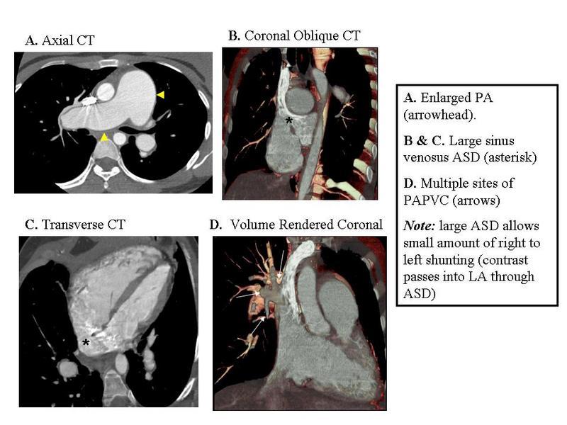
29 Pre-Operative PAPVC CT shows sinus venosus atrial septal defect (long arrow) allowing communication of the left atrium (LA) and superior vena cava (S). Anomalous pulmonary veins (short arrows) are also seen draining to the superior vena cava.
30 Pre-Operative PAPVC At a slightly higher level additional anomalous pulmonary veins (short arrows) are seen draining to the superior vena cava (long arrow).
31 Pre-Operative PAPVC CT shows persistent left superior vena cava (arrow).
32 Pre-Operative PAPVC Image through cardiac chambers shows dilated right atrium (RA) and right ventricle (RV) consistent with left to right shunt. Coronary sinus (C) is also dilated due to persistent left superior vena cava. At surgery the right superior vena cava was ligated above the junction with the most superior anomalous right pulmonary vein and the communication between the inferior superior vena cava and the right atrium was closed. Thus blood return from the anomalous pulmonary veins now drains to the inferior portion of the right superior vena cava and to the right atrium via the sinus venosus atrial septal defect.
33 Post-Operative PAPVC Post-operative CT scan shows anomalous pulmonary veins (short arrows) draining to inferior portion of superior vena cava (long arrow) to left atrium (LA) via sinus venosus atrial septal defect (asterisk).
34 Post-Operative PAPVC Reformatted coronal CT image shows anomalous pulmonary veins (P) draining to inferior segment of superior vena cava (S) to left atrium (LA) via sinus venosus atrial septal defect (asterisk).
35 Post-Operative PAPVC Reformatted coronal MIP image shows blood flow pattern above ligated portion of right superior vena cava. Superior portion of right superior vena cava (short arrow) communicates with left superior vena cava (long arrow) via collateral mediastinal veins. 55-year-old woman with PAPVC of left upper lobe pulmonary vein who had right pneumonectomy for lung cancer:
36 Post-Operative PAPVC Coronal reformatted MIP image shows anomalous left upper lobe pulmonary vein (arrow) draining to left brachiocephalic vein (B).
37 Post-Operative PAPVC Volume rendered view of vasculature from anterior perspective shows anomalous left upper lobe pulmonary vein (arrow) draining to left brachiocephalic vein (B). S = superior vena cava.
38 Post-Operative PAPVC CT Image shows that main pulmonary artery (P) is larger in diameter than aorta (A) consistent with increased pulmonary blood flow.
39 Post-Operative PAPVC Image of heart shows that right atrium (RA) and right ventricle (RV) are dilated compared to left-sided chambers consistent with significant left to right shunt. After right pneumonectomy, a larger percentage of pulmonary venous blood shunts to the right circulation through the anomalous left superior pulmonary vein.
40 Post-Operative PAPVC Patient with history of PAPVC and repair with anastamosis of anomalous vein to left atrium. Patient developed stenosis at junction [asterisk] of pulmonary vein and left atrium resulting in a large pulmonary varix [arrow].
41 Post-Operative PAPVC Volume rendered from anterior perspective shows large pulmonary varix extending throughout right lung [arrows]. 2. Conclusions After reviewing this exhibit, the participant should be familiar with the MDCT evaluation of partial anomalous pulmonary venous connection in adults. 3. References Broy C, Bennett S. Partial anomalous pulmonary venous return. Mil Med Jun;173(6):523-4 Moral S, Ortuño P, Aboal J. Multislice CT in Congenital Heart Disease: Partial Anomalous Pulmonary Venous Connection. Pediatr Cardiol Jun 28 Maillard JO, Cottin V, Etienne-Mastroïanni B, Frolet JM, Revel D, Cordier JF. Pulmonary varix mimicking
42 pulmonary arteriovenous malformation in a patient with Turner syndrome. Respiration. 2007;74(1): Sungur M, Ceyhan M, Baysal K. Partial anomalous pulmonary venous connection of left pulmonary veins to innominate vein evaluated by multislice CT. Heart Oct;93(10):1292. Haramati LB, Moche IE, Rivera VT, Patel PV, Heyneman L, McAdams HP, Issenberg HJ, White CS. Computed tomography of partial anomalous pulmonary venous connection in adults. J Comput Assist Tomogr Sep-Oct;27(5): Schatz SL, Ryvicker MJ, Deutsch AM, Cohen HR. Partial anomalous pulmonary venous drainage of the right lower lobe shown by CT scans. Radiology Apr;159(1):21-2. Greene R, Miller SW. Cross-sectional imaging of silent pulmonary venous anomalies. Radiology Apr;159(1): Personal Information Authors: Waseem A. Bhatti, M.S., M.D.¹ Pierre D. Maldjian M.D. ¹ Muhamed Saric M.D.² 1: Department of Radiology. New Jersey Medical School. University of Medicine and Dentistry of New Jersey. Newark, New Jersey. USA. 2: Department of Cardiology. New Jersey Medical School. University of Medicine and Dentistry of New Jersey. Newark, New Jersey. USA.

43 5. Mediafiles PAPVC Connection in Adults
![PAPVC Flow Direction CT in patient with heart failure from cardiomyopathy shows PAPVC of left upper lobe pulmonary veins (LUL PV) [arrows] to the left brachiocephalic vein (LBCV) [arrowhead].](/docs-images/81/83863996/images/44-0.jpg "Images were obtained early after injection of IV contrast as study was performed to exclude pulmonary embolism.")
44 PAPVC Flow Direction CT in patient with heart failure from cardiomyopathy shows PAPVC of left upper lobe pulmonary veins (LUL PV) [arrows] to the left brachiocephalic vein (LBCV) [arrowhead]. Images were obtained early after injection of IV contrast as study was performed to exclude pulmonary embolism. There is opacification of anomalous LUL PV before contrast has reached the remainder of the pulmonary veins and left sided cardiac chambers. (Prolonged circulation time from heart failure likely also contributes to lack of contrast in normal pulmonary veins.) Contrast must have entered anomalous pulmonary vein via retrograde flow from LBCV. We speculate that elevated systemic venous pressure from patient s cardiomyopathy also contributes to retrograde flow in anomalous pulmonary vein.
 [arrows] is very high due to undiluted contrast while the attenuation of the anomalous vessel [arrowheads] near its junction with the")
45 PAPVC Flow Direction With PAPVC of left upper lobe pulmonary vein, flow direction in the anomalous vessel is apparent if contrast in injected into the left arm. The attenuation of the left brachiocephalic vein (LBCV) [arrows] is very high due to undiluted contrast while the attenuation of the anomalous vessel [arrowheads] near its junction with the brachiocephalic vein is significantly lower. A low attenuation flow artifact may even be present in the LBCV due to inflow from the anomalous pulmonary vein.
![PAPVC Flow Direction CT scan in patient with history of tricuspid valve replacement for tricuspid valve endocarditis shows PAPVC [arrowhead] that forms conduit from LBCV to left inferior pulmonary](/docs-images/81/83863996/images/46-0.jpg "vein [arrow]. Flow direction in anomalous vessel is away from LBCV causing a right to left shunt.")
46 PAPVC Flow Direction CT scan in patient with history of tricuspid valve replacement for tricuspid valve endocarditis shows PAPVC [arrowhead] that forms conduit from LBCV to left inferior pulmonary vein [arrow]. Flow direction in anomalous vessel is away from LBCV causing a right to left shunt. We speculate that elevated systemic venous pressure from right heart disease is significantly contributory to this phenomenon. Note opacification of left ventricle before opacification of the right cardiac chambers.
 due to flow of contrast from LBCV to left atrium. Note the dense contrast in the anamalous pulmonary vein (curved arrow).")
47 PAPVC Flow Direction Axial image at level of main pulmonary artery shows early opacification of aorta (straight arrow) before opacification of pulmonary artery (PA) due to flow of contrast from LBCV to left atrium. Note the dense contrast in the anamalous pulmonary vein (curved arrow).
 to anomalous pulmonary vein connection (arrow).")
48 PAPVC Flow Direction Volume rendered view from the anterior prospective demonstrates the left brachiocephalic vein (L BCV) to anomalous pulmonary vein connection (arrow).

![same direction as flow in ascending aorta [arrowhead] (phase image on left with magnitude image on right).](/docs-images/81/83863996/images/49-1.jpg "PAPVC Flow Direction Note that attenuation of the anomalous pulmonary veins is less than that of the SVC")
49 PAPVC Flow Direction Oblique axial phase contrast MRI study on same patient performed after stabilization of heart failure shows direction of flow in anomalous vein [arrow] has normalized to cephalad, towards LBCV, in same direction as flow in ascending aorta [arrowhead] (phase image on left with magnitude image on right). PAPVC Flow Direction Note that attenuation of the anomalous pulmonary veins is less than that of the SVC (arrows).
![PAPVC Flow Direction Phase contrast image from MRI study confirms direction of flow in anomalous vessel [arrows] is caudad away from](/docs-images/81/83863996/images/50-0.jpg "LBCV, same direction as flow in descending thoracic aorta [arrowheads] ( phase contrast image on top with magnitude image on bottom).")
50 PAPVC Flow Direction Phase contrast image from MRI study confirms direction of flow in anomalous vessel [arrows] is caudad away from LBCV, same direction as flow in descending thoracic aorta [arrowheads] ( phase contrast image on top with magnitude image on bottom).
.")
51 PAPVC and ASD Volume rendering demonstrates extensive connections of pulmonary veins from the right lung to the superior vena cava (SVC).
52 PAPVC and ASD PAPVC and ASD

53 PAPVC and ASD CXR shows enlarged pulmonary arteries and right ventricular enlargement on lateral view.

54 PAPVC and Atrial Septal Defects Small sinus venosus ASD with dilated RA and RV

55 PAPVC of the LUL Pulmonary Vein vs. Persistent Left Superior Vena Cava Chest radiograph shows a vascular catheter coursing along the left mediastinum in a persistent left superior vena cava.

56 PAPVC of the LUL Pulmonary Vein vs. Persistent Left Superior Vena Cava Reformatted coronal MIP image shows persistent left superior vena cava (white arrow) draining to coronary sinus (black arrow).

57 PAPVC of the LUL Pulmonary Vein vs. Persistent Left Superior Vena Cava CT shows persistent left superior vena cava (arrow) anterior to the aortic arch.

58 PAPVC of the LUL Pulmonary Vein vs. Persistent Left Superior Vena Cava CT shows persistent left superior vena cava (straight arrow) anterior to left superior pulmonary vein (curved arrow).

59 PAPVC of the Left Upper Lobe Pulmonary Vein Volume rendering of PAPVC of the Left Upper Lobe Pulmonary Vein (arrowhead).
 of the Left Upper Lobe Pulmonary Vein on a chest")
60 PAPVC of the Left Upper Lobe Pulmonary Vein: CXR Findings PAPVC (arrow) of the Left Upper Lobe Pulmonary Vein on a chest radiology.

61 Partial Anomalous Pulmonary Venous Connection (PAPVC) of the Left Upper Lobe Pulmonary Vein Image at a lower level demonstrates that the left superior pulmonary vein is not seen in its normal location, anterior to the left upper lobe bronchus (asterisk).
 adjacent to the aortic arch.")
62 Partial Anomalous Pulmonary Venous Connection (PAPVC) of the Left Upper Lobe Pulmonary Vein 35-year-old man with PAPVC of the left superior pulmonary vein. CT demonstrates anomalous vessel (arrow) adjacent to the aortic arch.
 joining with left brachiocephalic vein (B).")
63 Partial Anomalous Pulmonary Venous Connection (PAPVC) of the Left Upper Lobe Pulmonary Vein Volume rendered view from anterior perspective shows anomalous left superior pulmonary vein (arrow) joining with left brachiocephalic vein (B). Arrowheads = tributaries of left upper lobe pulmonary veins.
 to left atrium (LA) via sinus venosus atrial septal defect")
64 Post-Operative PAPVC Reformatted coronal CT image shows anomalous pulmonary veins (P) draining to inferior segment of superior vena cava (S) to left atrium (LA) via sinus venosus atrial septal defect (asterisk).
 to left atrium (LA) via sinus venosus atrial septal defect (asterisk).")
65 Post-Operative PAPVC Post-operative CT scan shows anomalous pulmonary veins (short arrows) draining to inferior portion of superior vena cava (long arrow) to left atrium (LA) via sinus venosus atrial septal defect (asterisk).
 communicates with left superior vena cava (long arrow) via collateral")
66 Post-Operative PAPVC Reformatted coronal MIP image shows blood flow pattern above ligated portion of right superior vena cava. Superior portion of right superior vena cava (short arrow) communicates with left superior vena cava (long arrow) via collateral mediastinal veins.

67 Post-Operative PAPVC Volume rendered from anterior perspective shows large pulmonary varix extending throughout right lung [arrows].

68 Post-Operative PAPVC Image of heart shows that right atrium (RA) and right ventricle (RV) are dilated compared to left-sided chambers consistent with significant left to right shunt. After right pneumonectomy, a larger percentage of pulmonary venous blood shunts to the right circulation through the anomalous left superior pulmonary vein.
 draining to left brachiocephalic vein")
69 Post-Operative PAPVC Coronal reformatted MIP image shows anomalous left upper lobe pulmonary vein (arrow) draining to left brachiocephalic vein (B).

70 Post-Operative PAPVC Patient with history of PAPVC and repair with anastamosis of anomalous vein to left atrium. Patient developed stenosis at junction [asterisk] of pulmonary vein and left atrium resulting in a large pulmonary varix [arrow].
 consistent with increased pulmonary blood")
71 Post-Operative PAPVC CT Image shows that main pulmonary artery (P) is larger in diameter than aorta (A) consistent with increased pulmonary blood flow.
 draining to left brachiocephalic vein (B).")
72 Post-Operative PAPVC Volume rendered view of vasculature from anterior perspective shows anomalous left upper lobe pulmonary vein (arrow) draining to left brachiocephalic vein (B). S = superior vena cava.

73 Pre-Operative PAPVC Image through cardiac chambers shows dilated right atrium (RA) and right ventricle (RV) consistent with left to right shunt. Coronary sinus (C) is also dilated due to persistent left superior vena cava.
 are seen draining to the superior vena cava (long arrow).")
74 Pre-Operative PAPVC At a slightly higher level additional anomalous pulmonary veins (short arrows) are seen draining to the superior vena cava (long arrow).

75 Pre-Operative PAPVC CT shows persistent left superior vena cava (arrow).
 and superior vena cava (S).")
76 Pre-Operative PAPVC CT shows sinus venosus atrial septal defect (long arrow) allowing communication of the left atrium (LA) and superior vena cava (S). Anomalous pulmonary veins (short arrows) are also seen draining to the superior vena cava.

77 Strategies to detect PAPVC to SVC CXR shows cardiomegaly and enlarged pulmonary arteries.
 are more discernable with wider")
78 Strategies to detect PAPVC to SVC Note that connections of anamalous vein to SVC (arrows) are more discernable with wider windows.
.")
79 Strategies to detect PAPVC to SVC: Abrupt change in caliber of SVC CT shows dilated RA and RV, change in caliber SVC, multiple site of communication between pulmonary veins and SVC (arrows). Often with PAPVC to SVC there is more than one site of abnormal connection.
A pictorial review of normal anatomical appearences of Pericardial recesses on multislice Computed Tomography.
 A pictorial review of normal anatomical appearences of Pericardial recesses on multislice Computed Tomography. Poster No.: C-1787 Congress: ECR 2012 Type: Educational Exhibit Authors: N. Ahmed 1, G. Avery
High density thrombi of pulmonary embolism on precontrast CT scan: Is it dangerous?
 High density thrombi of pulmonary embolism on precontrast CT scan: Is it dangerous? Poster No.: C-1753 Congress: ECR 2013 Type: Authors: Keywords: DOI: Scientific Exhibit B. Y. Lee, H. R. KIM, J. I. Jung,
Coarctation of aorta in an adult-a case report
 Coarctation of aorta in an adult-a case report Poster No.: P-0057 Congress: ESTI 2014 Type: Educational Poster Authors: R. Challa, R. Ahmed; Bolton/UK Keywords: Imaging sequences, CT, Thorax, Congenital
Scientific Exhibit. Authors: D. Takenaka, Y. Ohno, Y. Onishi, K. Matsumoto, T.
 The feasibility of biphasic contrast-media-injection-protocol for chest imaging on 320-slice volume MDCT: Direct comparison of biphasic and bolus contrast-media injection protocols on 320-slice volume
64-MDCT imaging of the pancreas: Scan protocol optimisation by different scan delay regimes
 64-MDCT imaging of the pancreas: Scan protocol optimisation by different scan delay regimes Poster No.: C-051 Congress: ECR 2009 Type: Scientific Exhibit Topic: Abdominal and Gastrointestinal Authors:
Aetiologies of normal CT main pulmonary arterial (PA) measurements in patients with right heart catheter (RHC) confirmed pulmonary hypertension (PH)
 measurements in patients with right heart catheter (RHC) confirmed pulmonary hypertension (PH)") Aetiologies of normal CT main pulmonary arterial (PA) measurements in patients with right heart catheter (RHC) confirmed pulmonary hypertension (PH) Poster No.: C-0964 Congress: ECR 2010 Type: Scientific
Aetiologies of normal CT main pulmonary arterial (PA) measurements in patients with right heart catheter (RHC) confirmed pulmonary hypertension (PH) Poster No.: C-0964 Congress: ECR 2010 Type: Scientific
A Randomized Controlled Study to Compare Image Quality between Fenestrated and Non-Fenestrated Intravenous Catheters for Cardiac MDCT
 A Randomized Controlled Study to Compare Image Quality between Fenestrated and Non-Fenestrated Intravenous Catheters for Cardiac MDCT Poster No.: C-0623 Congress: ECR 2017 Type: Authors: Keywords: DOI:
AFib is the most common cardiac arrhythmia and its prevalence and incidence increases with age (Fuster V. et al. Circulation 2006).
.") Feasibility, image quality and radiation dose of coronary CT angiography (CCTA) in patients with atrial fibrillation using a new generation 256 multi-detector CT (MDCT) Poster No.: C-2378 Congress: ECR
Feasibility, image quality and radiation dose of coronary CT angiography (CCTA) in patients with atrial fibrillation using a new generation 256 multi-detector CT (MDCT) Poster No.: C-2378 Congress: ECR
Cruveilhier-Baumgarten syndrome: anatomical and pathologic imaging of periumbilical venous network
 Cruveilhier-Baumgarten syndrome: anatomical and pathologic imaging of periumbilical venous network Poster No.: C-0442 Congress: ECR 2014 Type: Educational Exhibit Authors: J. Isogai, H. Sakamoto ; Asahi/JP,
Monophasic versus biphasic contrast application in CT of patients with head and neck tumour
 Monophasic versus biphasic contrast application in CT of patients with head and neck tumour Poster No.: C-3331 Congress: ECR 2010 Type: Topic: Authors: Keywords: DOI: Scientific Exhibit Head and Neck G.
Contrast enhancement of the right ventricle during coronary CTA: is it necessary?
 Contrast enhancement of the right ventricle during coronary CTA: is it necessary? Poster No.: C-1545 Congress: ECR 2014 Type: Authors: Keywords: DOI: Scientific Exhibit M. Kok, C. Mihl, B. Kietselaer,
Preliminary experience of phlebographic studies in patients with multiple sclerosis and chronic cerebrospinal venous insufficiency
 Preliminary experience of phlebographic studies in patients with multiple sclerosis and chronic cerebrospinal venous insufficiency Poster No.: C-1715 Congress: ECR 2011 Type: Authors: Keywords: DOI: Scientific
Idiopathic dilatation of the pulmonary artery : radiographic and MDCT features in 6 cases
 Idiopathic dilatation of the pulmonary artery : radiographic and MDCT features in 6 cases Poster No.: P-0075 Congress: ESTI 2014 Type: Authors: Educational Poster J. J. Woo 1, K. Y. Lee 2, Y. Cho 1, J.
A rare case: Coronary sinus thrombosis
 A rare case: Coronary sinus thrombosis Poster No.: P-0085 Congress: ESTI 2014 Type: Educational Poster Authors: B. Özkul, N. Inan, Ö. Özkul, H. T. Sarisoy, G. Akansel, A. Akça, #. Çam; Kocaeli/TR Keywords:
Valsalva-manoeuvre or prone belly position for computed tomography (CT) scan when an orbita varix is suspected: a single-case study.
 scan when an orbita varix is suspected: a single-case study.") Valsalva-manoeuvre or prone belly position for computed tomography (CT) scan when an orbita varix is suspected: a single-case study. Poster No.: C-0512 Congress: ECR 2012 Type: Authors: Keywords: DOI:
Valsalva-manoeuvre or prone belly position for computed tomography (CT) scan when an orbita varix is suspected: a single-case study. Poster No.: C-0512 Congress: ECR 2012 Type: Authors: Keywords: DOI:
Primary epiploic appendagitis versus omental infarction : The role of MDCT
 Primary epiploic appendagitis versus omental infarction : The role of MDCT e-poster: EE-125 Congress: ESGAR 2010 Type: Educational Exhibit Topic: Diagnostic / Mesentery and Peritoneum Authors: P. Kraniotis,
Acute abdominal venous thromboses- the hyperdense noncontrast CT sign
 Acute abdominal venous thromboses- the hyperdense noncontrast CT sign Poster No.: C-1095 Congress: ECR 2011 Type: Educational Exhibit Authors: M. Goldstein, K. Jhaveri; Toronto, ON/CA Keywords: Abdomen,
Bolus administration of esmolol allows for safe and effective heart rate control during coronary computed tomography angiography
 Bolus administration of esmolol allows for safe and effective heart rate control during coronary computed tomography angiography Poster No.: C-1342 Congress: ECR 2013 Type: Scientific Exhibit Authors:
Treatment options for endoleaks: stents, embolizations and conversions
 Treatment options for endoleaks: stents, embolizations and conversions Poster No.: C-0861 Congress: ECR 2012 Type: Authors: Keywords: DOI: Scientific Exhibit G. Lombardi; napoli/it Arteries / Aorta, Abdomen,
Pulmonary infarction semiology in CT. Revision of 80 cases.
 Pulmonary infarction semiology in CT. Revision of 80 cases. Poster No.: C-0369 Congress: ECR 2012 Type: Scientific Exhibit Authors: M. González Vázquez, D. Castellon, J. Calatayud, N. Silva 1 2 1 1 1 1
Retrograde flow in the left ovarian vein is a shunt, not reflux
 Retrograde flow in the left ovarian vein is a shunt, not reflux Poster No.: C-0846 Congress: ECR 2013 Type: Scientific Exhibit Authors: R. Livsey; Brisbane/AU Keywords: Genital / Reproductive system female,
Spectrum of Findings of Sinus Venosus Atrial Septal Defect: CT and MR Findings
 Spectrum of Findings of Sinus Venosus Atrial Septal Defect: CT and MR Findings Poster No.: P-0026 Congress: ESCR 2015 Type: Scientific Poster Authors: J. M. Madrid, P. J. Mergo, P. Bartolomé, J. Phelan,
Feasibility of contrast agent volume reduction on 640-slice CT coronary angiography in patients with low heart rate
 Feasibility of contrast agent volume reduction on 640-slice CT coronary angiography in patients with low heart rate Poster No.: B-0742 Congress: ECR 2013 Type: Authors: Keywords: DOI: Scientific Paper
Lesions of the pancreaticoduodenal groove, a pictorial review
 Lesions of the pancreaticoduodenal groove, a pictorial review Poster No.: C-2131 Congress: ECR 2013 Type: Educational Exhibit Authors: E. Ni Mhurchu, L. Lavelle, I. Murphy, S. Skehan ; IE, Dublin/ IE Keywords:
Multidetector computed tomography in the evaluation of atrial septal defects
 Multidetector computed tomography in the evaluation of atrial septal defects Poster No.: C-0502 Congress: ECR 2010 Type: Educational Exhibit Topic: Cardiac Authors: S. Espejo, R. Ysamat, B. Cajal, M. Pan,
CT assessment of acute coalescent mastoiditis.
 CT assessment of acute coalescent mastoiditis. Poster No.: C-1794 Congress: ECR 2010 Type: Educational Exhibit Topic: Head and Neck Authors: A. Thomson, S. J. Thomas, A. Hutchings, E. Tilley; Portsmouth/UK
Normal and Abnormal Coronary Artery Anatomy: Is it significant?
 Normal and Abnormal Coronary Artery Anatomy: Is it significant? Poster No.: C-2112 Congress: ECR 2012 Type: Educational Exhibit Authors: M.-Y. Ng, S. Kumar, C. K. Liew, R. W. Bury; Blackpool/UK Keywords:
S. Inagawa, N. Yoshimura, Y. Ito; Niigata/JP spinal sacral areteriovenous fistulae, CTA, MRA /ecr2010/C-2581
 Localization of sacral spinal arteriovenous fistulae in reference to the dural structure with CTA and MRA of high spatial resolution: A pictorial essay Poster No.: C-2581 Congress: ECR 2010 Type: Educational
Spontaneous portosystemic venous shunts in liver cirrhosis: Anatomy, pathophysiology, hemodynamic changes and imaging findings
 Spontaneous portosystemic venous shunts in liver cirrhosis: Anatomy, pathophysiology, hemodynamic changes and imaging findings Poster No.: C-3193 Congress: ECR 2010 Type: Educational Exhibit Topic: Vascular
Tubes and lines in neonatal chest radiograph
 Tubes and lines in neonatal chest radiograph Poster No.: C-1008 Congress: ECR 2014 Type: Educational Exhibit Authors: R. TUMMA, N. AHMED, V. Prasad ; Hyderabad/IN, 1 2 1 1 2 HYDERABAD, ANDHRA PRADESH/IN
Computed tomography and Modified RECIST criteria for assessment of response in malignant pleural mesothelioma
 Computed tomography and Modified RECIST criteria for assessment of response in malignant pleural mesothelioma Poster No.: C-0729 Congress: ECR 2013 Type: Scientific Exhibit Authors: A. Marin, I. Pozek,
Anomalous origin of the right subclavian artery from main pulmonary artery
 Anomalous origin of the right subclavian artery from main pulmonary artery Award: AOSR Best Exhibit Prize - Bronze Poster No.: R-0178 Congress: RANZCR-AOCR 2012 Type: Educational Exhibit Authors: U. Chaumrattanakul,
Partial Anomalous Pulmonary Venous Drainage of the Left Upper Lobe vs Duplication of the Superior Vena Cava: Distinction
 375 Partial Anomalous Pulmonary Venous Drainage of the Left Upper Lobe vs Duplication of the Superior Vena Cava: Distinction Based on CT Findings Evan H. Dillon1 OBJECTIVE. Partial lobe Catharine Camputaro2
128-slice dual-source CT coronary angiography using highpitch scan protocols in 102 patients
 128-slice dual-source CT coronary angiography using highpitch scan protocols in 102 patients Poster No.: C-0634 Congress: ECR 2010 Type: Scientific Exhibit Topic: Cardiac Authors: Y. H. Choe, J. W. Lee,
Intraluminal gas in non-perforated acute appendicitis: a CT sign of gangrenous appendicitis
 Intraluminal gas in non-perforated acute appendicitis: a CT sign of gangrenous appendicitis Poster No.: C-978 Congress: ECR 202 Type: Scientific Exhibit Authors: D. Plata Ariza, E. MARTINEZ CHAMORRO, J.
Radiological features of Legionella Pneumophila Pneumonia
 Radiological features of Legionella Pneumophila Pneumonia Poster No.: E-0048 Congress: ESTI 2012 Type: Scientific Exhibit Authors: M. Vinciguerra, L. Stefanetti, E. Teti, G. Argentieri, L. G. 1 1 1 1 1
Individual Pulmonary Vein Atresia in Adults: Report of Two Cases
 Case Report DOI: 10.3348/kjr.2011.12.3.395 pissn 1229-6929 eissn 2005-8330 Korean J Radiol 2011;12(3):395-399 Individual Pulmonary Vein Atresia in Adults: Report of Two Cases Hyoung Nam Lee, MD, Young
Significance of MRI in diagnostics, outcome prognosis and definition the therapeutic tactics for cases of aseptic necrosis of the femoral head
 Significance of MRI in diagnostics, outcome prognosis and definition the therapeutic tactics for cases of aseptic necrosis of the femoral head Abstract: 539 Congress: ESMRMB 2013 Type: Scientific Poster
Surgical Management Of TAPVR. Daniel A. Velez, M.D. Congenital Cardiac Surgeon Phoenix Children s Hospital
 Surgical Management Of TAPVR Daniel A. Velez, M.D. Congenital Cardiac Surgeon Phoenix Children s Hospital No Disclosures Goals Review the embryology and anatomy Review Surgical Strategies for repair Discuss
Large veins of the thorax Brachiocephalic veins
 Large veins of the thorax Brachiocephalic veins Right brachiocephalic vein: formed at the root of the neck by the union of the right subclavian & the right internal jugular veins. Left brachiocephalic
ECG Gated CT Aorta in Transcatheter Aortic Valve Implantation
 ECG Gated CT Aorta in Transcatheter Aortic Valve Implantation Poster No.: C-2014 Congress: ECR 2014 Type: Educational Exhibit Authors: M. A. Ottesen; Oslo/NO Keywords: Cardiac, Arteries / Aorta, CT, CT-Angiography,
Bail out strategies after accidental Wallstent dislocation into the right atrium in patients with superior vena cava syndrome
 Bail out strategies after accidental Wallstent dislocation into the right atrium in patients with superior vena cava syndrome Poster No.: C-0613 Congress: ECR 2014 Type: Educational Exhibit Authors: P.
Postpancreatectomy Hemorrhage: Imaging and Interventional Radiological Treatment
 Postpancreatectomy Hemorrhage: Imaging and Interventional Radiological Treatment Poster No.: C-1422 Congress: ECR 2014 Type: Educational Exhibit Authors: T. Matsuura, K. Takase, T. Hasegawa, H. Ota, K.
Educational Exhibit Authors:
 Endoleaks in Abdominal Aortic Aneurysm Endoprosthesis: What radiologists need to know about Diagnostic, Characterization and Basic Management Strategies Poster No.: C-0150 Congress: ECR 2013 Type: Educational
The role of abdominal CT and MRI in detection of complications after transplantations of liver, kidney and pancreas.
 The role of abdominal CT and MRI in detection of complications after transplantations of liver, kidney and pancreas. Poster No.: C-1319 Congress: ECR 2015 Type: Educational Exhibit Authors: R. Muslimov,
Pulmonary veins CT: Imaging techniques, report and common ablation complications
 Pulmonary veins CT: Imaging techniques, report and common ablation complications Poster No.: P-0031 Congress: ESTI 2015 Type: Educational Poster Authors: E. Chavarri Ibañez, A. Caldera, P. Rodríguez Fernández,
Diffuse high-attenuation within mediastinal lymph nodes on non-enhanced CT scan: Usefulness in the prediction of benignancy
 Diffuse high-attenuation within mediastinal lymph nodes on non-enhanced CT scan: Usefulness in the prediction of benignancy Poster No.: C-1785 Congress: ECR 2012 Type: Authors: Keywords: DOI: Scientific
Lung cancer in patients with chronic empyema
 Lung cancer in patients with chronic empyema Poster No.: P-0025 Congress: ESTI 2015 Type: Scientific Poster Authors: Y. Lee, C.-K. Park; Guri/KR Keywords: Neoplasia, Biopsy, PET-CT, CT, Thorax, Lung DOI:
Ureteropelvic Junction Obstruction (UPJO) syndrome: imaging with Multidetector CT (MDCT) prior to minimally invasive treatment
 syndrome: imaging with Multidetector CT (MDCT) prior to minimally invasive treatment") Ureteropelvic Junction Obstruction (UPJO) syndrome: imaging with Multidetector CT (MDCT) prior to minimally invasive treatment Poster No.: C-1753 Congress: ECR 2011 Type: Scientific Exhibit Authors: E.
Ureteropelvic Junction Obstruction (UPJO) syndrome: imaging with Multidetector CT (MDCT) prior to minimally invasive treatment Poster No.: C-1753 Congress: ECR 2011 Type: Scientific Exhibit Authors: E.
Imaging features of VACTERL association
 Imaging features of VACTERL association Poster No.: C-2039 Congress: ECR 2013 Type: Authors: Keywords: DOI: Educational Exhibit J. Y. Kim; Dae-jeon/KR Pediatric, Cardiovascular system, Gastrointestinal
Optimal Site for Bone Graft Harvesting from the Iliac Bone
 Optimal Site for Bone Graft Harvesting from the Iliac Bone Poster No.: P-0095 Congress: ESSR 2015 Type: Scientific Poster Authors: B. Batohi 1, A. Isaac 1, J. Edwin 1, A. Hussain 1, J. Kumaraguru 1, L.
Venous Anomalies of the Thorax
 Venous Anomalies of the Thorax Poster No.: C-0266 Congress: ECR 2012 Type: Educational Exhibit Authors: L. M. GARCIA POSADA, A. Zuluaga, J. Mejía, N. Aldana ; 1 1 2 2 3 3 3 Envigado/CO, Medellin, Antioquia/CO,
Eponymous cardiovascular abnormalities- Imaging review and historical perspectives
 Eponymous cardiovascular abnormalities- Imaging review and historical perspectives Poster No.: C-2567 Congress: ECR 2015 Type: Educational Exhibit Authors: Y. Ahmed, P. Rajiah; Cleveland, Ohio/US Keywords:
in PAEDIATRIC CARDIOLOGY
 IMAGES in PAEDIATRIC CARDIOLOGY Morrison ML, 1 Sands AJ, 1 Paterson A. 2 Primitive hepatic venous plexus in a child with scimitar syndrome and pulmonary 1 Department of Paediatric Cardiology, Royal Belfast
ARDS - a must know. Page 1 of 14
 ARDS - a must know Poster No.: C-1683 Congress: ECR 2016 Type: Authors: Keywords: DOI: Educational Exhibit M. Cristian; Turda/RO Education and training, Edema, Acute, Localisation, Education, Digital radiography,
How to plan a Zenith AAA stent-graft from a CTA: Basic measurements and concepts explained
 How to plan a Zenith AAA stent-graft from a CTA: Basic measurements and concepts explained Poster No.: C-3077 Congress: ECR 2010 Type: Educational Exhibit Topic: Vascular Authors: D. V. Thomas; Northampton/UK
Seemingly isolated greater trochanter fractures do not exist
 Seemingly isolated greater trochanter fractures do not exist Poster No.: B-0950 Congress: ECR 2012 Type: Scientific Paper Authors: D. Dunker, J. H. Göthlin, M. Geijer ; Gothenburg/SE, Lund/SE Keywords:
Utility of CT angiography for pre-operative evaluation of robotic-assisted minimally invasive mitral valve surgery.
 Utility of CT angiography for pre-operative evaluation of robotic-assisted minimally invasive mitral valve surgery. Poster No.: C-2214 Congress: ECR 2014 Type: Educational Exhibit Authors: M. Muthuvelu,
Audit of CT Pulmonary Angiogram in suspected pulmonary embolism patients
 Audit of CT Pulmonary Angiogram in suspected pulmonary embolism patients Poster No.: C-2511 Congress: ECR 2012 Type: Scientific Exhibit Authors: N. D. Gupta, M. K. Heir, P. Bradding; Leicester/UK Keywords:
CT evaluation of small bowel carcinoid tumors
 CT evaluation of small bowel carcinoid tumors Poster No.: C-0060 Congress: ECR 2015 Type: Educational Exhibit Authors: N. V. V. P. Costa, L. Nascimento, T. Bilhim ; Estoril/PT, PT, 1 2 3 1 2 3 Lisbon/PT
Single ventricle on cardiac MRI
 Single ventricle on cardiac MRI Poster No.: C-0414 Congress: ECR 2011 Type: Scientific Exhibit Authors: F. Secchi, V. G. Nardella, A. Giardino, G. Di Leo, F. 1 2 1 1 1 1 2 Sardanelli ; Milano/IT, Milan/IT
The "whirl sign". Diagnostic accuracy for intestinal volvulus.
 The "whirl sign". Diagnostic accuracy for intestinal volvulus. Poster No.: C-0670 Congress: ECR 2014 Type: Scientific Exhibit Authors: M. Pire, M. Marti, A. Borobia, A. Verón; Madrid/ES Keywords: Abdomen,
Popliteal pterygium syndrome
 Popliteal pterygium syndrome Poster No.: C-1816 Congress: ECR 2011 Type: Educational Exhibit Authors: L. B. S. Santos, J. L. D. O. Schiavon, O. O. Guimaraes Neto, 1 1 2 3 1 1 C. A. P. Braga, R. S. LEMOS,
CT Demonstration of the Extracardiac Anastomoses of the Coronary Veins in Superior Vena Cava or Left Brachiocephalic Vein Obstruction
 Case Report http://dx.doi.org/10.3348/kjr.2013.14.1.132 pissn 1229-6929 eissn 2005-8330 Korean J Radiol 2013;14(1):132-137 CT Demonstration of the Extracardiac Anastomoses of the Coronary Veins in Superior
"Ultrasound measurements of the lateral ventricles in neonates: A comparison of multiple measurements methods."
 "Ultrasound measurements of the lateral ventricles in neonates: A comparison of multiple measurements methods." Poster No.: C-1557 Congress: ECR 2014 Type: Authors: Keywords: DOI: Educational Exhibit I.
Bottom up cardiac CT for CABG assessment to resolve breathing artefact
 Bottom up cardiac CT for CABG assessment to resolve breathing artefact Poster No.: C-0589 Congress: ECR 2010 Type: Educational Exhibit Topic: Cardiac Authors: P. Glass, P. Donnelly, P. Hanley, D. Higginson,
Audit of CT Pulmonary Angiogram in suspected pulmonary embolism patients
 Audit of CT Pulmonary Angiogram in suspected pulmonary embolism patients Poster No.: C-2511 Congress: ECR 2012 Type: Scientific Exhibit Authors: N. D. Gupta, M. K. Heir, P. Bradding; Leicester/UK Keywords:
Slowly growing malignant nodules and rapidly growing benign nodules: Evaluation of the value of volume doubling time
 Slowly growing malignant nodules and rapidly growing benign nodules: Evaluation of the value of volume doubling time Poster No.: C-208 Congress: ECR 2009 Type: Educational Exhibit Topic: Chest Authors:
BI-RADS 3, 4 and 5 lesions on US: Five categories and their diagnostic efficacy and pitfalls in interpretation
 BI-RADS 3, 4 and 5 lesions on US: Five categories and their diagnostic efficacy and pitfalls in interpretation e-poster: C-118 Congress: ECR 2008 Type: Educational Exhibit Topic: Breast / Ultrasound Authors:
Percutaneous transluminal angioplasty in the treatment of stenosis of hemodialysis arteriovenous fistulae: our experience
 Percutaneous transluminal angioplasty in the treatment of stenosis of hemodialysis arteriovenous fistulae: our experience Poster No.: C-3355 Congress: ECR 2010 Type: Scientific Exhibit Topic: Interventional
Our experience in the endovascular treatment of female varicocele
 Our experience in the endovascular treatment of female varicocele Poster No.: C-0347 Congress: ECR 2013 Type: Authors: Keywords: DOI: Scientific Exhibit A. Sáez de Ocáriz García, M. M. Mendigana Ramos,
Ultrasonic evaluation of superior mesenteric vein in cancer of the pancreatic head
 Ultrasonic evaluation of superior mesenteric vein in cancer of the pancreatic head Poster No.: C-1430 Congress: ECR 2012 Type: Authors: Keywords: DOI: Scientific Exhibit E. Fisenko, N. Vetsheva, E. Pershina;
Excavated pulmonary nodule: steps to diagnosis?
 Excavated pulmonary nodule: steps to diagnosis? Poster No.: C-1044 Congress: ECR 2014 Type: Authors: Keywords: DOI: Educational Exhibit W. Mnari, M. MAATOUK, A. Zrig, B. Hmida, M. GOLLI; Monastir/ TN Metastases,
Scientific Exhibit Authors: M. Sugiyama, Y. Takehara, T. Saito, N. Ooishi, M. Alley,
 Abnormal flow dynamics within the ascending aorta of the patients with aortic valve stenosis. Assessments with phase resolved three dimensional phase contrast MR image (4DFlow). Poster No.: C-2504 Congress:
Purpose. Methods and Materials. Results
 Prevalence and significance of hypoattenuating hepatic lesions deemed too small to characterise: How are we following up these lesions and what are the outcomes? Poster No.: C-014 Congress: ECR 2009 Type:
Comparison of Cardiac MDCT with MRI and Echocardiography in the Assessement of Left Ventricular Function
 Comparison of Cardiac MDCT with MRI and Echocardiography in the Assessement of Left Ventricular Function Poster No.: C-0969 Congress: ECR 2012 Type: Scientific Exhibit Authors: B. Kara, Y. Paksoy, C. Erol,
What every radiologist should know about cardiac CT: A case-based pictorial review
 What every radiologist should know about cardiac CT: A case-based pictorial review Poster No.: C-0555 Congress: ECR 2010 Type: Educational Exhibit Topic: Cardiac Authors: C. M. Capuñay, P. Carrascosa,
Cavitary lung lesion: Two different diagnosis with similar appearence
 Cavitary lung lesion: Two different diagnosis with similar appearence Poster No.: P-0043 Congress: ESTI 2015 Type: Educational Poster Authors: M. Yesildag, H. Kalkan, K. Ödev; Konya/TR Keywords: Infection,
Artifact in Head CT Images Due to Air Bubbles in X-Ray Tube Oil
 Artifact in Head CT Images Due to Air Bubbles in X-Ray Tube Oil Poster No.: C-0671 Congress: ECR 2016 Type: Educational Exhibit Authors: H. Patel 1, W. Liu 2, J. DeSanto 2, S. Meagher 2, M. Zagardo 2,
Low-dose computed tomography (CT) protocol in the screening of patients with social exposure to asbestos
 protocol in the screening of patients with social exposure to asbestos") Low-dose computed tomography (CT) protocol in the screening of patients with social exposure to asbestos Poster No.: C-3032 Congress: ECR 2010 Type: Scientific Exhibit Topic: Radiographers Authors: P.
Low-dose computed tomography (CT) protocol in the screening of patients with social exposure to asbestos Poster No.: C-3032 Congress: ECR 2010 Type: Scientific Exhibit Topic: Radiographers Authors: P.
Diffuse Alveolar Hemorrhage: Initial and Follow-up HRCT Features
 Diffuse Alveolar Hemorrhage: Initial and Follow-up HRCT Features Poster No.: E-0037 Congress: ESTI 2012 Type: Authors: Keywords: Scientific Exhibit M. Y. Kim; Seoul/KR Lung, CT-High Resolution, CT, Computer
Application of a flow chart to evaluate placement of umbilical venous catheters on frontal radiogram in radiologist resident
 Application of a flow chart to evaluate placement of umbilical venous catheters on frontal radiogram in radiologist resident Poster No.: C-0883 Congress: ECR 2015 Type: Scientific Exhibit Authors: S. Salerno,
Identification and numbering of lumbar vertebrae using various anatomical landmarks on MRI of lumbosacral spine
 Identification and numbering of lumbar vertebrae using various anatomical landmarks on MRI of lumbosacral spine Poster No.: C-2125 Congress: ECR 2015 Type: Authors: Scientific Exhibit S. patil 1, A. M.
Application of three-dimensional angiography in elderly patients with meningioma
 Application of three-dimensional angiography in elderly patients with meningioma Poster No.: C-0123 Congress: ECR 2012 Type: Scientific Paper Authors: X. Han, J. Chen, K. Shi; Haikou/CN Keywords: Neuroradiology
Chapter 2 Cardiac Interpretation of Pediatric Chest X-Ray
 Chapter 2 Cardiac Interpretation of Pediatric Chest X-Ray Ra-id Abdulla and Douglas M. Luxenberg Key Facts The cardiac silhouette occupies 50 55% of the chest width on an anterior posterior chest X-ray
3D ultrasound applied to abdominal aortic aneurysm: preliminary evaluation of diameter measurement accuracy
 3D ultrasound applied to abdominal aortic aneurysm: preliminary evaluation of diameter measurement accuracy Poster No.: C-0493 Congress: ECR 2011 Type: Authors: Keywords: DOI: Scientific Paper A. LONG
MDCT signs differentiating retroperitoneal and intraperitoneal lesions- diagnostic pearls
 MDCT signs differentiating retroperitoneal and intraperitoneal lesions- diagnostic pearls Poster No.: C-0987 Congress: ECR 2015 Type: Educational Exhibit Authors: D. V. Bhargavi, R. Avantsa, P. Kala; Bangalore/IN
A time-honored but almost forgotten sign of COPD: sabersheath trachea as a marker of severe airflow obstruction
 A time-honored but almost forgotten sign of COPD: sabersheath trachea as a marker of severe airflow obstruction Poster No.: C-0958 Congress: ECR 2013 Type: Scientific Exhibit Authors: F. Ciccarese, A.
Pulmonary changes induced by radiotherapy. HRCT findings
 Pulmonary changes induced by radiotherapy. HRCT findings Poster No.: C-2299 Congress: ECR 2015 Type: Educational Exhibit Authors: R. E. Correa Soto, M. Albert Antequera, K. Müller Campos, D. 1 2 4 3 1
Scientific Exhibit Authors:
 Clinical Audit on Optimization of Radiation Dose from MDCT: Effect on Diagnostic Reference Levels for Brain, Sinus, Cervical Spine, Chest, Abdomen-Pelvis, and Lumbar Spine Examinations and on Nationwide
Biliary tree dilation - and now what?
 Biliary tree dilation - and now what? Poster No.: C-1767 Congress: ECR 2012 Type: Educational Exhibit Authors: I. Ferreira, A. B. Ramos, S. Magalhães, M. Certo; Porto/PT Keywords: Pathology, Diagnostic
Duret hemorraghe caused by traumatic brain injury: what the radiologist should know.
 Duret hemorraghe caused by traumatic brain injury: what the radiologist should know. Poster No.: C-1270 Congress: ECR 2012 Type: Educational Exhibit Authors: P. Dewachter 1, T. Vanderhasselt 1, K. De Smet
Integrating CT in minimally invasive treatment of the coronary arteries
 Integrating CT in minimally invasive treatment of the coronary arteries e-poster: C-200 Congress: ECR 2008 Type: Scientific Exhibit Topic: Cardiac / Miscellaneous MeSH: Keywords: Coronary Vessels [A07.231.114.269]
When to suspect Wegener Granulomatosis: A radiologic review
 When to suspect Wegener Granulomatosis: A radiologic review Poster No.: P-0038 Congress: ESTI 2015 Type: Educational Poster Authors: A. Tilve Gómez, R. Díez Bandera, P. Rodríguez Fernández, M. Garcia Vazquez-Noguerol,
This is the left, right?
 This is the left, right? Poster No.: C-1214 Congress: ECR 2013 Type: Educational Exhibit Authors: L.-L. Huang, L. Mitchell, S. Andronikou, F. Suleman, Z. I. Lockhat; Pretoria/ZA Keywords: Congenital, Diagnostic
Overview of physiological post-mortem alterations in totalbody imaging of 100 in-hospital deceased patients
 Overview of physiological post-mortem alterations in totalbody imaging of 100 in-hospital deceased patients Poster No.: C-1234 Congress: ECR 2016 Type: Scientific Exhibit Authors: I. Wagensveld, W. Oosterhuis,
PI-RADS classification: prognostic value for prostate cancer grading
 PI-RADS classification: prognostic value for prostate cancer grading Poster No.: C-1622 Congress: ECR 2014 Type: Scientific Exhibit Authors: I. Platzek, A. Borkowetz, T. Paulus, T. Brauer, M. Wirth, M.
Computed tomographic dacryocystography as compared with X-ray dacryocystography in patients with dacryostenosis
 Computed tomographic dacryocystography as compared with X-ray dacryocystography in patients with dacryostenosis Poster No.: C-1887 Congress: ECR 2016 Type: Authors: Keywords: DOI: Educational Exhibit M.
Purpose. Methods and Materials
 Thin-section CT findings in peripheral lung cancer of 3 cm or smaller: are there any characteristic features for predicting tumor histology or do they depend only on tumor size? Poster No.: C-1893 Congress:
Comparison of multidetector-row computed tomography findings of IgG4-related sclerosing cholangitis and cholangiocarcinoma
 Comparison of multidetector-row computed tomography findings of IgG4-related sclerosing cholangitis and cholangiocarcinoma Poster No.: C-0245 Congress: ECR 2014 Type: Scientific Exhibit Authors: M. Yata,