Myocardial Strain Imaging in Cardiac Diseases and Cardiomyopathies.
|
|
|
- Lesley McCormick
- 5 years ago
- Views:
Transcription
1 Myocardial Strain Imaging in Cardiac Diseases and Cardiomyopathies. Session: Cardiomyopathy Tarun Pandey MD, FRCR. Associate Professor University of Arkansas for Medical Sciences
2 Disclosures No relevant financial disclosures Research Support Circle CVI PIA Medical
3 Outline/Educational Objectives What is myocardial strain? Why measure myocardial strain? How do we measure myocardial strain? Echo (TDI versus Speckle tracking) MRI Pearls and Pitfalls Clinical Utility & Applications Current literature and Utility of Strain Imaging
4 What is myocardial strain? Strain is defined as the percent change in myocardial deformation. Compression (Shortening) Distraction (Lengthening) This deformation can be conveniently divided into the basic components: longitudinal, radial, circumferential motion.

5 Myocardial Deformation & Strain Theodore P. Abraham et al. Circulation. 2007;116: Copyright American Heart Association, Inc. All rights reserved.
6 Myocardial strain Several strain derivatives can also be calculated: Tissue velocity (velocity of deformation) Strain rate (rate of myocardial deformation) Displacement (displacement of the myocardium during deformation)
7 Why do we need to measure myocardial strain? 1. Detection of altered myocardial performance beyond EF Ejection Fraction Myocardial Strain/Strain rate Limited as a measure of contractility due to load dependency. Limited prognostic value in low-normal or higher range. By the time EF decreases, it is too late! Less Preload dependent Greater utility in those with preserved EF Allows better understanding of progression of preclinical disease to overt heart failure.
8 Why do we need to measure myocardial strain? 1. Detection of altered myocardial performance beyond EF Advanced Age Obesity Hypertension Renal Failure Diabetes Atrial fibrillation Cardiomyopathies Carriers of HCM mutations Duchenne Muscular Dys. Fabry s Valvular heart disease Severe AS AR MS Acute CAD After MI, global strain values may predict subsequent remodeling and function recovery
9 How to measure myocardial strain? Can be measured on Echo and MRI most experience with echocardiography. Two main methods on Echo: Tissue Doppler Imaging Conversion of tissue velocity data to strain and strain rate Speckle tracking Tracking of echo speckles using computer post processing
10 Strain Rate: TDI ε = V1-V2/L ε=strain rate, V1=velocity at point 1, V2=velocity at point 2, and L=length, usually set at 10 mm.

11 TDI: Myocardial strain & Derivatives
 divided by the original length (L0) and expressed as a percentage.")
12 Strain: Speckle Tracking Strain is calculated as the change in length (L) divided by the original length (L0) and expressed as a percentage.
13 Echocardiographic Myocardial Strain Techniques TDI Indirect measure of strain Derived from myocardial velocities Based on Doppler principle Dependent on angle of insonation Speckle Tracking Direct measure of strain Measures myocardial displacement Based on computer-based tracking of speckles Independent on angle of insonation.
14 Myocardial Strain: MRI techniques Grid tagged MRI Feature Tracking


15 Grid Tagged MRI Fading of grid tags Needs prospective application of tags Cannot use regular Cine Images
16


17 MRI Feature Tracking: LV
18 MRI Feature Tracking: RV


19 RV Strain: Feature Tracking
20 Myocardial strain: Considerations LV Strain Easier to perform Thicker LV walls LV chamber is simpler Greater utility in health and disease RV Strain Technically difficult Thin RV walls Hyper-trabeculated RV More irregular shape and volume Lesser clinical applications No reliable non-invasive surrogate
21 LA Strain

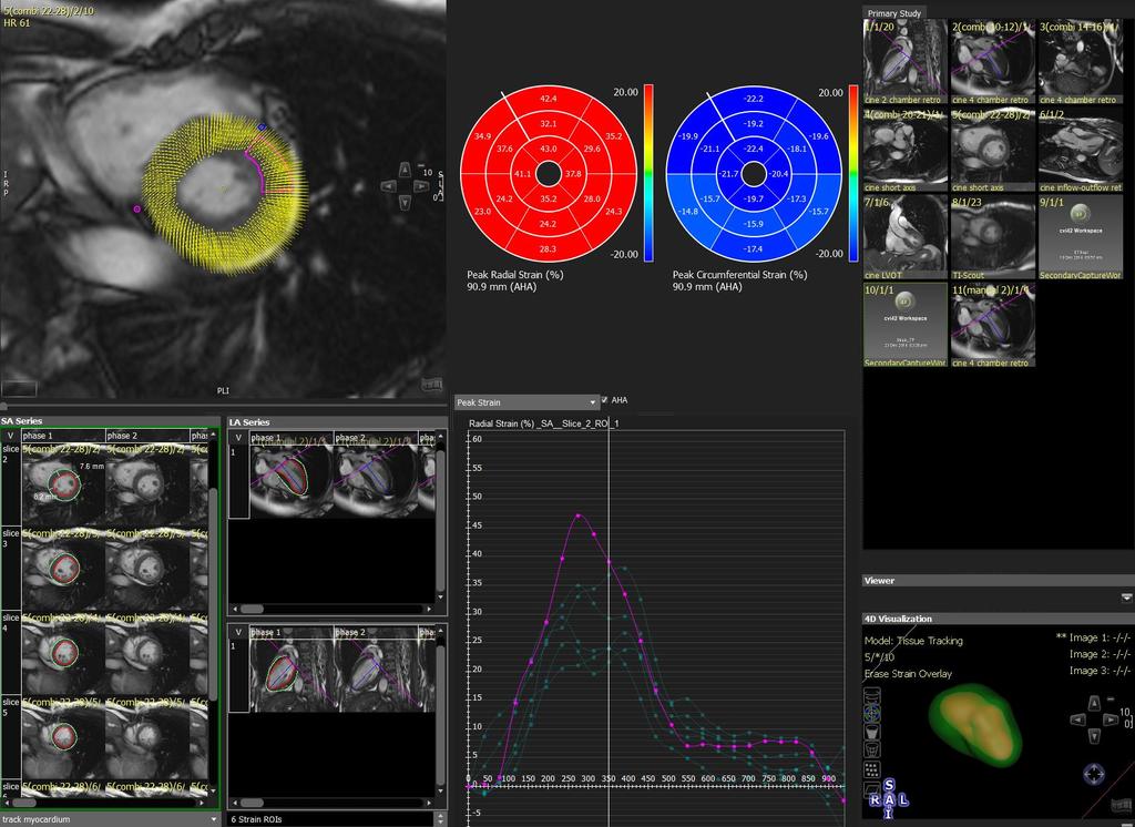
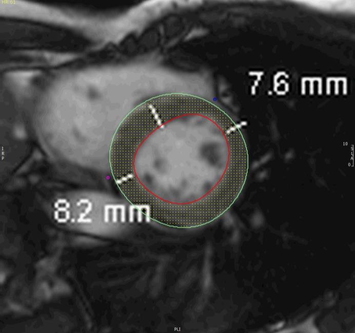
22 Strain: MRI Technique, Pearls & Pitfalls Image Selection Contouring Quality Assurance


23 Strain: MRI Technique, Pearls & Pitfalls Image Selection Use series composer tool to create a full SAX stack Exclude basal images with significant tricuspid or pulmonic systolic valve presence. Exclude apical slices with too much through-plane motion. Select 4CH image most central to the tricuspid valve and which has the clearest myocardium

, but not the epicardial.")
24 Strain: MRI Technique, Pearls & Pitfalls Contouring All images contoured at end diastole LAX: The septum is excluded. The basal bulge is included as long as it was able to be tracked as well as the free wall myocardium. A LAX RV extent contour (orange) is also used to define the systolic excursion of the tricuspid valve. SAX: The septum is included in the endocardial contour (since it must be a closed surface), but not the epicardial. Nonetheless, like with the 4CH view, only the free wall myocardium is tracked for strain analysis.

25 Strain: MRI Technique, Pearls & Pitfalls Quality Assurance Lengthiest process Use of the cinematic loop is essential to QA. Enable boundary points overlay to assess this. The boundary points reflect the movement of the tracking algorithm, and any time this does not appear to reflect the actual motion of the myocardium, the contours are adjusted to improve the tracking

26 Strain: MRI Technique, Pearls & Pitfalls Quality Assurance If the boundary points move too far in, and end up in the blood pool at systole (green circle), then the contours must be adjusted outwardly. If the boundary points are too far, out, ending up extracardiac during systole (green circle), then the contours must be adjusted inwardly.
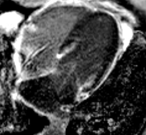
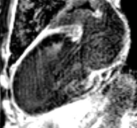
27 Strain: MRI Technique, Pearls & Pitfalls Quality Assurance Exclude large artifacts that obscure entire portions of myocardium, yielding nonsensical results from analysis: brightness from the fat obscures the free wall anterolaterally, giving results which are clearly erroneous, with negative radial strain rates in that region.

28 Strain: MRI Technique, Pearls & Pitfalls Quality Assurance Exclude large artifacts that obscure entire portions of myocardium, yielding nonsensical results from analysis: brightness from the fat obscures the free wall anterolaterally, giving results which are clearly erroneous, with negative radial strain rates in that region. The purple region in the ES image, with very poor tracking, can be seen in this regional radial strain graph as a purple trend line, giving paradoxical results
29 Strain: MRI Technique, Pearls & Pitfalls Quality Assurance Exclude large artifacts : As a solution, the entire anterolateral portion of the RV myocardium should be excluded (ED and ES) Caveat: exclusion will render segmental strain analysis incorrect
30 Myocardial Strain: Clinical Applications


31 Gorcsan J et al. J Am Coll Cardiol 2011;58: #1. Heart Failure Normal Heart Failure

32 Longitudinal RV strain in Heart Failure Normal Heart Failure


33 Circ. RV strain in Heart Failure Normal Heart Failure


34 Radial RV strain in Heart Failure Normal Heart Failure


35 Gorcsan J et al. J Am Coll Cardiol 2011;58: #2. Ventricular Dyssynchrony (Radial LV Strain) Normal Heart failure with LBBB

")
36 Gorcsan J et al. J Am Coll Cardiol 2011;58: Ventricular Dyssynchrony (Circumferential LV Strain) Normal Heart failure with LBBB


37 Strain: Ventricular Dyssynchrony Normal Heart Failure Radial Circumferential
38 #3. Cardiomyopathy

39 Myocardial Strain in Cardiomyopathies Normal Myocardial Amyloidosis
40 LV Strain in Cardiac Amyloidosis Amyloid Normal Amyloid Normal Evaluation of Myocardial Strain in Patients With Amyloidosis Using Cardiac Magnetic Resonance Feature Tracking. Pandey et al, CPDR, 2016
41 RV strain in Myocardial Amyloidosis p= p= p< UAMS Data


42 Apical Sparing in Cardiac Amyloidosis
43 #4: Myocarditis/Pericarditis
44 #4 Myocarditis Vs Pericarditis


45 #4 Myocarditis Vs Pericarditis Myocarditis Pericarditis
46 #4 Myocarditis Vs Pericarditis Pericarditis Myocarditis


47 #4 Myocarditis Vs Pericarditis Myocarditis Pericarditis
48 Conclusions Myocardial strain evaluation with CMR is feasible Evolving field with several challenges Technique Paucity of normative Data Vendor variability Large scale data on clinical utility is lacking. Offers a great opportunity for research and with further refinement it will establish as a useful technique.
49 Thank you
50 Acknowledgement PIA Medical Imaging Sam Alkek. Circle CVI Ron Skostad
Global left ventricular circumferential strain is a marker for both systolic and diastolic myocardial function
 Global left ventricular circumferential strain is a marker for both systolic and diastolic myocardial function Toshinari Onishi 1, Samir K. Saha 2, Daniel Ludwig 1, Erik B. Schelbert 1, David Schwartzman
Alicia Armour, MA, BS, RDCS
 Alicia Armour, MA, BS, RDCS No disclosures Review 2D Speckle Strain (briefly) Discuss some various patient populations & disease pathways where Strain can be helpful Discuss how to acquire images for Strain
2/2/2011. Strain and Strain Rate Imaging How, Why and When? Movement vs Deformation. Doppler Myocardial Velocities. Movement. Deformation.
 Strain and Strain Rate Imaging How, Why and When? João L. Cavalcante, MD Advanced Cardiac Imaging Fellow Cleveland Clinic Foundation Disclosures: No conflicts of interest Movement vs Deformation Movement
Restrictive Cardiomyopathy
 ESC Congress 2011, Paris Imaging Unusual Causes of Cardiomyopathy Restrictive Cardiomyopathy Kazuaki Tanabe, MD, PhD Professor of Medicine Chair, Division of Cardiology Izumo, Japan I Have No Disclosures
VECTORS OF CONTRACTION
 1/3/216 Strain, Strain Rate, and Torsion: Myocardial Mechanics Simplified and Applied VECTORS OF CONTRACTION John Gorcsan, MD University of Pittsburgh, Pittsburgh, PA Shortening Thickening Twisting No
Velocity Vector Imaging as a new approach for cardiac magnetic resonance: Comparison with echocardiography
 Velocity Vector Imaging as a new approach for cardiac magnetic resonance: Comparison with echocardiography Toshinari Onishi 1, Samir K. Saha 2, Daniel Ludwig 1, Erik B. Schelbert 1, David Schwartzman 1,
Strain and Strain Rate Imaging How, Why and When?
 Strain and Strain Rate Imaging How, Why and When? João L. Cavalcante, MD Advanced Cardiac Imaging Fellow Cleveland Clinic Foundation Disclosures: No conflicts of interest Movement vs Deformation Movement
Imaging in Heart Failure: A Multimodality Approach. Thomas Ryan, MD
 Imaging in Heart Failure: A Multimodality Approach Thomas Ryan, MD Heart Failure HFrEF HFpEF EF50% Lifetime risk 20% Prevalence 6M Americans Societal costs - $30B 50% 5-year survival 1 Systolic
HYPERTROPHY: Behind the curtain. V. Yotova St. Radboud Medical University Center, Nijmegen
 HYPERTROPHY: Behind the curtain V. Yotova St. Radboud Medical University Center, Nijmegen Disclosure of interest: none Relative wall thickness (cm) M 0.22 0.42 0.43 0.47 0.48 0.52 0.53 F 0.24 0.42 0.43
Incorporating the New Echo Guidelines Into Everyday Practice
 Incorporating the New Echo Guidelines Into Everyday Practice Clinical Case RIGHT VENTRICULAR FAILURE Gustavo Restrepo MD President Elect Interamerican Society of Cardiology Director Fellowship Training
LV FUNCTION ASSESSMENT: WHAT IS BEYOND EJECTION FRACTION
 LV FUNCTION ASSESSMENT: WHAT IS BEYOND EJECTION FRACTION Jamilah S AlRahimi Assistant Professor, KSU-HS Consultant Noninvasive Cardiology KFCC, MNGHA-WR Introduction LV function assessment in Heart Failure:
Velocity, strain and strain rate: Doppler and Non-Doppler methods. Thoraxcentre, Erasmus MC,Rotterdam
 Velocity, strain and strain rate: Doppler and Non-Doppler methods J Roelandt J. Roelandt Thoraxcentre, Erasmus MC,Rotterdam Basics of tissue Doppler imaging Instantaneous annular velocity profiles IVCT
Tissue Doppler and Strain Imaging. Steven J. Lester MD, FRCP(C), FACC, FASE
, FACC, FASE") Tissue Doppler and Strain Imaging Steven J. Lester MD, FRCP(C), FACC, FASE Relevant Financial Relationship(s) None Off Label Usage None a. Turn the wall filters on and turn down the receiver gain. b. Turn
Tissue Doppler and Strain Imaging Steven J. Lester MD, FRCP(C), FACC, FASE Relevant Financial Relationship(s) None Off Label Usage None a. Turn the wall filters on and turn down the receiver gain. b. Turn
Three-dimensional Wall Motion Tracking:
 Three-dimensional Wall Motion Tracking: A Novel Echocardiographic Method for the Assessment of Ventricular Volumes, Strain and Dyssynchrony Jeffrey C. Hill, BS, RDCS, FASE Jennifer L. Kane, RCS Gerard
Feasibility and limitations of 2D speckle tracking echocardiography
 ORIGINAL ARTICLE 204 A prospective study in daily clinical practice Feasibility and limitations of 2D speckle tracking echocardiography Lina Melzer, Anja Faeh-Gunz, Barbara Naegeli, Burkhardt Seifert*,
Nancy Goldman Cutler, MD Beaumont Children s Hospital Royal Oak, Mi
 Nancy Goldman Cutler, MD Beaumont Children s Hospital Royal Oak, Mi Identify increased LV wall thickness (WT) Understand increased WT in athletes Understand hypertrophic cardiomyopathy (HCM) Enhance understanding
How To Perform Strain Imaging; Step By Step Approach. Maryam Bo Khamseen Echotechnoligist II EACVI, ARDMS, RCS King Abdulaziz Cardiac Center- Riyadh
 How To Perform Strain Imaging; Step By Step Approach Maryam Bo Khamseen Echotechnoligist II EACVI, ARDMS, RCS King Abdulaziz Cardiac Center- Riyadh Outlines: Introduction Describe the basic of myocardium
Strain Imaging: Myocardial Mechanics Simplified and Applied
 9/28/217 Strain Imaging: Myocardial Mechanics Simplified and Applied John Gorcsan III, MD Professor of Medicine Director of Clinical Research Division of Cardiology VECTORS OF CONTRACTION Shortening Thickening
Novel echocardiographic modalities: 3D echo, speckle tracking and strain rate imaging. Potential roles in sports cardiology. Stefano Caselli, MD, PhD
 Novel echocardiographic modalities: 3D echo, speckle tracking and strain rate imaging. Potential roles in sports cardiology. Stefano Caselli, MD, PhD Ospedale San Pietro Fatebenefratelli Rome, Italy Differential
DISCLOSURE. Myocardial Mechanics. Relevant Financial Relationship(s) Off Label Usage
 Off Label Usage") 7th Annual Team Echocardiography: The Heart of Cardiovascular Medicine Tissue Doppler, Strain, Speckle: What? How? Christopher J Kramer RDCS Aurora Medical Group Advanced Cardiovascular Services, Aurora
7th Annual Team Echocardiography: The Heart of Cardiovascular Medicine Tissue Doppler, Strain, Speckle: What? How? Christopher J Kramer RDCS Aurora Medical Group Advanced Cardiovascular Services, Aurora
Quantitation of right ventricular dimensions and function
 SCCS Basics of cardiac assessment Quantitation of right ventricular dimensions and function Tomasz Kukulski, MD PhD Dept of Cardiology, Congenital Heart Disease and Electrotherapy Silesian Medical University
Strain/Untwisting/Diastolic Suction
 What Is Diastole and How to Assess It? Strain/Untwisting/Diastolic Suction James D. Thomas, M.D., F.A.C.C. Cardiovascular Imaging Center Department of Cardiology Cleveland Clinic Foundation Cleveland,
RIGHT VENTRICULAR SIZE AND FUNCTION
 RIGHT VENTRICULAR SIZE AND FUNCTION Edwin S. Tucay, MD, FPCC, FPCC, FPSE Philippine Society of Echocardiography Quezon City, Philippines Echo Mission, BRTTH, Legaspi City, July 1-2, 2016 NO DISCLOSURE
Cardiac Chamber Quantification by Echocardiography
 Cardiac Chamber Quantification by Echocardiography Maryam Bokhamseen, RCS, RCDS, EACVI Echotechnologist ǁ, Non invasive Cardiac Laboratory King Abdulaziz Cardiac Center. Outline: Introduction. Background
Ιπποκράτειες μέρες καρδιολογίας Θεσσαλονίκη, 9-10 Μαρτίου Φωτεινή Α. Λαζαρίδου Επιμελήτρια Α Γενικό Νοσοκομείο Αγιος Παύλος, Θεσσαλονίκη
 Ιπποκράτειες μέρες καρδιολογίας Θεσσαλονίκη, 9-10 Μαρτίου 2018 Φωτεινή Α. Λαζαρίδου Επιμελήτρια Α Γενικό Νοσοκομείο Αγιος Παύλος, Θεσσαλονίκη RV shape Triangular shape in frontal plane crescent shape in
How NOT to miss Hypertrophic Cardiomyopathy? Adaya Weissler-Snir, MD University Health Network, University of Toronto
 How NOT to miss Hypertrophic Cardiomyopathy? Adaya Weissler-Snir, MD University Health Network, University of Toronto Introduction Hypertrophic cardiomyopathy is the most common genetic cardiomyopathy,
DELAYED ENHANCEMENT IMAGING IN CHILDREN
 NASCI 38 TH ANNUAL MEENG, SEATLE October 3-5, 21 1. DELAYED ENHANCEMENT IN CHILDREN Shi-Joon Yoo, MD Lars Grosse-Wortmann, MD University of Toronto Canada -1. 1. 1. Magnitude image Magnitude images -1.
Cardiac Health in Duchenne: What we are Learning from Cardiac MRI
 Cardiac Health in Duchenne: What we are Learning from Cardiac MRI 24 th Annual Duchenne Connect Conference Parent Project Muscular Dystrophy Scottsdale, AZ June 29, 2018 Kan N. Hor, MD Director of Cardiac
Left atrial function. Aliakbar Arvandi MD
 In the clinic Left atrial function Abstract The left atrium (LA) is a left posterior cardiac chamber which is located adjacent to the esophagus. It is separated from the right atrium by the inter-atrial
Right Ventricular Strain in Normal Healthy Adult Filipinos: A Retrospective, Cross- Sectional Pilot Study
 Right Ventricular Strain in Normal Healthy Adult Filipinos: A Retrospective, Cross- Sectional Pilot Study By Julius Caesar D. de Vera, MD Jonnah Fatima B. Pelat, MD Introduction Right ventricle contributes
Tissue Doppler and Strain Imaging
 Tissue Doppler and Strain Imaging Steven J. Lester MD, FRCP(C), FACC, FASE Relevant Financial Relationship(s) None Off Label Usage None 1 Objective way with which to quantify the minor amplitude and temporal
Tissue Doppler and Strain Imaging
 Tissue Doppler and Strain Imaging Steven J. Lester MD, FRCP(C), FACC, FASE Relevant Financial Relationship(s) None Off Label Usage None 1 Objective way with which to quantify the minor amplitude and temporal
10/7/2013. Systolic Function How to Measure, How Accurate is Echo, Role of Contrast. Thanks to our Course Director: Neil J.
 Systolic Function How to Measure, How Accurate is Echo, Role of Contrast Neil J. Weissman, MD MedStar Health Research Institute & Professor of Medicine Georgetown University Washington, D.C. No Disclosures
Tissue Doppler Imaging in Congenital Heart Disease
 Tissue Doppler Imaging in Congenital Heart Disease L. Youngmin Eun, M.D. Department of Pediatrics, Division of Pediatric Cardiology, Kwandong University College of Medicine The potential advantage of ultrasound
Utility of Echocardiography
 Hypertrophic Cardiomyopathy and Beyond- Echo Hawaii 2018 Lawrence Rudski MD FRCPC FACC FASE Professor of Medicine Director, Division of Cardiology and Azrieli Heart Center Jewish General Hospital, McGill
Athlete s Heart vs. Cardiomyopathy
 Athlete s Heart vs. Cardiomyopathy Linda D. Gillam, MD, MPH, FASE Chair, Department of Cardiovascular Medicine Medical Director, Cardiovascular Service Line Former Team Cardiologist to the New York Jets
22 nd Annual Conference of the Saudi Heart Association Riyadh, Saudi Arabia
 22 nd Annual Conference of the Saudi Heart Association Riyadh, Saudi Arabia New Echocardiographic Modalities to Evaluate Ventricular Function in Congenital Heart Disease: Tissue Doppler & Strain Rate Imaging
L ecocardiografia nello Scompenso Cardiaco Acuto e cronico: vecchi dogmi e nuovi trends.
 V SESSIONE SCOMPENSO CARDIACO 2015 Genova, 13-14 Novembre 2015 L ecocardiografia nello Scompenso Cardiaco Acuto e cronico: vecchi dogmi e nuovi trends. Gian Paolo Bezante, MD, FACC UOC Clinica di Malattie
Echocardiographic assessment of the right ventricle in paediatric pulmonary hypertension.
 Echocardiographic assessment of the right ventricle in paediatric pulmonary hypertension. Mark K. Friedberg, MD No disclosures Outline RV response to increased afterload Echo assessment of RV function
Review Article Transthoracic Echocardiography in Children and Young Adults with Congenital Heart Disease
 International Scholarly Research Network ISRN Pediatrics Volume 2012, Article ID 753481, 15 pages doi:10.5402/2012/753481 Review Article Transthoracic Echocardiography in Children and Young Adults with
Little is known about the degree and time course of
 Differential Changes in Regional Right Ventricular Function Before and After a Bilateral Lung Transplantation: An Ultrasonic Strain and Strain Rate Study Virginija Dambrauskaite, MD, Lieven Herbots, MD,
Chamber Quantitation Guidelines - Update II
 Chamber Quantitation Guidelines - Update II Right Heart Measurements Steven A. Goldstein MD FACC FASE Professor of Medicine Georgetown University Medical Center MedStar Heart Institute Washington Hospital
Chamber Quantitation Guidelines II Right Heart Measurements
 Chamber Quantitation Guidelines II Right Heart Measurements Steven A. Goldstein MD FACC FASE Director, Noninvasive Cardiology MedStar Heart Institute Washington Hospital Center Sunday, October 9, 2016
WHAT DO ELECTROPHYSIOLOGISTS WANT TO KNOW FROM ECHOCARDIOGRAPHERS BEFORE, DURING&AFTER CARDIAC RESYNCHRONIZATION THERAPY?
 WHAT DO ELECTROPHYSIOLOGISTS WANT TO KNOW FROM ECHOCARDIOGRAPHERS BEFORE, DURING&AFTER CARDIAC RESYNCHRONIZATION THERAPY? Mary Ong Go, MD, FPCP, FPCC, FACC OUTLINE What is CRT Who needs CRT What does the
Evalua&on)of)Le-)Ventricular)Diastolic) Dysfunc&on)by)Echocardiography:) Role)of)Ejec&on)Frac&on)
of)Le-)Ventricular)Diastolic) Dysfunc&on)by)Echocardiography:) Role)of)Ejec&on)Frac&on)") Evalua&on)of)Le-)Ventricular)Diastolic) Dysfunc&on)by)Echocardiography:) Role)of)Ejec&on)Frac&on) N.Koutsogiannis) Department)of)Cardiology) University)Hospital)of)Patras)! I have no conflicts of interest
Evalua&on)of)Le-)Ventricular)Diastolic) Dysfunc&on)by)Echocardiography:) Role)of)Ejec&on)Frac&on) N.Koutsogiannis) Department)of)Cardiology) University)Hospital)of)Patras)! I have no conflicts of interest
Stress, strain and contrast. UK available agents. Safety 13/06/2018. Which enhancing agent do you use? Ultrasound enhancing agents.
 Stress, strain and contrast Stephen Glen Ultrasound enhancing agents Safety Effectiveness during stress Perfusion / myocardial contrast UK available agents Which enhancing agent do you use? Name Bubble
The importance of left atrium in LV diastolic function
 II Baltic Heart Failure Meeting and Congress of Latvian Society of Cardiology The importance of left atrium in LV diastolic function Dr. Artem Kalinin Eastern Clinical University Hospital Riga 30.09.2010.
MYOCARDIAL STRAIN MEASUREMENTS WITH MRI USING FEATURE TRACKING
 MYOCARDIAL STRAIN MEASUREMENTS WITH MRI USING FEATURE TRACKING Mercy Kataike Victoria MSc in Physics Submission date: May 2016 Supervisor: Pål Erik Goa, IFY Norwegian University of Science and Technology
Managing Hypertrophic Cardiomyopathy with Imaging. Gisela C. Mueller University of Michigan Department of Radiology
 Managing Hypertrophic Cardiomyopathy with Imaging Gisela C. Mueller University of Michigan Department of Radiology Disclosures Gadolinium contrast material for cardiac MRI Acronyms Afib CAD Atrial fibrillation
Athlete s Heart vs. Cardiomyopathy
 Athlete s Heart vs. Cardiomyopathy Linda D. Gillam, MD, MPH, FASE Chair, Department of Cardiovascular Medicine Medical Director, Cardiovascular Service Line Former Team Cardiologist to the New York Jets
Pulmonary Hypertension: Echocardiographic Evaluation of Pulmonary Hypertension and Right Ventricular Function. Irmina Gradus-Pizlo, MD
 Pulmonary Hypertension: Echocardiographic Evaluation of Pulmonary Hypertension and Right Ventricular Function Irmina Gradus-Pizlo, MD Disclosures: Nothing to disclose Overview Is pulmonary hypertension
Imaging to Measure Cardiac Contractility: Current and Future. Safety Pharmacology Society 2012 Jon Heyen on behalf of Bob Coatney
 Imaging to Measure Cardiac Contractility: Current and Future Safety Pharmacology Society 2012 Jon Heyen on behalf of Bob Coatney Background / Context / Scope How do we bridge? Contractility = Force generated
HFPEF Echo with Strain vs. MRI T1 Mapping
 HFPEF Echo with Strain vs. MRI T1 Mapping Erik Schelbert, MD MS Director, Cardiovascular Magnetic Resonance Assistant Professor of Medicine Heart & Vascular Institute University of Pittsburgh Disclosures
Review of Cardiac Imaging Modalities in the Renal Patient. George Youssef
 Review of Cardiac Imaging Modalities in the Renal Patient George Youssef ECHO Left ventricular hypertrophy (LVH) assessment Diastolic dysfunction Stress ECHO Cardiac CT angiography Echocardiography - positives
Advanced imaging of the left atrium - strain, CT, 3D, MRI -
 Advanced imaging of the left atrium - strain, CT, 3D, MRI - Monica Rosca, MD Carol Davila University of Medicine and Pharmacy, Bucharest, Romania Declaration of interest: I have nothing to declare Case
Dr. Dermot Phelan MB BCh BAO PhD European Society of Cardiology 2012
 Relative Apical Sparing of Longitudinal Strain Using 2- Dimensional Speckle-Tracking Echocardiography is Both Sensitive and Specific for the Diagnosis of Cardiac Amyloidosis. Dr. Dermot Phelan MB BCh BAO
What s New in Cardiac MRI
 What s New in Cardiac MRI Katie M. Hawthorne, MD Director, Cardiac MRI Main Line Health Philadelphia Cardiovascular Summit November 18, 2017 Cardiac MRI: Disclosure 2 Disclosures No financial disclosures
Η ηχωκαρδιολογία στην διάγνωση κα πρόγνωση της καρδιακής ανεπάρκειας µε µειωµένο και φυσιολογικό κλάσµα εξώθησης
 Η ηχωκαρδιολογία στην διάγνωση κα πρόγνωση της καρδιακής ανεπάρκειας µε µειωµένο και φυσιολογικό κλάσµα εξώθησης Βασίλειος Σαχπεκίδης Επιµελητής Β Καρδιολογίας Γ.Ν. Παπαγεωργίου Θεσσαλονίκη ESC Guidelines
Mechanisms of False Positive Exercise Electrocardiography: Is False Positive Test Truly False?
 Mechanisms of False Positive Exercise Electrocardiography: Is False Positive Test Truly False? Masaki Izumo a, Kengo Suzuki b, Hidekazu Kikuchi b, Seisyo Kou b, Keisuke Kida b, Yu Eguchi b, Nobuyuki Azuma
Evaluation of Left Ventricular Diastolic Dysfunction by Doppler and 2D Speckle-tracking Imaging in Patients with Primary Pulmonary Hypertension
 ESC Congress 2011.No 85975 Evaluation of Left Ventricular Diastolic Dysfunction by Doppler and 2D Speckle-tracking Imaging in Patients with Primary Pulmonary Hypertension Second Department of Internal
Value of echocardiography in chronic dyspnea
 Value of echocardiography in chronic dyspnea Jahrestagung Schweizerische Gesellschaft für /Schweizerische Gesellschaft für Pneumologie B. Kaufmann 16.06.2016 Chronic dyspnea Shortness of breath lasting
Constrictive Pericarditis Pitfalls in MR Diagnosis Cylen Javidan-Nejad Associate Professor Mallinckrodt Institute of Radiology Washington University
 Constrictive Pericarditis Pitfalls in MR Diagnosis Cylen Javidan-Nejad Associate Professor Mallinckrodt Institute of Radiology Washington University in St. Louis Goal o To review the imaging criteria of
Echocardiographic Assessment of the Left Ventricle
 Echocardiographic Assessment of the Left Ventricle Theodora Zaglavara, MD, PhD, BSCI/BSCCT Department of Cardiovascular Imaging INTERBALKAN EUROPEAN MEDICAL CENTER 2015 The quantification of cardiac chamber
Basic Assessment of Left Ventricular Systolic Function
 WINFOCUS BASIC ECHO (WBE) Basic Assessment of Left Ventricular Systolic Function Ritesh Dhar, MD Director, Echocardiography Lab and Staff Cardiologist Intermountain Medical Center Murray, Utah Outline
Echocardiographic Evaluation of the Cardiomyopathies. Stephanie Coulter, MD, FACC, FASE April, 2016
 Echocardiographic Evaluation of the Cardiomyopathies Stephanie Coulter, MD, FACC, FASE April, 2016 Cardiomyopathies (CMP) primary disease intrinsic to cardiac muscle Dilated CMP Hypertrophic CMP Infiltrative
Disclosure of Interests. No financial relationships to disclose concerning the content of this presentation or session.
 Comparison of Free Breathing Cardiac MRI Radial Technique to the Standard Multi Breath-Hold Cine SSFP CMR Technique For the Assessment of LV Volumes and Function Shimon Kolker, Giora Weisz, Naama Bogot,
Heart Failure in Women: Dr Goh Ping Ping Cardiologist Asian Heart & Vascular Centre
 Heart Failure in Women: More than EF? Dr Goh Ping Ping Cardiologist Asian Heart & Vascular Centre Overview Review pathophysiology as it relates to diagnosis and management Rational approach to workup:
좌심실수축기능평가 Cardiac Function
 Basic Echo Review Course 좌심실수축기능평가 Cardiac Function Seonghoon Choi Cardiology Hallym university LV systolic function Systolic function 좌심실수축기능 - 심근의수축으로심실에서혈액을대동맥으로박출하는기능 실제임상에서 LV function 의의미 1Diagnosis
Global and Regional Myocardial Function Quantification by Two-Dimensional Strain Application in Hypertrophic Cardiomyopathy
 Journal of the American College of Cardiology Vol. 47, No. 6, 2006 2006 by the American College of Cardiology Foundation ISSN 0735-1097/06/$32.00 Published by Elsevier Inc. doi:10.1016/j.jacc.2005.10.061
Pulmonary arteriovenous fistula
 International Journal of Medical Imaging 2014; 2(2): 34-38 Published online April 10, 2014 (http://www.sciencepublishinggroup.com/j/ijmi) doi: 10.11648/j.ijmi.20140202.16 Pulmonary arteriovenous fistula
Coronary artery disease (CAD) risk factors
 risk factors") Background Coronary artery disease (CAD) risk factors CAD Risk factors Hypertension Insulin resistance /diabetes Dyslipidemia Smoking /Obesity Male gender/ Old age Atherosclerosis Arterial stiffness precedes
Background Coronary artery disease (CAD) risk factors CAD Risk factors Hypertension Insulin resistance /diabetes Dyslipidemia Smoking /Obesity Male gender/ Old age Atherosclerosis Arterial stiffness precedes
Quantification of Cardiac Chamber Size
 2017 KSE 2017-11-25 Quantification of Cardiac Chamber Size Division of Cardiology Keimyung University Dongsan Medical Center In-Cheol Kim M.D., Ph.D. LV size and function Internal linear dimensions PLX
Cardiac Magnetic Resonance in pregnant women
 Cardiac Magnetic Resonance in pregnant women Chen SSM, Leeton L, Dennis AT Royal Women s Hospital and The University of Melbourne, Parkville, Australia alicia.dennis@thewomens.org.au Quantification of
HIGHLIGHT SESSION. Imaging. J. L. Zamorano Gomez (Madrid, ES) Disclosures: Speaker Philips
 Disclosures: Speaker Philips") Imaging. J. L. Zamorano Gomez (Madrid, ES) Disclosures: Speaker Philips Agenda ECHO Diagnosis & Prognosis : Functional MR Severity Aortic Stenosis CT How to select pts for TAVI Adding prognostic info to
Imaging. J. L. Zamorano Gomez (Madrid, ES) Disclosures: Speaker Philips Agenda ECHO Diagnosis & Prognosis : Functional MR Severity Aortic Stenosis CT How to select pts for TAVI Adding prognostic info to
PRESENTER DISCLOSURE INFORMATION. There are no potential conflicts of interest regarding current presentation
 PRESENTER DISCLOSURE INFORMATION There are no potential conflicts of interest regarding current presentation Better synchrony and diastolic function for septal versus apical right ventricular permanent
Pathophysiology and Current Evidence for Detection of Dyssynchrony
 Editorial Cardiol Res. 2017;8(5):179-183 Pathophysiology and Current Evidence for Detection of Dyssynchrony Michael Spartalis a, d, Eleni Tzatzaki a, Eleftherios Spartalis b, Christos Damaskos b, Antonios
Echo assessment of the failing heart
 Echo assessment of the failing heart Mark K. Friedberg, MD The Labatt Family Heart Center The Hospital for Sick Children Toronto, Ontario, Canada Cardiac function- definitions Cardiovascular function:
Assessment of right ventricular contraction by speckle tracking echocardiography in pulmonary hypertension patients.
 Biomedical Research 2017; 28 (1): 173-177 ISSN 0970-938X www.biomedres.info Assessment of right ventricular contraction by speckle tracking echocardiography in pulmonary hypertension patients. Yudong Peng,
Stephen G. Worthley. Cardiovascular Research Centre, Royal Adelaide Hospital and University of Adelaide, Adelaide, South Australia 5000, Australia
 RIGHT VENTRICULAR SPECKLE TRACKING STRAIN HAS A CLOSER CORRELATION WITH RIGHT VENTRICULAR EJECTION FRACTION THAN OTHER ECHOCARDIOGRAPHIC INDICES OF RIGHT VENTRICULAR FUNCTION: A COMPARISON WITH CARDIAC
Left Ventricular Dyssynchrony in Patients Showing Diastolic Dysfunction without Overt Symptoms of Heart Failure
 ORIGINAL ARTICLE DOI: 10.3904/kjim.2010.25.3.246 Left Ventricular Dyssynchrony in Patients Showing Diastolic Dysfunction without Overt Symptoms of Heart Failure Jae Hoon Kim, Hee Sang Jang, Byung Seok
DECLARATION OF CONFLICT OF INTEREST. None
 DECLARATION OF CONFLICT OF INTEREST None Hot Topics in Echocardiography: The position of the EAE EAE / ASE recommendation about Echo Assessment of Cardiac Mechanics Jens-Uwe Voigt Dpt. of Cardiovascular
Role of CMR in heart failure and cardiomyopathy
 Role of CMR in heart failure and cardiomyopathy Hajime Sakuma Department of Radiology, Mie University Late gadolinium enhancement (LGE) LGE MRI can demonstrate site of necrosis, fibrosis or deposition
Current Guidelines for Diagnosis of AMI Chest pain ST change on EKG Cardiac Enzymes
 Noninvasive Cardiac Imaging in Myocardial Infarction Sangchol Lee Sungkyunkwan University Samsung Medical Center Current Guidelines for Diagnosis of AMI Chest pain ST change on EKG Cardiac Enzymes Do We
Μαρία Μπόνου Διευθύντρια ΕΣΥ, ΓΝΑ Λαϊκό
 Μαρία Μπόνου Διευθύντρια ΕΣΥ, ΓΝΑ Λαϊκό Diastolic HF DD: Diastolic Dysfunction DHF: Diastolic HF HFpEF: HF with preserved EF DD Pathophysiologic condition: impaired relaxation, LV compliance, LV filling
Cardiac MRI: Cardiomyopathy
 Cardiac MRI: Cardiomyopathy Laura E. Heyneman, MD I do not have any relevant financial relationships with any commercial interests Cardiac MRI: Cardiomyopathy Laura E. Heyneman, MD Duke University Medical
Imaging and heart failure
 Imaging and heart failure Jeroen J Bax Dept of Cardiology Leiden Univ Medical Center The Netherlands Davos, feb 2013 Research grants: Medtronic, Biotronik, Boston, St Jude, BMS imaging, GE Healthcare,
Impaired Regional Myocardial Function Detection Using the Standard Inter-Segmental Integration SINE Wave Curve On Magnetic Resonance Imaging
 Original Article Impaired Regional Myocardial Function Detection Using the Standard Inter-Segmental Integration Ngam-Maung B, RT email : chaothawee@yahoo.com Busakol Ngam-Maung, RT 1 Lertlak Chaothawee,
How to Assess Dyssynchrony
 How to Assess Dyssynchrony Otto A. Smiseth, Professor, MD, PhD Oslo University Hospital None Conflicts of interest Cardiac resynchronization therapy effect on mortality Cleland JG et al, N Engl J Med
Index. K Knobology, TTE artifact, image resolution, ultrasound, 14
 A Acute aortic regurgitation (AR), 124 128 Acute aortic syndrome (AAS) classic aortic dissection diagnosis, 251 263 evolutive patterns, 253 255 pathology, 250 251 classifications, 247 248 incomplete aortic
Radiologic Assessment of Myocardial Viability
 November 2001 Radiologic Assessment of Myocardial Viability Joshua Moss, Harvard Medical School Year III Patient EF 66yo female with a 3-year history of intermittent chest pain previously relieved by sublingual
Right ventricular adaptation in endurance athletes. António Freitas. No conflict of interest
 The role of echocardiography in sports cardiology Right ventricular adaptation in endurance athletes. António Freitas Cardiology Department - Fernando Fonseca Hospital Lisbon Sports Medicine Centre - Lisbon
Advanced Multi-Layer Speckle Strain Permits Transmural Myocardial Function Analysis in Health and Disease:
 Advanced Multi-Layer Speckle Strain Permits Transmural Myocardial Function Analysis in Health and Disease: Clinical Case Examples Jeffrey C. Hill, BS, RDCS Echocardiography Laboratory, University of Massachusetts
When is strain assessment mandatory?
 When is strain assessment mandatory? Geneviève Derumeaux University of Lyon France Presenter Disclosure Information Geneviève Derumeaux When is strain assessment mandatory? DISCLOSURE INFORMATION: None
Chamber Quantitation Guidelines: What is New?
 Chamber Quantitation Guidelines: What is New? Roberto M Lang, MD J AM Soc Echocardiogr 2005; 18:1440-1463 1 Approximately 10,000 citations iase in itune Cardiac Chamber Quantification: What is New? Database
THE first objective of this thesis was to explore possible shape parameterizations
 8 SUMMARY Columbus is not the only person who has discovered a new continent. So too have I. Anak Semua Bangsa (Child of All Nations) PRAMOEDYA ANANTA TOER 8.1 Myocardial wall motion modeling THE first
3/27/2014. Introduction.
 Introduction. Myocardial perfusion & contractility becomes abnormal immediately after the onset of ischaemia, even before the development of the symptoms & ST segment changes. 1 Myocardial Wall Motion
Early Regional Myocardial Dysfunction in Young Patients With Duchenne Muscular Dystrophy
 Early Regional Myocardial Dysfunction in Young Patients With Duchenne Muscular Dystrophy Luc Mertens, MD, PhD, Javier Ganame, MD, PhD, Piet Claus, PhD, Nathalie Goemans, MD, Daisy Thijs, MSc, Bénédicte
Cardiac magnetic resonance imaging in rheumatoid arthritis: promising or misleading? Sophie Mavrogeni MD FESC
 Cardiac magnetic resonance imaging in rheumatoid arthritis: promising or misleading? Sophie Mavrogeni MD FESC Onassis Cardiac Surgery Center Athens Greece Nothing to disclose Financial disclosure Cardiac
Hypertrophic Cardiomyopathy
 Hypertrophic Cardiomyopathy From Genetics to ECHO Alexandra A Frogoudaki Second Cardiology Department ATTIKON University Hospital Athens University Athens, Greece EUROECHO 2010, Copenhagen, 11/12/2010
Cardiac Resynchronization Therapy Selection therapy Echocardiography
 Cardiac Resynchronization Therapy Selection therapy Echocardiography Prof. Patrizio LANCELLOTTI CHU Sart Tilman, Liège April, 2010 Candidates for CRT : class IA NYHA Functional Class III or IV (Subjective)